Filters
▼Clonality
▼Type
▼Reactivity
▼Gene Name
▼Isotype
▼Host
▼Application
▼Clone
▼Polyclonal Antibodies
At AAA Biotech also known as AAA Bio or AAABio, we provide a broad range of purified polyclonal antibodies (pAbs) that are able to all be browsed online through our website. Due to their high specificity and strong binding affinity, these antibodies are ideal for wide swathes of research and experimental applications.
Our polyclonal antibodies can easily support your work, whether you use them for Western Blotting, Immunocytochemistry (with or without Immunofluorescence used in conjunction), Immunohistochemistry, Immunoprecipitation, and ELISA tests. We highly encourage you to browse our range of pAbs and choose the one that best suits your experimental model.
Viewing 3000-3050 of 96812 product results
 Application Data
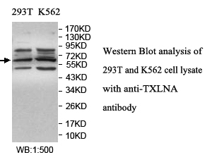
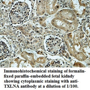
TXLNA, Polyclonal Antibody (Cat# AAA112041)
Predicted: Mouse, Rat
 Application Data
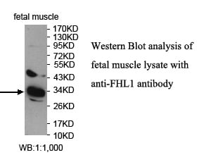
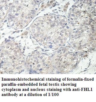
FHL1, Polyclonal Antibody (Cat# AAA112050)
Predicted: Mouse, Rat
 Application Data
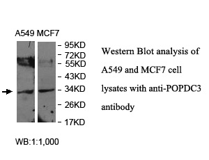
POPDC3, Polyclonal Antibody (Cat# AAA112060)
Predicted: Mouse, Rat; Not tested in other species
 Application Data
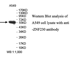
ZNF230, Polyclonal Antibody (Cat# AAA112065)
Predicted: Mouse, Rat
 Application Data
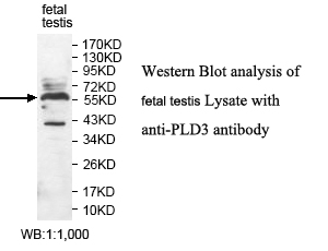
PLD3, Polyclonal Antibody (Cat# AAA112077)
Predicted: Mouse, Rat
 Application Data
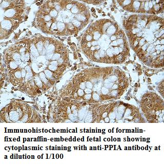
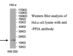
PPIA, Polyclonal Antibody (Cat# AAA112092)
Predicted: Mouse, Rat
 Application Data
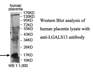
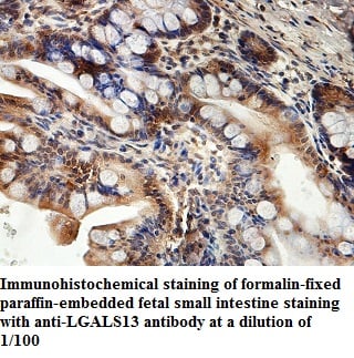
LGALS13, Polyclonal Antibody (Cat# AAA112096)
Predicted: Mouse, Rat
 Application Data
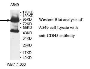
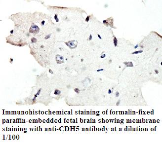
CDH5, Polyclonal Antibody (Cat# AAA112097)
Predicted: Mouse, Rat
 Application Data
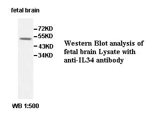
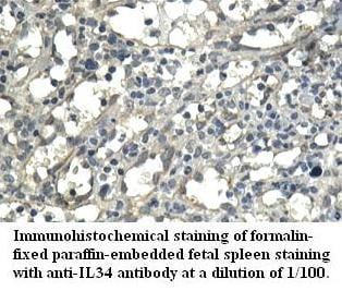
IL34, Polyclonal Antibody (Cat# AAA111452)
Predicted: Mouse, Rat
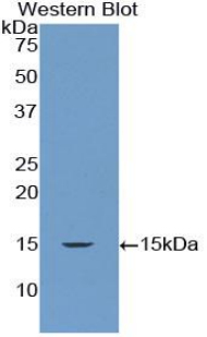
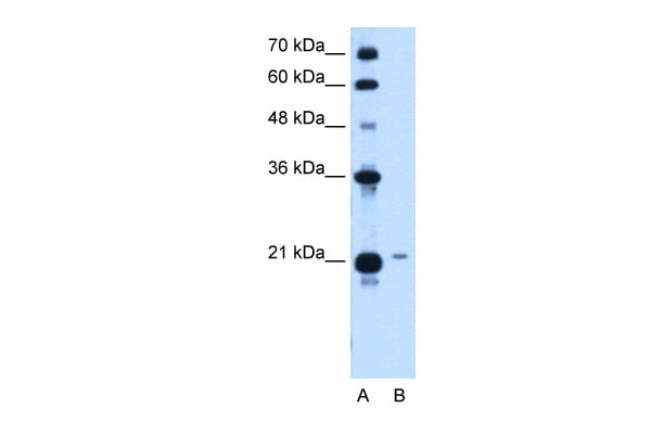
 WB (Western Blot)
((Figure. Western Blot; Sample: Recombinant protein.))
WB (Western Blot)
((Figure. Western Blot; Sample: Recombinant protein.))
S100 Calcium Binding Protein B (S100B), Polyclonal Antibody (Cat# AAA135372)
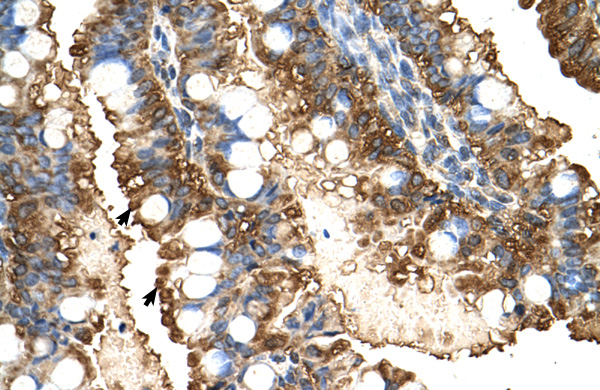
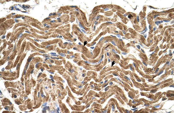
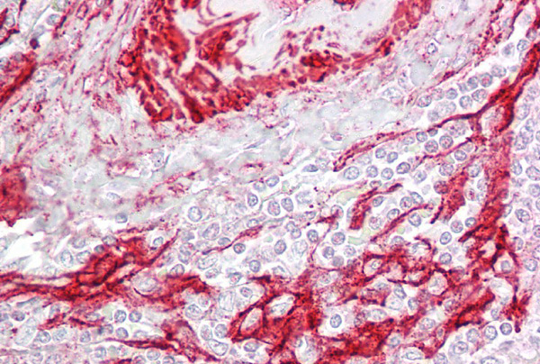
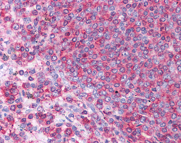
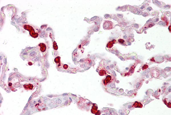
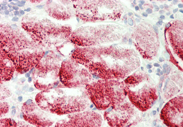
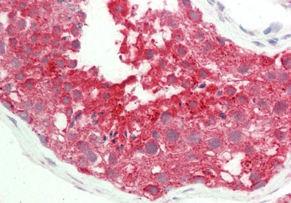
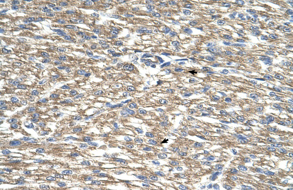
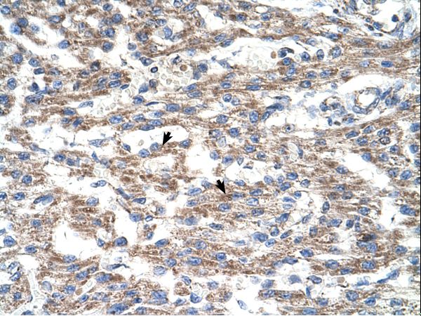
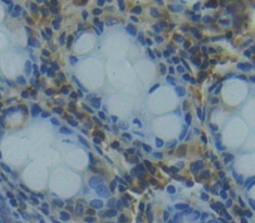 IHC (Immunohiostchemistry)
(DABstainingonIHC-P.Samples:HumanTissue))
IHC (Immunohiostchemistry)
(DABstainingonIHC-P.Samples:HumanTissue))
Ribonuclease III, Nuclear (RNASEN), Polyclonal Antibody (Cat# AAA135378)
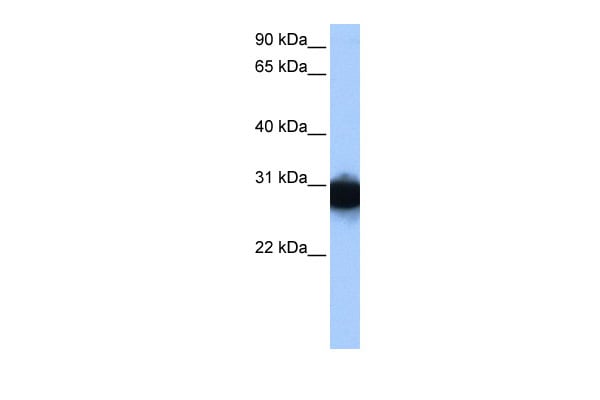
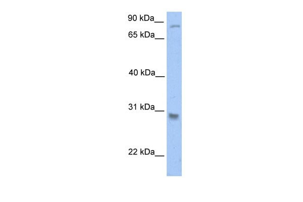
 WB (Western Blot)
(WB Suggested Anti-PGK1 Antibody Titration: 1.25ug/mlPositive Control: HepG2 cell lysate)
WB (Western Blot)
(WB Suggested Anti-PGK1 Antibody Titration: 1.25ug/mlPositive Control: HepG2 cell lysate)
PGK1, Polyclonal Antibody (Cat# AAA199868)
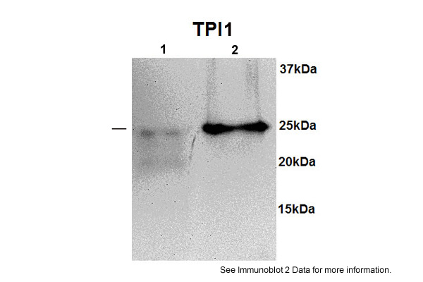
 WB (Western Blot)
(WB Suggested Anti-TPI1 Antibody Titration: 0.2-1 ug/mlPositive Control: HepG2 cell lysate)
WB (Western Blot)
(WB Suggested Anti-TPI1 Antibody Titration: 0.2-1 ug/mlPositive Control: HepG2 cell lysate)
TPI1, Polyclonal Antibody (Cat# AAA199869)
 WB (Western Blot)
(WB Suggested Anti-PTGDS Antibody Titration: 1 ug/mlPositive Control: HepG2 cell lysate)
WB (Western Blot)
(WB Suggested Anti-PTGDS Antibody Titration: 1 ug/mlPositive Control: HepG2 cell lysate)
PTGDS, Polyclonal Antibody (Cat# AAA199874)
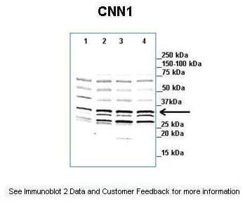
 WB (Western Blot)
(WB Suggested Anti-CNN1 Antibody Titration: 0.2-1 ug/mlELISA Titer: 1:1562500Positive Control: Hela cell lysate)
WB (Western Blot)
(WB Suggested Anti-CNN1 Antibody Titration: 0.2-1 ug/mlELISA Titer: 1:1562500Positive Control: Hela cell lysate)
CNN1, Polyclonal Antibody (Cat# AAA199875)
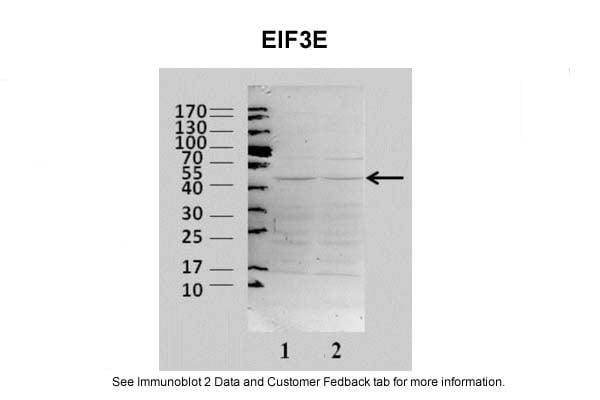
 WB (Western Blot)
(WB Suggested Anti-EIF3E Antibody Titration: 0.2-1 ug/mlELISA Titer: 1:312500Positive Control: 293T cell lysateEIF3E is supported by BioGPS gene expression data to be expressed in HEK293T)
WB (Western Blot)
(WB Suggested Anti-EIF3E Antibody Titration: 0.2-1 ug/mlELISA Titer: 1:312500Positive Control: 293T cell lysateEIF3E is supported by BioGPS gene expression data to be expressed in HEK293T)
EIF3E, Polyclonal Antibody (Cat# AAA199878)
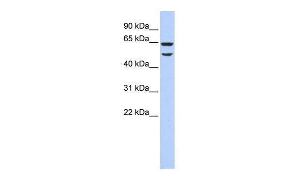
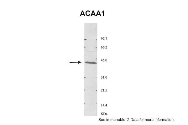 WB (Western Blot)
(Rat purified peroxisomes)
WB (Western Blot)
(Rat purified peroxisomes)
ACAA1, Polyclonal Antibody (Cat# AAA199880)
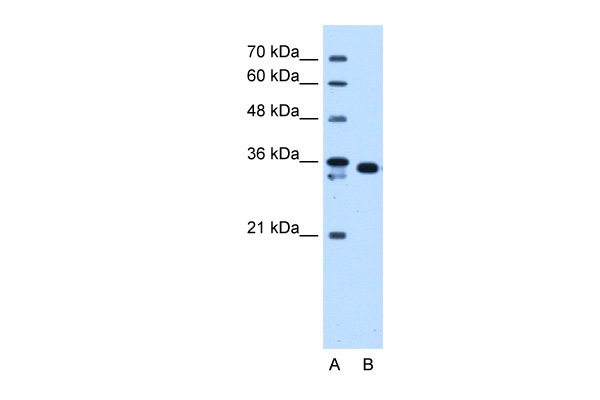
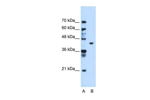
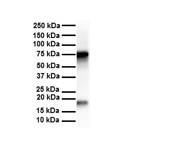
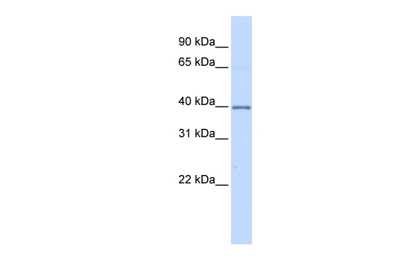
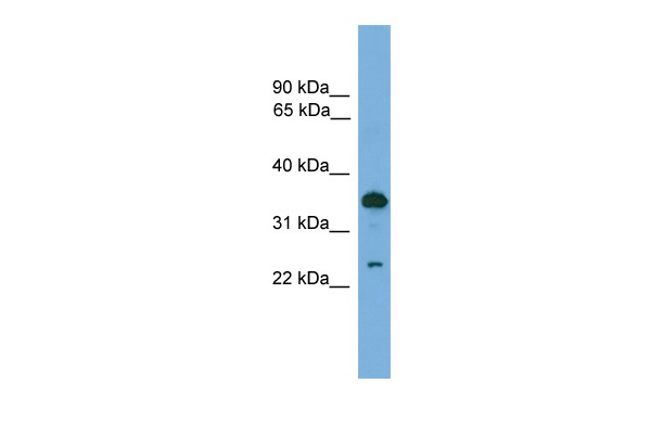
 SDS-PAGE
(25 ug of the indicated Human whole cell or tissue extracts was loaded onto a 12% SDS-PAGE gel. 1 ug/mL of the antibody was used in this experiment.)
SDS-PAGE
(25 ug of the indicated Human whole cell or tissue extracts was loaded onto a 12% SDS-PAGE gel. 1 ug/mL of the antibody was used in this experiment.)
AKR1B1, Polyclonal Antibody (Cat# AAA199882)
Predicted Species Reactivity: Human, Mouse, Rat, Cow, Dog, Guinea Pig, Horse, Pig, Rabbit, Zebrafish
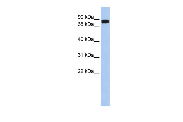

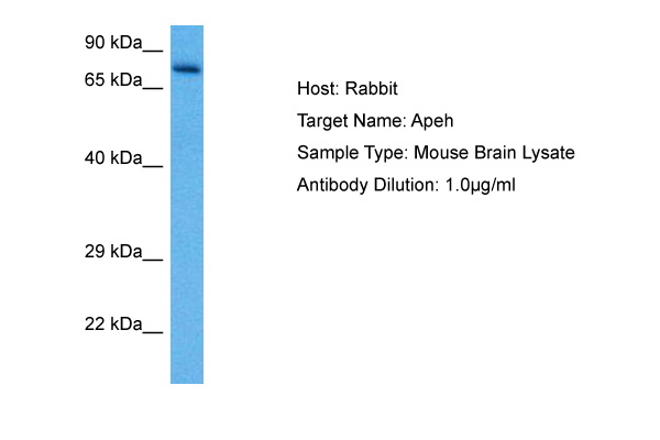
 WB (Western Blot)
(WB Suggested Anti-APEH antibody Titration: 1 ug/mLSample Type: Human liver)
WB (Western Blot)
(WB Suggested Anti-APEH antibody Titration: 1 ug/mLSample Type: Human liver)
APEH, Polyclonal Antibody (Cat# AAA199883)
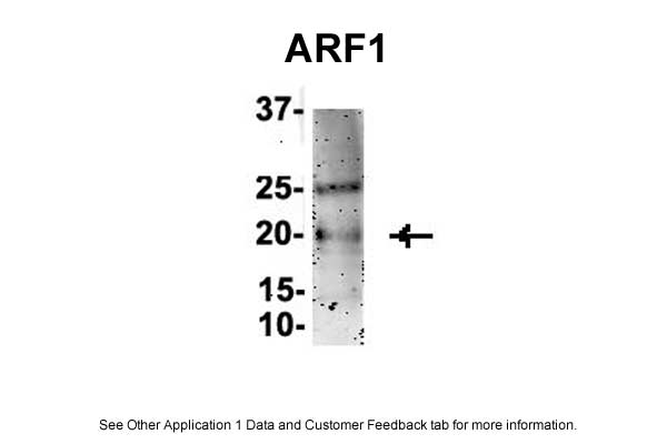
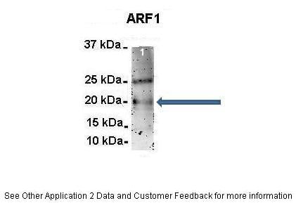
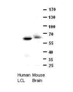
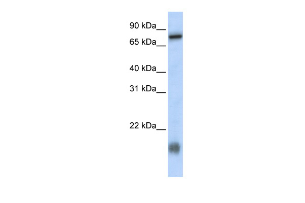
 WB (Western Blot)
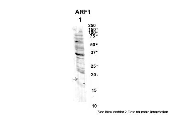
(Sample Type: 1. Human NT-2 cells (60ug)2. mouse brain extracts (80ug)Primary antibody dilution: 2ug/mlSecondary antibody: IRDye 800CW goat anti-rabbit from Li-COR BioscienceSecondary antibody dilution: 1: 20,000Image Submitted by: Yuzhi ChenUniversity of Arkansas for Medical Science)
WB (Western Blot)
(Sample Type: 1. Human NT-2 cells (60ug)2. mouse brain extracts (80ug)Primary antibody dilution: 2ug/mlSecondary antibody: IRDye 800CW goat anti-rabbit from Li-COR BioscienceSecondary antibody dilution: 1: 20,000Image Submitted by: Yuzhi ChenUniversity of Arkansas for Medical Science)
ARF1, Polyclonal Antibody (Cat# AAA199884)
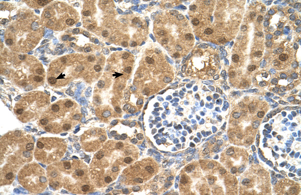
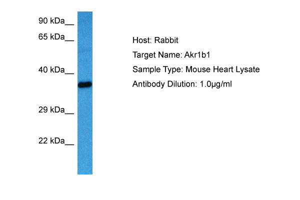
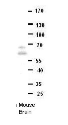
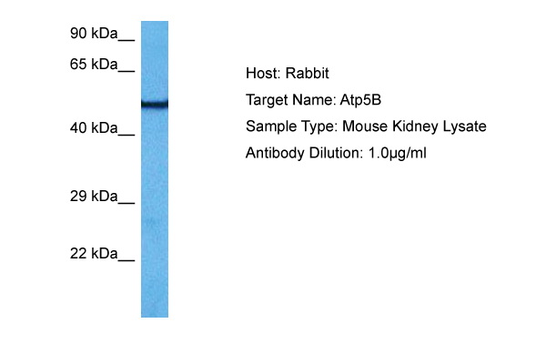 WB (Western Blot)
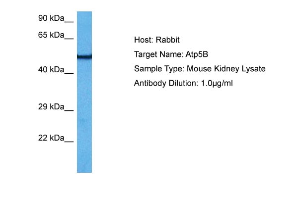
(Host: RabbitTarget Name: ATP5BSample Tissue: Mouse KidneyAntibody Dilution: 1ug/ml)
WB (Western Blot)
(Host: RabbitTarget Name: ATP5BSample Tissue: Mouse KidneyAntibody Dilution: 1ug/ml)
ATP5F1B, Polyclonal Antibody (Cat# AAA199885)
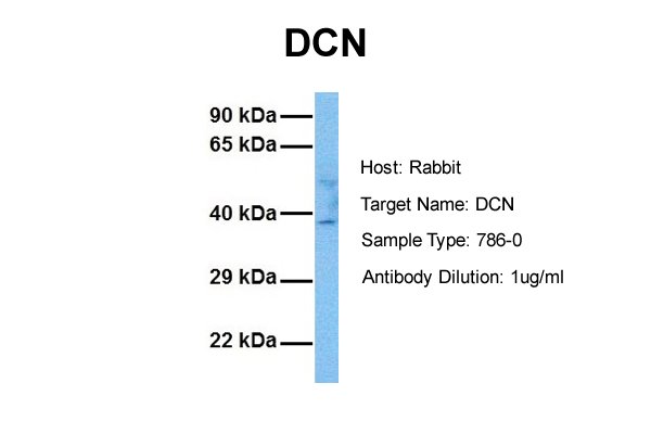
 WB (Western Blot)
(WB Suggested Anti-DCN Antibody Titration: 0.2-1 ug/mlELISA Titer: 1:12500Positive Control: Human heart)
WB (Western Blot)
(WB Suggested Anti-DCN Antibody Titration: 0.2-1 ug/mlELISA Titer: 1:12500Positive Control: Human heart)
DCN, Polyclonal Antibody (Cat# AAA199890)
 WB (Western Blot)
(WB Suggested Anti-Tagln Antibody Titration: 1 ug/mlPositive Control: Rat Lung lysate)
WB (Western Blot)
(WB Suggested Anti-Tagln Antibody Titration: 1 ug/mlPositive Control: Rat Lung lysate)
Tagln, Polyclonal Antibody (Cat# AAA199897)
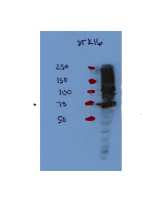
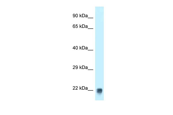
 WB (Western Blot)
(WB Suggested Anti-STK16 Antibody Titration: 0.2-1 ug/mlELISA Titer: 1:12500Positive Control: Human brain)
WB (Western Blot)
(WB Suggested Anti-STK16 Antibody Titration: 0.2-1 ug/mlELISA Titer: 1:12500Positive Control: Human brain)
STK16, Polyclonal Antibody (Cat# AAA199900)
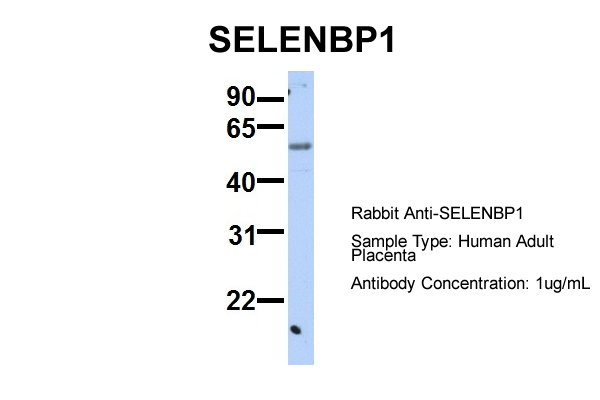
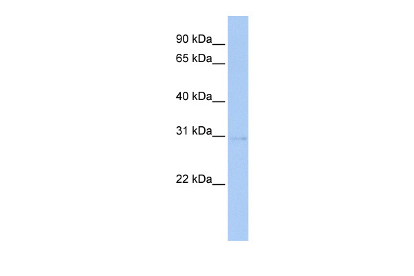
 WB (Western Blot)
(WB Suggested Anti-SELENBP1 Antibody Titration: 0.2-1 ug/mlPositive Control: 721_B cell lysate)
WB (Western Blot)
(WB Suggested Anti-SELENBP1 Antibody Titration: 0.2-1 ug/mlPositive Control: 721_B cell lysate)
SELENBP1, Polyclonal Antibody (Cat# AAA199902)
 WB (Western Blot)
(Host: RabbitTarget Name: sep15Sample Type: Rat Thymus lysatesAntibody Dilution: 1.0ug/ml)
WB (Western Blot)
(Host: RabbitTarget Name: sep15Sample Type: Rat Thymus lysatesAntibody Dilution: 1.0ug/ml)
SELENOF, Polyclonal Antibody (Cat# AAA199903)
Predicted Species Reactivity: Human, Mouse, Rat, Cow, Pig, Zebrafish
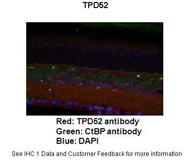
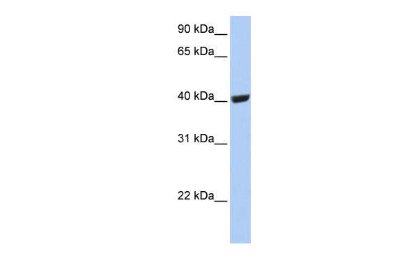
 WB (Western Blot)
(WB Suggested Anti-TPD52 Antibody Titration: 0.2-1 ug/mlELISA Titer: 1:312500Positive Control: THP-1 cell lysate)
WB (Western Blot)
(WB Suggested Anti-TPD52 Antibody Titration: 0.2-1 ug/mlELISA Titer: 1:312500Positive Control: THP-1 cell lysate)
TPD52, Polyclonal Antibody (Cat# AAA199904)
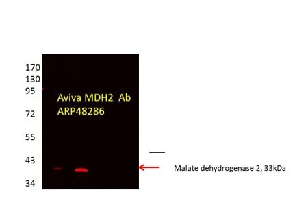
 WB (Western Blot)
(WB Suggested Anti-MDH2 Antibody Titration: 2.5ug/mlPositive Control: HepG2 cell lysate)
WB (Western Blot)
(WB Suggested Anti-MDH2 Antibody Titration: 2.5ug/mlPositive Control: HepG2 cell lysate)
MDH2, Polyclonal Antibody (Cat# AAA199908)
 WB (Western Blot)
(WB Suggested Anti-RABL4 Antibody Titration: 0.2-1 ug/mlPositive Control: 721_B cell lysateIFT27 is supported by BioGPS gene expression data to be expressed in 721_B)
WB (Western Blot)
(WB Suggested Anti-RABL4 Antibody Titration: 0.2-1 ug/mlPositive Control: 721_B cell lysateIFT27 is supported by BioGPS gene expression data to be expressed in 721_B)
RABL4, Polyclonal Antibody (Cat# AAA199911)
Predicted Species Reactivity: Human, Mouse, Rat, Cow, Dog, Pig, Sheep
 WB (Western Blot)
(WB Suggested Anti-RSU1 Antibody Titration: 1.25ug/mlPositive Control: Jurkat cell lysate)
WB (Western Blot)
(WB Suggested Anti-RSU1 Antibody Titration: 1.25ug/mlPositive Control: Jurkat cell lysate)
RSU1, Polyclonal Antibody (Cat# AAA199915)
 WB (Western Blot)
(WB Suggested Anti-EPSTI1 Antibody Titration: 0.2-1 ug/mlELISA Titer: 1:62500Positive Control: MCF7 cell lysate)
WB (Western Blot)
(WB Suggested Anti-EPSTI1 Antibody Titration: 0.2-1 ug/mlELISA Titer: 1:62500Positive Control: MCF7 cell lysate)
EPSTI1, Polyclonal Antibody (Cat# AAA199917)
Predicted Species Reactivity: Human, Mouse, Rat, Cow, Dog, Horse, Rabbit
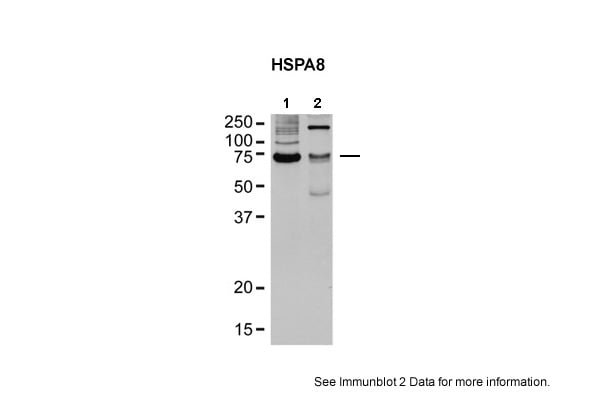
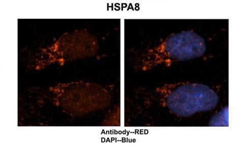
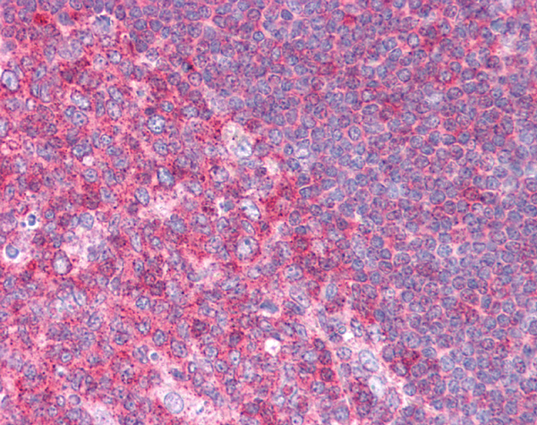
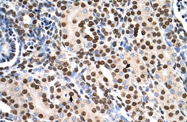
 WB (Western Blot)
(WB Suggested Anti-HSPA8 Antibody Titration: 0.2-1 ug/mlELISA Titer: 1:312500Positive Control: COLO205 cell lysate)
WB (Western Blot)
(WB Suggested Anti-HSPA8 Antibody Titration: 0.2-1 ug/mlELISA Titer: 1:312500Positive Control: COLO205 cell lysate)
HSPA8, Polyclonal Antibody (Cat# AAA199919)
 WB (Western Blot)
(WB Suggested Anti-GSTT1 Antibody Titration: 0.2-1 ug/mlPositive Control: Jurkat cell lysate)
WB (Western Blot)
(WB Suggested Anti-GSTT1 Antibody Titration: 0.2-1 ug/mlPositive Control: Jurkat cell lysate)
GSTT1, Polyclonal Antibody (Cat# AAA199927)
 WB (Western Blot)
(WB Suggested Anti-GGPS1 Antibody Titration: 0.2-1 ug/mlELISA Titer: 1:62500Positive Control: 293T cell lysateGGPS1 is strongly supported by BioGPS gene expression data to be expressed in Human HEK293T cells)
WB (Western Blot)
(WB Suggested Anti-GGPS1 Antibody Titration: 0.2-1 ug/mlELISA Titer: 1:62500Positive Control: 293T cell lysateGGPS1 is strongly supported by BioGPS gene expression data to be expressed in Human HEK293T cells)
GGPS1, Polyclonal Antibody (Cat# AAA199930)
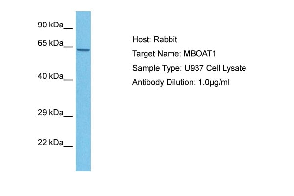
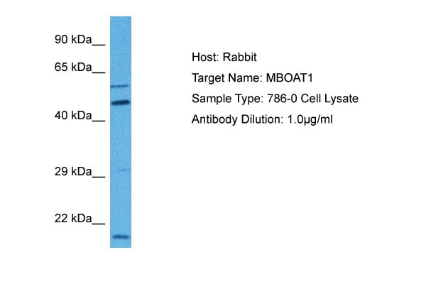
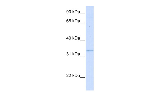
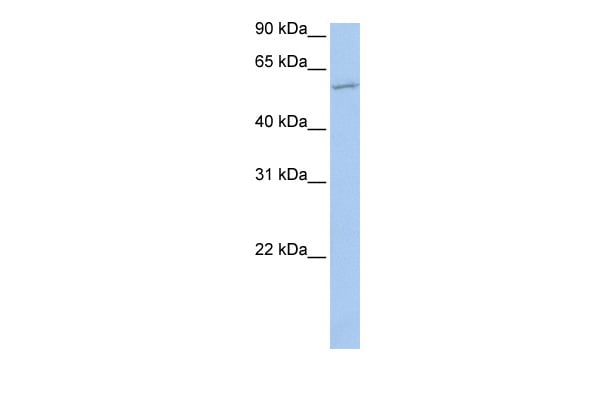 WB (Western Blot)
(WB Suggested Anti-MBOAT1 Antibody Titration: 0.2-1 ug/mlPositive Control: Jurkat cell lysate)
WB (Western Blot)
(WB Suggested Anti-MBOAT1 Antibody Titration: 0.2-1 ug/mlPositive Control: Jurkat cell lysate)
MBOAT1, Polyclonal Antibody (Cat# AAA199932)
Predicted: Horse, Human, Mouse, Pig, Rabbit, Rat
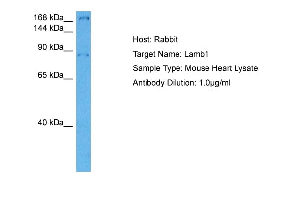

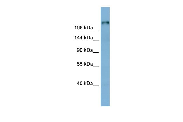 WB (Western Blot)
(WB Suggested Anti-LAMB1 Antibody Titration: 0.2-1 ug/mlELISA Titer: 1:62500Positive Control: Human Stomach)
WB (Western Blot)
(WB Suggested Anti-LAMB1 Antibody Titration: 0.2-1 ug/mlELISA Titer: 1:62500Positive Control: Human Stomach)
LAMB1, Polyclonal Antibody (Cat# AAA199935)
 WB (Western Blot)
(WB Suggested Anti-Gyg Antibody Titration: 0.2-1 ug/mlELISA Titer: 1:62500Positive Control: Mouse Kidney)
WB (Western Blot)
(WB Suggested Anti-Gyg Antibody Titration: 0.2-1 ug/mlELISA Titer: 1:62500Positive Control: Mouse Kidney)
Gyg, Polyclonal Antibody (Cat# AAA199937)
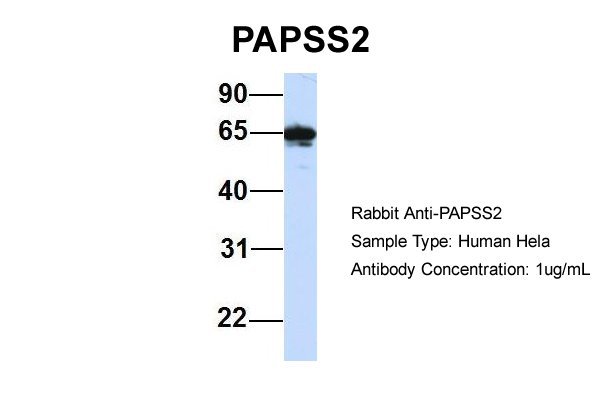
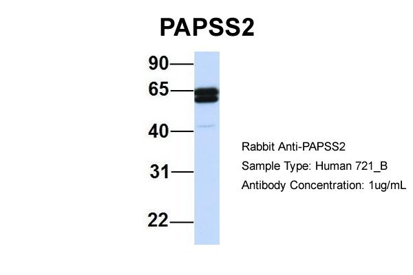
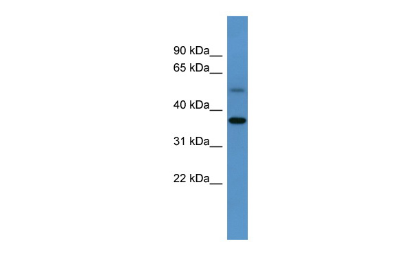
 WB (Western Blot)
(WB Suggested Anti-PAPSS2 Antibody Titration: 0.2-1 ug/mlELISA Titer: 1:312500Positive Control: 293T cell lysate)
WB (Western Blot)
(WB Suggested Anti-PAPSS2 Antibody Titration: 0.2-1 ug/mlELISA Titer: 1:312500Positive Control: 293T cell lysate)
PAPSS2, Polyclonal Antibody (Cat# AAA199939)
 WB (Western Blot)
(WB Suggested Anti-GLYAT Antibody Titration: 0.2-1 ug/mlELISA Titer: 1:312500Positive Control: HepG2 cell lysate)
WB (Western Blot)
(WB Suggested Anti-GLYAT Antibody Titration: 0.2-1 ug/mlELISA Titer: 1:312500Positive Control: HepG2 cell lysate)
GLYAT, Polyclonal Antibody (Cat# AAA199941)
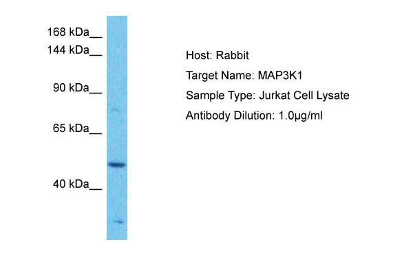
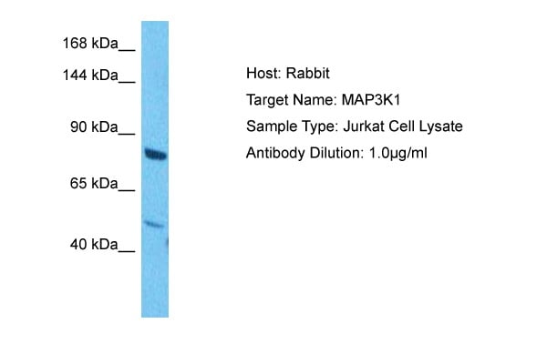
 WB (Western Blot)
(WB Suggested Anti-MAP3K1 Antibody Titration: 0.2-1 ug/mlPositive Control: 721_B cell lysate)
WB (Western Blot)
(WB Suggested Anti-MAP3K1 Antibody Titration: 0.2-1 ug/mlPositive Control: 721_B cell lysate)
MAP3K1, Polyclonal Antibody (Cat# AAA199942)
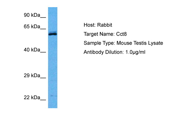

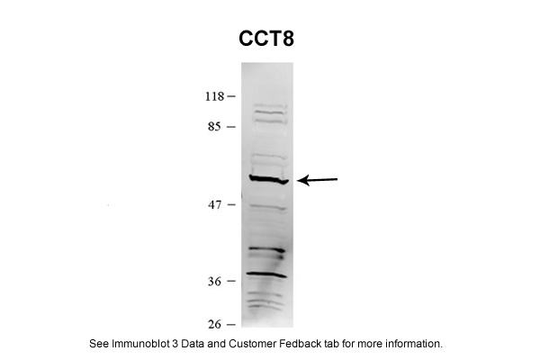
 WB (Western Blot)
(WB Suggested Anti-CCT8 Antibody Titration: 0.2-1 ug/mlPositive Control: Jurkat cell lysate.CCT8 is strongly supported by BioGPS gene expression data to be expressed in Jurkat)
WB (Western Blot)
(WB Suggested Anti-CCT8 Antibody Titration: 0.2-1 ug/mlPositive Control: Jurkat cell lysate.CCT8 is strongly supported by BioGPS gene expression data to be expressed in Jurkat)
CCT8, Polyclonal Antibody (Cat# AAA199628)
 WB (Western Blot)
(WB Suggested Anti-DONSON Antibody Titration: 0.2-1 ug/mlPositive Control: HepG2 cell lysateDONSON is supported by BioGPS gene expression data to be expressed in HepG2)
WB (Western Blot)
(WB Suggested Anti-DONSON Antibody Titration: 0.2-1 ug/mlPositive Control: HepG2 cell lysateDONSON is supported by BioGPS gene expression data to be expressed in HepG2)
DONSON, Polyclonal Antibody (Cat# AAA199634)
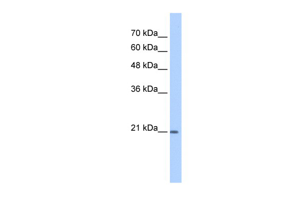
 WB (Western Blot)
(WB Suggested Anti-TCP10L Antibody Titration: 1.25ug/mlPositive Control: HepG2 cell lysate)
WB (Western Blot)
(WB Suggested Anti-TCP10L Antibody Titration: 1.25ug/mlPositive Control: HepG2 cell lysate)
TCP10L, Polyclonal Antibody (Cat# AAA199638)
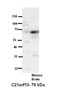 WB (Western Blot)
(WB Suggested Anti-C21orf13 Antibody Titration: 5% MilkELISA Titer: dilution: 1:500Positive Control: Mouse Brain lysate)
WB (Western Blot)
(WB Suggested Anti-C21orf13 Antibody Titration: 5% MilkELISA Titer: dilution: 1:500Positive Control: Mouse Brain lysate)
C21orf13, Polyclonal Antibody (Cat# AAA199639)
 WB (Western Blot)
(WB Suggested Anti-SNF1LK Antibody Titration: 1.25ug/mlPositive Control: Jurkat cell lysate)
WB (Western Blot)
(WB Suggested Anti-SNF1LK Antibody Titration: 1.25ug/mlPositive Control: Jurkat cell lysate)
SNF1LK, Polyclonal Antibody (Cat# AAA199640)
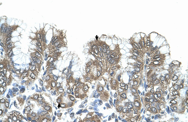
 WB (Western Blot)
(WB Suggested Anti-GUCY1B3 Antibody Titration: 0.2-1 ug/mlELISA Titer: 1:12500Positive Control: 293T cell lysateGUCY1B3 is supported by BioGPS gene expression data to be expressed in HEK293T)
WB (Western Blot)
(WB Suggested Anti-GUCY1B3 Antibody Titration: 0.2-1 ug/mlELISA Titer: 1:12500Positive Control: 293T cell lysateGUCY1B3 is supported by BioGPS gene expression data to be expressed in HEK293T)
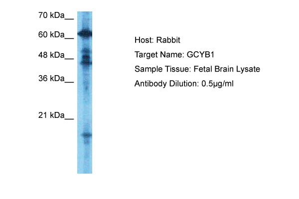
GUCY1B1, Polyclonal Antibody (Cat# AAA199641)
 WB (Western Blot)
(WB Suggested Anti-DUT Antibody Titration: 1 ug/mlPositive Control: Fetal Small Intestine cell lysate)
WB (Western Blot)
(WB Suggested Anti-DUT Antibody Titration: 1 ug/mlPositive Control: Fetal Small Intestine cell lysate)
DUT, Polyclonal Antibody (Cat# AAA199645)
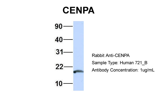
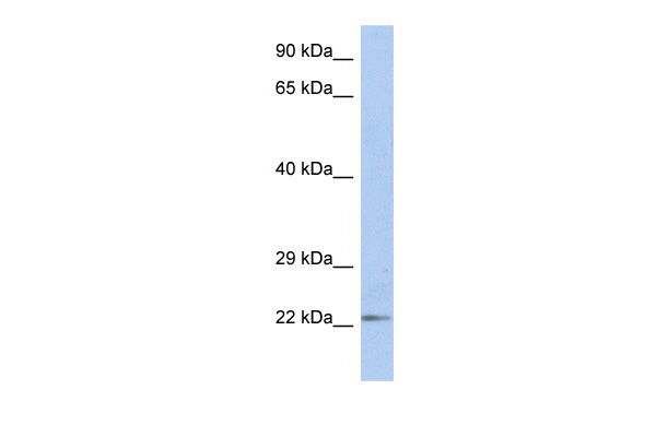
 WB (Western Blot)
(WB Suggested Anti-CENPA Antibody Titration: 0.2-1 ug/mlELISA Titer: 1:62500Positive Control: Hela cell lysate.CENPA is strongly supported by BioGPS gene expression data to be expressed in HeLa)
WB (Western Blot)
(WB Suggested Anti-CENPA Antibody Titration: 0.2-1 ug/mlELISA Titer: 1:62500Positive Control: Hela cell lysate.CENPA is strongly supported by BioGPS gene expression data to be expressed in HeLa)
CENPA, Polyclonal Antibody (Cat# AAA199647)
Predicted Species Reactivity: Human, Mouse, Rat, Cow, Dog, Goat, Guinea Pig, Horse, Rabbit, Zebrafish
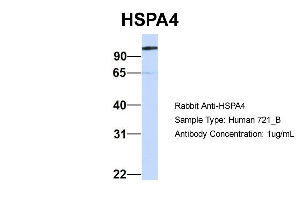
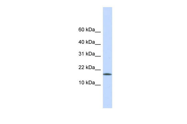
 WB (Western Blot)
(WB Suggested Anti-HSPA4 Antibody Titration: 0.2-1 ug/mlELISA Titer: 1:312500Positive Control: Human brain)
WB (Western Blot)
(WB Suggested Anti-HSPA4 Antibody Titration: 0.2-1 ug/mlELISA Titer: 1:312500Positive Control: Human brain)
HSPA4, Polyclonal Antibody (Cat# AAA199651)
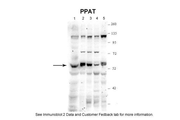
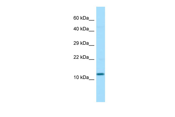
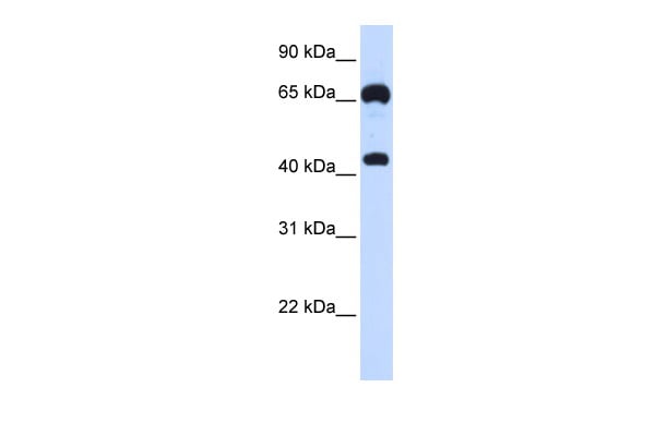
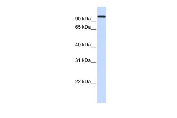
 WB (Western Blot)
(PPAT antibody - N-terminal region validated by WB using HepG2 Cell Lysate at 0.25ug/ml.PPAT is supported by BioGPS gene expression data to be expressed in HepG2)
WB (Western Blot)
(PPAT antibody - N-terminal region validated by WB using HepG2 Cell Lysate at 0.25ug/ml.PPAT is supported by BioGPS gene expression data to be expressed in HepG2)
PPAT, Polyclonal Antibody (Cat# AAA199653)
What are Polyclonal Antibodies?
Polyclonal antibodies are antibodies that come from multiple B cell clones of a host animal. The typical hosts used for the majority of polyclonal antibody production are rabbits, goats, sheep, and donkeys. These polyclonal antibodies, once having identified their target, will bind to different epitopes located at different regions or sequences on the same protein/antigen. As a result, they are ideal at locating and binding to the target, even if the target is in very low concentrations (due to many different antibodies being able to bind to the same target molecule, which allows for significant amplification of a downstream signal).
Polyclonal antibodies are typically produced by injecting an antigen into a host animal, which causes the animal’s immune system to attack the foreign antigen by mass generating antibodies against it. After a period of time, serum is collected from the animal and purified using physicochemical fractionation, class-specific affinity purification, and/or antigen-affinity purification.
Key Uses of Polyclonal Antibodies
- Western Blotting: This method is used to find specific proteins in biological samples after separating them by size.
- Immunohistochemistry: IHC helps visualize the location of proteins in tissue sections using various staining techniques.
- ELISA: (Enzyme-Linked Immunosorbent Assay) is typically used to identify specific protein quantities in a sample. ELISAs can be either “Quantitative” or “Qualitative”.
- Flow Cytometry: technique that identifies and measures the specific protein on the surface or inside the cells in a fluid suspension.
- Immunoprecipitation: IP isolates and studies a specific protein from a complex mixture using antibodies.
Why Buy Polyclonal Antibodies from AAA Biotech?
1. Ideal for Various Applications
Our antibodies are generally going to be validated for use in multiple types of assays, including ELISA, Western Blotting, Immunohistochemistry, Immunoprecipitation, amongst others. They are ideal for a wide range of research applications.
2. Rigorous Quality Control
All of the antibodies in our catalog undergo strict quality testing to ensure specificity, sensitivity, and consistent performance. We are confident in the ability of our antibodies to provide you with accurate results.
3. Wide Assortment of Antibodies
Antibodies in are catalog can be found for both common and exotic species, and these antibodies are also available in both conjugated and recombinant forms to suit many diverse experimental needs.
4. Highly Purified
Our antibodies are available in purified forms with over 85% purity, as confirmed by SDS-PAGE. They are also available with tags such as His, Flag, GST, or MBP. We cater to customers worldwide.
FAQ
1. How are polyclonal antibodies produced?
Traditionally, polyclonal antibodies are produced by injecting an antigen into a host animal (such as a rabbit or goat), which then triggers an immune response from the host animal. The animal’s B cells produce antibodies that will recognize different parts of the injected antigen. These antibodies are then collected from the animal’s blood and purified for use.
2. How do polyclonal antibodies differ from monoclonal antibodies?
Polyclonal antibodies are a mix of antibodies that bind to different locations (epitopes) of the same antigen, while monoclonal antibodies are identical and bind to just one specific epitope. This makes polyclonal antibodies more versatile and better at detecting proteins that may be present in low quantities or in altered/modified forms.
3. How should I store polyclonal antibodies?
Polyclonal antibodies should be stored at 4°C for short-term use (up to a few weeks) and at -20°C or -80°C for long-term storage. Avoid repeated freeze-thaw cycles by dividing them into small aliquots. Always check the datasheet for specific storage instructions.