Filters
▼Clonality
▼Type
▼Reactivity
▼Gene Name
▼Isotype
▼Host
▼Application
▼Clone
▼Polyclonal Antibodies
At AAA Biotech also known as AAA Bio or AAABio, we provide a broad range of purified polyclonal antibodies (pAbs) that are able to all be browsed online through our website. Due to their high specificity and strong binding affinity, these antibodies are ideal for wide swathes of research and experimental applications.
Our polyclonal antibodies can easily support your work, whether you use them for Western Blotting, Immunocytochemistry (with or without Immunofluorescence used in conjunction), Immunohistochemistry, Immunoprecipitation, and ELISA tests. We highly encourage you to browse our range of pAbs and choose the one that best suits your experimental model.
Viewing 3200-3250 of 96805 product results
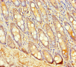
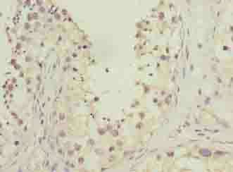
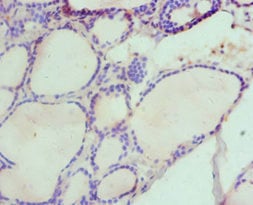
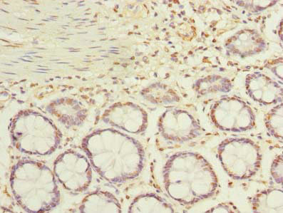
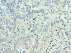
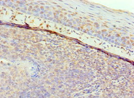
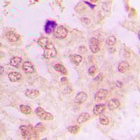
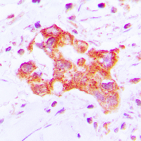
 IHC (Immunohiostchemistry)
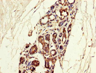
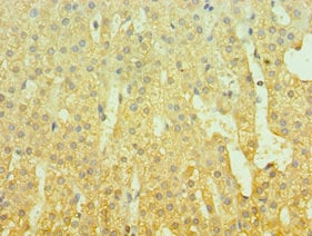
(Immunohistochemistry of paraffin-embedded human colon cancer using AAA114293 at dilution of 1:100)
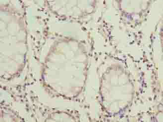
IHC (Immunohiostchemistry)
(Immunohistochemistry of paraffin-embedded human colon cancer using AAA114293 at dilution of 1:100)
UGT2A3, Polyclonal Antibody (Cat# AAA114293)
RNASE3, Polyclonal Antibody (Cat# AAA114298)
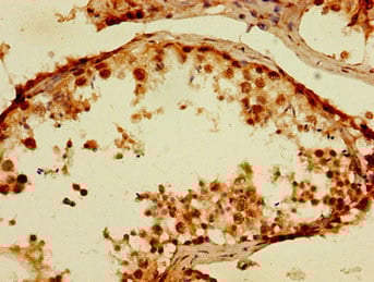
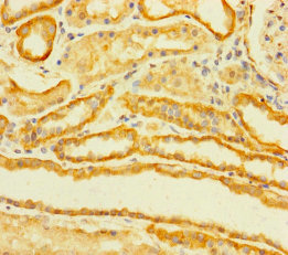
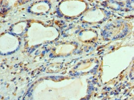
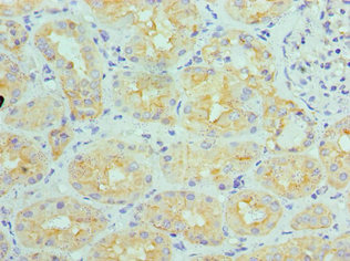
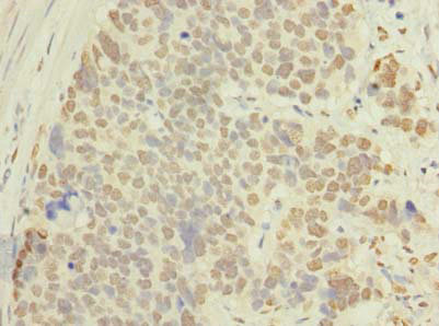
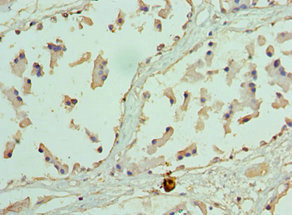
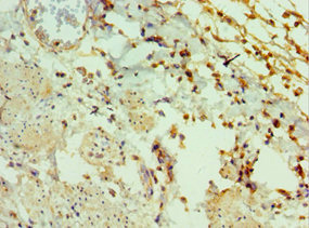
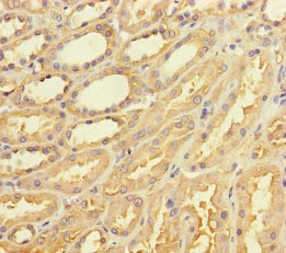 IHC (Immunohiostchemistry)
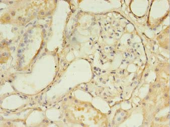
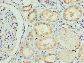
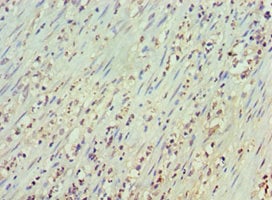
(Immunohistochemistry of paraffin-embedded human kidney tissue using AAA114299 at dilution of 1:100)
IHC (Immunohiostchemistry)
(Immunohistochemistry of paraffin-embedded human kidney tissue using AAA114299 at dilution of 1:100)
TAS2R42, Polyclonal Antibody (Cat# AAA114299)
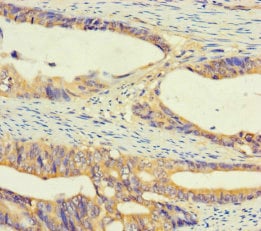
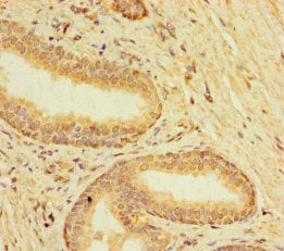
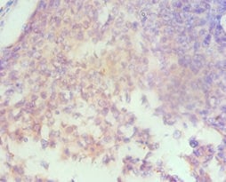
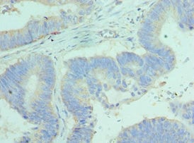
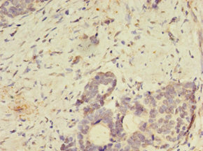
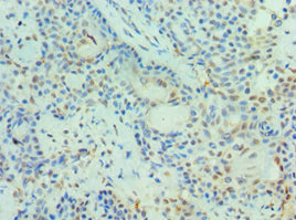
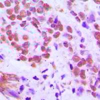
 IHC (Immunohistochemisry)
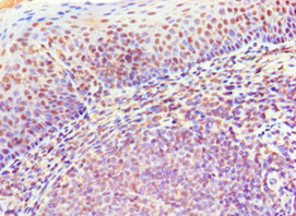
(Immunohistochemistry of paraffin-embedded human colon cancer using AAA114302 at dilution of 1:100)
IHC (Immunohistochemisry)
(Immunohistochemistry of paraffin-embedded human colon cancer using AAA114302 at dilution of 1:100)
HOOK1, Polyclonal Antibody (Cat# AAA114302)
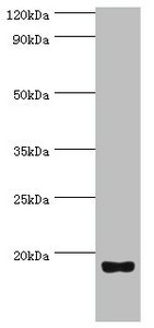
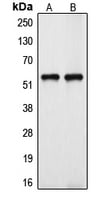
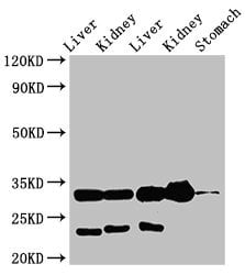 WB (Western Blot)
(Western BlotPositive WB detected in: Rat liver tissue,Rat kidney tissue,Mouse liver tissue,Mouse kidney tissue,Mouse stomach tissueAll lanes: TPSB2 antibody at 3ug/mlSecondaryGoat polyclonal to rabbit IgG at 1/50000 dilutionPredicted band size: 31 KDaObserved band size: 31 KDa)
WB (Western Blot)
(Western BlotPositive WB detected in: Rat liver tissue,Rat kidney tissue,Mouse liver tissue,Mouse kidney tissue,Mouse stomach tissueAll lanes: TPSB2 antibody at 3ug/mlSecondaryGoat polyclonal to rabbit IgG at 1/50000 dilutionPredicted band size: 31 KDaObserved band size: 31 KDa)
TPSB2, Polyclonal Antibody (Cat# AAA114303)
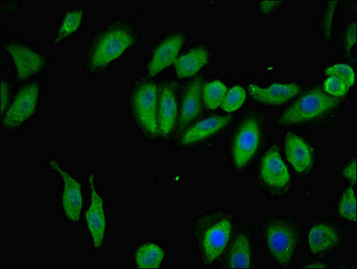 IHC (Immunohistochemisry)
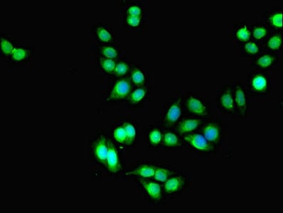
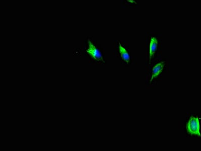
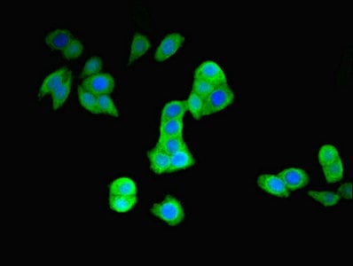
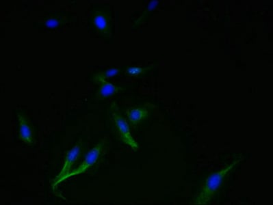
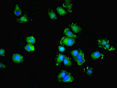
(Immunofluorescent analysis of A549 cells using AAA114306 at a dilution of 1:100 and Alexa Fluor 488-congugated AffiniPure Goat Anti-Rabbit IgG(H+L))
IHC (Immunohistochemisry)
(Immunofluorescent analysis of A549 cells using AAA114306 at a dilution of 1:100 and Alexa Fluor 488-congugated AffiniPure Goat Anti-Rabbit IgG(H+L))
TMEM120B, Polyclonal Antibody (Cat# AAA114306)
 IF (Immunofluorescence)
(Immunofluorescent analysis of PC3 cells using AAA114161 at a dilution of 1:100 and Alexa Fluor 488-congugated AffiniPure Goat Anti-Rabbit IgG(H+L))
IF (Immunofluorescence)
(Immunofluorescent analysis of PC3 cells using AAA114161 at a dilution of 1:100 and Alexa Fluor 488-congugated AffiniPure Goat Anti-Rabbit IgG(H+L))
DLEU1, Polyclonal Antibody (Cat# AAA114161)
 IHC (Immunohistochemisry)
(Immunofluorescent analysis of U251 cells using AAA114166 at a dilution of 1:100 and Alexa Fluor 488-congugated AffiniPure Goat Anti-Rabbit IgG(H+L))
IHC (Immunohistochemisry)
(Immunofluorescent analysis of U251 cells using AAA114166 at a dilution of 1:100 and Alexa Fluor 488-congugated AffiniPure Goat Anti-Rabbit IgG(H+L))
Hyaluronan synthase 3, Polyclonal Antibody (Cat# AAA114166)
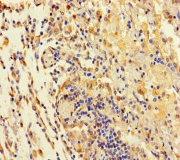
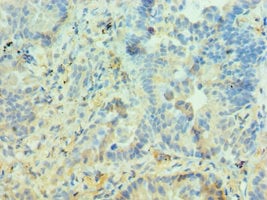
 IHC (Immunohiostchemistry)
(Immunohistochemistry of paraffin-embedded human gastric cancer using AAA114169 at dilution of 1:100)
IHC (Immunohiostchemistry)
(Immunohistochemistry of paraffin-embedded human gastric cancer using AAA114169 at dilution of 1:100)
GTPBP6, Polyclonal Antibody (Cat# AAA114169)

 IHC (Immunohistochemisry)
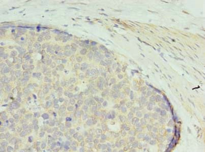
(Immunohistochemistry of paraffin-embedded human breast cancer tissue using AAA114174 at dilution 1:100)
IHC (Immunohistochemisry)
(Immunohistochemistry of paraffin-embedded human breast cancer tissue using AAA114174 at dilution 1:100)
CA8, Polyclonal Antibody (Cat# AAA114174)
 IF (Immunofluorescence)
(Immunofluorescent analysis of PC3 cells using AAA114176 at a dilution of 1:100 and Alexa Fluor 488-congugated AffiniPure Goat Anti-Rabbit IgG(H+L))
IF (Immunofluorescence)
(Immunofluorescent analysis of PC3 cells using AAA114176 at a dilution of 1:100 and Alexa Fluor 488-congugated AffiniPure Goat Anti-Rabbit IgG(H+L))
E3 ubiquitin-protein ligase parkin, Polyclonal Antibody (Cat# AAA114176)
 IHC (Immunohiostchemistry)
(Immunohistochemistry of paraffin-embedded human colon cancer using AAA114177 at dilution 1:100)
IHC (Immunohiostchemistry)
(Immunohistochemistry of paraffin-embedded human colon cancer using AAA114177 at dilution 1:100)
Apoptosis facilitator Bcl-2-like protein 14, Polyclonal Antibody (Cat# AAA114177)
 IHC (Immunohistochemisry)
(Immunohistochemistry of paraffin-embedded human kidney using AAA114179 at dilution 1:100)
IHC (Immunohistochemisry)
(Immunohistochemistry of paraffin-embedded human kidney using AAA114179 at dilution 1:100)
SOST, Polyclonal Antibody (Cat# AAA114179)
Ribonucleoside-diphosphate reductase 1 subunit beta, Polyclonal Antibody (Cat# AAA114180)
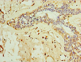
 IHC (Immunohiostchemistry)
(IHC image of AAA114181 diluted at 1:300 and staining in paraffin-embedded human breast cancer performed on a Leica Bond system. After dewaxing and hydration, antigen retrieval was mediated by high pressure in a citrate buffer (pH 6.0). Section was blocked with 10% normal goat serum 30min at RT. Then primary antibody (1% BSA) was incubated at 4 degree C overnight. The primary is detected by a biotinylated secondary antibody and visualized using an HRP conjugated SP system.)
IHC (Immunohiostchemistry)
(IHC image of AAA114181 diluted at 1:300 and staining in paraffin-embedded human breast cancer performed on a Leica Bond system. After dewaxing and hydration, antigen retrieval was mediated by high pressure in a citrate buffer (pH 6.0). Section was blocked with 10% normal goat serum 30min at RT. Then primary antibody (1% BSA) was incubated at 4 degree C overnight. The primary is detected by a biotinylated secondary antibody and visualized using an HRP conjugated SP system.)
Matrix Gla protein, Polyclonal Antibody (Cat# AAA114181)
 IF (Immunofluorescence)
(Immunofluorescent analysis of Hela cells using AAA114183 at a dilution of 1:100 and Alexa Fluor 488-congugated AffiniPure Goat Anti-Rabbit IgG(H+L))
IF (Immunofluorescence)
(Immunofluorescent analysis of Hela cells using AAA114183 at a dilution of 1:100 and Alexa Fluor 488-congugated AffiniPure Goat Anti-Rabbit IgG(H+L))
SMIM14, Polyclonal Antibody (Cat# AAA114183)
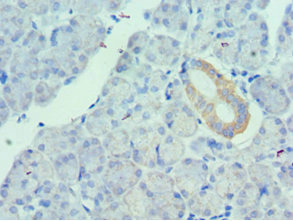
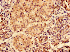 IHC (Immunohiostchemistry)
(Immunohistochemistry of paraffin-embedded human pancreatic tissue using AAA114188 at dilution of 1:100)
IHC (Immunohiostchemistry)
(Immunohistochemistry of paraffin-embedded human pancreatic tissue using AAA114188 at dilution of 1:100)
Uncharacterized protein KIAA1377, Polyclonal Antibody (Cat# AAA114188)
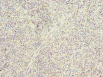
 IHC (Immunohiostchemistry)
(Immunohistochemistry of paraffin-embedded human pancreas using AAA114190 at dilution 1:100)
IHC (Immunohiostchemistry)
(Immunohistochemistry of paraffin-embedded human pancreas using AAA114190 at dilution 1:100)
Urotensin-2, Polyclonal Antibody (Cat# AAA114190)
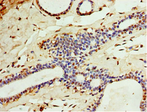
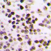
 IHC (Immunohistochemistry)
(Immunohistochemistry of paraffin-embedded human tonsil using AAA114191 at dilution of 1:100)
IHC (Immunohistochemistry)
(Immunohistochemistry of paraffin-embedded human tonsil using AAA114191 at dilution of 1:100)
TMTC4, Polyclonal Antibody (Cat# AAA114191)
coronavirus Spike glycoprotein, Polyclonal Antibody (Cat# AAA114194)
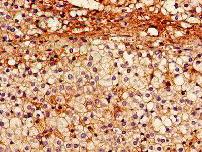
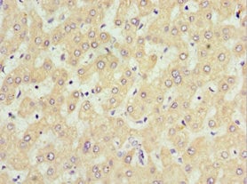
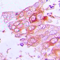
 IHC (Immunohiostchemistry)
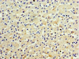
(Immunohistochemistry of paraffin-embedded human liver cancer using AAA114198 at dilution 1:100)
IHC (Immunohiostchemistry)
(Immunohistochemistry of paraffin-embedded human liver cancer using AAA114198 at dilution 1:100)
IFNL3, Polyclonal Antibody (Cat# AAA114198)
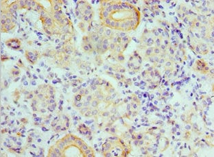
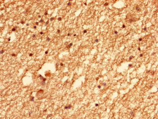
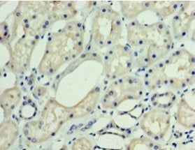
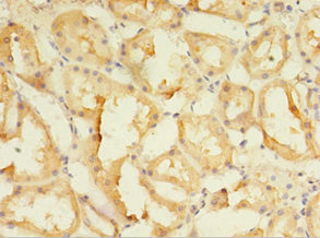
 IHC (Immunohiostchemistry)
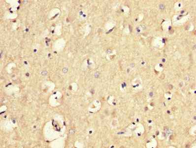
(Immunohistochemistry of paraffin-embedded human kidney using AAA114202 at dilution 1:100)
IHC (Immunohiostchemistry)
(Immunohistochemistry of paraffin-embedded human kidney using AAA114202 at dilution 1:100)
V-type proton ATPase subunit B, Polyclonal Antibody (Cat# AAA114202)
 IHC (Immunohiostchemistry)
(Immunohistochemistry of paraffin-embedded human gastric cancer using AAA114203 at dilution of 1:100)
IHC (Immunohiostchemistry)
(Immunohistochemistry of paraffin-embedded human gastric cancer using AAA114203 at dilution of 1:100)
ACTRT1, Polyclonal Antibody (Cat# AAA114203)
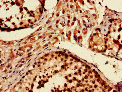
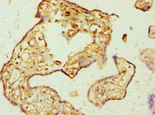 IHC (Immunohiostchemistry)
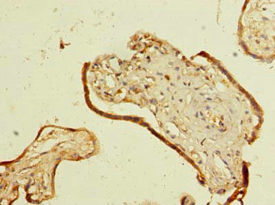
(Immunohistochemistry of paraffin-embedded human placenta using AAA114204 at dilution 1:100)
IHC (Immunohiostchemistry)
(Immunohistochemistry of paraffin-embedded human placenta using AAA114204 at dilution 1:100)
AMP deaminase 3, Polyclonal Antibody (Cat# AAA114204)
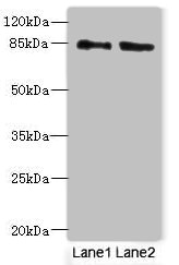
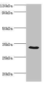
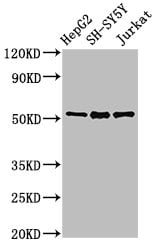
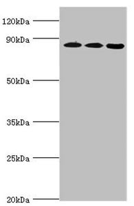
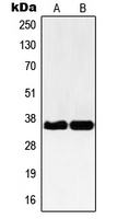
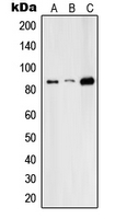
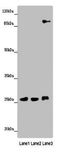 WB (Western Blot)
(Western blotAll lanes: DNAJC30 antibody at 10ug/mlLane 1: 293T whole cell lysateLane 2: U87 whole cell lysateLane 3: SH-SY5Y whole cell lysateSecondaryGoat polyclonal to rabbit IgG at 1/10000 dilutionPredicted band size: 26 kDaObserved band size: 26,90 kDa)
WB (Western Blot)
(Western blotAll lanes: DNAJC30 antibody at 10ug/mlLane 1: 293T whole cell lysateLane 2: U87 whole cell lysateLane 3: SH-SY5Y whole cell lysateSecondaryGoat polyclonal to rabbit IgG at 1/10000 dilutionPredicted band size: 26 kDaObserved band size: 26,90 kDa)
DnaJ homolog subfamily C member 30, Polyclonal Antibody (Cat# AAA114205)
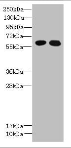
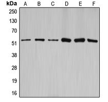
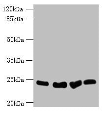 WB (Western Blot)
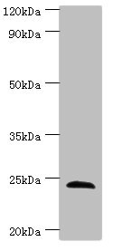
(Western blotAll lanes: Socs3 antibody at 12ug/mlLane 1: K562 whole cell lysateLane 2: A375 whole cell lysateLane 3: Hela whole cell lysateLane 4: HepG-2 whole cell lysateSecondaryGoat polyclonal to Rabbit IgG at 1/10000 dilutionPredicted band size: 25 kDaObserved band size: 25 kDa)
WB (Western Blot)
(Western blotAll lanes: Socs3 antibody at 12ug/mlLane 1: K562 whole cell lysateLane 2: A375 whole cell lysateLane 3: Hela whole cell lysateLane 4: HepG-2 whole cell lysateSecondaryGoat polyclonal to Rabbit IgG at 1/10000 dilutionPredicted band size: 25 kDaObserved band size: 25 kDa)
Socs3, Polyclonal Antibody (Cat# AAA114208)
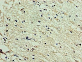
 IHC (Immunohiostchemistry)
(Immunohistochemistry of paraffin-embedded human placenta using AAA114209 at dilution 1:100)
IHC (Immunohiostchemistry)
(Immunohistochemistry of paraffin-embedded human placenta using AAA114209 at dilution 1:100)
14-3-3 protein theta, Polyclonal Antibody (Cat# AAA114209)
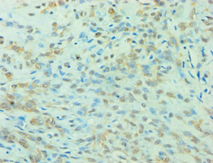
 IHC (Immunohiostchemistry)
(Immunohistochemistry of paraffin-embedded human lung cancer using AAA114210 at dilution 1:100)
IHC (Immunohiostchemistry)
(Immunohistochemistry of paraffin-embedded human lung cancer using AAA114210 at dilution 1:100)
Histone-lysine N-methyltransferase EZH1, Polyclonal Antibody (Cat# AAA114210)
 IHC (Immunohistochemisry)
(Immunofluorescent analysis of MCF-7 cells using AAA114211 at a dilution of 1:100 and Alexa Fluor 488-congugated AffiniPure Goat Anti-Rabbit IgG(H+L))
IHC (Immunohistochemisry)
(Immunofluorescent analysis of MCF-7 cells using AAA114211 at a dilution of 1:100 and Alexa Fluor 488-congugated AffiniPure Goat Anti-Rabbit IgG(H+L))
SPIRE2, Polyclonal Antibody (Cat# AAA114211)
Interferon tau-1, Polyclonal Antibody (Cat# AAA114212)
 IHC (Immunohiostchemistry)
(Immunohistochemistry of paraffin-embedded human epityphlon using AAA114216 at dilution 1:100)
IHC (Immunohiostchemistry)
(Immunohistochemistry of paraffin-embedded human epityphlon using AAA114216 at dilution 1:100)
B-lymphocyte antigen CD20, Polyclonal Antibody (Cat# AAA114216)
 IHC (Immunohistochemisry)
(Immunohistochemistry of paraffin-embedded human epityphlon using AAA114222 at dilution 1:100)
IHC (Immunohistochemisry)
(Immunohistochemistry of paraffin-embedded human epityphlon using AAA114222 at dilution 1:100)
Galanin peptides, Polyclonal Antibody (Cat# AAA114222)
 IHC (Immunohiostchemistry)
(Immunohistochemistry of paraffin-embedded human lung cancer using AAA114227 at dilution 1:100)
IHC (Immunohiostchemistry)
(Immunohistochemistry of paraffin-embedded human lung cancer using AAA114227 at dilution 1:100)
E3 SUMO-protein ligase PIAS3, Polyclonal Antibody (Cat# AAA114227)
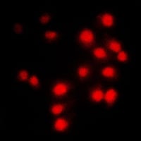
 IF (Immunofluorescence)
(Immunofluorescent analysis of NF-kappaB p65 staining in MCF7 cells. Formalin-fixed cells were permeabilized with 0.1% Triton X-100 in TBS for 5-10 minutes and blocked with 3% BSA-PBS for 30 minutes at room temperature. Cells were probed with the primary antibody in 3% BSA-PBS and incubated overnight at 4 °C in a humidified chamber. Cells were washed with PBST and incubated with a DyLight 594-conjugated secondary antibody (red) in PBS at room temperature in the dark.)
IF (Immunofluorescence)
(Immunofluorescent analysis of NF-kappaB p65 staining in MCF7 cells. Formalin-fixed cells were permeabilized with 0.1% Triton X-100 in TBS for 5-10 minutes and blocked with 3% BSA-PBS for 30 minutes at room temperature. Cells were probed with the primary antibody in 3% BSA-PBS and incubated overnight at 4 °C in a humidified chamber. Cells were washed with PBST and incubated with a DyLight 594-conjugated secondary antibody (red) in PBS at room temperature in the dark.)
NF-kappaB p65, Polyclonal Antibody (Cat# AAA104910)
 IHC (Immunohiostchemistry)
NBPF-pan, Polyclonal Antibody (Cat# AAA104955)
 IF (Immunofluorescence)
(Immunofluorescent analysis of SYNE3 staining in HepG2 cells. Formalin-fixed cells were permeabilized with 0.1% Triton X-100 in TBS for 5-10 minutes and blocked with 3% BSA-PBS for 30 minutes at room temperature. Cells were probed with the primary antibody in 3% BSA-PBS and incubated overnight at 4 °C in a humidified chamber. Cells were washed with PBST and incubated with a DyLight 594-conjugated secondary antibody (red) in PBS at room temperature in the dark.)
IF (Immunofluorescence)
(Immunofluorescent analysis of SYNE3 staining in HepG2 cells. Formalin-fixed cells were permeabilized with 0.1% Triton X-100 in TBS for 5-10 minutes and blocked with 3% BSA-PBS for 30 minutes at room temperature. Cells were probed with the primary antibody in 3% BSA-PBS and incubated overnight at 4 °C in a humidified chamber. Cells were washed with PBST and incubated with a DyLight 594-conjugated secondary antibody (red) in PBS at room temperature in the dark.)
SYNE3, Polyclonal Antibody (Cat# AAA104958)
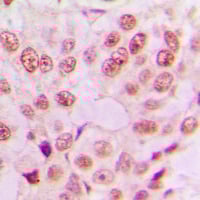
 IHC (Immunohiostchemistry)
(Immunohistochemical analysis of c-Jun (pY170) staining in human breast cancer formalin fixed paraffin embedded tissue section. The section was pre-treated using heat mediated antigen retrieval with sodium citrate buffer (pH 6.0). The section was then incubated with the antibody at room temperature and detected using an HRP conjugated compact polymer system. DAB was used as the chromogen. The section was then counterstained with haematoxylin and mounted with DPX.)
IHC (Immunohiostchemistry)
(Immunohistochemical analysis of c-Jun (pY170) staining in human breast cancer formalin fixed paraffin embedded tissue section. The section was pre-treated using heat mediated antigen retrieval with sodium citrate buffer (pH 6.0). The section was then incubated with the antibody at room temperature and detected using an HRP conjugated compact polymer system. DAB was used as the chromogen. The section was then counterstained with haematoxylin and mounted with DPX.)
c-Jun (pY170), Polyclonal Antibody (Cat# AAA104965)
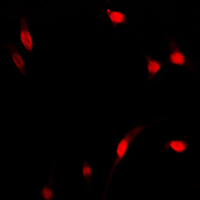
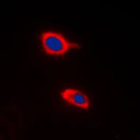
 IF (Immunofluorescence)
(Immunofluorescent analysis of COMT staining in Raw264.7 cells. Formalin-fixed cells were permeabilized with 0.1% Triton X-100 in TBS for 5-10 minutes and blocked with 3% BSA-PBS for 30 minutes at room temperature. Cells were probed with the primary antibody in 3% BSA-PBS and incubated overnight at 4 °C in a humidified chamber. Cells were washed with PBST and incubated with a DyLight 594-conjugated secondary antibody (red) in PBS at room temperature in the dark. DAPI was used to stain the cell nuclei (blue).)
IF (Immunofluorescence)
(Immunofluorescent analysis of COMT staining in Raw264.7 cells. Formalin-fixed cells were permeabilized with 0.1% Triton X-100 in TBS for 5-10 minutes and blocked with 3% BSA-PBS for 30 minutes at room temperature. Cells were probed with the primary antibody in 3% BSA-PBS and incubated overnight at 4 °C in a humidified chamber. Cells were washed with PBST and incubated with a DyLight 594-conjugated secondary antibody (red) in PBS at room temperature in the dark. DAPI was used to stain the cell nuclei (blue).)
COMT, Polyclonal Antibody (Cat# AAA104977)
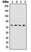
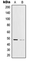
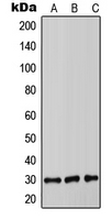
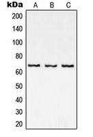
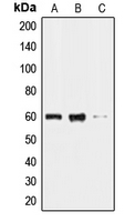
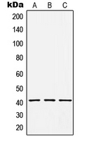
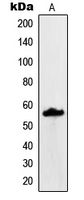
 WB (Western Blot)
(Western blot analysis of Carbonic Anhydrase 14 expression in CT26 (A), PC12 (B), MCF7 (C) whole cell lysates.)
WB (Western Blot)
(Western blot analysis of Carbonic Anhydrase 14 expression in CT26 (A), PC12 (B), MCF7 (C) whole cell lysates.)
Carbonic Anhydrase 14, Polyclonal Antibody (Cat# AAA104981)
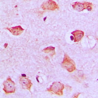
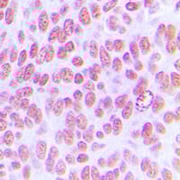
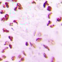
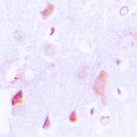
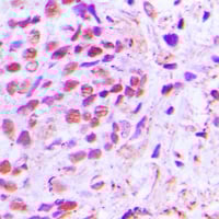 IHC (Immunohiostchemistry)
(Immunohistochemical analysis of NF-kappaB p65 staining in human breast cancer formalin fixed paraffin embedded tissue section. The section was pre-treated using heat mediated antigen retrieval with sodium citrate buffer (pH 6.0). The section was then incubated with the antibody at room temperature and detected using an HRP conjugated compact polymer system. DAB was used as the chromogen. The section was then counterstained with haematoxylin and mounted with DPX.)
IHC (Immunohiostchemistry)
(Immunohistochemical analysis of NF-kappaB p65 staining in human breast cancer formalin fixed paraffin embedded tissue section. The section was pre-treated using heat mediated antigen retrieval with sodium citrate buffer (pH 6.0). The section was then incubated with the antibody at room temperature and detected using an HRP conjugated compact polymer system. DAB was used as the chromogen. The section was then counterstained with haematoxylin and mounted with DPX.)
NF-kappaB p65, Polyclonal Antibody (Cat# AAA104738)
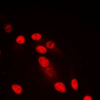
 IF (Immunofluorescence)
(Immunofluorescent analysis of ARRDC1 staining in MDAMB231 cells. Formalin-fixed cells were permeabilized with 0.1% Triton X-100 in TBS for 5-10 minutes and blocked with 3% BSA-PBS for 30 minutes at room temperature. Cells were probed with the primary antibody in 3% BSA-PBS and incubated overnight at 4 °C in a humidified chamber. Cells were washed with PBST and incubated with a DyLight 594-conjugated secondary antibody (red) in PBS at room temperature in the dark. DAPI was used to stain the cell nuclei (blue).)
IF (Immunofluorescence)
(Immunofluorescent analysis of ARRDC1 staining in MDAMB231 cells. Formalin-fixed cells were permeabilized with 0.1% Triton X-100 in TBS for 5-10 minutes and blocked with 3% BSA-PBS for 30 minutes at room temperature. Cells were probed with the primary antibody in 3% BSA-PBS and incubated overnight at 4 °C in a humidified chamber. Cells were washed with PBST and incubated with a DyLight 594-conjugated secondary antibody (red) in PBS at room temperature in the dark. DAPI was used to stain the cell nuclei (blue).)
ARRDC1, Polyclonal Antibody (Cat# AAA104741)
 IHC (Immunohiostchemistry)
(Immunohistochemical analysis of CLK2 staining in human lung cancer formalin fixed paraffin embedded tissue section. The section was pre-treated using heat mediated antigen retrieval with sodium citrate buffer (pH 6.0). The section was then incubated with the antibody at room temperature and detected using an HRP conjugated compact polymer system. DAB was used as the chromogen. The section was then counterstained with haematoxylin and mounted with DPX.)
IHC (Immunohiostchemistry)
(Immunohistochemical analysis of CLK2 staining in human lung cancer formalin fixed paraffin embedded tissue section. The section was pre-treated using heat mediated antigen retrieval with sodium citrate buffer (pH 6.0). The section was then incubated with the antibody at room temperature and detected using an HRP conjugated compact polymer system. DAB was used as the chromogen. The section was then counterstained with haematoxylin and mounted with DPX.)
CLK2, Polyclonal Antibody (Cat# AAA104773)

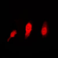 IF (Immunofluorescence)
(Immunofluorescent analysis of Histone Deacetylase 6 staining in HeLa cells. Formalin-fixed cells were permeabilized with 0.1% Triton X-100 in TBS for 5-10 minutes and blocked with 3% BSA-PBS for 30 minutes at room temperature. Cells were probed with the primary antibody in 3% BSA-PBS and incubated overnight at 4 °C in a humidified chamber. Cells were washed with PBST and incubated with a DyLight 594-conjugated secondary antibody (red) in PBS at room temperature in the dark.)
IF (Immunofluorescence)
(Immunofluorescent analysis of Histone Deacetylase 6 staining in HeLa cells. Formalin-fixed cells were permeabilized with 0.1% Triton X-100 in TBS for 5-10 minutes and blocked with 3% BSA-PBS for 30 minutes at room temperature. Cells were probed with the primary antibody in 3% BSA-PBS and incubated overnight at 4 °C in a humidified chamber. Cells were washed with PBST and incubated with a DyLight 594-conjugated secondary antibody (red) in PBS at room temperature in the dark.)
Histone Deacetylase 6, Polyclonal Antibody (Cat# AAA104774)
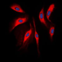
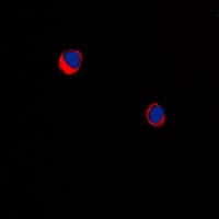
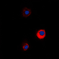
 IF (Immunofluorescence)
(Immunofluorescent analysis of HCCS staining in Raw264.7 cells. Formalin-fixed cells were permeabilized with 0.1% Triton X-100 in TBS for 5-10 minutes and blocked with 3% BSA-PBS for 30 minutes at room temperature. Cells were probed with the primary antibody in 3% BSA-PBS and incubated overnight at 4 °C in a humidified chamber. Cells were washed with PBST and incubated with a DyLight 594-conjugated secondary antibody (red) in PBS at room temperature in the dark. DAPI was used to stain the cell nuclei (blue).)
IF (Immunofluorescence)
(Immunofluorescent analysis of HCCS staining in Raw264.7 cells. Formalin-fixed cells were permeabilized with 0.1% Triton X-100 in TBS for 5-10 minutes and blocked with 3% BSA-PBS for 30 minutes at room temperature. Cells were probed with the primary antibody in 3% BSA-PBS and incubated overnight at 4 °C in a humidified chamber. Cells were washed with PBST and incubated with a DyLight 594-conjugated secondary antibody (red) in PBS at room temperature in the dark. DAPI was used to stain the cell nuclei (blue).)
HCCS, Polyclonal Antibody (Cat# AAA104792)
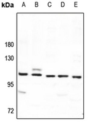
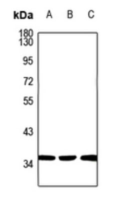
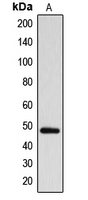
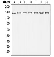
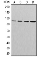
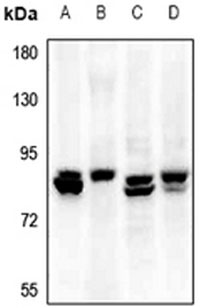 WB (Western Blot)
(Western blot analysis of Elastin expression in rat lung (A), mouse lung (B), A375 (C), A549 (D) whole cell lysates.)
WB (Western Blot)
(Western blot analysis of Elastin expression in rat lung (A), mouse lung (B), A375 (C), A549 (D) whole cell lysates.)
Elastin, Polyclonal Antibody (Cat# AAA104815)
 IF (Immunofluorescence)
(Immunofluorescent analysis of TBX15/18 staining in HepG2 cells. Formalin-fixed cells were permeabilized with 0.1% Triton X-100 in TBS for 5-10 minutes and blocked with 3% BSA-PBS for 30 minutes at room temperature. Cells were probed with the primary antibody in 3% BSA-PBS and incubated overnight at 4 °C in a humidified chamber. Cells were washed with PBST and incubated with a DyLight 594-conjugated secondary antibody (red) in PBS at room temperature in the dark.)
IF (Immunofluorescence)
(Immunofluorescent analysis of TBX15/18 staining in HepG2 cells. Formalin-fixed cells were permeabilized with 0.1% Triton X-100 in TBS for 5-10 minutes and blocked with 3% BSA-PBS for 30 minutes at room temperature. Cells were probed with the primary antibody in 3% BSA-PBS and incubated overnight at 4 °C in a humidified chamber. Cells were washed with PBST and incubated with a DyLight 594-conjugated secondary antibody (red) in PBS at room temperature in the dark.)
Cyclin A1/2, Polyclonal Antibody (Cat# AAA105252)
 IHC (Immunohiostchemistry)
(Immunohistochemical analysis of Cullin 3 staining in human tonsil formalin fixed paraffin embedded tissue section. The section was pre-treated using heat mediated antigen retrieval with sodium citrate buffer (pH 6.0). The section was then incubated with the antibody at room temperature and detected using an HRP conjugated compact polymer system. DAB was used as the chromogen. The section was then counterstained with haematoxylin and mounted with DPX.)
IHC (Immunohiostchemistry)
(Immunohistochemical analysis of Cullin 3 staining in human tonsil formalin fixed paraffin embedded tissue section. The section was pre-treated using heat mediated antigen retrieval with sodium citrate buffer (pH 6.0). The section was then incubated with the antibody at room temperature and detected using an HRP conjugated compact polymer system. DAB was used as the chromogen. The section was then counterstained with haematoxylin and mounted with DPX.)
Cullin 3, Polyclonal Antibody (Cat# AAA105268)
 IF (Immunofluorescence)
(Immunofluorescent analysis of MUC13 staining in KNRK cells. Formalin-fixed cells were permeabilized with 0.1% Triton X-100 in TBS for 5-10 minutes and blocked with 3% BSA-PBS for 30 minutes at room temperature. Cells were probed with the primary antibody in 3% BSA-PBS and incubated overnight at 4 °C in a humidified chamber. Cells were washed with PBST and incubated with a DyLight 594-conjugated secondary antibody (red) in PBS at room temperature in the dark. DAPI was used to stain the cell nuclei (blue).)
IF (Immunofluorescence)
(Immunofluorescent analysis of MUC13 staining in KNRK cells. Formalin-fixed cells were permeabilized with 0.1% Triton X-100 in TBS for 5-10 minutes and blocked with 3% BSA-PBS for 30 minutes at room temperature. Cells were probed with the primary antibody in 3% BSA-PBS and incubated overnight at 4 °C in a humidified chamber. Cells were washed with PBST and incubated with a DyLight 594-conjugated secondary antibody (red) in PBS at room temperature in the dark. DAPI was used to stain the cell nuclei (blue).)
MUC13, Polyclonal Antibody (Cat# AAA105284)
 IHC (Immunohiostchemistry)
(Immunohistochemical analysis of Cytochrome P450 39A1 staining in human lung cancer formalin fixed paraffin embedded tissue section. The section was pre-treated using heat mediated antigen retrieval with sodium citrate buffer (pH 6.0). The section was then incubated with the antibody at room temperature and detected using an HRP conjugated compact polymer system. DAB was used as the chromogen. The section was then counterstained with haematoxylin and mounted with DPX.)
IHC (Immunohiostchemistry)
(Immunohistochemical analysis of Cytochrome P450 39A1 staining in human lung cancer formalin fixed paraffin embedded tissue section. The section was pre-treated using heat mediated antigen retrieval with sodium citrate buffer (pH 6.0). The section was then incubated with the antibody at room temperature and detected using an HRP conjugated compact polymer system. DAB was used as the chromogen. The section was then counterstained with haematoxylin and mounted with DPX.)
Cytochrome P450 39A1, Polyclonal Antibody (Cat# AAA105330)
 IHC (Immunohiostchemistry)
(Immunohistochemical analysis of RFX3 staining in human breast cancer formalin fixed paraffin embedded tissue section. The section was pre-treated using heat mediated antigen retrieval with sodium citrate buffer (pH 6.0). The section was then incubated with the antibody at room temperature and detected using an HRP conjugated compact polymer system. DAB was used as the chromogen. The section was then counterstained with haematoxylin and mounted with DPX.)
IHC (Immunohiostchemistry)
(Immunohistochemical analysis of RFX3 staining in human breast cancer formalin fixed paraffin embedded tissue section. The section was pre-treated using heat mediated antigen retrieval with sodium citrate buffer (pH 6.0). The section was then incubated with the antibody at room temperature and detected using an HRP conjugated compact polymer system. DAB was used as the chromogen. The section was then counterstained with haematoxylin and mounted with DPX.)
RFX3, Polyclonal Antibody (Cat# AAA105333)
What are Polyclonal Antibodies?
Polyclonal antibodies are antibodies that come from multiple B cell clones of a host animal. The typical hosts used for the majority of polyclonal antibody production are rabbits, goats, sheep, and donkeys. These polyclonal antibodies, once having identified their target, will bind to different epitopes located at different regions or sequences on the same protein/antigen. As a result, they are ideal at locating and binding to the target, even if the target is in very low concentrations (due to many different antibodies being able to bind to the same target molecule, which allows for significant amplification of a downstream signal).
Polyclonal antibodies are typically produced by injecting an antigen into a host animal, which causes the animal’s immune system to attack the foreign antigen by mass generating antibodies against it. After a period of time, serum is collected from the animal and purified using physicochemical fractionation, class-specific affinity purification, and/or antigen-affinity purification.
Key Uses of Polyclonal Antibodies
- Western Blotting: This method is used to find specific proteins in biological samples after separating them by size.
- Immunohistochemistry: IHC helps visualize the location of proteins in tissue sections using various staining techniques.
- ELISA: (Enzyme-Linked Immunosorbent Assay) is typically used to identify specific protein quantities in a sample. ELISAs can be either “Quantitative” or “Qualitative”.
- Flow Cytometry: technique that identifies and measures the specific protein on the surface or inside the cells in a fluid suspension.
- Immunoprecipitation: IP isolates and studies a specific protein from a complex mixture using antibodies.
Why Buy Polyclonal Antibodies from AAA Biotech?
1. Ideal for Various Applications
Our antibodies are generally going to be validated for use in multiple types of assays, including ELISA, Western Blotting, Immunohistochemistry, Immunoprecipitation, amongst others. They are ideal for a wide range of research applications.
2. Rigorous Quality Control
All of the antibodies in our catalog undergo strict quality testing to ensure specificity, sensitivity, and consistent performance. We are confident in the ability of our antibodies to provide you with accurate results.
3. Wide Assortment of Antibodies
Antibodies in are catalog can be found for both common and exotic species, and these antibodies are also available in both conjugated and recombinant forms to suit many diverse experimental needs.
4. Highly Purified
Our antibodies are available in purified forms with over 85% purity, as confirmed by SDS-PAGE. They are also available with tags such as His, Flag, GST, or MBP. We cater to customers worldwide.
FAQ
1. How are polyclonal antibodies produced?
Traditionally, polyclonal antibodies are produced by injecting an antigen into a host animal (such as a rabbit or goat), which then triggers an immune response from the host animal. The animal’s B cells produce antibodies that will recognize different parts of the injected antigen. These antibodies are then collected from the animal’s blood and purified for use.
2. How do polyclonal antibodies differ from monoclonal antibodies?
Polyclonal antibodies are a mix of antibodies that bind to different locations (epitopes) of the same antigen, while monoclonal antibodies are identical and bind to just one specific epitope. This makes polyclonal antibodies more versatile and better at detecting proteins that may be present in low quantities or in altered/modified forms.
3. How should I store polyclonal antibodies?
Polyclonal antibodies should be stored at 4°C for short-term use (up to a few weeks) and at -20°C or -80°C for long-term storage. Avoid repeated freeze-thaw cycles by dividing them into small aliquots. Always check the datasheet for specific storage instructions.