Filters
▼Clonality
▼Type
▼Reactivity
▼Gene Name
▼Isotype
▼Host
▼Application
▼Clone
▼Polyclonal Antibodies
At AAA Biotech also known as AAA Bio or AAABio, we provide a broad range of purified polyclonal antibodies (pAbs) that are able to all be browsed online through our website. Due to their high specificity and strong binding affinity, these antibodies are ideal for wide swathes of research and experimental applications.
Our polyclonal antibodies can easily support your work, whether you use them for Western Blotting, Immunocytochemistry (with or without Immunofluorescence used in conjunction), Immunohistochemistry, Immunoprecipitation, and ELISA tests. We highly encourage you to browse our range of pAbs and choose the one that best suits your experimental model.
Viewing 3050-3100 of 96805 product results
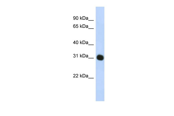
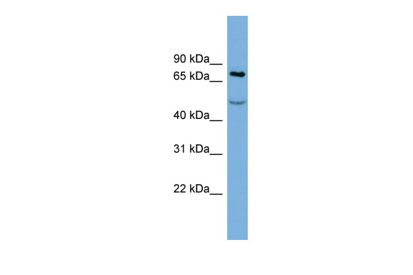
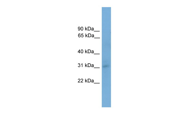
 WB (Western Blot)
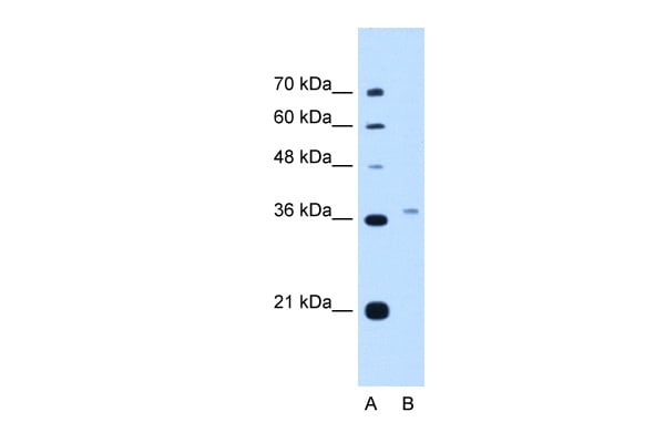
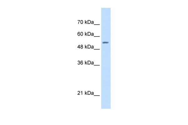
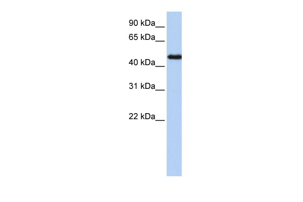
(WB Suggested Anti-PEX3 Antibody Titration: 5.0ug/mlPositive Control: HepG2 cell lysateThere is BioGPS gene expression data showing that PEX3 is expressed in HepG2)
WB (Western Blot)
(WB Suggested Anti-PEX3 Antibody Titration: 5.0ug/mlPositive Control: HepG2 cell lysateThere is BioGPS gene expression data showing that PEX3 is expressed in HepG2)
PEX3, Polyclonal Antibody (Cat# AAA199656)
 WB (Western Blot)
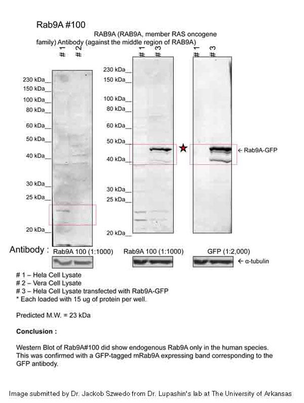
(WB Suggested Anti-RAB9A Antibody Titration: 0.2-1 ug/mlELISA Titer: 1:62500Positive Control: Jurkat cell lysate)
WB (Western Blot)
(WB Suggested Anti-RAB9A Antibody Titration: 0.2-1 ug/mlELISA Titer: 1:62500Positive Control: Jurkat cell lysate)
RAB9A, Polyclonal Antibody (Cat# AAA199660)
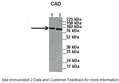
 WB (Western Blot)
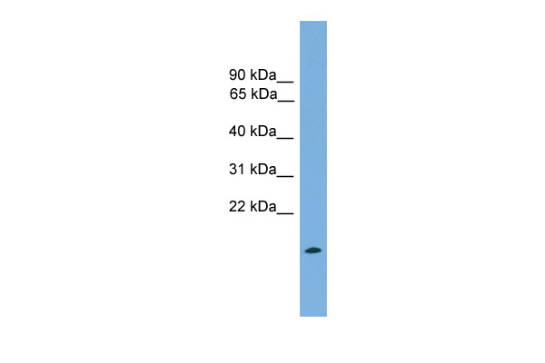
(WB Suggested Anti-CAD Antibody Titration: 0.2-1 ug/mlELISA Titer: 1:62500Positive Control: 293T cell lysate)
WB (Western Blot)
(WB Suggested Anti-CAD Antibody Titration: 0.2-1 ug/mlELISA Titer: 1:62500Positive Control: 293T cell lysate)
CAD, Polyclonal Antibody (Cat# AAA199661)
 WB (Western Blot)
(WB Suggested Anti-DFNA5 Antibody Titration: 1.25 ug/mlPositive Control: HepG2 cell lysateDFNA5 is supported by BioGPS gene expression data to be expressed in HepG2)
WB (Western Blot)
(WB Suggested Anti-DFNA5 Antibody Titration: 1.25 ug/mlPositive Control: HepG2 cell lysateDFNA5 is supported by BioGPS gene expression data to be expressed in HepG2)
GSDME, Polyclonal Antibody (Cat# AAA199662)
 WB (Western Blot)
(NOLC1 antibody - C-terminal region validated by WB using HepG2 cell lysate at 2.5ug/ml.NOLC1 is strongly supported by BioGPS gene expression data to be expressed in Human HepG2 cells)
WB (Western Blot)
(NOLC1 antibody - C-terminal region validated by WB using HepG2 cell lysate at 2.5ug/ml.NOLC1 is strongly supported by BioGPS gene expression data to be expressed in Human HepG2 cells)
NOLC1, Polyclonal Antibody (Cat# AAA199664)
 WB (Western Blot)
(WB Suggested Anti-SHMT2 Antibody Titration: 0.5ug/mlPositive Control: HepG2 cell lysate)
WB (Western Blot)
(WB Suggested Anti-SHMT2 Antibody Titration: 0.5ug/mlPositive Control: HepG2 cell lysate)
SHMT2, Polyclonal Antibody (Cat# AAA199671)
 WB (Western Blot)
(WB Suggested Anti-LMNB1 Antibody Titration: 0.2-1 ug/mlELISA Titer: 1:1562500Positive Control: SK-MEL-2 cell lysateLMNB1 is supported by BioGPS gene expression data to be expressed in SKMEL2)
WB (Western Blot)
(WB Suggested Anti-LMNB1 Antibody Titration: 0.2-1 ug/mlELISA Titer: 1:1562500Positive Control: SK-MEL-2 cell lysateLMNB1 is supported by BioGPS gene expression data to be expressed in SKMEL2)
LMNB1, Polyclonal Antibody (Cat# AAA199672)
 WB (Western Blot)
(WB Suggested Anti-PSME3 Antibody Titration: 1 ug/mlPositive Control: HepG2 cell lysatePSME3 is strongly supported by BioGPS gene expression data to be expressed in Human HepG2 cells)
WB (Western Blot)
(WB Suggested Anti-PSME3 Antibody Titration: 1 ug/mlPositive Control: HepG2 cell lysatePSME3 is strongly supported by BioGPS gene expression data to be expressed in Human HepG2 cells)
PSME3, Polyclonal Antibody (Cat# AAA199674)
 WB (Western Blot)
(WB Suggested Anti-MAT2A Antibody Titration: 0.2-1 ug/mlELISA Titer: 1:312500Positive Control: Hela cell lysateMAT2A is strongly supported by BioGPS gene expression data to be expressed in Human HeLa cells)
WB (Western Blot)
(WB Suggested Anti-MAT2A Antibody Titration: 0.2-1 ug/mlELISA Titer: 1:312500Positive Control: Hela cell lysateMAT2A is strongly supported by BioGPS gene expression data to be expressed in Human HeLa cells)
MAT2A, Polyclonal Antibody (Cat# AAA199675)
 WB (Western Blot)
(WB Suggested Anti-IFI44L Antibody Titration: 5.0ug/mlPositive Control: Jurkat cell lysate)
WB (Western Blot)
(WB Suggested Anti-IFI44L Antibody Titration: 5.0ug/mlPositive Control: Jurkat cell lysate)
IFI44L, Polyclonal Antibody (Cat# AAA199679)
 WB (Western Blot)
(WB Suggested Anti-RFC5 Antibody Titration: 1 ug/mlPositive Control: Jurkat cell lysateRFC5 is strongly supported by BioGPS gene expression data to be expressed in Human Jurkat cells)
WB (Western Blot)
(WB Suggested Anti-RFC5 Antibody Titration: 1 ug/mlPositive Control: Jurkat cell lysateRFC5 is strongly supported by BioGPS gene expression data to be expressed in Human Jurkat cells)
RFC5, Polyclonal Antibody (Cat# AAA199680)
 WB (Western Blot)
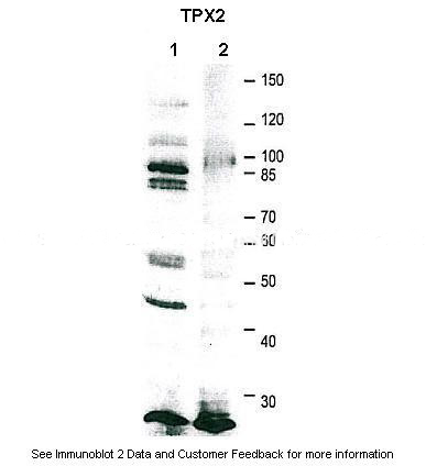
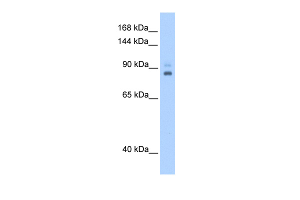
(WB Suggested Anti-TPX2 Antibody Titration: 0.2-1 ug/mlPositive Control: 721_B cell lysateTPX2 is supported by BioGPS gene expression data to be expressed in 721_B)
WB (Western Blot)
(WB Suggested Anti-TPX2 Antibody Titration: 0.2-1 ug/mlPositive Control: 721_B cell lysateTPX2 is supported by BioGPS gene expression data to be expressed in 721_B)
TPX2, Polyclonal Antibody (Cat# AAA199681)
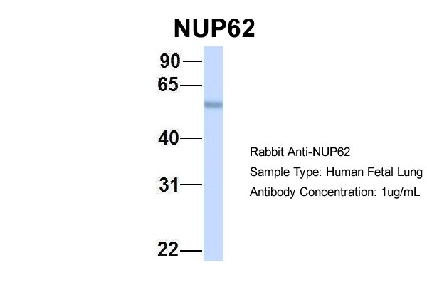
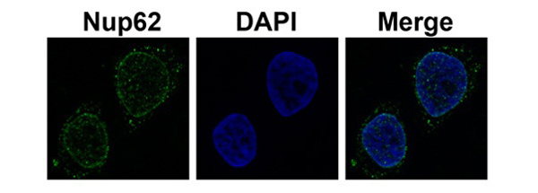
 WB (Western Blot)
(WB Suggested Anti-NUP62 Antibody Titration: 0.2-1 ug/mlELISA Titer: 1:312500Positive Control: Human brain)
WB (Western Blot)
(WB Suggested Anti-NUP62 Antibody Titration: 0.2-1 ug/mlELISA Titer: 1:312500Positive Control: Human brain)
NUP62, Polyclonal Antibody (Cat# AAA199682)
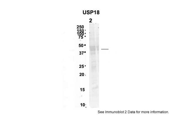
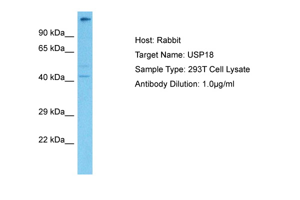
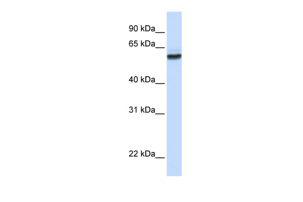
 WB (Western Blot)
(WB Suggested Anti-USP18 Antibody Titration: 0.2-1 ug/mlELISA Titer: 1:62500Positive Control: Hela cell lysate)
WB (Western Blot)
(WB Suggested Anti-USP18 Antibody Titration: 0.2-1 ug/mlELISA Titer: 1:62500Positive Control: Hela cell lysate)
USP18, Polyclonal Antibody (Cat# AAA199686)
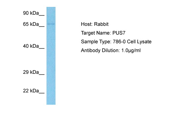
 WB (Western Blot)
(WB Suggested Anti-PUS7 Antibody Titration: 1.25ug/mlPositive Control: Transfected 293T)
WB (Western Blot)
(WB Suggested Anti-PUS7 Antibody Titration: 1.25ug/mlPositive Control: Transfected 293T)
PUS7, Polyclonal Antibody (Cat# AAA199687)
 WB (Western Blot)
(WB Suggested Anti-XTP3TPA Antibody Titration: 0.5ug/mlPositive Control: Transfected 293T)
WB (Western Blot)
(WB Suggested Anti-XTP3TPA Antibody Titration: 0.5ug/mlPositive Control: Transfected 293T)
XTP3TPA, Polyclonal Antibody (Cat# AAA199692)
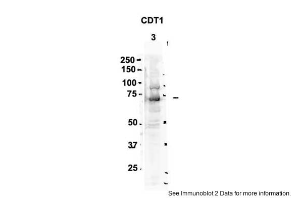
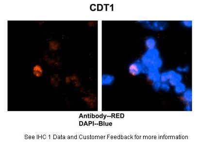
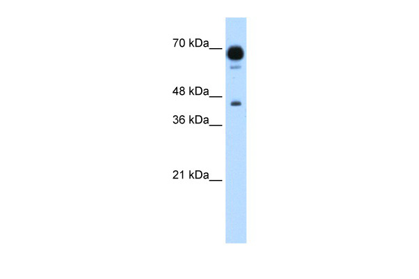
 WB (Western Blot)
(WB Suggested Anti-CDT1 Antibody Titration: 0.2-1 ug/mlPositive Control: HepG2 cell lysate)
WB (Western Blot)
(WB Suggested Anti-CDT1 Antibody Titration: 0.2-1 ug/mlPositive Control: HepG2 cell lysate)
CDT1, Polyclonal Antibody (Cat# AAA199694)
 WB (Western Blot)
(WB Suggested Anti-ST3GAL5 Antibody Titration: 1.25ug/mlPositive Control: Jurkat cell lysate)
WB (Western Blot)
(WB Suggested Anti-ST3GAL5 Antibody Titration: 1.25ug/mlPositive Control: Jurkat cell lysate)
ST3GAL5, Polyclonal Antibody (Cat# AAA199698)
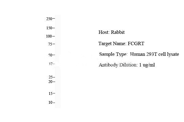
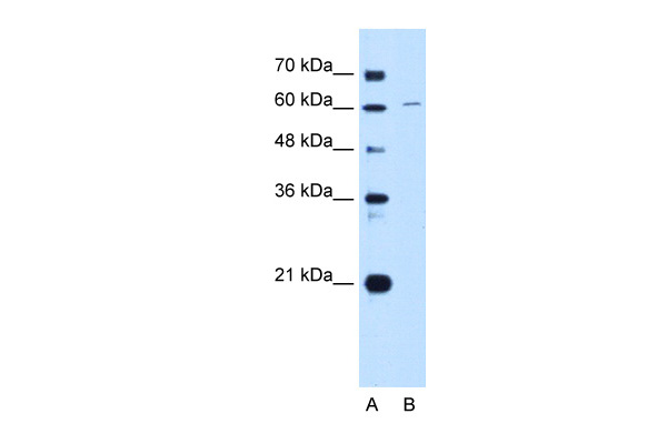
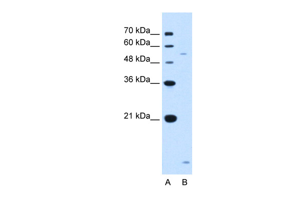
 WB (Western Blot)
(WB Suggested Anti-FCGRT Antibody Titration: 0.2-1 ug/mlPositive Control: HepG2 cell lysateFCGRT is supported by BioGPS gene expression data to be expressed in HepG2)
WB (Western Blot)
(WB Suggested Anti-FCGRT Antibody Titration: 0.2-1 ug/mlPositive Control: HepG2 cell lysateFCGRT is supported by BioGPS gene expression data to be expressed in HepG2)
FCGRT, Polyclonal Antibody (Cat# AAA199700)
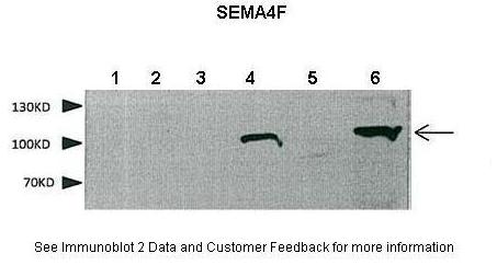
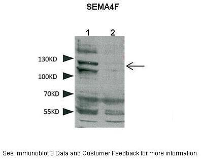
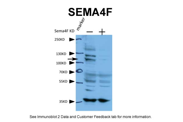
 WB (Western Blot)
(WB Suggested Anti-SEMA4F Antibody Titration: 0.2-1 ug/mlELISA Titer: 1:312500Positive Control: Transfected 293T)
WB (Western Blot)
(WB Suggested Anti-SEMA4F Antibody Titration: 0.2-1 ug/mlELISA Titer: 1:312500Positive Control: Transfected 293T)
SEMA4F, Polyclonal Antibody (Cat# AAA199703)
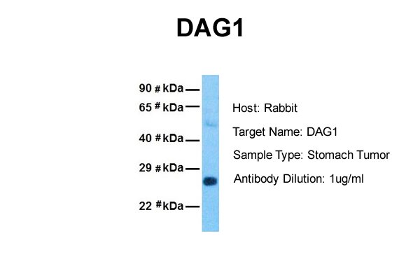
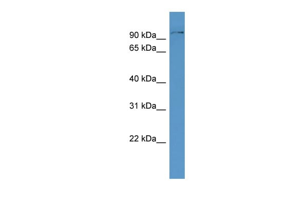
 WB (Western Blot)
(WB Suggested Anti-Alpha-DAG1 Antibody Titration: 0.2-1 ug/mlELISA Titer: 1:312500Positive Control: Human Stomach)
WB (Western Blot)
(WB Suggested Anti-Alpha-DAG1 Antibody Titration: 0.2-1 ug/mlELISA Titer: 1:312500Positive Control: Human Stomach)
DAG1, Polyclonal Antibody (Cat# AAA199704)
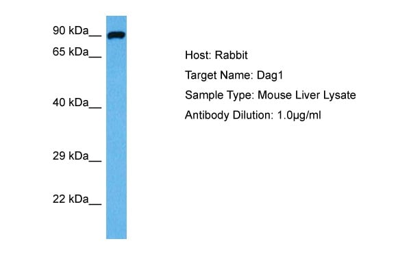
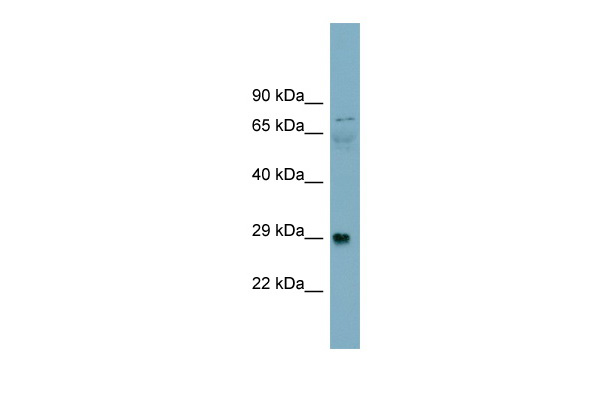
 WB (Western Blot)
(WB Suggested Anti-Dag1 Antibody Titration: 0.2-1 ug/mlELISA Titer: 1:12500Positive Control: Mouse Heart)
WB (Western Blot)
(WB Suggested Anti-Dag1 Antibody Titration: 0.2-1 ug/mlELISA Titer: 1:12500Positive Control: Mouse Heart)
Dag1, Polyclonal Antibody (Cat# AAA199705)

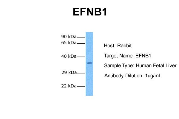
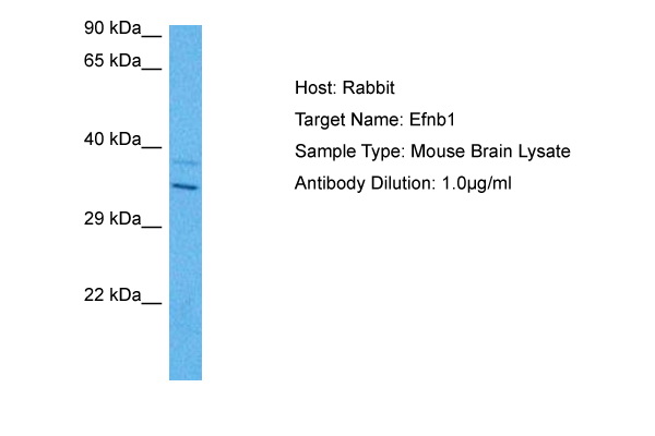
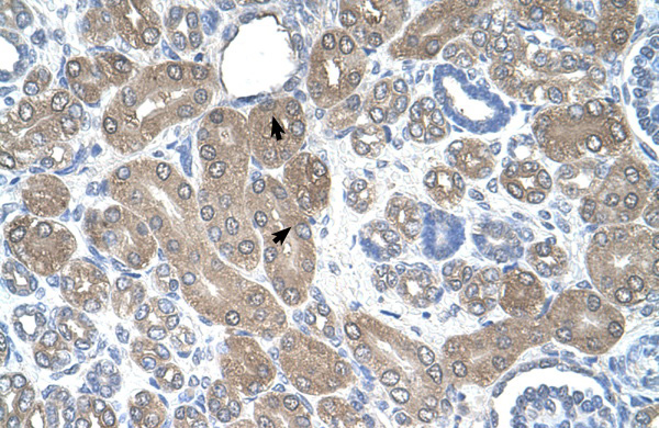
 WB (Western Blot)
(WB Suggested Anti-EFNB1 Antibody Titration: 0.2-1 ug/mlPositive Control: Human kidney)
WB (Western Blot)
(WB Suggested Anti-EFNB1 Antibody Titration: 0.2-1 ug/mlPositive Control: Human kidney)
EFNB1, Polyclonal Antibody (Cat# AAA199707)
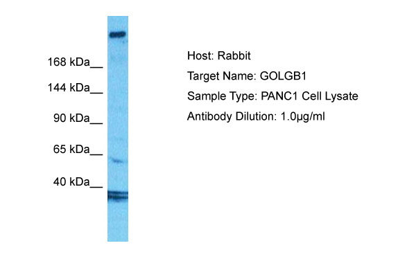
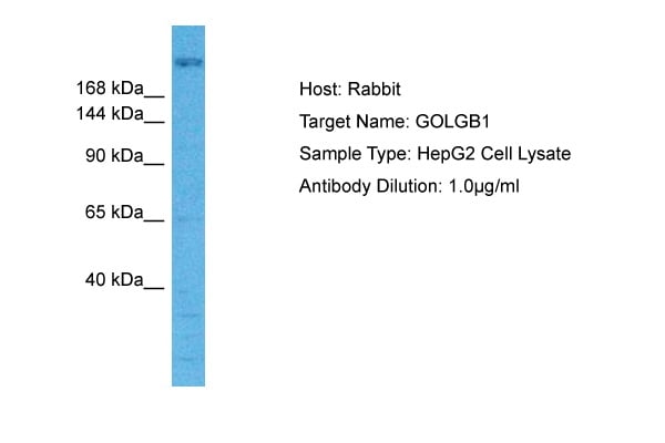
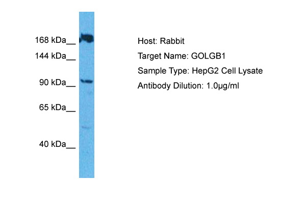
 WB (Western Blot)
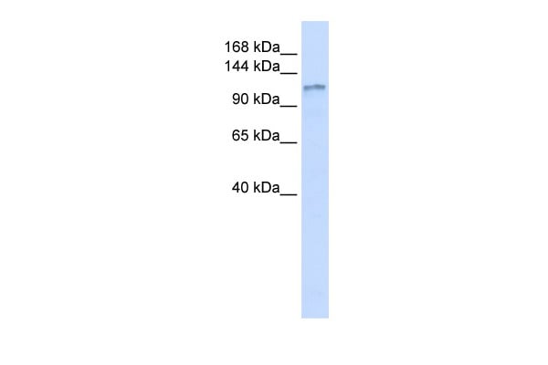
(WB Suggested Anti-GOLGB1 Antibody Titration: 0.2-1 ug/mlELISA Titer: 1:12500Positive Control: 721_B cell lysateGOLGB1 is supported by BioGPS gene expression data to be expressed in 721_B)
WB (Western Blot)
(WB Suggested Anti-GOLGB1 Antibody Titration: 0.2-1 ug/mlELISA Titer: 1:12500Positive Control: 721_B cell lysateGOLGB1 is supported by BioGPS gene expression data to be expressed in 721_B)
GOLGB1, Polyclonal Antibody (Cat# AAA199709)
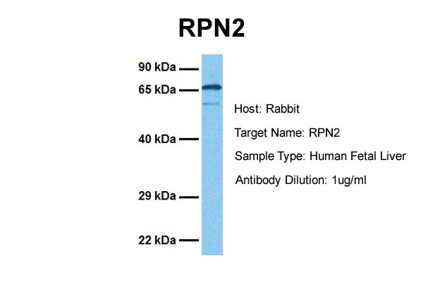
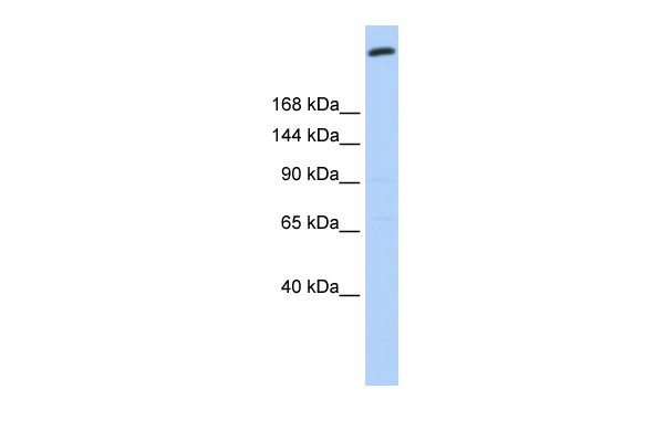
 WB (Western Blot)
(WB Suggested Anti-RPN2 Antibody Titration: 0.25ug/mlPositive Control: HepG2 cell lysateRPN2 is strongly supported by BioGPS gene expression data to be expressed in Human HepG2 cells)
WB (Western Blot)
(WB Suggested Anti-RPN2 Antibody Titration: 0.25ug/mlPositive Control: HepG2 cell lysateRPN2 is strongly supported by BioGPS gene expression data to be expressed in Human HepG2 cells)
RPN2, Polyclonal Antibody (Cat# AAA199547)
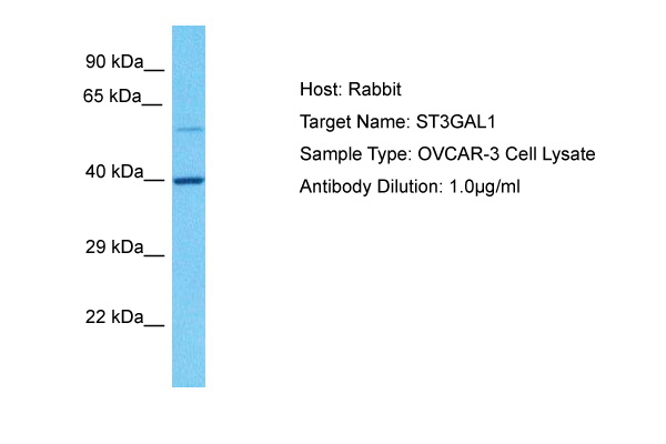
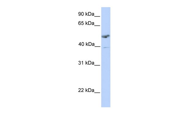 WB (Western Blot)
(WB Suggested Anti-ST3GAL1 Antibody Titration: 0.2-1 ug/mlPositive Control: Human Placenta)
WB (Western Blot)
(WB Suggested Anti-ST3GAL1 Antibody Titration: 0.2-1 ug/mlPositive Control: Human Placenta)
ST3GAL1, Polyclonal Antibody (Cat# AAA199548)
 WB (Western Blot)
(WB Suggested Anti-SSR2 Antibody Titration: 1.25ug/mlPositive Control: Jurkat cell lysate)
WB (Western Blot)
(WB Suggested Anti-SSR2 Antibody Titration: 1.25ug/mlPositive Control: Jurkat cell lysate)
SSR2, Polyclonal Antibody (Cat# AAA199550)
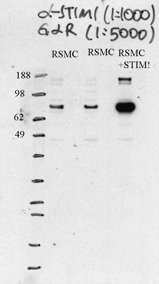
 WB (Western Blot)
(WB Suggested Anti-STIM1 Antibody Titration: 0.2-1 ug/mlELISA Titer: 1:12500Positive Control: PANC1 cell lysateSTIM1 is supported by BioGPS gene expression data to be expressed in PANC1)
WB (Western Blot)
(WB Suggested Anti-STIM1 Antibody Titration: 0.2-1 ug/mlELISA Titer: 1:12500Positive Control: PANC1 cell lysateSTIM1 is supported by BioGPS gene expression data to be expressed in PANC1)
STIM1, Polyclonal Antibody (Cat# AAA199551)
 WB (Western Blot)
(WB Suggested Anti-TAPBP Antibody Titration: 0.2-1 ug/mlPositive Control: 293T cell lysate)
WB (Western Blot)
(WB Suggested Anti-TAPBP Antibody Titration: 0.2-1 ug/mlPositive Control: 293T cell lysate)
TAPBP, Polyclonal Antibody (Cat# AAA199552)
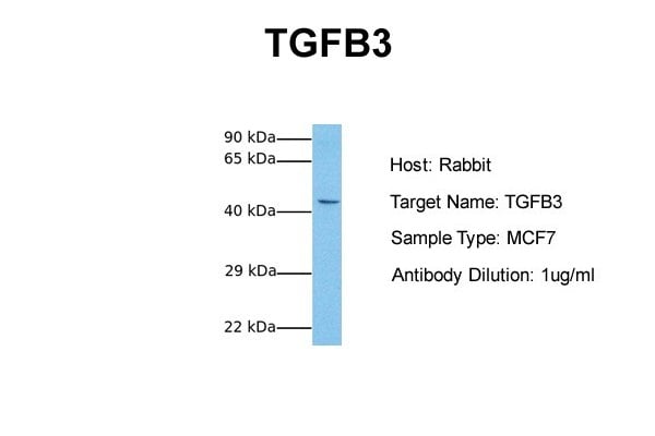
 WB (Western Blot)
(WB Suggested Anti-TGFB3 Antibody Titration: 0.2-1 ug/mlELISA Titer: 1:12500Positive Control: SH-SYSY cell lysateTGFB3 is supported by BioGPS gene expression data to be expressed in SHSY5Y)
WB (Western Blot)
(WB Suggested Anti-TGFB3 Antibody Titration: 0.2-1 ug/mlELISA Titer: 1:12500Positive Control: SH-SYSY cell lysateTGFB3 is supported by BioGPS gene expression data to be expressed in SHSY5Y)
TGFB3, Polyclonal Antibody (Cat# AAA199559)
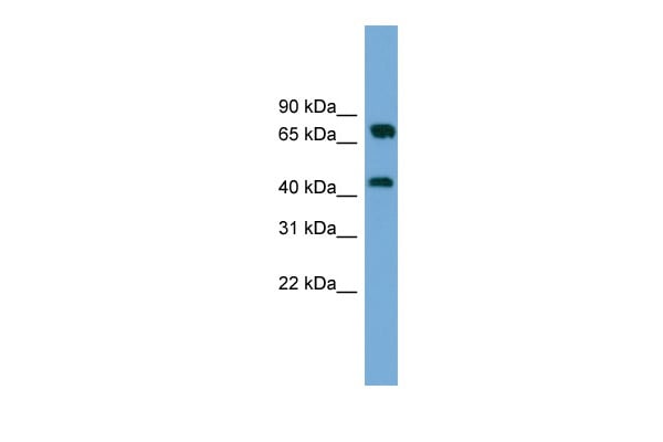
 WB (Western Blot)
(Host: RabbitTarget Name: NR4A1Sample Type: Liver Tumor lysatesAntibody Dilution: 1.0ug/ml)
WB (Western Blot)
(Host: RabbitTarget Name: NR4A1Sample Type: Liver Tumor lysatesAntibody Dilution: 1.0ug/ml)
NR4A1, Polyclonal Antibody (Cat# AAA199575)
Predicted Species Reactivity: Human, Mouse, Rat, Cow, Dog, Guinea Pig, Horse, Rabbit, Zebrafish
 WB (Western Blot)
(WB Suggested Anti-NR2F6 Antibody Titration: 0.2-1 ug/mlPositive Control: HepG2 cell lysateNR2F6 is strongly supported by BioGPS gene expression data to be expressed in Human HepG2 cells)
WB (Western Blot)
(WB Suggested Anti-NR2F6 Antibody Titration: 0.2-1 ug/mlPositive Control: HepG2 cell lysateNR2F6 is strongly supported by BioGPS gene expression data to be expressed in Human HepG2 cells)
NR2F6, Polyclonal Antibody (Cat# AAA199582)
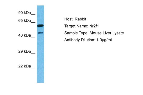
 WB (Western Blot)
(WB Suggested Anti-NR2F1 Antibody Titration: 0.2-1 ug/mlELISA Titer: 1:62500Positive Control: Transfected 293T)
WB (Western Blot)
(WB Suggested Anti-NR2F1 Antibody Titration: 0.2-1 ug/mlELISA Titer: 1:62500Positive Control: Transfected 293T)
NR2F1, Polyclonal Antibody (Cat# AAA199583)
 WB (Western Blot)
(WB Suggested Anti-NR0B2 Antibody Titration: 0.2-1 ug/mlELISA Titer: 1:312500Positive Control: Transfected 293T)
WB (Western Blot)
(WB Suggested Anti-NR0B2 Antibody Titration: 0.2-1 ug/mlELISA Titer: 1:312500Positive Control: Transfected 293T)
NR0B2, Polyclonal Antibody (Cat# AAA199584)
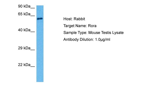
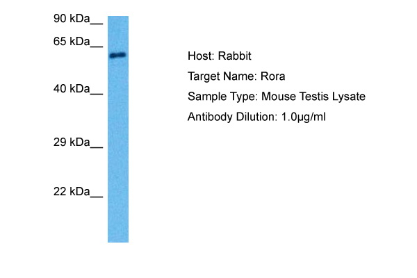
 WB (Western Blot)
(Host: RabbitTarget Name: RoraSample Type: Mouse Lung lysatesAntibody Dilution: 1.0ug/ml)
WB (Western Blot)
(Host: RabbitTarget Name: RoraSample Type: Mouse Lung lysatesAntibody Dilution: 1.0ug/ml)
Rora, Polyclonal Antibody (Cat# AAA199586)
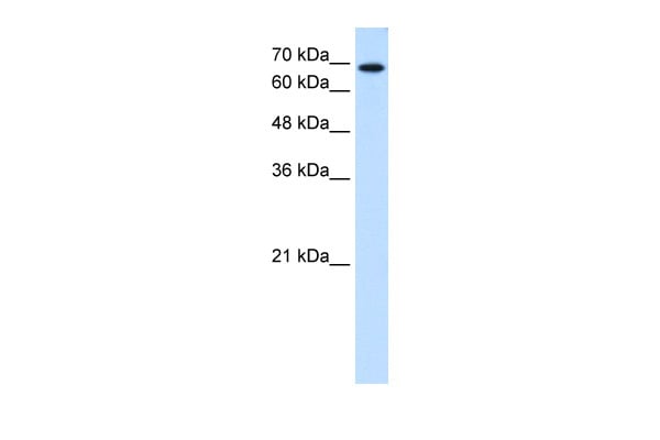
 WB (Western Blot)
(WB Suggested Anti-MBL2 Antibody Titration: 0.2-1 ug/mlPositive Control: Hela cell lysate)
WB (Western Blot)
(WB Suggested Anti-MBL2 Antibody Titration: 0.2-1 ug/mlPositive Control: Hela cell lysate)
MBL2, Polyclonal Antibody (Cat# AAA199591)
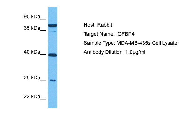
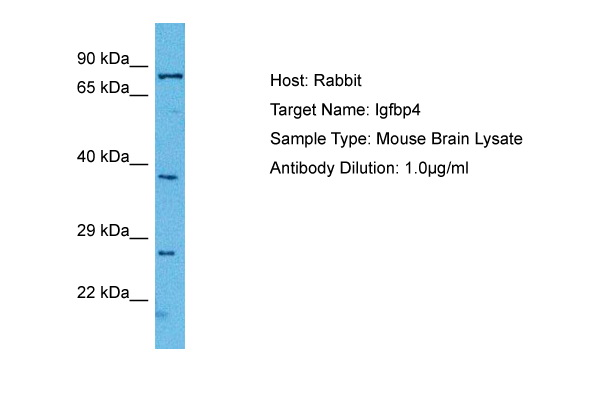
 WB (Western Blot)
(WB Suggested Anti-IGFBP4 Antibody Titration: 1 ug/mlPositive Control: Fetal Brain Lysate)
WB (Western Blot)
(WB Suggested Anti-IGFBP4 Antibody Titration: 1 ug/mlPositive Control: Fetal Brain Lysate)
IGFBP4, Polyclonal Antibody (Cat# AAA199593)
 WB (Western Blot)
(WB Suggested Anti-BAAT Antibody Titration: 0.2-1 ug/mlELISA Titer: 1:62500Positive Control: Human Liver)
WB (Western Blot)
(WB Suggested Anti-BAAT Antibody Titration: 0.2-1 ug/mlELISA Titer: 1:62500Positive Control: Human Liver)
BAAT, Polyclonal Antibody (Cat# AAA199594)
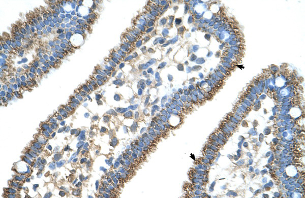
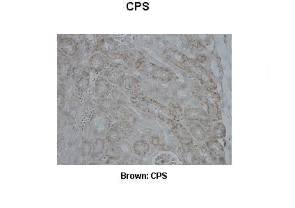
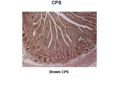
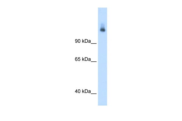 WB (Western Blot)
(WB Suggested Anti-CPS1 Antibody Titration: 1.25 ug/mlPositive Control: Fetal liver cell lysate)
WB (Western Blot)
(WB Suggested Anti-CPS1 Antibody Titration: 1.25 ug/mlPositive Control: Fetal liver cell lysate)
CPS1, Polyclonal Antibody (Cat# AAA199595)
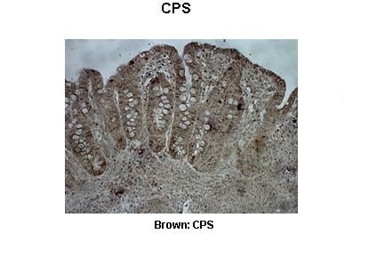
 WB (Western Blot)
(WB Suggested Anti-CPS1 Antibody Titration: 5.0ug/mlPositive Control: Fetal liver cell lysate)
WB (Western Blot)
(WB Suggested Anti-CPS1 Antibody Titration: 5.0ug/mlPositive Control: Fetal liver cell lysate)
CPS1, Polyclonal Antibody (Cat# AAA199596)
 WB (Western Blot)
(WB Suggested Anti-GPT Antibody Titration: 5.0ug/mlPositive Control: Fetal liver cell lysate)
WB (Western Blot)
(WB Suggested Anti-GPT Antibody Titration: 5.0ug/mlPositive Control: Fetal liver cell lysate)
GPT, Polyclonal Antibody (Cat# AAA199600)
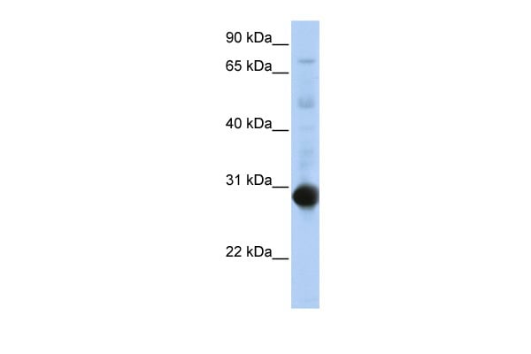
 WB (Western Blot)
(WB Suggested Antibody Titration: 2.5ug/mlPositive Control: Jurkat)
WB (Western Blot)
(WB Suggested Antibody Titration: 2.5ug/mlPositive Control: Jurkat)
GPT, Polyclonal Antibody (Cat# AAA199601)


 WB (Western Blot)
(WB Suggested Anti-MST1 Antibody Titration: 0.2-1 ug/mlELISA Titer: 1:312500Positive Control: Human Lung)
WB (Western Blot)
(WB Suggested Anti-MST1 Antibody Titration: 0.2-1 ug/mlELISA Titer: 1:312500Positive Control: Human Lung)
MST1, Polyclonal Antibody (Cat# AAA199608)
 WB (Western Blot)
(WB Suggested Anti-SGPP2 Antibody Titration: 2.5ug/mlPositive Control: HepG2 cell lysate)
WB (Western Blot)
(WB Suggested Anti-SGPP2 Antibody Titration: 2.5ug/mlPositive Control: HepG2 cell lysate)
SGPP2, Polyclonal Antibody (Cat# AAA199610)
Predicted Species Reactivity: Human, Mouse, Rat, Cow, Guinea Pig, Horse, Rabbit
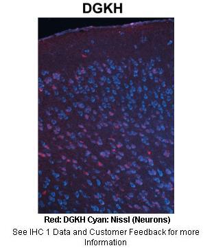
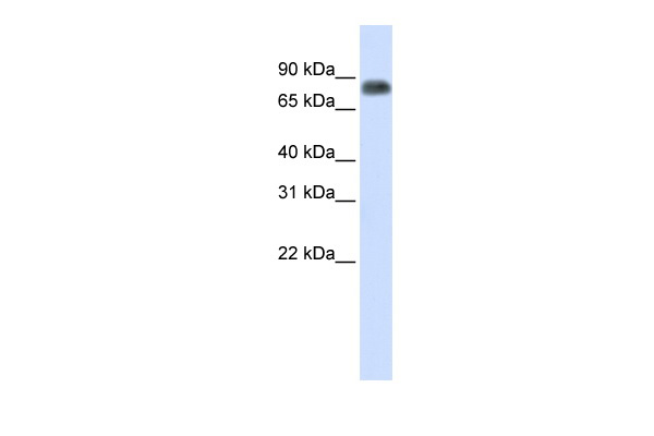
 WB (Western Blot)
(WB Suggested Anti-DGKH Antibody Titration: 0.2-1 ug/mlELISA Titer: 1:1562500Positive Control: Human heart)
WB (Western Blot)
(WB Suggested Anti-DGKH Antibody Titration: 0.2-1 ug/mlELISA Titer: 1:1562500Positive Control: Human heart)
DGKH, Polyclonal Antibody (Cat# AAA199611)
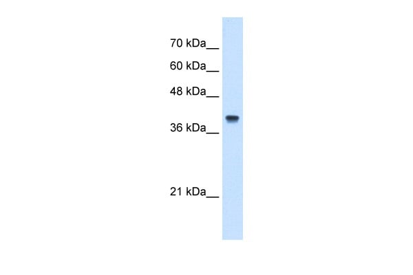
 WB (Western Blot)
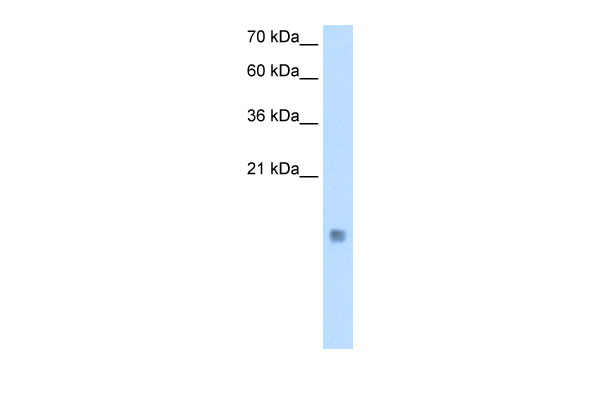
(CSTB antibody - middle region validated by WB using Transfected 293T cell lysate at 0.25ug/ml.)
WB (Western Blot)
(CSTB antibody - middle region validated by WB using Transfected 293T cell lysate at 0.25ug/ml.)
CSTB, Polyclonal Antibody (Cat# AAA199612)
 WB (Western Blot)
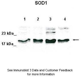
(WB Suggested Antibody Titration: 2.5 ug/mlPositive Control: JurkatSOD1 is supported by BioGPS gene expression data to be expressed in Jurkat)
WB (Western Blot)
(WB Suggested Antibody Titration: 2.5 ug/mlPositive Control: JurkatSOD1 is supported by BioGPS gene expression data to be expressed in Jurkat)
SOD1, Polyclonal Antibody (Cat# AAA199614)
 WB (Western Blot)
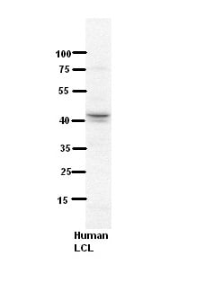
(WB Suggested Anti-NDUFV3 Antibody Titration: 5% MilkELISA Titer: dilution: 1:500Positive Control: Human LCL)
WB (Western Blot)
(WB Suggested Anti-NDUFV3 Antibody Titration: 5% MilkELISA Titer: dilution: 1:500Positive Control: Human LCL)
NDUFV3, Polyclonal Antibody (Cat# AAA199615)
 WB (Western Blot)
(WB Suggested Anti-SH3BGR Antibody Titration: 0.2-1 ug/mlPositive Control: HepG2 cell lysate)
WB (Western Blot)
(WB Suggested Anti-SH3BGR Antibody Titration: 0.2-1 ug/mlPositive Control: HepG2 cell lysate)
SH3BGR, Polyclonal Antibody (Cat# AAA199617)
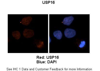
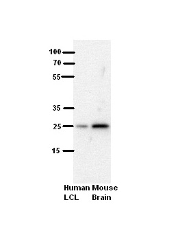
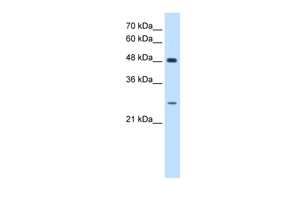
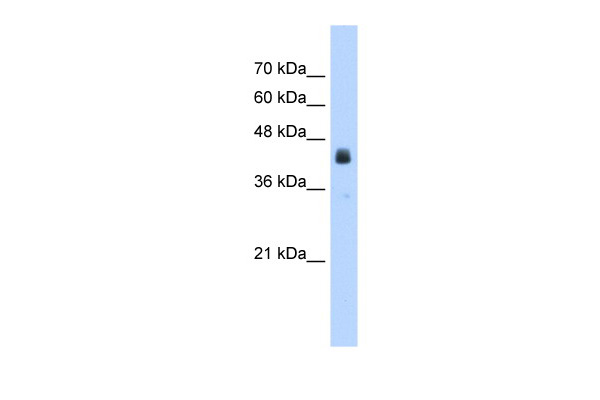 WB (Western Blot)
(WB Suggested Anti-USP16 Antibody Titration: 2.5ug/mlPositive Control: HepG2 cell lysate)
WB (Western Blot)
(WB Suggested Anti-USP16 Antibody Titration: 2.5ug/mlPositive Control: HepG2 cell lysate)
USP16, Polyclonal Antibody (Cat# AAA199619)
What are Polyclonal Antibodies?
Polyclonal antibodies are antibodies that come from multiple B cell clones of a host animal. The typical hosts used for the majority of polyclonal antibody production are rabbits, goats, sheep, and donkeys. These polyclonal antibodies, once having identified their target, will bind to different epitopes located at different regions or sequences on the same protein/antigen. As a result, they are ideal at locating and binding to the target, even if the target is in very low concentrations (due to many different antibodies being able to bind to the same target molecule, which allows for significant amplification of a downstream signal).
Polyclonal antibodies are typically produced by injecting an antigen into a host animal, which causes the animal’s immune system to attack the foreign antigen by mass generating antibodies against it. After a period of time, serum is collected from the animal and purified using physicochemical fractionation, class-specific affinity purification, and/or antigen-affinity purification.
Key Uses of Polyclonal Antibodies
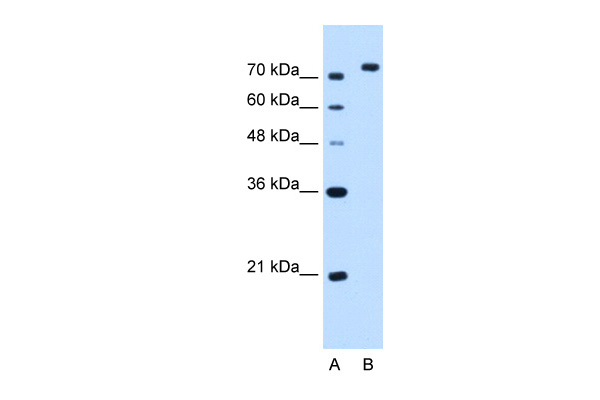
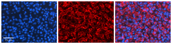
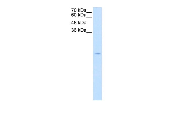
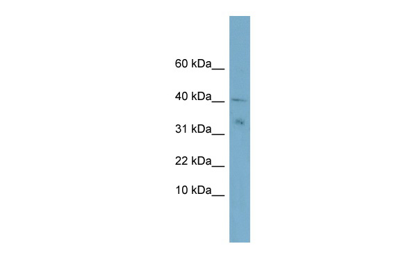
- Western Blotting: This method is used to find specific proteins in biological samples after separating them by size.
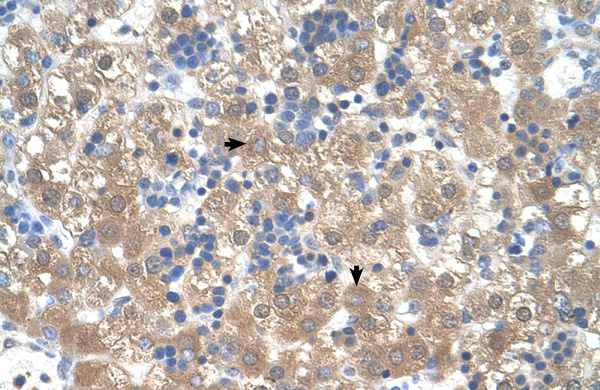
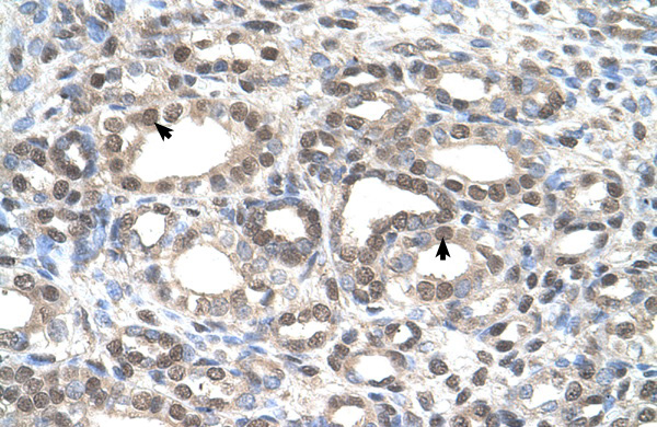
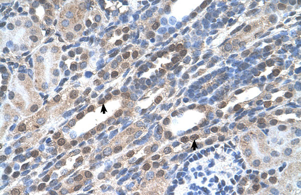
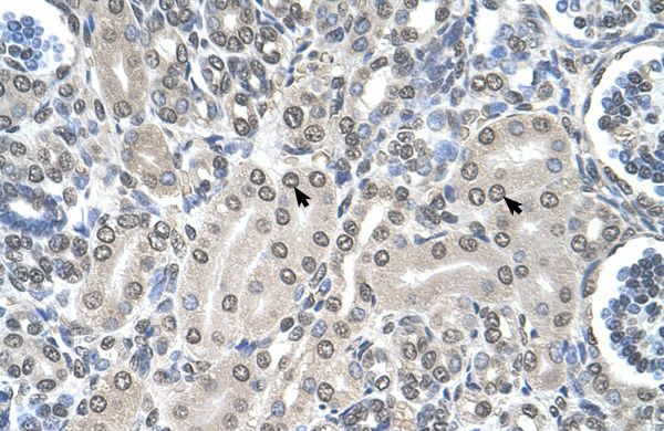
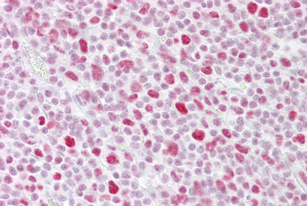
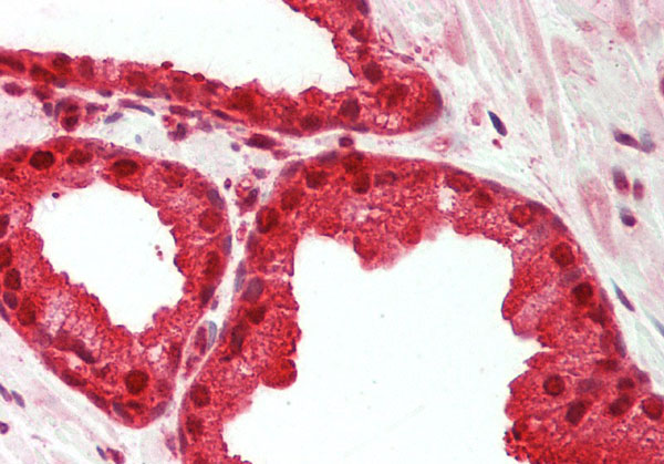
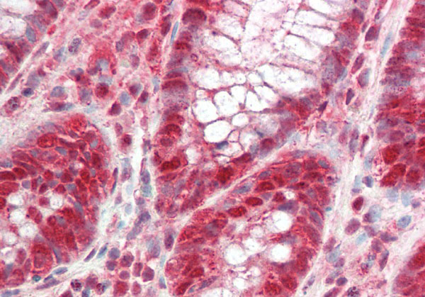
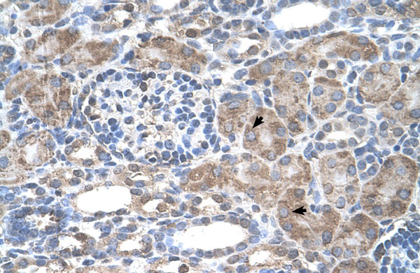
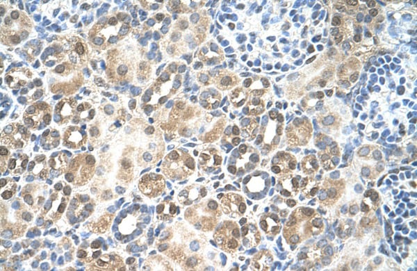
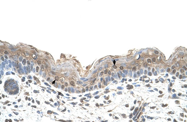
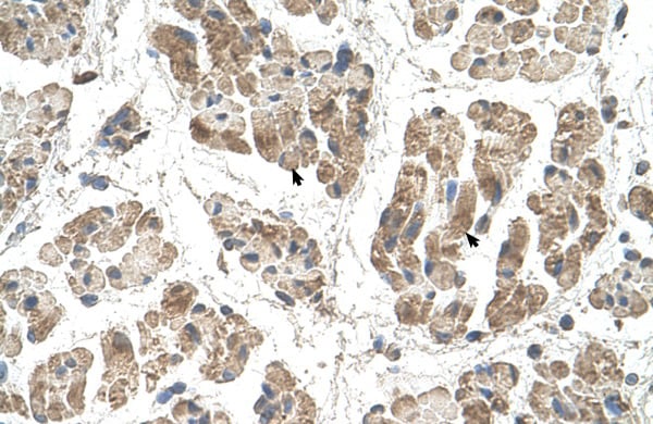
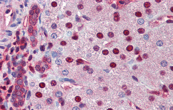
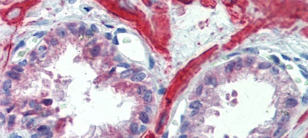
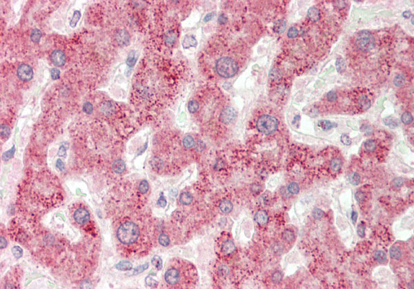
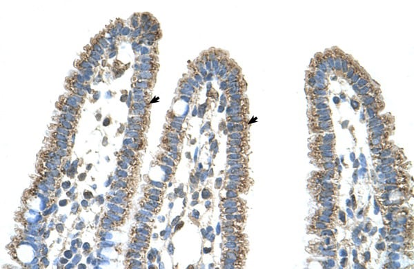
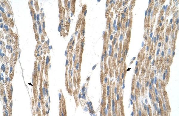
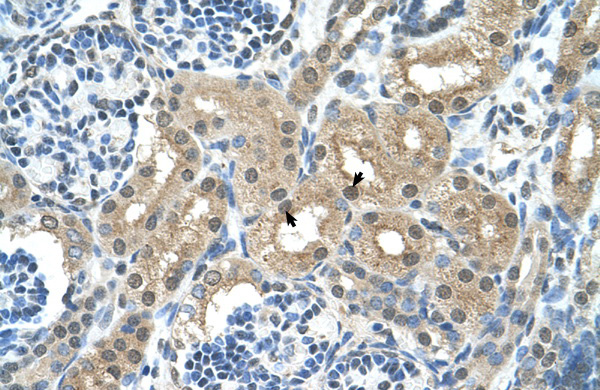
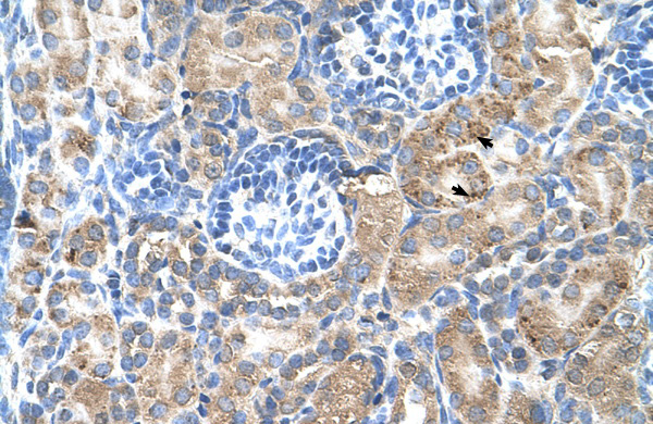
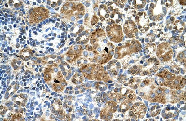
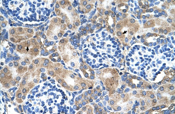
- Immunohistochemistry: IHC helps visualize the location of proteins in tissue sections using various staining techniques.
- ELISA: (Enzyme-Linked Immunosorbent Assay) is typically used to identify specific protein quantities in a sample. ELISAs can be either “Quantitative” or “Qualitative”.
- Flow Cytometry: technique that identifies and measures the specific protein on the surface or inside the cells in a fluid suspension.
- Immunoprecipitation: IP isolates and studies a specific protein from a complex mixture using antibodies.
Why Buy Polyclonal Antibodies from AAA Biotech?
1. Ideal for Various Applications
Our antibodies are generally going to be validated for use in multiple types of assays, including ELISA, Western Blotting, Immunohistochemistry, Immunoprecipitation, amongst others. They are ideal for a wide range of research applications.
2. Rigorous Quality Control
All of the antibodies in our catalog undergo strict quality testing to ensure specificity, sensitivity, and consistent performance. We are confident in the ability of our antibodies to provide you with accurate results.
3. Wide Assortment of Antibodies
Antibodies in are catalog can be found for both common and exotic species, and these antibodies are also available in both conjugated and recombinant forms to suit many diverse experimental needs.
4. Highly Purified
Our antibodies are available in purified forms with over 85% purity, as confirmed by SDS-PAGE. They are also available with tags such as His, Flag, GST, or MBP. We cater to customers worldwide.
FAQ
1. How are polyclonal antibodies produced?
Traditionally, polyclonal antibodies are produced by injecting an antigen into a host animal (such as a rabbit or goat), which then triggers an immune response from the host animal. The animal’s B cells produce antibodies that will recognize different parts of the injected antigen. These antibodies are then collected from the animal’s blood and purified for use.
2. How do polyclonal antibodies differ from monoclonal antibodies?
Polyclonal antibodies are a mix of antibodies that bind to different locations (epitopes) of the same antigen, while monoclonal antibodies are identical and bind to just one specific epitope. This makes polyclonal antibodies more versatile and better at detecting proteins that may be present in low quantities or in altered/modified forms.
3. How should I store polyclonal antibodies?
Polyclonal antibodies should be stored at 4°C for short-term use (up to a few weeks) and at -20°C or -80°C for long-term storage. Avoid repeated freeze-thaw cycles by dividing them into small aliquots. Always check the datasheet for specific storage instructions.