Filters
▼Clonality
▼Type
▼Reactivity
▼Gene Name
▼Isotype
▼Host
▼Application
▼Clone
▼Polyclonal Antibodies
At AAA Biotech also known as AAA Bio or AAABio, we provide a broad range of purified polyclonal antibodies (pAbs) that are able to all be browsed online through our website. Due to their high specificity and strong binding affinity, these antibodies are ideal for wide swathes of research and experimental applications.
Our polyclonal antibodies can easily support your work, whether you use them for Western Blotting, Immunocytochemistry (with or without Immunofluorescence used in conjunction), Immunohistochemistry, Immunoprecipitation, and ELISA tests. We highly encourage you to browse our range of pAbs and choose the one that best suits your experimental model.
Viewing 2950-3000 of 96812 product results
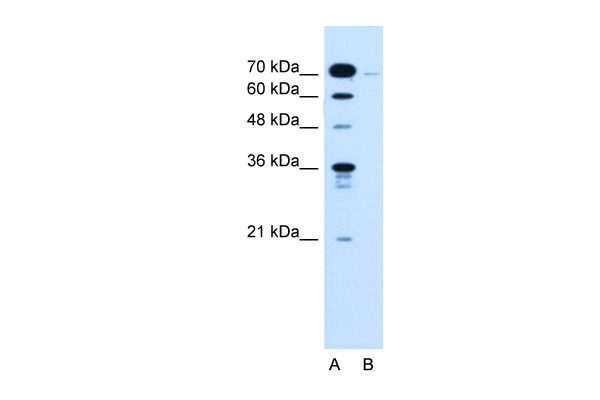
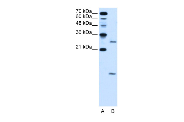
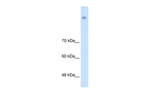
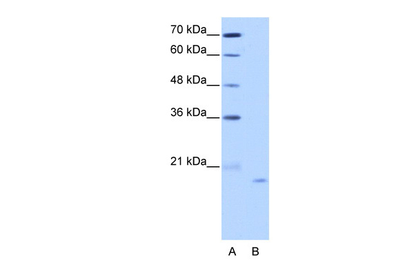
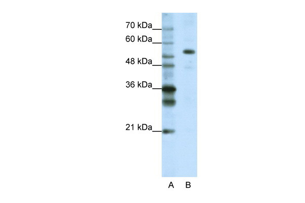
 WB (Western Blot)
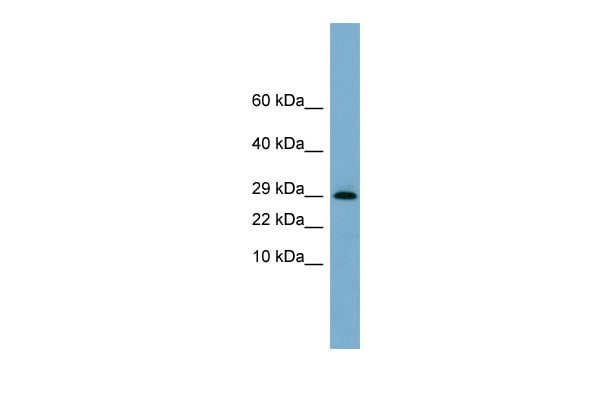
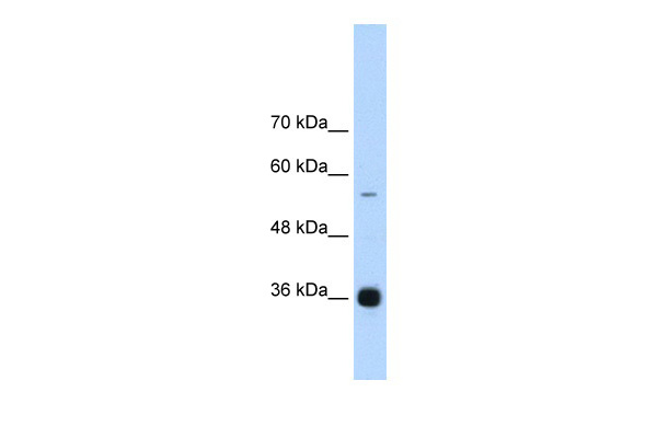
(WB Suggested Anti-DNAJB12 Antibody Titration: 0.2-1 ug/mlELISA Titer: 1:1562500Positive Control: Human Spleen)
WB (Western Blot)
(WB Suggested Anti-DNAJB12 Antibody Titration: 0.2-1 ug/mlELISA Titer: 1:1562500Positive Control: Human Spleen)
DNAJB12, Polyclonal Antibody (Cat# AAA199446)
 WB (Western Blot)
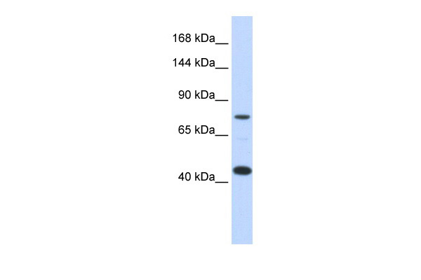
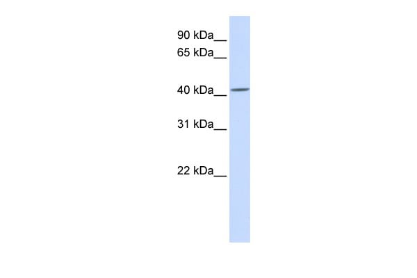
(WB Suggested Anti-UCRC Antibody Titration: 0.2-1 ug/mlELISA Titer: 1:62500Positive Control: MCF7 cell lysateUQCR10 is supported by BioGPS gene expression data to be expressed in MCF7)
WB (Western Blot)
(WB Suggested Anti-UCRC Antibody Titration: 0.2-1 ug/mlELISA Titer: 1:62500Positive Control: MCF7 cell lysateUQCR10 is supported by BioGPS gene expression data to be expressed in MCF7)
UCRC, Polyclonal Antibody (Cat# AAA199450)
 WB (Western Blot)
(WB Suggested Anti-KLHL31 Antibody Titration: 0.2-1 ug/mlPositive Control: Jurkat cell lysate)
WB (Western Blot)
(WB Suggested Anti-KLHL31 Antibody Titration: 0.2-1 ug/mlPositive Control: Jurkat cell lysate)
KLHL31, Polyclonal Antibody (Cat# AAA199454)
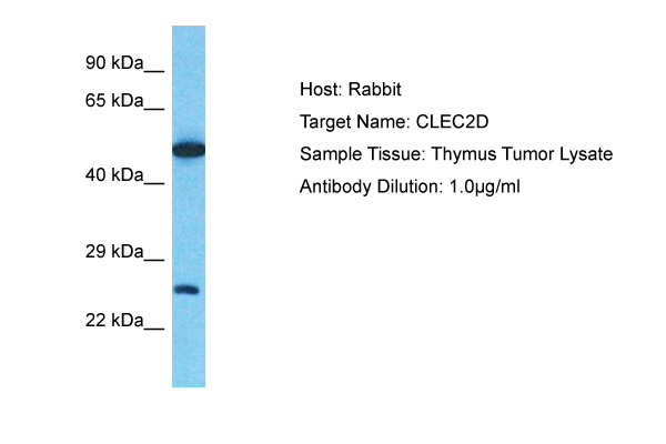 WB (Western Blot)
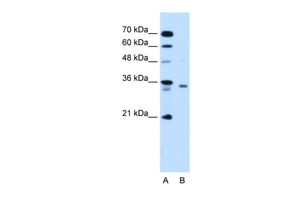
(Host: RabbitTarget Name: CLEC2DSample Type: Thymus Tumor lysatesAntibody Dilution: 1.0ug/ml)
WB (Western Blot)
(Host: RabbitTarget Name: CLEC2DSample Type: Thymus Tumor lysatesAntibody Dilution: 1.0ug/ml)
CLEC2D, Polyclonal Antibody (Cat# AAA199455)
Predicted Species Reactivity: Human
 WB (Western Blot)
(WB Suggested Anti-GPNMB Antibody Titration: 1 ug/mlPositive Control: 293T cells lysate)
WB (Western Blot)
(WB Suggested Anti-GPNMB Antibody Titration: 1 ug/mlPositive Control: 293T cells lysate)
GPNMB, Polyclonal Antibody (Cat# AAA199457)
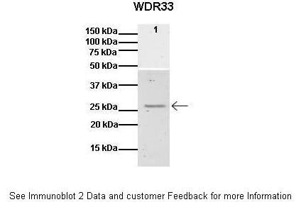
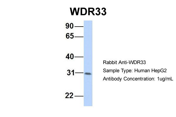
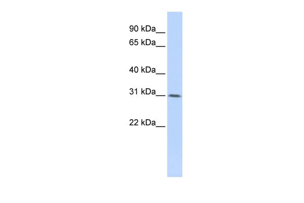 WB (Western Blot)
(WB Suggested Anti-WDR33 Antibody Titration: 0.2-1 ug/mlELISA Titer: 1:62500Positive Control: HepG2 cell lysateWDR33 is supported by BioGPS gene expression data to be expressed in HepG2)
WB (Western Blot)
(WB Suggested Anti-WDR33 Antibody Titration: 0.2-1 ug/mlELISA Titer: 1:62500Positive Control: HepG2 cell lysateWDR33 is supported by BioGPS gene expression data to be expressed in HepG2)
WDR33, Polyclonal Antibody (Cat# AAA199459)
 WB (Western Blot)
(WB Suggested Anti-PDPN Antibody Titration: 0.2-1 ug/mlPositive Control: Jurkat cell lysate)
WB (Western Blot)
(WB Suggested Anti-PDPN Antibody Titration: 0.2-1 ug/mlPositive Control: Jurkat cell lysate)
PDPN, Polyclonal Antibody (Cat# AAA199460)
 WB (Western Blot)
(WB Suggested Anti-TM9SF1 Antibody Titration: 2.5ug/mlPositive Control: Jurkat cell lysate)
WB (Western Blot)
(WB Suggested Anti-TM9SF1 Antibody Titration: 2.5ug/mlPositive Control: Jurkat cell lysate)
TM9SF1, Polyclonal Antibody (Cat# AAA199472)
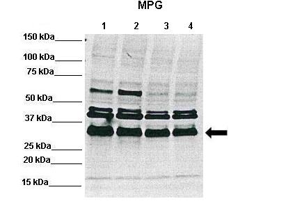
 WB (Western Blot)
(WB Suggested Anti-MPG Antibody Titration: 0.2-1 ug/mlPositive Control: Jurkat cell lysate)
WB (Western Blot)
(WB Suggested Anti-MPG Antibody Titration: 0.2-1 ug/mlPositive Control: Jurkat cell lysate)
MPG, Polyclonal Antibody (Cat# AAA199473)
 WB (Western Blot)
(WB Suggested Anti-VMA21 Antibody Titration: 5.0ug/mlPositive Control: HepG2 cell lysate)
WB (Western Blot)
(WB Suggested Anti-VMA21 Antibody Titration: 5.0ug/mlPositive Control: HepG2 cell lysate)
VMA21, Polyclonal Antibody (Cat# AAA199476)
 WB (Western Blot)
(WB Suggested Anti-LRP8 Antibody Titration: 0.2-1 ug/mlELISA Titer: 1:1562500Positive Control: Transfected 293T)
WB (Western Blot)
(WB Suggested Anti-LRP8 Antibody Titration: 0.2-1 ug/mlELISA Titer: 1:1562500Positive Control: Transfected 293T)
LRP8, Polyclonal Antibody (Cat# AAA199477)
 WB (Western Blot)
(WB Suggested Anti-LETMD1 Antibody Titration: 0.2-1 ug/mlELISA Titer: 1:312500Positive Control: PANC1 cell lysate)
WB (Western Blot)
(WB Suggested Anti-LETMD1 Antibody Titration: 0.2-1 ug/mlELISA Titer: 1:312500Positive Control: PANC1 cell lysate)
LETMD1, Polyclonal Antibody (Cat# AAA199479)
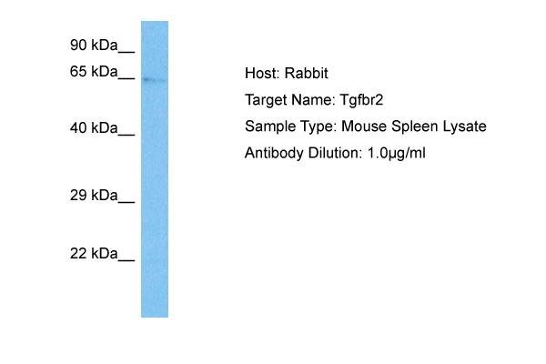
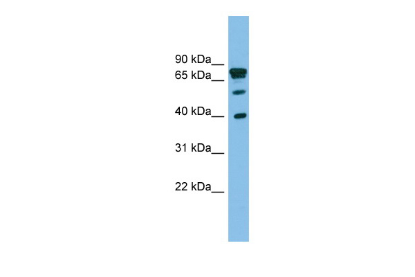
 WB (Western Blot)
(WB Suggested Anti-TGFBR2 Antibody Titration: 5.0ug/mlPositive Control: HepG2 cell lysateTGFBR2 is strongly supported by BioGPS gene expression data to be expressed in Human HepG2 cells)
WB (Western Blot)
(WB Suggested Anti-TGFBR2 Antibody Titration: 5.0ug/mlPositive Control: HepG2 cell lysateTGFBR2 is strongly supported by BioGPS gene expression data to be expressed in Human HepG2 cells)
TGFBR2, Polyclonal Antibody (Cat# AAA199480)
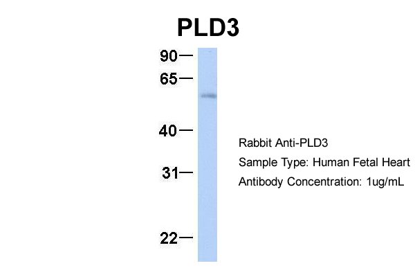
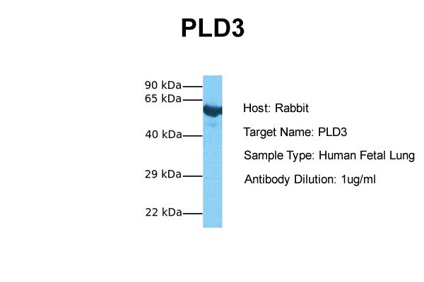
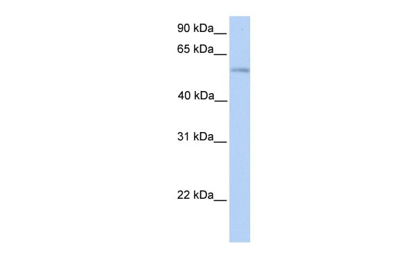 WB (Western Blot)
(WB Suggested Anti-PLD3 Antibody Titration: 0.2-1 ug/mlPositive Control: Human heart)
WB (Western Blot)
(WB Suggested Anti-PLD3 Antibody Titration: 0.2-1 ug/mlPositive Control: Human heart)
PLD3, Polyclonal Antibody (Cat# AAA199482)
 WB (Western Blot)
(WB Suggested Anti-RCE1 Antibody Titration: 1.25ug/mlPositive Control: HepG2 cell lysateRCE1 is supported by BioGPS gene expression data to be expressed in HepG2)
WB (Western Blot)
(WB Suggested Anti-RCE1 Antibody Titration: 1.25ug/mlPositive Control: HepG2 cell lysateRCE1 is supported by BioGPS gene expression data to be expressed in HepG2)
RCE1, Polyclonal Antibody (Cat# AAA199484)
 WB (Western Blot)
(WB Suggested Anti-RHOT1 antibody Titration: 1 ug/mLSample Type: HEK293)
WB (Western Blot)
(WB Suggested Anti-RHOT1 antibody Titration: 1 ug/mLSample Type: HEK293)
RHOT1, Polyclonal Antibody (Cat# AAA199487)
 WB (Western Blot)
(WB Suggested Anti-ERP29 Antibody Titration: 0.2-1 ug/mlPositive Control: ACHN cell lysateERP29 is supported by BioGPS gene expression data to be expressed in ACHN)
WB (Western Blot)
(WB Suggested Anti-ERP29 Antibody Titration: 0.2-1 ug/mlPositive Control: ACHN cell lysateERP29 is supported by BioGPS gene expression data to be expressed in ACHN)
ERP29, Polyclonal Antibody (Cat# AAA199488)
 WB (Western Blot)
(WB Suggested Anti-NRCAM Antibody Titration: 2.5ug/mlPositive Control: HepG2 cell lysate)
WB (Western Blot)
(WB Suggested Anti-NRCAM Antibody Titration: 2.5ug/mlPositive Control: HepG2 cell lysate)
NRCAM, Polyclonal Antibody (Cat# AAA199491)
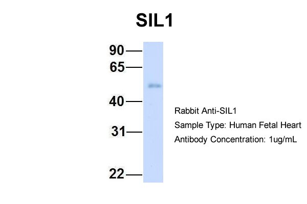
 WB (Western Blot)
(WB Suggested Anti-SIL1 antibody Titration: 1 ug/mLSample Type: Human heart)
WB (Western Blot)
(WB Suggested Anti-SIL1 antibody Titration: 1 ug/mLSample Type: Human heart)
SIL1, Polyclonal Antibody (Cat# AAA199494)
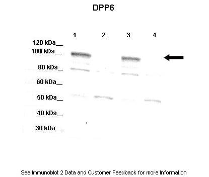
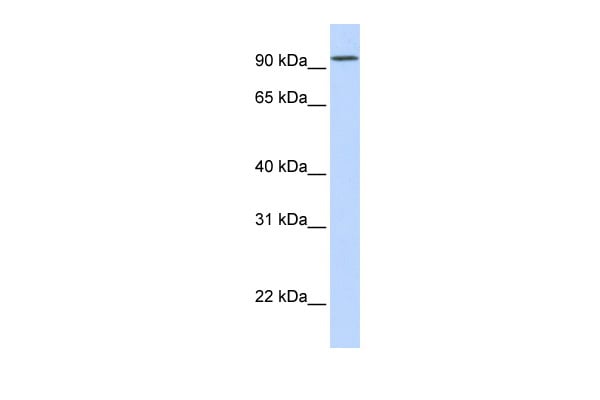 WB (Western Blot)
(WB Suggested Anti-DPP6 Antibody Titration: 0.2-1 ug/mlELISA Titer: 1:312500Positive Control: Human Muscle)
WB (Western Blot)
(WB Suggested Anti-DPP6 Antibody Titration: 0.2-1 ug/mlELISA Titer: 1:312500Positive Control: Human Muscle)
DPP6, Polyclonal Antibody (Cat# AAA199498)
 WB (Western Blot)
(WB Suggested Anti-KIAA0317 Antibody Titration: 0.2-1 ug/mlPositive Control: HT1080 cell lysateAREL1 is supported by BioGPS gene expression data to be expressed in HT1080)
WB (Western Blot)
(WB Suggested Anti-KIAA0317 Antibody Titration: 0.2-1 ug/mlPositive Control: HT1080 cell lysateAREL1 is supported by BioGPS gene expression data to be expressed in HT1080)
KIAA0317, Polyclonal Antibody (Cat# AAA199499)
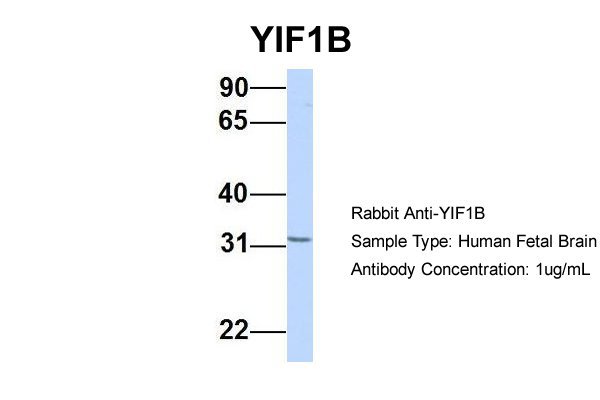
 WB (Western Blot)
(WB Suggested Anti-YIF1B Antibody Titration: 0.2-1 ug/mlELISA Titer: 1:62500Positive Control: 721_B cell lysateYIF1B is supported by BioGPS gene expression data to be expressed in 721_B)
WB (Western Blot)
(WB Suggested Anti-YIF1B Antibody Titration: 0.2-1 ug/mlELISA Titer: 1:62500Positive Control: 721_B cell lysateYIF1B is supported by BioGPS gene expression data to be expressed in 721_B)
YIF1B, Polyclonal Antibody (Cat# AAA199500)
 WB (Western Blot)
(WB Suggested Anti-ASAHL Antibody Titration: 0.2-1 ug/mlELISA Titer: 1:62500Positive Control: 721_B cell lysate)
WB (Western Blot)
(WB Suggested Anti-ASAHL Antibody Titration: 0.2-1 ug/mlELISA Titer: 1:62500Positive Control: 721_B cell lysate)
ASAHL, Polyclonal Antibody (Cat# AAA199503)
Predicted Species Reactivity: Cow, Guinea Pig, Horse, Human, Mouse, Pig, Rabbit, Rat
 WB (Western Blot)
(WB Suggested Anti-C19orf28 Antibody Titration: 0.2-1 ug/mlPositive Control: HepG2 cell lysate)
WB (Western Blot)
(WB Suggested Anti-C19orf28 Antibody Titration: 0.2-1 ug/mlPositive Control: HepG2 cell lysate)
C19orf28, Polyclonal Antibody (Cat# AAA199504)
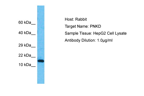 WB (Western Blot)
(Host: RabbitTarget Name: PNKDSample Type: HepG2 Whole Cell lysatesAntibody Dilution: 1.0ug/ml)
WB (Western Blot)
(Host: RabbitTarget Name: PNKDSample Type: HepG2 Whole Cell lysatesAntibody Dilution: 1.0ug/ml)
PNKD, Polyclonal Antibody (Cat# AAA199507)
Predicted Species Reactivity: Human, Mouse, Rat, Cow, Dog, Goat, Guinea Pig, Horse, Rabbit
 WB (Western Blot)
(WB Suggested Anti-FLJ22167 Antibody Titration: 1.25ug/mlPositive Control: HepG2 cell lysate)
WB (Western Blot)
(WB Suggested Anti-FLJ22167 Antibody Titration: 1.25ug/mlPositive Control: HepG2 cell lysate)
FLJ22167, Polyclonal Antibody (Cat# AAA199509)


 WB (Western Blot)
(WB Suggested Anti-ACVR1 Antibody Titration: 0.2-1 ug/mlELISA Titer: 1:312500Positive Control: Transfected 293T)
WB (Western Blot)
(WB Suggested Anti-ACVR1 Antibody Titration: 0.2-1 ug/mlELISA Titer: 1:312500Positive Control: Transfected 293T)
ACVR1, Polyclonal Antibody (Cat# AAA199514)
 WB (Western Blot)
(WB Suggested Anti-MIF4GD Antibody Titration: 1.25ug/mlPositive Control: HepG2 cell lysate)
WB (Western Blot)
(WB Suggested Anti-MIF4GD Antibody Titration: 1.25ug/mlPositive Control: HepG2 cell lysate)
MIF4GD, Polyclonal Antibody (Cat# AAA198794)
 WB (Western Blot)
(WB Suggested Anti-EIF4H Antibody Titration: 0.2-1 ug/mlELISA Titer: 1:62500Positive Control: 721_B cell lysateEIF4H is supported by BioGPS gene expression data to be expressed in 721_B)
WB (Western Blot)
(WB Suggested Anti-EIF4H Antibody Titration: 0.2-1 ug/mlELISA Titer: 1:62500Positive Control: 721_B cell lysateEIF4H is supported by BioGPS gene expression data to be expressed in 721_B)
EIF4H, Polyclonal Antibody (Cat# AAA198797)
 WB (Western Blot)
(WB Suggested Anti-NOL6 Antibody Titration: 2.5ug/mlPositive Control: HepG2 cell lysateNOL6 is supported by BioGPS gene expression data to be expressed in HepG2)
WB (Western Blot)
(WB Suggested Anti-NOL6 Antibody Titration: 2.5ug/mlPositive Control: HepG2 cell lysateNOL6 is supported by BioGPS gene expression data to be expressed in HepG2)
NOL6, Polyclonal Antibody (Cat# AAA198801)
 WB (Western Blot)
(WB Suggested Anti-FLJ12529 Antibody Titration: 1.25ug/mlPositive Control: HepG2 cell lysateCPSF7 is supported by BioGPS gene expression data to be expressed in HepG2)
WB (Western Blot)
(WB Suggested Anti-FLJ12529 Antibody Titration: 1.25ug/mlPositive Control: HepG2 cell lysateCPSF7 is supported by BioGPS gene expression data to be expressed in HepG2)
FLJ12529, Polyclonal Antibody (Cat# AAA198804)
 WB (Western Blot)
(WB Suggested Anti-BXDC5 Antibody Titration: 0.2-1 ug/mlPositive Control: K562 cell lysate)
WB (Western Blot)
(WB Suggested Anti-BXDC5 Antibody Titration: 0.2-1 ug/mlPositive Control: K562 cell lysate)
BXDC5, Polyclonal Antibody (Cat# AAA198806)
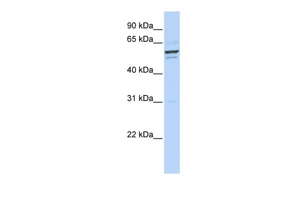
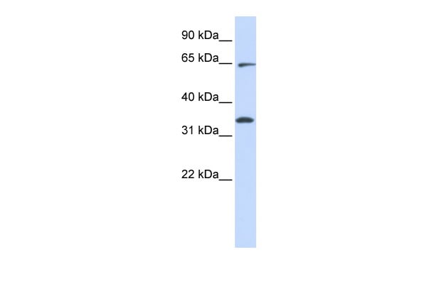
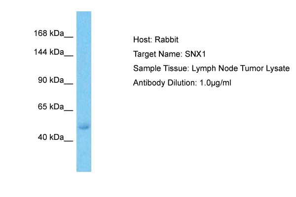 WB (Western Blot)
(Host: RabbitTarget Name: SNX1Sample Type: Lymph Node Tumor lysatesAntibody Dilution: 1.0ug/ml)
WB (Western Blot)
(Host: RabbitTarget Name: SNX1Sample Type: Lymph Node Tumor lysatesAntibody Dilution: 1.0ug/ml)
SNX1, Polyclonal Antibody (Cat# AAA198807)

 WB (Western Blot)
(WB Suggested Anti-HNRPAB Antibody Titration: 1.25ug/mlPositive Control: Daudi cell lysateHNRNPAB is supported by BioGPS gene expression data to be expressed in Daudi)
WB (Western Blot)
(WB Suggested Anti-HNRPAB Antibody Titration: 1.25ug/mlPositive Control: Daudi cell lysateHNRNPAB is supported by BioGPS gene expression data to be expressed in Daudi)
HNRPAB, Polyclonal Antibody (Cat# AAA198812)
 WB (Western Blot)
(WB Suggested Anti-EIF2A Antibody Titration: 2.5ug/mlPositive Control: Human Thymus)
WB (Western Blot)
(WB Suggested Anti-EIF2A Antibody Titration: 2.5ug/mlPositive Control: Human Thymus)
EIF2A, Polyclonal Antibody (Cat# AAA198814)
 WB (Western Blot)
(WB Suggested Anti-SFRS2B Antibody Titration: 0.2-1 ug/mlELISA Titer: 1:62500Positive Control: 721_B cell lysateSRSF8 is strongly supported by BioGPS gene expression data to be expressed in Human 721_B cells)
WB (Western Blot)
(WB Suggested Anti-SFRS2B Antibody Titration: 0.2-1 ug/mlELISA Titer: 1:62500Positive Control: 721_B cell lysateSRSF8 is strongly supported by BioGPS gene expression data to be expressed in Human 721_B cells)
SFRS2B, Polyclonal Antibody (Cat# AAA198815)
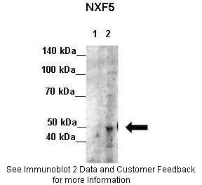
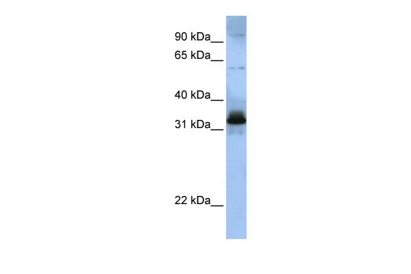
 WB (Western Blot)
(WB Suggested Anti-NXF5 Antibody Titration: 2.5ug/mlPositive Control: Jurkat cell lysate)
WB (Western Blot)
(WB Suggested Anti-NXF5 Antibody Titration: 2.5ug/mlPositive Control: Jurkat cell lysate)
NXF5, Polyclonal Antibody (Cat# AAA198818)
 WB (Western Blot)
(WB Suggested Anti-RAVER1 Antibody Titration: 1.25ug/mlPositive Control: HepG2 cell lysate)
WB (Western Blot)
(WB Suggested Anti-RAVER1 Antibody Titration: 1.25ug/mlPositive Control: HepG2 cell lysate)
RAVER1, Polyclonal Antibody (Cat# AAA198821)
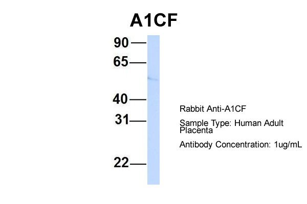
 WB (Western Blot)
(WB Suggested Anti-A1CF Antibody Titration: 0.2-1 ug/mlELISA Titer: 1:1562500Positive Control: HepG2 cell lysateA1CF is supported by BioGPS gene expression data to be expressed in HepG2)
WB (Western Blot)
(WB Suggested Anti-A1CF Antibody Titration: 0.2-1 ug/mlELISA Titer: 1:1562500Positive Control: HepG2 cell lysateA1CF is supported by BioGPS gene expression data to be expressed in HepG2)
A1CF, Polyclonal Antibody (Cat# AAA198823)
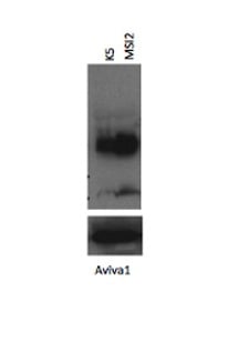
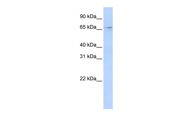
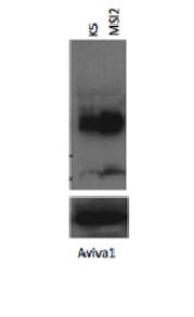
 WB (Western Blot)
(WB Suggested Anti-MSI2 Antibody Titration: 0.2-1 ug/mlELISA Titer: 1:1562500Positive Control: Hela cell lysate)
WB (Western Blot)
(WB Suggested Anti-MSI2 Antibody Titration: 0.2-1 ug/mlELISA Titer: 1:1562500Positive Control: Hela cell lysate)
MSI2, Polyclonal Antibody (Cat# AAA198824)
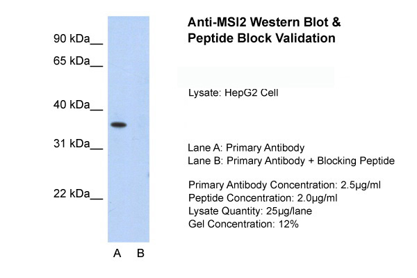
 WB (Western Blot)
(WB Suggested Anti-MSI2 Antibody Titration: 1.25ug/mlELISA Titer: 1:1562500Positive Control: HepG2 cell lysate)
WB (Western Blot)
(WB Suggested Anti-MSI2 Antibody Titration: 1.25ug/mlELISA Titer: 1:1562500Positive Control: HepG2 cell lysate)
MSI2, Polyclonal Antibody (Cat# AAA198825)
 WB (Western Blot)
(WB Suggested Anti-RG9MTD3 Antibody Titration: 0.2-1 ug/mlPositive Control: Jurkat cell lysate)
WB (Western Blot)
(WB Suggested Anti-RG9MTD3 Antibody Titration: 0.2-1 ug/mlPositive Control: Jurkat cell lysate)
RG9MTD3, Polyclonal Antibody (Cat# AAA198827)
 WB (Western Blot)
(WB Suggested Anti-MGC42174 Antibody Titration: 1.25ug/mlELISA Titer: 1:62500Positive Control: Jurkat cell lysate)
WB (Western Blot)
(WB Suggested Anti-MGC42174 Antibody Titration: 1.25ug/mlELISA Titer: 1:62500Positive Control: Jurkat cell lysate)
MGC42174, Polyclonal Antibody (Cat# AAA198830)
 WB (Western Blot)
(WB Suggested Anti-APOBEC3D Antibody Titration: 0.2-1 ug/mlPositive Control: HepG2 cell lysate)
WB (Western Blot)
(WB Suggested Anti-APOBEC3D Antibody Titration: 0.2-1 ug/mlPositive Control: HepG2 cell lysate)
APOBEC3D, Polyclonal Antibody (Cat# AAA198832)
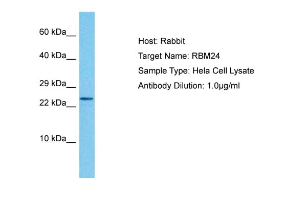
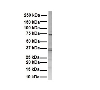
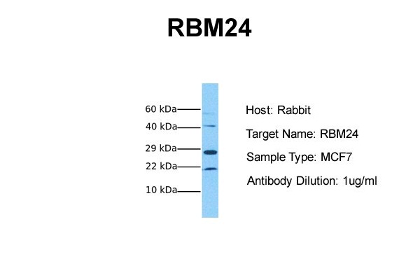 WB (Western Blot)
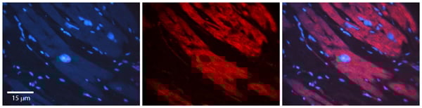
(Host: RabbitTarget Name: RBM24Sample Tissue: Human MCF7Antibody Dilution: 1.0ug/ml)
WB (Western Blot)
(Host: RabbitTarget Name: RBM24Sample Tissue: Human MCF7Antibody Dilution: 1.0ug/ml)
RBM24, Polyclonal Antibody (Cat# AAA198833)
 WB (Western Blot)
(WB Suggested Anti-DAZAP1 Antibody Titration: 0.2-1 ug/mlELISA Titer: 1:312500Positive Control: HepG2 cell lysateDAZAP1 is supported by BioGPS gene expression data to be expressed in HepG2)
WB (Western Blot)
(WB Suggested Anti-DAZAP1 Antibody Titration: 0.2-1 ug/mlELISA Titer: 1:312500Positive Control: HepG2 cell lysateDAZAP1 is supported by BioGPS gene expression data to be expressed in HepG2)
DAZAP1, Polyclonal Antibody (Cat# AAA198835)
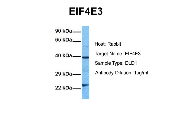
 WB (Western Blot)
(WB Suggested Anti-EIF4E3 Antibody Titration: 0.2-1 ug/mlPositive Control: Human Liver)
WB (Western Blot)
(WB Suggested Anti-EIF4E3 Antibody Titration: 0.2-1 ug/mlPositive Control: Human Liver)
EIF4E3, Polyclonal Antibody (Cat# AAA198836)
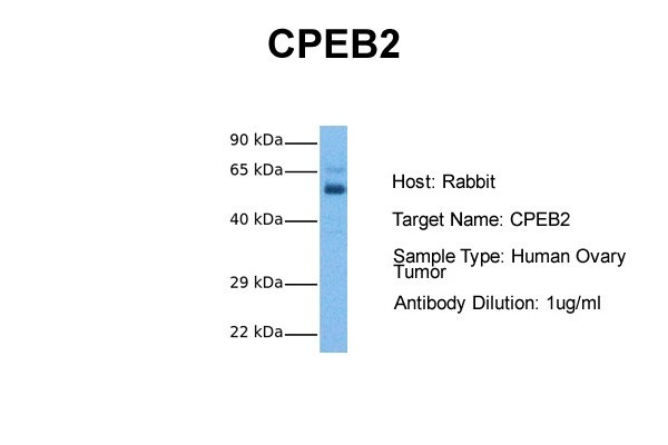
 WB (Western Blot)
(WB Suggested Anti-CPEB2 Antibody Titration: 0.2-1 ug/mlELISA Titer: 1:312500Positive Control: Jurkat cell lysate)
WB (Western Blot)
(WB Suggested Anti-CPEB2 Antibody Titration: 0.2-1 ug/mlELISA Titer: 1:312500Positive Control: Jurkat cell lysate)
CPEB2, Polyclonal Antibody (Cat# AAA198837)
 WB (Western Blot)
(WB Suggested Anti-HNRPA3 Antibody Titration: 5.0ug/mlELISA Titer: 1:62500Positive Control: Jurkat cell lysateHNRNPA3 is strongly supported by BioGPS gene expression data to be expressed in Human Jurkat cells)
WB (Western Blot)
(WB Suggested Anti-HNRPA3 Antibody Titration: 5.0ug/mlELISA Titer: 1:62500Positive Control: Jurkat cell lysateHNRNPA3 is strongly supported by BioGPS gene expression data to be expressed in Human Jurkat cells)
HNRPA3, Polyclonal Antibody (Cat# AAA198839)
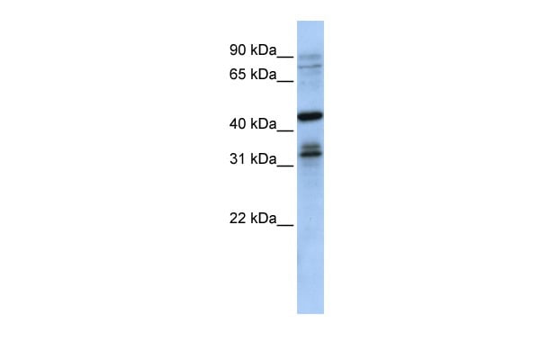
 WB (Western Blot)
(WB Suggested Anti-SF1 Antibody Titration: 1.25ug/mlELISA Titer: 1:312500Positive Control: HepG2 cell lysateSF1 is supported by BioGPS gene expression data to be expressed in HepG2)
WB (Western Blot)
(WB Suggested Anti-SF1 Antibody Titration: 1.25ug/mlELISA Titer: 1:312500Positive Control: HepG2 cell lysateSF1 is supported by BioGPS gene expression data to be expressed in HepG2)
SF1, Polyclonal Antibody (Cat# AAA198844)
What are Polyclonal Antibodies?
Polyclonal antibodies are antibodies that come from multiple B cell clones of a host animal. The typical hosts used for the majority of polyclonal antibody production are rabbits, goats, sheep, and donkeys. These polyclonal antibodies, once having identified their target, will bind to different epitopes located at different regions or sequences on the same protein/antigen. As a result, they are ideal at locating and binding to the target, even if the target is in very low concentrations (due to many different antibodies being able to bind to the same target molecule, which allows for significant amplification of a downstream signal).
Polyclonal antibodies are typically produced by injecting an antigen into a host animal, which causes the animal’s immune system to attack the foreign antigen by mass generating antibodies against it. After a period of time, serum is collected from the animal and purified using physicochemical fractionation, class-specific affinity purification, and/or antigen-affinity purification.
Key Uses of Polyclonal Antibodies
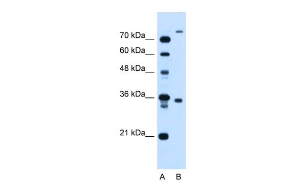
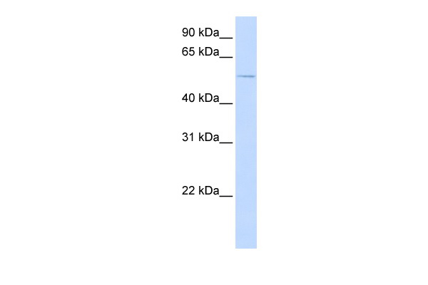
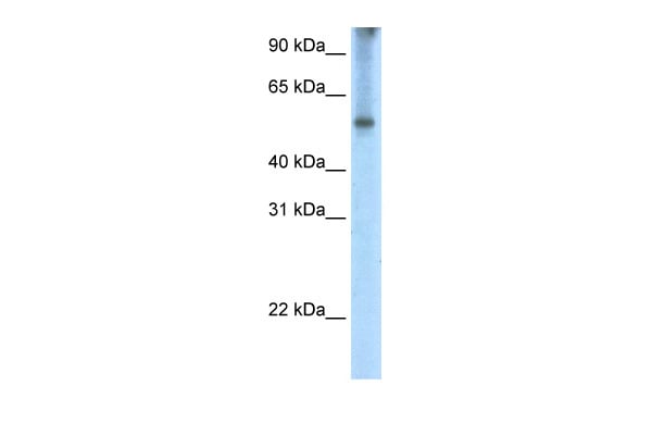
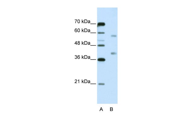
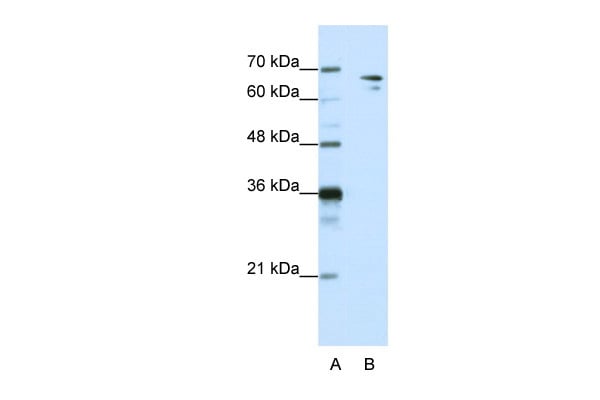
- Western Blotting: This method is used to find specific proteins in biological samples after separating them by size.
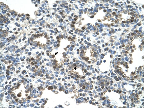
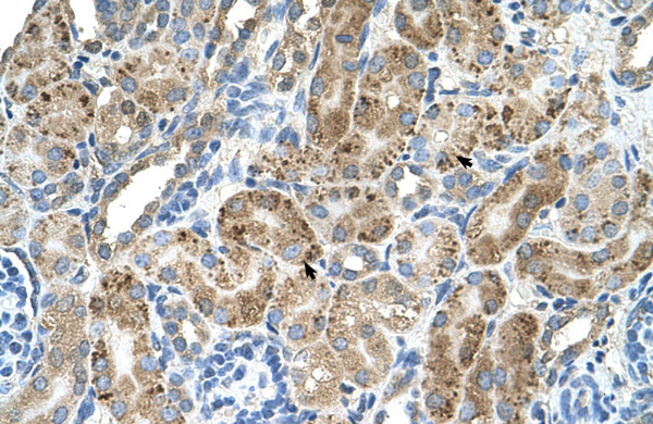
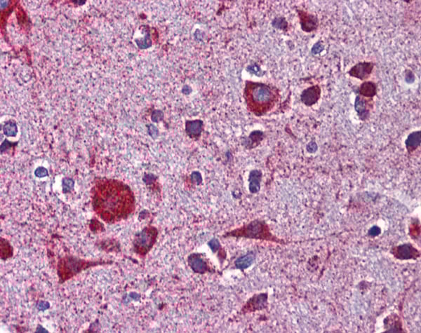
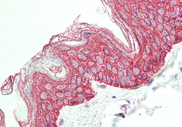
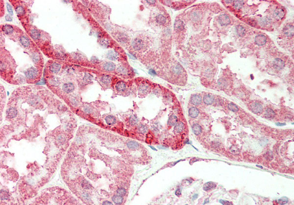
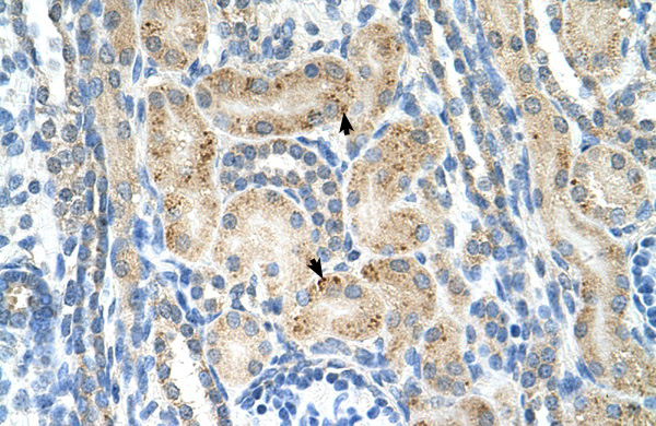
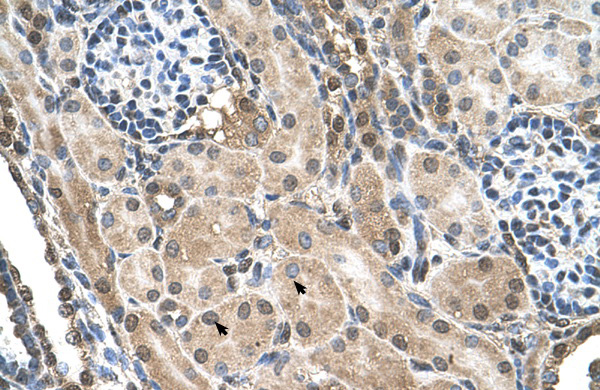
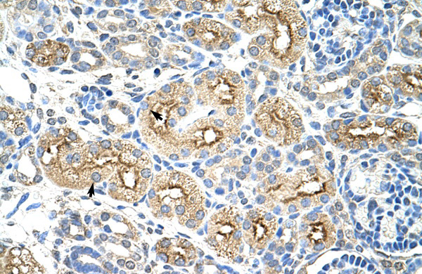
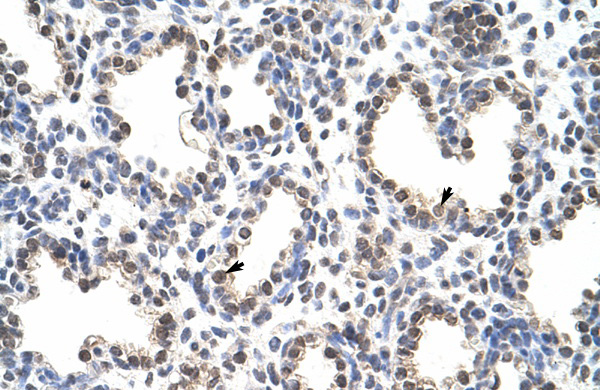
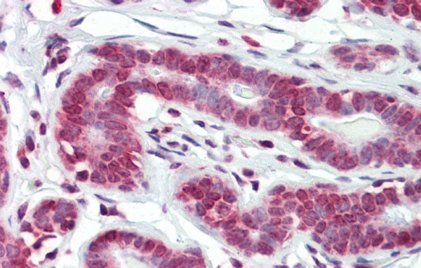
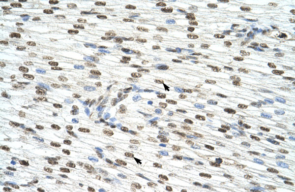
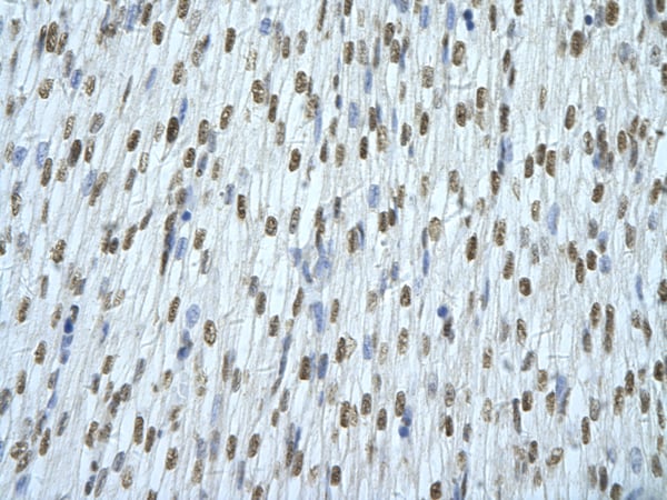
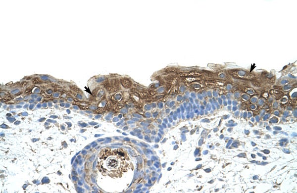
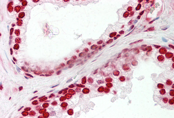
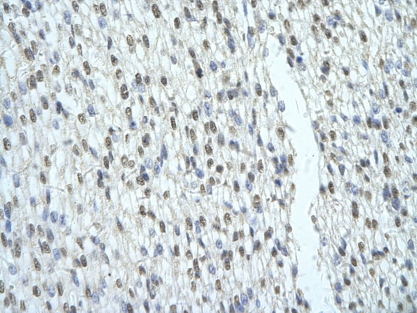
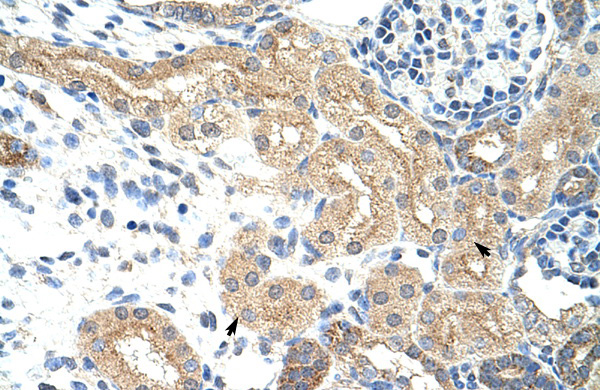
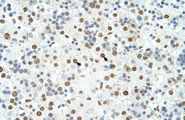
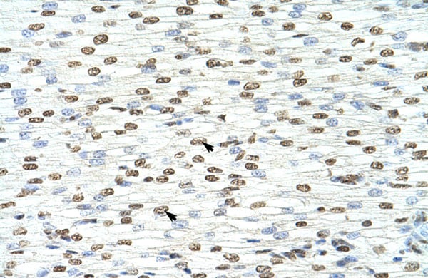
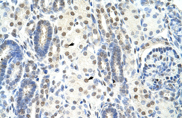
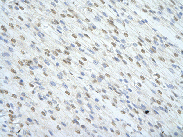
- Immunohistochemistry: IHC helps visualize the location of proteins in tissue sections using various staining techniques.
- ELISA: (Enzyme-Linked Immunosorbent Assay) is typically used to identify specific protein quantities in a sample. ELISAs can be either “Quantitative” or “Qualitative”.
- Flow Cytometry: technique that identifies and measures the specific protein on the surface or inside the cells in a fluid suspension.
- Immunoprecipitation: IP isolates and studies a specific protein from a complex mixture using antibodies.
Why Buy Polyclonal Antibodies from AAA Biotech?
1. Ideal for Various Applications
Our antibodies are generally going to be validated for use in multiple types of assays, including ELISA, Western Blotting, Immunohistochemistry, Immunoprecipitation, amongst others. They are ideal for a wide range of research applications.
2. Rigorous Quality Control
All of the antibodies in our catalog undergo strict quality testing to ensure specificity, sensitivity, and consistent performance. We are confident in the ability of our antibodies to provide you with accurate results.
3. Wide Assortment of Antibodies
Antibodies in are catalog can be found for both common and exotic species, and these antibodies are also available in both conjugated and recombinant forms to suit many diverse experimental needs.
4. Highly Purified
Our antibodies are available in purified forms with over 85% purity, as confirmed by SDS-PAGE. They are also available with tags such as His, Flag, GST, or MBP. We cater to customers worldwide.
FAQ
1. How are polyclonal antibodies produced?
Traditionally, polyclonal antibodies are produced by injecting an antigen into a host animal (such as a rabbit or goat), which then triggers an immune response from the host animal. The animal’s B cells produce antibodies that will recognize different parts of the injected antigen. These antibodies are then collected from the animal’s blood and purified for use.
2. How do polyclonal antibodies differ from monoclonal antibodies?
Polyclonal antibodies are a mix of antibodies that bind to different locations (epitopes) of the same antigen, while monoclonal antibodies are identical and bind to just one specific epitope. This makes polyclonal antibodies more versatile and better at detecting proteins that may be present in low quantities or in altered/modified forms.
3. How should I store polyclonal antibodies?
Polyclonal antibodies should be stored at 4°C for short-term use (up to a few weeks) and at -20°C or -80°C for long-term storage. Avoid repeated freeze-thaw cycles by dividing them into small aliquots. Always check the datasheet for specific storage instructions.