Filters
▼Clonality
▼Type
▼Reactivity
▼Gene Name
▼Isotype
▼Host
▼Application
▼Clone
▼Polyclonal Antibodies
At AAA Biotech also known as AAA Bio or AAABio, we provide a broad range of purified polyclonal antibodies (pAbs) that are able to all be browsed online through our website. Due to their high specificity and strong binding affinity, these antibodies are ideal for wide swathes of research and experimental applications.
Our polyclonal antibodies can easily support your work, whether you use them for Western Blotting, Immunocytochemistry (with or without Immunofluorescence used in conjunction), Immunohistochemistry, Immunoprecipitation, and ELISA tests. We highly encourage you to browse our range of pAbs and choose the one that best suits your experimental model.
Viewing 4550-4600 of 96812 product results

 WB (Western Blot)
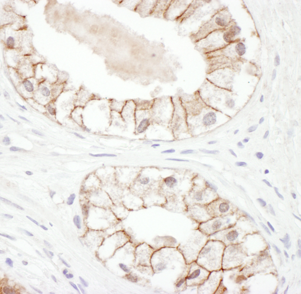
(Detection of human Plakoglobin by western blot and immunoprecipitation. Samples: Whole cell lysate from HeLa (15 and 50 ug for WB; 1 mg for IP, 20% of IP loaded), HEK293T (T; 50 ug) and Jurkat (J; 50 ug) cells. Antibodies: Affinity purified rabbit anti-Plakoglobin antibody AAA212271 used for WB at 0.1 ug/ml (A) and 1 ug/ml (B) and used for IP at 6 ug/mg lysate. Plakoglobin was also immunoprecipitated by rabbit anti-Plakoglobin antibody which recognizes an upstream epitope. Detection: Chemiluminescence with exposure times of 10 seconds (A) and 3 seconds (B).)
WB (Western Blot)
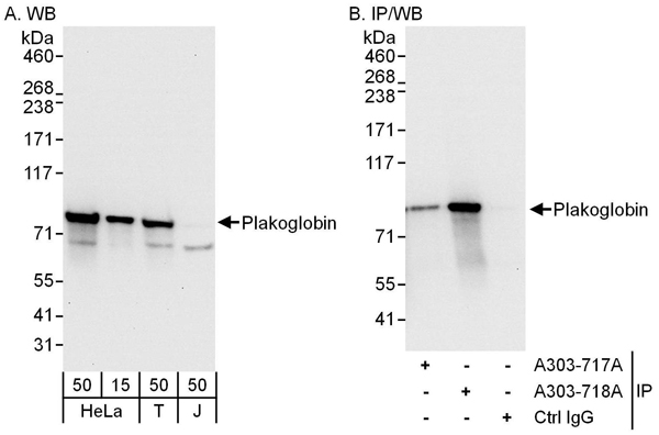
(Detection of human Plakoglobin by western blot and immunoprecipitation. Samples: Whole cell lysate from HeLa (15 and 50 ug for WB; 1 mg for IP, 20% of IP loaded), HEK293T (T; 50 ug) and Jurkat (J; 50 ug) cells. Antibodies: Affinity purified rabbit anti-Plakoglobin antibody AAA212271 used for WB at 0.1 ug/ml (A) and 1 ug/ml (B) and used for IP at 6 ug/mg lysate. Plakoglobin was also immunoprecipitated by rabbit anti-Plakoglobin antibody which recognizes an upstream epitope. Detection: Chemiluminescence with exposure times of 10 seconds (A) and 3 seconds (B).)
Plakoglobin, Polyclonal Antibody (Cat# AAA212271)
 WB (Western Blot)
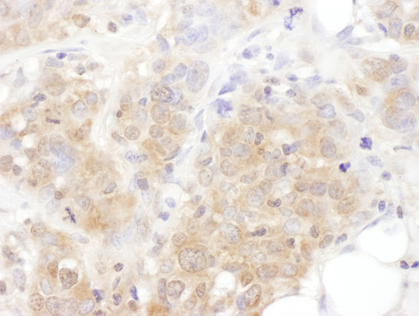
(Detection of human AS160 by western blot and immunoprecipitation. Samples: Whole cell lysate from HeLa (15 and 50 ug for WB; 1 mg for IP, 20% of IP loaded), HEK293T (T; 50 ug) and Jurkat (J; 50 ug) cells. Antibodies: Affinity purified rabbit anti-AS160 antibody AAA212273 used for WB at 0.1 ug/ml (A) and 1 ug/ml (B) and used for IP at 6 ug/mg lysate. AS160 was also immunoprecipitated by rabbit anti-AS160 antibodies and which recognize other epitopes. Detection: Chemiluminescence with exposure times of 10 seconds (A and B).)
WB (Western Blot)
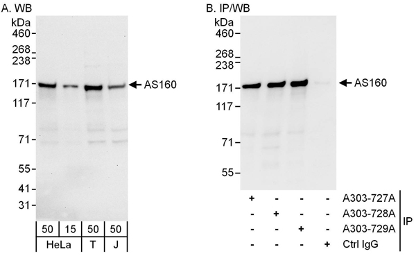
(Detection of human AS160 by western blot and immunoprecipitation. Samples: Whole cell lysate from HeLa (15 and 50 ug for WB; 1 mg for IP, 20% of IP loaded), HEK293T (T; 50 ug) and Jurkat (J; 50 ug) cells. Antibodies: Affinity purified rabbit anti-AS160 antibody AAA212273 used for WB at 0.1 ug/ml (A) and 1 ug/ml (B) and used for IP at 6 ug/mg lysate. AS160 was also immunoprecipitated by rabbit anti-AS160 antibodies and which recognize other epitopes. Detection: Chemiluminescence with exposure times of 10 seconds (A and B).)
AS160, Polyclonal Antibody (Cat# AAA212273)
 IP (Immunoprecipitation)
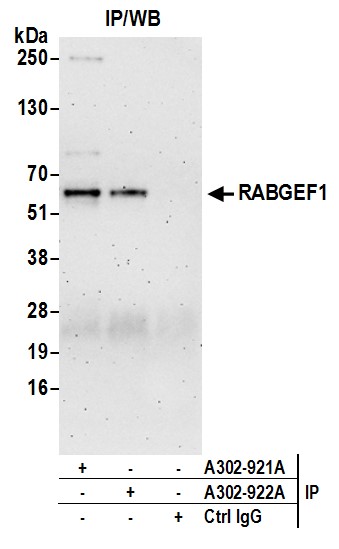
(Detection of human RABGEF1 by western blot of immunoprecipitates. Samples:Whole cell lysate (1.0 mg per IP reaction; 20% of IP loaded) from HeLa cells prepared using NETN lysis buffer. Antibodies: Affinity purified rabbit anti-RABGEF1 antibody AAA212071 (lot AAA212071-2) used for IP at 3 ug per reaction. RABGEF1 was also immunoprecipitated by rabbit anti-RABGEF1 antibody For blotting immunoprecipitated RABGEF1, was used at 1 ug/ml. Detection:Chemiluminescence with an exposure time of 3 minutes.)
IP (Immunoprecipitation)
(Detection of human RABGEF1 by western blot of immunoprecipitates. Samples:Whole cell lysate (1.0 mg per IP reaction; 20% of IP loaded) from HeLa cells prepared using NETN lysis buffer. Antibodies: Affinity purified rabbit anti-RABGEF1 antibody AAA212071 (lot AAA212071-2) used for IP at 3 ug per reaction. RABGEF1 was also immunoprecipitated by rabbit anti-RABGEF1 antibody For blotting immunoprecipitated RABGEF1, was used at 1 ug/ml. Detection:Chemiluminescence with an exposure time of 3 minutes.)
RABGEF1, Polyclonal Antibody (Cat# AAA212071)
 WB (Western Blot)
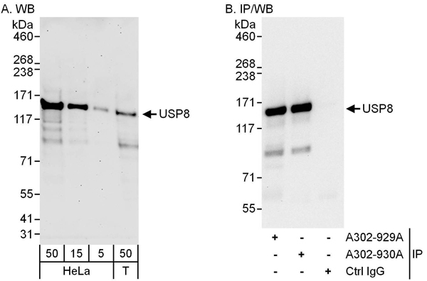
(Detection of human USP8 by western blot and immunoprecipitation. Samples: Whole cell lysate from HeLa (5, 15 and 50 ug for WB; 1 mg for IP, 20% of IP loaded) and HEK293T (T; 50 ug) cells. Antibodies: Affinity purified rabbit anti-USP8 antibody AAA212075 used for WB at 0.04 ug/ml (A) and 0.4 ug/ml (B) and used for IP at 3 ug/mg lysate. USP8 was also immunoprecipitated by rabbit anti-USP8 antibody which recognizes a downstream epitope. Detection: Chemiluminescence with exposure times of 3 minutes (A) and 10 seconds (B).)
WB (Western Blot)
(Detection of human USP8 by western blot and immunoprecipitation. Samples: Whole cell lysate from HeLa (5, 15 and 50 ug for WB; 1 mg for IP, 20% of IP loaded) and HEK293T (T; 50 ug) cells. Antibodies: Affinity purified rabbit anti-USP8 antibody AAA212075 used for WB at 0.04 ug/ml (A) and 0.4 ug/ml (B) and used for IP at 3 ug/mg lysate. USP8 was also immunoprecipitated by rabbit anti-USP8 antibody which recognizes a downstream epitope. Detection: Chemiluminescence with exposure times of 3 minutes (A) and 10 seconds (B).)
USP8, Polyclonal Antibody (Cat# AAA212075)
 WB (Western Blot)
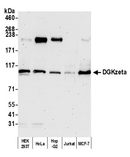
(Detection of human DGKzeta by western blot. Samples: Whole cell lysate (2 to 5 ug) from HEK293T, HeLa, Hep-G2, Jurkat, and MCF-7 cells prepared using NETN lysis buffer. Antibody: Affinity purified rabbit anti-DGKzeta antibody AAA212082 lot 2 used for WB at 0.04 ug/ml. Detection: Chemiluminescence with an exposure time of 75 seconds.)
WB (Western Blot)
(Detection of human DGKzeta by western blot. Samples: Whole cell lysate (2 to 5 ug) from HEK293T, HeLa, Hep-G2, Jurkat, and MCF-7 cells prepared using NETN lysis buffer. Antibody: Affinity purified rabbit anti-DGKzeta antibody AAA212082 lot 2 used for WB at 0.04 ug/ml. Detection: Chemiluminescence with an exposure time of 75 seconds.)
DGKzeta, Polyclonal Antibody (Cat# AAA212082)
 WB (Western Blot)
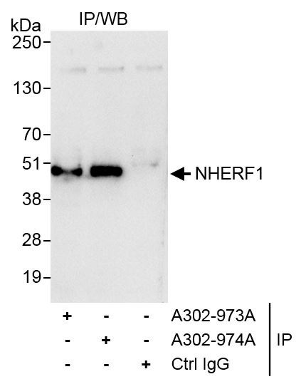
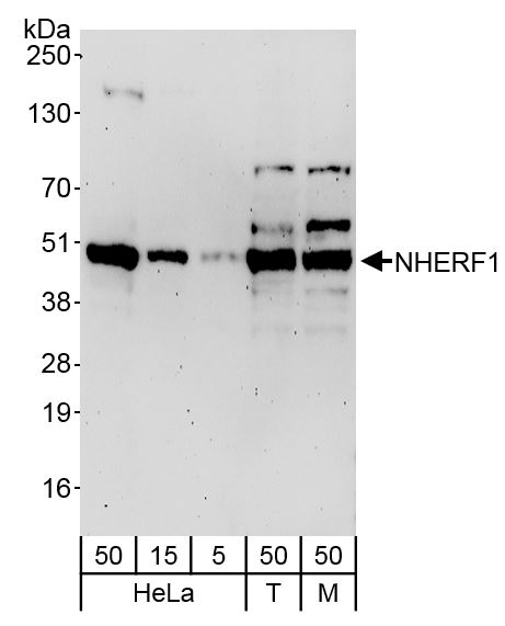
(Detection of human and mouse NHERF1 by western blot. Samples: Whole cell lysate from HeLa (5, 15 and 50 ug), HEK293T (T; 50 ug) and mouse NIH 3T3 (M; 50 ug) cells. Antibodies: Affinity purified rabbit anti-NHERF1 antibody AAA212085 used at 0.04 ug/ml. Detection: Chemiluminescence with exposure time of 3 minutes.)
WB (Western Blot)
(Detection of human and mouse NHERF1 by western blot. Samples: Whole cell lysate from HeLa (5, 15 and 50 ug), HEK293T (T; 50 ug) and mouse NIH 3T3 (M; 50 ug) cells. Antibodies: Affinity purified rabbit anti-NHERF1 antibody AAA212085 used at 0.04 ug/ml. Detection: Chemiluminescence with exposure time of 3 minutes.)
NHERF1, Polyclonal Antibody (Cat# AAA212085)
 WB (Western Blot)
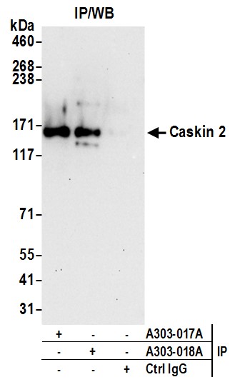
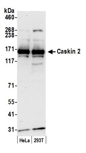
(Detection of human Caskin 2 by western blot. Samples: Whole cell lysate (50 ug) from HeLa and HEK293T cells prepared using NETN lysis buffer. Antibody: Affinity purified rabbit anti-Caskin 2 antibody AAA212097 (lot AAA212097-1) used for WB at 0.4 ug/ml. Detection: Chemiluminescence with an exposure time of 30 seconds.)
WB (Western Blot)
(Detection of human Caskin 2 by western blot. Samples: Whole cell lysate (50 ug) from HeLa and HEK293T cells prepared using NETN lysis buffer. Antibody: Affinity purified rabbit anti-Caskin 2 antibody AAA212097 (lot AAA212097-1) used for WB at 0.4 ug/ml. Detection: Chemiluminescence with an exposure time of 30 seconds.)
Caskin 2, Polyclonal Antibody (Cat# AAA212097)
 WB (Western Blot)
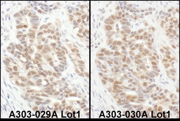
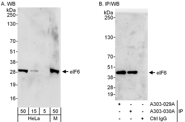
(Detection of human and mouse eIF6 by western blot (h&m) and immunoprecipitation (h). Samples: Whole cell lysate from HeLa (5, 15 and 50 ug for WB; 1 mg for IP, 20% of IP loaded) and mouse NIH 3T3 (M; 50 ug) cells. Antibodies: Affinity purified rabbit anti-eIF6 antibody AAA212099 used for WB at 0.1 ug/ml (A) and 1 ug/ml (B) and used for IP at 6 ug/mg lysate. eIF6 was also immunoprecipitated by rabbit anti-eIF6 antibody which recognizes an upstream epitope. Detection: Chemiluminescence with exposure times of 3 minutes (A) and 10 seconds (B).)
WB (Western Blot)
(Detection of human and mouse eIF6 by western blot (h&m) and immunoprecipitation (h). Samples: Whole cell lysate from HeLa (5, 15 and 50 ug for WB; 1 mg for IP, 20% of IP loaded) and mouse NIH 3T3 (M; 50 ug) cells. Antibodies: Affinity purified rabbit anti-eIF6 antibody AAA212099 used for WB at 0.1 ug/ml (A) and 1 ug/ml (B) and used for IP at 6 ug/mg lysate. eIF6 was also immunoprecipitated by rabbit anti-eIF6 antibody which recognizes an upstream epitope. Detection: Chemiluminescence with exposure times of 3 minutes (A) and 10 seconds (B).)
eIF6, Polyclonal Antibody (Cat# AAA212099)
 IP (Immunoprecipitation)
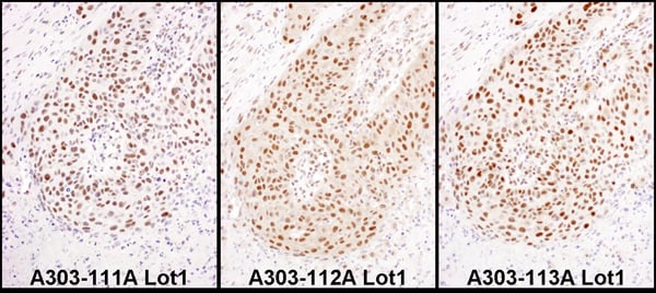
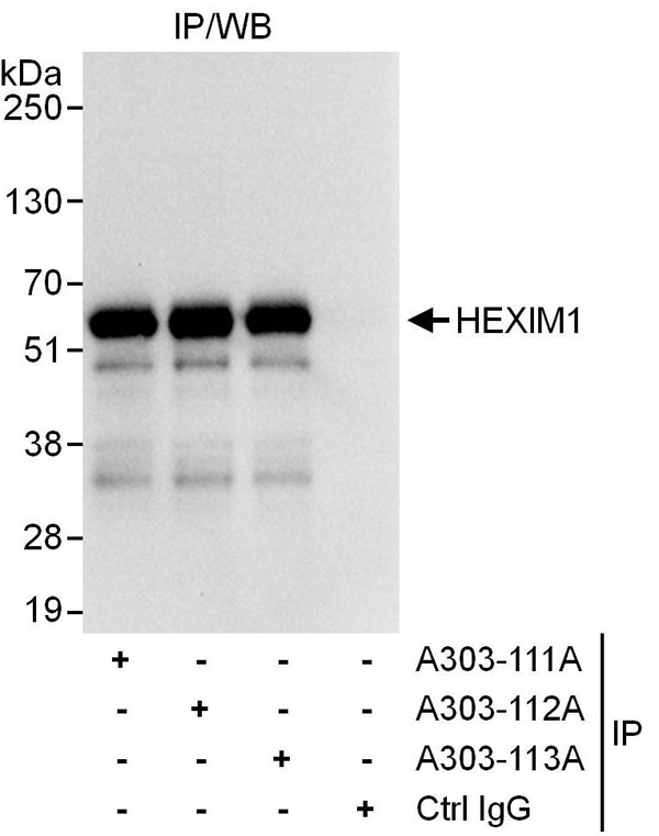
(Detection of human HEXIM1 by western blot of immunoprecipitates. Samples: Whole cell lysate (1 mg for IP, 20% of IP loaded) from HeLa cells. Antibodies: Affinity purified rabbit anti-HEXIM1 antibody AAA212112 used for IP at 6 ug/mg lysate. HEXIM1 was also immunoprecipitated by rabbit anti-HEXIM1 antibodies and which recognize downstream epitopes. For blotting immunoprecipitated HEXIM1, was used at 1 ug/ml. Detection: Chemiluminescence with an exposure time of 3 seconds.)
IP (Immunoprecipitation)
(Detection of human HEXIM1 by western blot of immunoprecipitates. Samples: Whole cell lysate (1 mg for IP, 20% of IP loaded) from HeLa cells. Antibodies: Affinity purified rabbit anti-HEXIM1 antibody AAA212112 used for IP at 6 ug/mg lysate. HEXIM1 was also immunoprecipitated by rabbit anti-HEXIM1 antibodies and which recognize downstream epitopes. For blotting immunoprecipitated HEXIM1, was used at 1 ug/ml. Detection: Chemiluminescence with an exposure time of 3 seconds.)
HEXIM1, Polyclonal Antibody (Cat# AAA212112)
 WB (Western Blot)
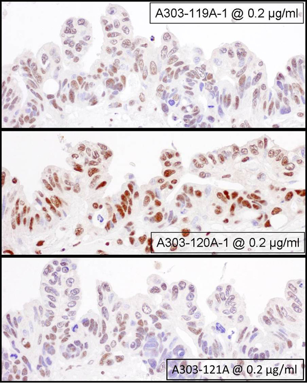
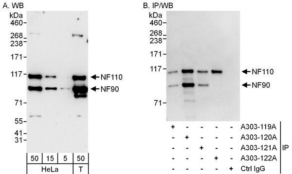
(Detection of human NF90 and NF110 by western blot and immunoprecipitation. Samples: Whole cell lysate from HeLa (5, 15 and 50 ug for WB; 1 mg for IP, 20% of IP loaded) and HEK293T (T; 50 ug) cells. Antibodies: Affinity purified rabbit anti-NF90/NF110 antibody AAA212116 used for WB at 0.04 ug/ml (A) and 1 ug/ml (B) and used for IP at 6 ug/mg lysate. NF90 and/or NF110 were also immunoprecipitated by rabbit anti-NF90/NF110 antibodies and as well as anti-NF110 antibody each of which recognizes a different epitope. Detection: Chemiluminescence with exposure times of 30 seconds (A) and 1 second (B).)
WB (Western Blot)
(Detection of human NF90 and NF110 by western blot and immunoprecipitation. Samples: Whole cell lysate from HeLa (5, 15 and 50 ug for WB; 1 mg for IP, 20% of IP loaded) and HEK293T (T; 50 ug) cells. Antibodies: Affinity purified rabbit anti-NF90/NF110 antibody AAA212116 used for WB at 0.04 ug/ml (A) and 1 ug/ml (B) and used for IP at 6 ug/mg lysate. NF90 and/or NF110 were also immunoprecipitated by rabbit anti-NF90/NF110 antibodies and as well as anti-NF110 antibody each of which recognizes a different epitope. Detection: Chemiluminescence with exposure times of 30 seconds (A) and 1 second (B).)
NF90/NF110, Polyclonal Antibody (Cat# AAA212116)
 IP (Immunoprecipitation)
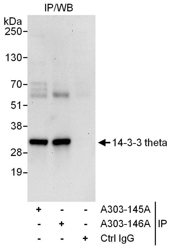
(Detection of human 14-3-3-theta by western blot of immunoprecipitates. Samples: Whole cell lysate (1 mg for IP, 20% of IP loaded) from HeLa cells. Antibodies: Affinity purified rabbit anti-14-3-3-theta antibody AAA212124 used for IP at 6 ug/mg lysate. 14-3-3-theta was also immunoprecipitated by rabbit anti-14-3-3-theta antibody which recognizes a downstream epitope. For blotting immunoprecipitated 14-3-3-theta, was used at 1 ug/ml. Detection: Chemiluminescence with an exposure time of 30 seconds.)
IP (Immunoprecipitation)
(Detection of human 14-3-3-theta by western blot of immunoprecipitates. Samples: Whole cell lysate (1 mg for IP, 20% of IP loaded) from HeLa cells. Antibodies: Affinity purified rabbit anti-14-3-3-theta antibody AAA212124 used for IP at 6 ug/mg lysate. 14-3-3-theta was also immunoprecipitated by rabbit anti-14-3-3-theta antibody which recognizes a downstream epitope. For blotting immunoprecipitated 14-3-3-theta, was used at 1 ug/ml. Detection: Chemiluminescence with an exposure time of 30 seconds.)
14-3-3 theta, Polyclonal Antibody (Cat# AAA212124)
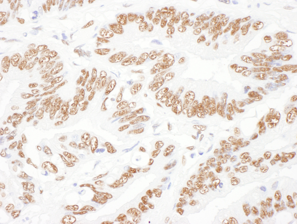
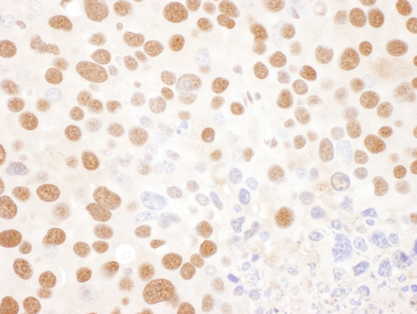
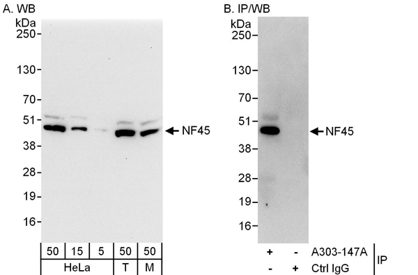 WB (Western Blot)
(Detection of human and mouse NF45 by western blot (h & m) and immunoprecipitation (h). Samples: Whole cell lysate from HeLa (5, 15 and 50 ug for WB; 1 mg for IP, 20% of IP loaded), HEK293T (T; 50 ug) and mouse NIH 3T3 (M; 50 ug) cells. Antibodies: Affinity purified rabbit anti-NF45 antibody AAA212126 used for WB at 0.1 ug/ml (A) and 1 ug/ml (B) and used for IP at 6 ug/mg lysate. Detection: Chemiluminescence with exposure times of 10 seconds (A and B).)
WB (Western Blot)
(Detection of human and mouse NF45 by western blot (h & m) and immunoprecipitation (h). Samples: Whole cell lysate from HeLa (5, 15 and 50 ug for WB; 1 mg for IP, 20% of IP loaded), HEK293T (T; 50 ug) and mouse NIH 3T3 (M; 50 ug) cells. Antibodies: Affinity purified rabbit anti-NF45 antibody AAA212126 used for WB at 0.1 ug/ml (A) and 1 ug/ml (B) and used for IP at 6 ug/mg lysate. Detection: Chemiluminescence with exposure times of 10 seconds (A and B).)
NF45, Polyclonal Antibody (Cat# AAA212126)
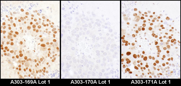
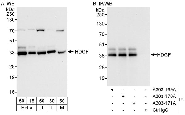 WB (Western Blot)
(Detection of human and mouse HDGF by western blot (h and m) and immunoprecipitation (h). Samples: Whole cell lysate from HeLa (15 and 50 ug for WB; 1 mg for IP, 20% of IP loaded), Jurkat (J; 50 ug), HEK293T (T; 50 ug), and mouse NIH 3T3 (M; 50 ug) cells. Antibodies: Affinity purified rabbit anti-HDGF antibody AAA212130 used for WB at 0.04 ug/ml (A) and 1 ug/ml (B) and used for IP at 6 ug/mg lysate. HDGF was also immunoprecipitated by rabbit anti-HDGF antibodies and which recognize upstream epitopes. Detection: Chemiluminescence with exposure times of 10 seconds (A) and 3 seconds (B).)
WB (Western Blot)
(Detection of human and mouse HDGF by western blot (h and m) and immunoprecipitation (h). Samples: Whole cell lysate from HeLa (15 and 50 ug for WB; 1 mg for IP, 20% of IP loaded), Jurkat (J; 50 ug), HEK293T (T; 50 ug), and mouse NIH 3T3 (M; 50 ug) cells. Antibodies: Affinity purified rabbit anti-HDGF antibody AAA212130 used for WB at 0.04 ug/ml (A) and 1 ug/ml (B) and used for IP at 6 ug/mg lysate. HDGF was also immunoprecipitated by rabbit anti-HDGF antibodies and which recognize upstream epitopes. Detection: Chemiluminescence with exposure times of 10 seconds (A) and 3 seconds (B).)
HDGF, Polyclonal Antibody (Cat# AAA212130)
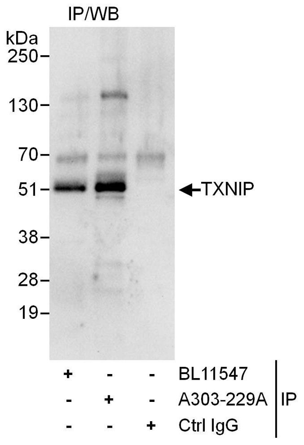 IP (Immunoprecipitation)
(Detection of human TXNIP by western blot of immunoprecipitates. Samples: Whole cell lysate (1 mg for IP, 20% of IP loaded) from HeLa cells. Antibodies: Affinity purified rabbit anti-TXNIP antibody AAA212142 used for IP at 6 ug/mg lysate. TXNIP was also immunoprecipitated by rabbit anti-TXNIP antibody BL11547, which recognizes an upstream epitope. For blotting immunoprecipitated TXNIP, AAA212142 was used at 1 ug/ml. Detection: Chemiluminescence with an exposure time of 10 seconds.)
IP (Immunoprecipitation)
(Detection of human TXNIP by western blot of immunoprecipitates. Samples: Whole cell lysate (1 mg for IP, 20% of IP loaded) from HeLa cells. Antibodies: Affinity purified rabbit anti-TXNIP antibody AAA212142 used for IP at 6 ug/mg lysate. TXNIP was also immunoprecipitated by rabbit anti-TXNIP antibody BL11547, which recognizes an upstream epitope. For blotting immunoprecipitated TXNIP, AAA212142 was used at 1 ug/ml. Detection: Chemiluminescence with an exposure time of 10 seconds.)
TXNIP, Polyclonal Antibody (Cat# AAA212142)
 WB (Western Blot)
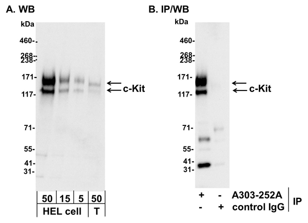
(Detection of human c-Kit by western blot and immunoprecipitation. Samples: Whole cell lysate from human erythroleukemia (HEL) cells (50, 15, and 5 ug for WB; 1 mg for IP, 20% of IP loaded and HEK293T (T; 50 ug). Antibodies: Affinity purified rabbit anti-c-Kit antibody AAA212145 used for WB at 0.1 ug/ml (A) and 1.0 ug/ml (B) and used for IP at 3 ug/mg lysate. Detection: Chemiluminescence with exposure times of 10 seconds (A) and 3 seconds (B).)
WB (Western Blot)
(Detection of human c-Kit by western blot and immunoprecipitation. Samples: Whole cell lysate from human erythroleukemia (HEL) cells (50, 15, and 5 ug for WB; 1 mg for IP, 20% of IP loaded and HEK293T (T; 50 ug). Antibodies: Affinity purified rabbit anti-c-Kit antibody AAA212145 used for WB at 0.1 ug/ml (A) and 1.0 ug/ml (B) and used for IP at 3 ug/mg lysate. Detection: Chemiluminescence with exposure times of 10 seconds (A) and 3 seconds (B).)
c-Kit, Polyclonal Antibody (Cat# AAA212145)
 WB (Western Blot)
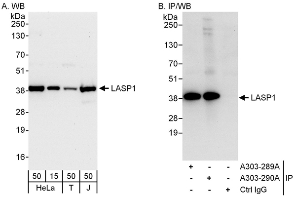
(Detection of human LASP1 by western blot and immunoprecipitation. Samples: Whole cell lysate from HeLa (15 and 50 ug for WB; 1 mg for IP, 20% of IP loaded), HEK293T (T; 50 ug) and Jurkat (J; 50 ug) cells. Antibodies: Affinity purified rabbit anti-LASP1 antibody AAA212148 used for WB at 0.04 ug/ml (A) and 1 ug/ml (B) and used for IP at 6 ug/mg lysate. LASP1 was also immunoprecipitated by rabbit anti-LASP1 antibody which recognizes an upstream epitope. Detection: Chemiluminescence with exposure times of 30 seconds (A) and 10 seconds (B).)
WB (Western Blot)
(Detection of human LASP1 by western blot and immunoprecipitation. Samples: Whole cell lysate from HeLa (15 and 50 ug for WB; 1 mg for IP, 20% of IP loaded), HEK293T (T; 50 ug) and Jurkat (J; 50 ug) cells. Antibodies: Affinity purified rabbit anti-LASP1 antibody AAA212148 used for WB at 0.04 ug/ml (A) and 1 ug/ml (B) and used for IP at 6 ug/mg lysate. LASP1 was also immunoprecipitated by rabbit anti-LASP1 antibody which recognizes an upstream epitope. Detection: Chemiluminescence with exposure times of 30 seconds (A) and 10 seconds (B).)
LASP1, Polyclonal Antibody (Cat# AAA212148)
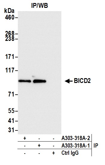
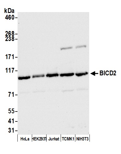 WB (Western Blot)
(Detection of human BICD2 by western blot. Samples: Whole cell lysate (50 ug) from HeLa, HEK293T, Jurkat, TCMK-1, and NIH 3T3 cells prepared using NETN lysis buffer. Antibody: Affinity purified rabbit anti-BICD2 antibody AAA212153 (lot AAA212153-3) used for WB at 0.04 ug/ml. Detection: Chemiluminescence with an exposure time of 10 seconds.)
WB (Western Blot)
(Detection of human BICD2 by western blot. Samples: Whole cell lysate (50 ug) from HeLa, HEK293T, Jurkat, TCMK-1, and NIH 3T3 cells prepared using NETN lysis buffer. Antibody: Affinity purified rabbit anti-BICD2 antibody AAA212153 (lot AAA212153-3) used for WB at 0.04 ug/ml. Detection: Chemiluminescence with an exposure time of 10 seconds.)
BICD2, Polyclonal Antibody (Cat# AAA212153)
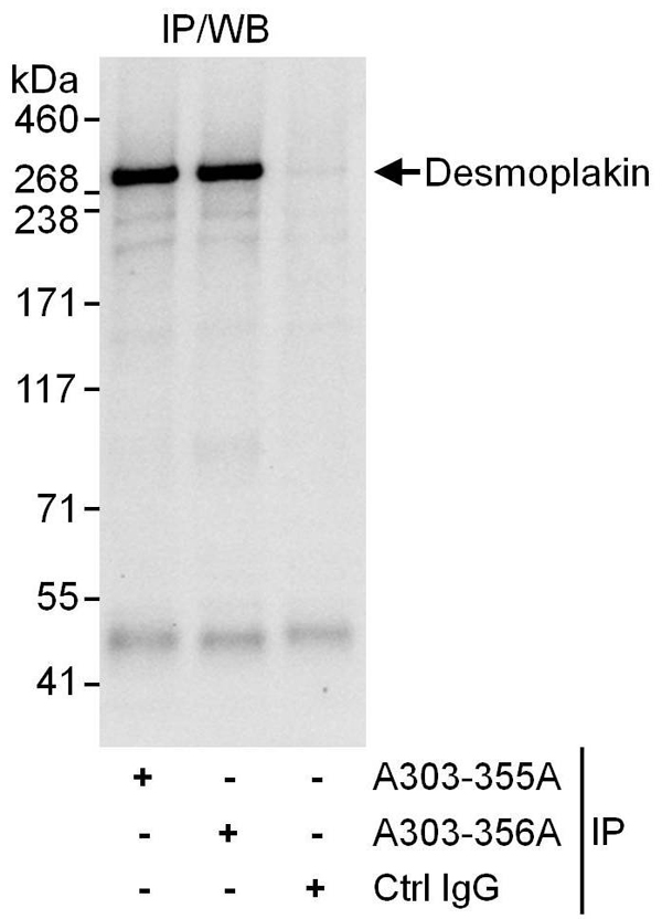 IP (Immunoprecipitation)
(Detection of human Desmoplakin by western blot of immunoprecipitates. Samples: Whole cell lysate (1 mg for IP, 20% of IP loaded) from HeLa cells. Antibodies: Affinity purified rabbit anti-Desmoplakin antibody AAA212155 used for IP at 6 ug/mg lysate. Desmoplakin was also immunoprecipitated by rabbit anti-Desmoplakin antibody which recognizes an upstream epitope. For blotting immunoprecipitated Desmoplakin, was used at 1 ug/ml. Detection: Chemiluminescence with an exposure time of 10 seconds.)
IP (Immunoprecipitation)
(Detection of human Desmoplakin by western blot of immunoprecipitates. Samples: Whole cell lysate (1 mg for IP, 20% of IP loaded) from HeLa cells. Antibodies: Affinity purified rabbit anti-Desmoplakin antibody AAA212155 used for IP at 6 ug/mg lysate. Desmoplakin was also immunoprecipitated by rabbit anti-Desmoplakin antibody which recognizes an upstream epitope. For blotting immunoprecipitated Desmoplakin, was used at 1 ug/ml. Detection: Chemiluminescence with an exposure time of 10 seconds.)
Desmoplakin, Polyclonal Antibody (Cat# AAA212155)
 IP (Immunoprecipitation)
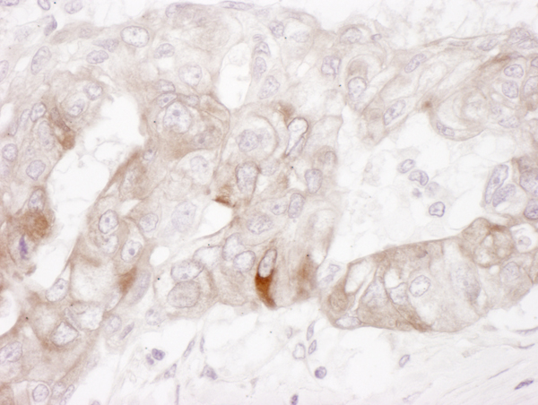
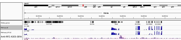
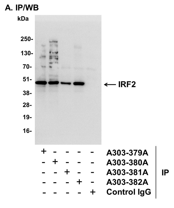
(Detection of human IRF2 by western blot of immunoprecipitates. Samples: Whole cell lysate (1 mg for IP, 20% of IP loaded) from human erythroleukemia (HEL) cells. Antibodies: Affinity purified rabbit anti-IRF2 antibody AAA212159 used for IP at 6 ug/mg lysate. IRF2 was also immunoprecipitated by rabbit anti-IRF2 antibodies)
IP (Immunoprecipitation)
(Detection of human IRF2 by western blot of immunoprecipitates. Samples: Whole cell lysate (1 mg for IP, 20% of IP loaded) from human erythroleukemia (HEL) cells. Antibodies: Affinity purified rabbit anti-IRF2 antibody AAA212159 used for IP at 6 ug/mg lysate. IRF2 was also immunoprecipitated by rabbit anti-IRF2 antibodies)
IRF2, Polyclonal Antibody (Cat# AAA212159)
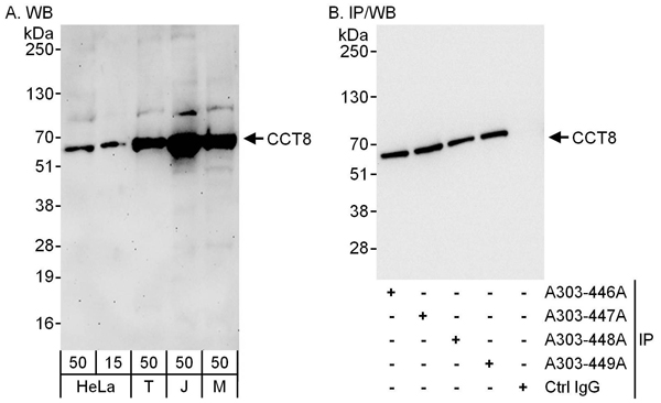 WB (Western Blot)
(Detection of human and mouse CCT8 by western blot (h and m) and immunoprecipitation (h). Samples: Whole cell lysate from HeLa (15 and 50 ug for WB; 1 mg for IP, 20% of IP loaded), HEK293T (T; 50 ug), Jurkat (J; 50 ug) and mouse NIH 3T3 (M; 50 ug) cells. Antibodies: Affinity purified rabbit anti-CCT8 antibody AAA212169 used for WB at 0.04 ug/ml (A) and 0.4 ug/ml (B) and used for IP at 6 ug/mg lysate. CCT8 was also immunoprecipitated by rabbit anti-CCT8 antibodies A303-448 and which recognize downstream epitopes. Detection: Chemiluminescence with exposure times of 3 minutes (A) and 3 seconds (B).)
WB (Western Blot)
(Detection of human and mouse CCT8 by western blot (h and m) and immunoprecipitation (h). Samples: Whole cell lysate from HeLa (15 and 50 ug for WB; 1 mg for IP, 20% of IP loaded), HEK293T (T; 50 ug), Jurkat (J; 50 ug) and mouse NIH 3T3 (M; 50 ug) cells. Antibodies: Affinity purified rabbit anti-CCT8 antibody AAA212169 used for WB at 0.04 ug/ml (A) and 0.4 ug/ml (B) and used for IP at 6 ug/mg lysate. CCT8 was also immunoprecipitated by rabbit anti-CCT8 antibodies A303-448 and which recognize downstream epitopes. Detection: Chemiluminescence with exposure times of 3 minutes (A) and 3 seconds (B).)
CCT8, Polyclonal Antibody (Cat# AAA212169)
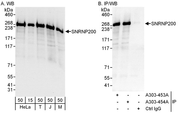 WB (Western Blot)
(Detection of human and mouse SNRNP200 by western blot (h and m) and immunoprecipitation (h). Samples: Whole cell lysate from HeLa (15 and 50 ug for WB; 1 mg for IP, 20% of IP loaded), HEK293T (T; 50 ug), Jurkat (J; 50 ug) and mouse NIH 3T3 (M; 50 ug) cells. Antibodies: Affinity purified rabbit anti-SNRNP200 antibody AAA212172 used for WB at 0.04 ug/ml (A) and 0.4 ug/ml (B) and used for IP at 6 ug/mg lysate. SNRNP200 was also immunoprecipitated by rabbit anti-SNRNP200 antibody which recognizes a downstream epitope. Detection: Chemiluminescence with exposure times of 10 seconds (A) and 3 seconds (B).)
WB (Western Blot)
(Detection of human and mouse SNRNP200 by western blot (h and m) and immunoprecipitation (h). Samples: Whole cell lysate from HeLa (15 and 50 ug for WB; 1 mg for IP, 20% of IP loaded), HEK293T (T; 50 ug), Jurkat (J; 50 ug) and mouse NIH 3T3 (M; 50 ug) cells. Antibodies: Affinity purified rabbit anti-SNRNP200 antibody AAA212172 used for WB at 0.04 ug/ml (A) and 0.4 ug/ml (B) and used for IP at 6 ug/mg lysate. SNRNP200 was also immunoprecipitated by rabbit anti-SNRNP200 antibody which recognizes a downstream epitope. Detection: Chemiluminescence with exposure times of 10 seconds (A) and 3 seconds (B).)
SNRNP200, Polyclonal Antibody (Cat# AAA212172)
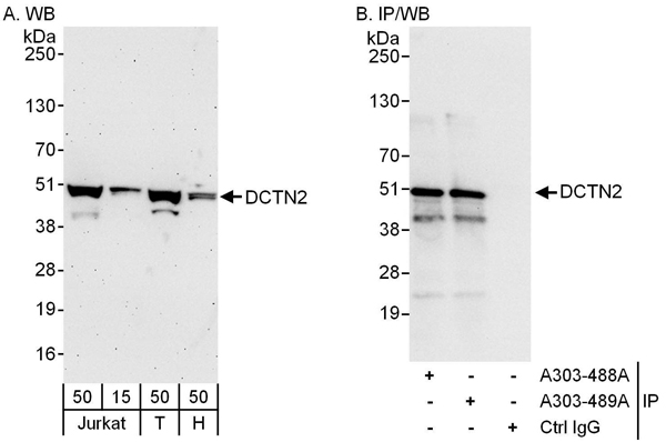 WB (Western Blot)
(Detection of human DCTN2 by western blot and immunoprecipitation. Samples: Whole cell lysate from Jurkat (15 and 50 ug for WB; 1 mg for IP, 20% of IP loaded), HEK293T (T; 50 ug) and HeLa (H; 50 ug) cells. Antibodies: Affinity purified rabbit anti-DCTN2 antibody AAA212183 used for WB at 0.04 ug/ml (A) and 1 ug/ml (B) and used for IP at 6 ug/mg lysate. DCTN2 was also immunoprecipitated by rabbit anti-DCTN2 antibody which recognizes an upstream epitope. Detection: Chemiluminescence with exposure times of 3 minutes (A) and 3 seconds (B).)
WB (Western Blot)
(Detection of human DCTN2 by western blot and immunoprecipitation. Samples: Whole cell lysate from Jurkat (15 and 50 ug for WB; 1 mg for IP, 20% of IP loaded), HEK293T (T; 50 ug) and HeLa (H; 50 ug) cells. Antibodies: Affinity purified rabbit anti-DCTN2 antibody AAA212183 used for WB at 0.04 ug/ml (A) and 1 ug/ml (B) and used for IP at 6 ug/mg lysate. DCTN2 was also immunoprecipitated by rabbit anti-DCTN2 antibody which recognizes an upstream epitope. Detection: Chemiluminescence with exposure times of 3 minutes (A) and 3 seconds (B).)
DCTN2, Polyclonal Antibody (Cat# AAA212183)
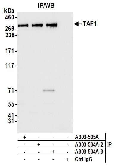
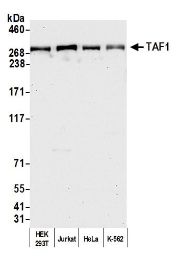 WB (Western Blot)
(Detection of human TAF1 by western blot. Samples: Whole cell lysate (50 ug) from HEK293T, Jurkat, HeLa, and K-562 cells prepared using NETN lysis buffer. Antibody: Affinity purified rabbit anti-TAF1 antibody AAA212195 lot 3 used for WB at 0.04 ug/ml. Detection: Chemiluminescence with an exposure time of 30 seconds.)
WB (Western Blot)
(Detection of human TAF1 by western blot. Samples: Whole cell lysate (50 ug) from HEK293T, Jurkat, HeLa, and K-562 cells prepared using NETN lysis buffer. Antibody: Affinity purified rabbit anti-TAF1 antibody AAA212195 lot 3 used for WB at 0.04 ug/ml. Detection: Chemiluminescence with an exposure time of 30 seconds.)
TAF1, Polyclonal Antibody (Cat# AAA212195)
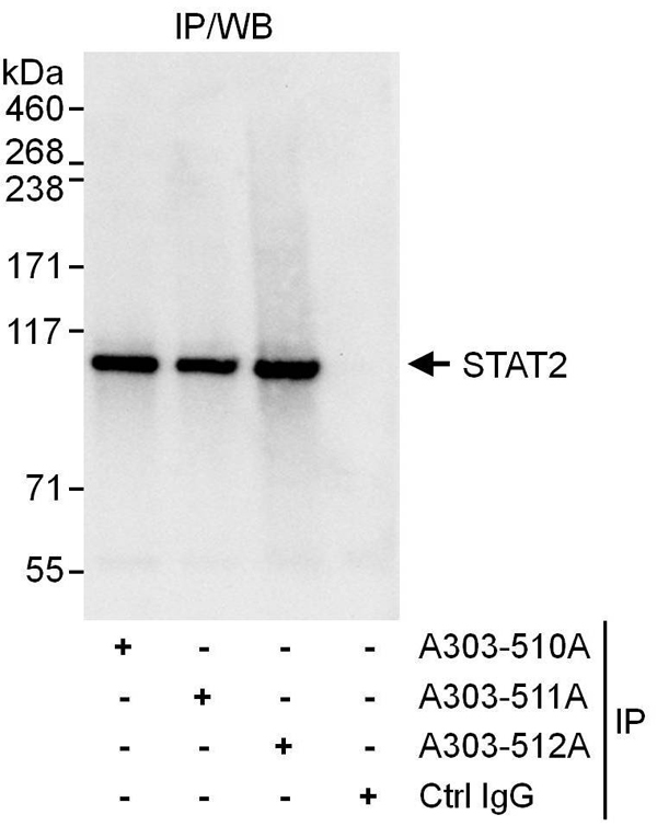 IP (Immunoprecipitation)
(Detection of human STAT2 by western blot of immunoprecipitates. Samples: Whole cell lysate (1 mg for IP, 20% of IP loaded) from Jurkat cells. Antibodies: Affinity purified rabbit anti-STAT2 antibody AAA212197 used for IP at 6 ug/mg lysate. STAT2 was also immunoprecipitated by rabbit anti-STAT2 antibodies and which recognize downstream epitopes. For blotting immunoprecipitated STAT2, was used at 1 ug/ml. Detection: Chemiluminescence with an exposure time of 3 seconds.)
IP (Immunoprecipitation)
(Detection of human STAT2 by western blot of immunoprecipitates. Samples: Whole cell lysate (1 mg for IP, 20% of IP loaded) from Jurkat cells. Antibodies: Affinity purified rabbit anti-STAT2 antibody AAA212197 used for IP at 6 ug/mg lysate. STAT2 was also immunoprecipitated by rabbit anti-STAT2 antibodies and which recognize downstream epitopes. For blotting immunoprecipitated STAT2, was used at 1 ug/ml. Detection: Chemiluminescence with an exposure time of 3 seconds.)
STAT2, Polyclonal Antibody (Cat# AAA212197)
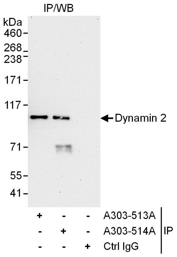 IP (Immunoprecipitation)
(Detection of human Dynamin 2 by western blot of immunoprecipitates. Samples: Whole cell lysate (1 mg for IP, 20% of IP loaded) from HeLa cells. Antibodies: Affinity purified rabbit anti-Dynamin 2 antibody AAA212200 used for IP at 6 ug/mg lysate. Dynamin 2 was also immunoprecipitated by rabbit anti-Dynamin 2 antibody which recognizes a downstream epitope. For blotting immunoprecipitated Dynamin 2, was used at 1 ug/ml. Detection: Chemiluminescence with an exposure time of 30 seconds.)
IP (Immunoprecipitation)
(Detection of human Dynamin 2 by western blot of immunoprecipitates. Samples: Whole cell lysate (1 mg for IP, 20% of IP loaded) from HeLa cells. Antibodies: Affinity purified rabbit anti-Dynamin 2 antibody AAA212200 used for IP at 6 ug/mg lysate. Dynamin 2 was also immunoprecipitated by rabbit anti-Dynamin 2 antibody which recognizes a downstream epitope. For blotting immunoprecipitated Dynamin 2, was used at 1 ug/ml. Detection: Chemiluminescence with an exposure time of 30 seconds.)
Dynamin 2, Polyclonal Antibody (Cat# AAA212200)
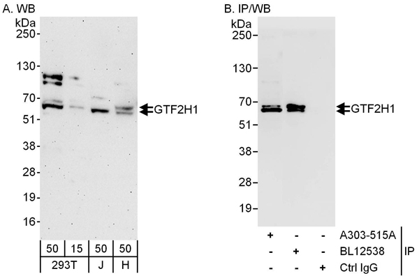 WB (Western Blot)
(Detection of human GTF2H1 by western blot and immunoprecipitation. Samples: Whole cell lysate from HEK293T (15 and 50 ug for WB; 1 mg for IP, 20% of IP loaded), Jurkat (J; 50 ug) and HeLa (H; 50 ug) cells. Antibodies: Affinity purified rabbit anti-GTF2H1 antibody AAA212201 used for WB at 0.1 ug/ml (A) and 1 ug/ml (B) and used for IP at 6 ug/mg lysate. GTF2H1 was also immunoprecipitated by rabbit anti-GTF2H1 antibody BL12538, which recognizes a downstream epitope. Detection: Chemiluminescence with exposure times of 3 minutes (A) and 10 seconds (B).)
WB (Western Blot)
(Detection of human GTF2H1 by western blot and immunoprecipitation. Samples: Whole cell lysate from HEK293T (15 and 50 ug for WB; 1 mg for IP, 20% of IP loaded), Jurkat (J; 50 ug) and HeLa (H; 50 ug) cells. Antibodies: Affinity purified rabbit anti-GTF2H1 antibody AAA212201 used for WB at 0.1 ug/ml (A) and 1 ug/ml (B) and used for IP at 6 ug/mg lysate. GTF2H1 was also immunoprecipitated by rabbit anti-GTF2H1 antibody BL12538, which recognizes a downstream epitope. Detection: Chemiluminescence with exposure times of 3 minutes (A) and 10 seconds (B).)
GTF2H1, Polyclonal Antibody (Cat# AAA212201)
 WB (Western Blot)
(Detection of human FOXC1 by western blot and immunoprecipitation. Samples: Whole cell lysate from HeLa (15 and 50 ug for WB; 1 mg for IP, 20% of IP loaded), HEK293T (T; 50 ug) and Jurkat (J; 50 ug) cells. Antibodies: Affinity purified rabbit anti-FOXC1 antibody AAA212203 used for WB at 0.04 ug/ml (A) and 1 ug/ml (B) and used for IP at 6 ug/mg lysate. FOXC1 was also immunoprecipitated by rabbit anti-FOXC1 antibody which recognizes an upstream epitope. Detection: Chemiluminescence with exposure times of 10 seconds (A and B).)
WB (Western Blot)
(Detection of human FOXC1 by western blot and immunoprecipitation. Samples: Whole cell lysate from HeLa (15 and 50 ug for WB; 1 mg for IP, 20% of IP loaded), HEK293T (T; 50 ug) and Jurkat (J; 50 ug) cells. Antibodies: Affinity purified rabbit anti-FOXC1 antibody AAA212203 used for WB at 0.04 ug/ml (A) and 1 ug/ml (B) and used for IP at 6 ug/mg lysate. FOXC1 was also immunoprecipitated by rabbit anti-FOXC1 antibody which recognizes an upstream epitope. Detection: Chemiluminescence with exposure times of 10 seconds (A and B).)
FOXC1, Polyclonal Antibody (Cat# AAA212203)
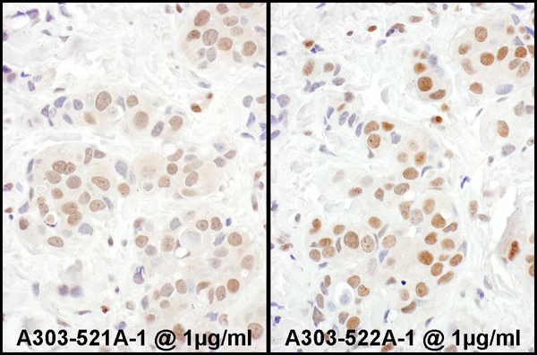
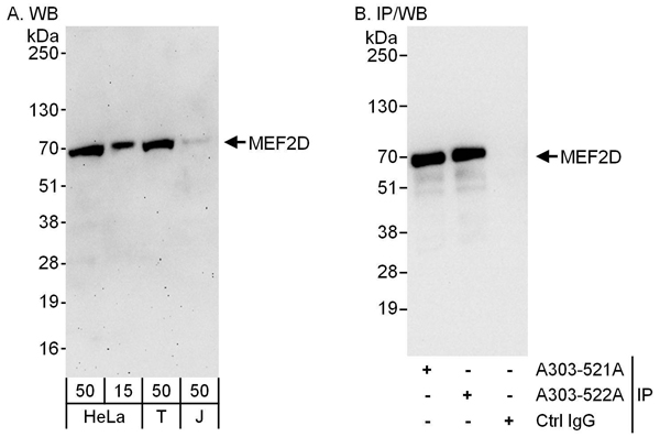 WB (Western Blot)
(Detection of human MEF2D by western blot and immunoprecipitation. Samples: Whole cell lysate from HeLa (15 and 50 ug for WB; 1 mg for IP, 20% of IP loaded), HEK293T (T; 50 ug) and Jurkat (J; 50 ug) cells. Antibodies: Affinity purified rabbit anti-MEF2D antibody AAA212204 used for WB at 0.04 ug/ml (A) and 1 ug/ml (B) and used for IP at 6 ug/mg lysate. MEF2D was also immunoprecipitated by rabbit anti-MEF2D antibody which recognizes a downstream epitope. Detection: Chemiluminescence with exposure times of 3 minutes (A) and 10 seconds (B).)
WB (Western Blot)
(Detection of human MEF2D by western blot and immunoprecipitation. Samples: Whole cell lysate from HeLa (15 and 50 ug for WB; 1 mg for IP, 20% of IP loaded), HEK293T (T; 50 ug) and Jurkat (J; 50 ug) cells. Antibodies: Affinity purified rabbit anti-MEF2D antibody AAA212204 used for WB at 0.04 ug/ml (A) and 1 ug/ml (B) and used for IP at 6 ug/mg lysate. MEF2D was also immunoprecipitated by rabbit anti-MEF2D antibody which recognizes a downstream epitope. Detection: Chemiluminescence with exposure times of 3 minutes (A) and 10 seconds (B).)
MEF2D, Polyclonal Antibody (Cat# AAA212204)
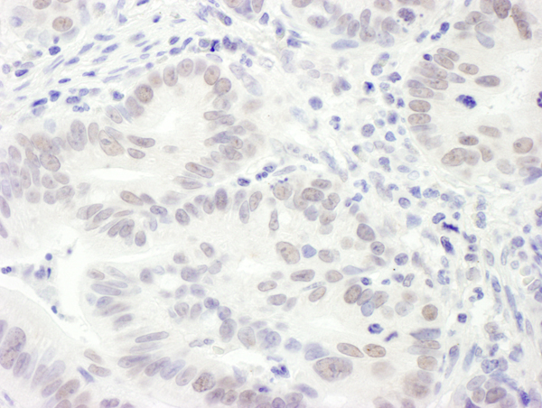
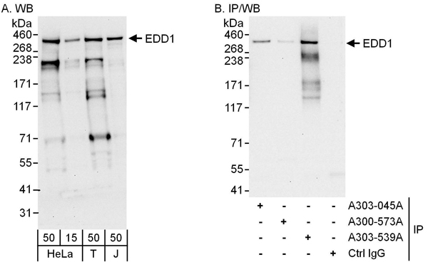 WB (Western Blot)
(Detection of human EDD1 by western blot and immunoprecipitation. Samples: Whole cell lysate from HeLa (15 and 50 ug for WB; 1 mg for IP, 20% of IP loaded), HEK293T (T; 50 ug) and Jurkat (J; 50 ug) cells. Antibodies: Affinity purified rabbit anti-EDD1 antibody AAA212214 used for WB at 0.4 ug/ml (A) and 1 ug/ml (B) and used for IP at 6 ug/mg lysate. EDD1 was also immunoprecipitated by rabbit anti-EDD1 antibodies which recognizes a downstream epitope, and which recognizes a similar epitope. Detection: Chemiluminescence with exposure times of 10 seconds (A and B).)
WB (Western Blot)
(Detection of human EDD1 by western blot and immunoprecipitation. Samples: Whole cell lysate from HeLa (15 and 50 ug for WB; 1 mg for IP, 20% of IP loaded), HEK293T (T; 50 ug) and Jurkat (J; 50 ug) cells. Antibodies: Affinity purified rabbit anti-EDD1 antibody AAA212214 used for WB at 0.4 ug/ml (A) and 1 ug/ml (B) and used for IP at 6 ug/mg lysate. EDD1 was also immunoprecipitated by rabbit anti-EDD1 antibodies which recognizes a downstream epitope, and which recognizes a similar epitope. Detection: Chemiluminescence with exposure times of 10 seconds (A and B).)
EDD1, Polyclonal Antibody (Cat# AAA212214)
 WB (Western Blot)
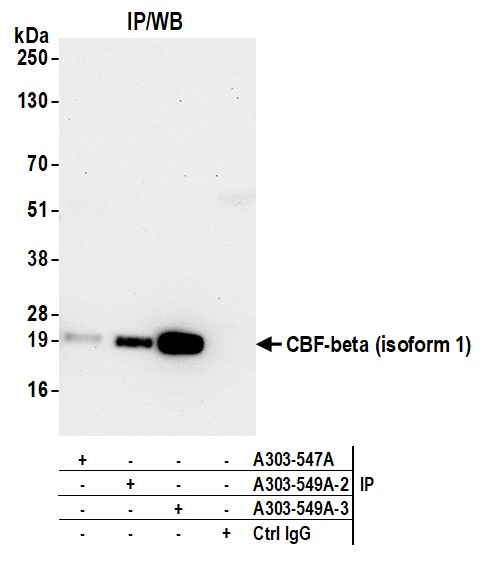
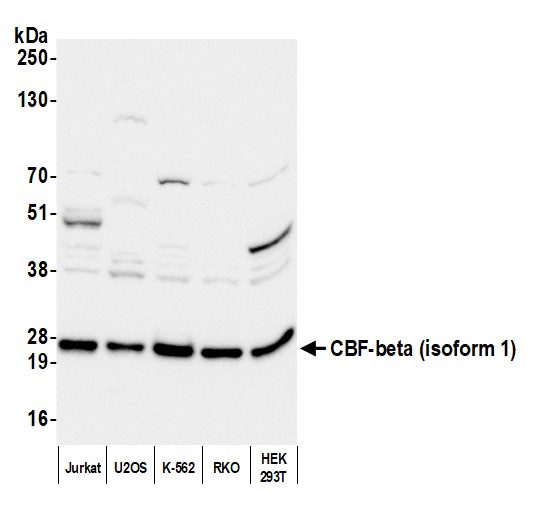
(Detection of human CBF-beta (isoform 1) by western blot. Samples: Whole cell lysate (25 ug) from Jurkat, U2OS, K-562, RKO, and HEK293T cells prepared using NETN lysis buffer. Antibody: Affinity purified rabbit anti-CBF-beta (isoform 1) antibody (AAA212219 lot 3) used for WB at 0.1 ug/ml. Detection: Chemiluminescence with an exposure time of 3 seconds.)
WB (Western Blot)
(Detection of human CBF-beta (isoform 1) by western blot. Samples: Whole cell lysate (25 ug) from Jurkat, U2OS, K-562, RKO, and HEK293T cells prepared using NETN lysis buffer. Antibody: Affinity purified rabbit anti-CBF-beta (isoform 1) antibody (AAA212219 lot 3) used for WB at 0.1 ug/ml. Detection: Chemiluminescence with an exposure time of 3 seconds.)
CBF-beta (isoform 1), Polyclonal Antibody (Cat# AAA212219)
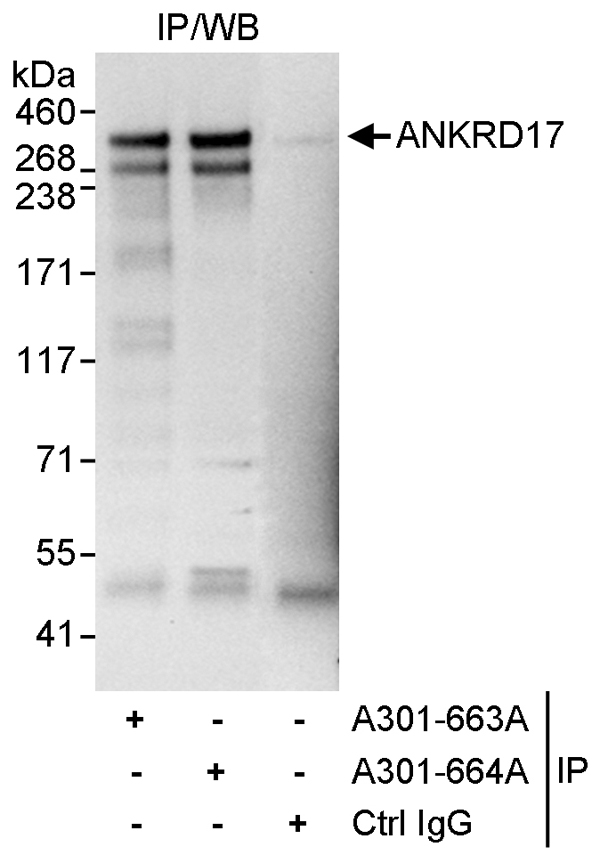 IP (Immunoprecipitation)
(Detection of human ANKRD17 by western blot of immunoprecipitates. Samples: Whole cell lysate (1 mg for IP, 20% of IP loaded) from HeLa cells. Antibodies: Affinity purified rabbit anti-ANKRD17 antibody AAA211579 used for IP at 3 ug/mg lysate. ANKRD17 was also immunoprecipitated by rabbit anti-ANKRD17 antibody which recognizes an upstream epitope. For blotting immunoprecipitated ANKRD17, was used at 1 ug/ml. Detection: Chemiluminescence with an exposure time 10 seconds.)
IP (Immunoprecipitation)
(Detection of human ANKRD17 by western blot of immunoprecipitates. Samples: Whole cell lysate (1 mg for IP, 20% of IP loaded) from HeLa cells. Antibodies: Affinity purified rabbit anti-ANKRD17 antibody AAA211579 used for IP at 3 ug/mg lysate. ANKRD17 was also immunoprecipitated by rabbit anti-ANKRD17 antibody which recognizes an upstream epitope. For blotting immunoprecipitated ANKRD17, was used at 1 ug/ml. Detection: Chemiluminescence with an exposure time 10 seconds.)
ANKRD17, Polyclonal Antibody (Cat# AAA211579)
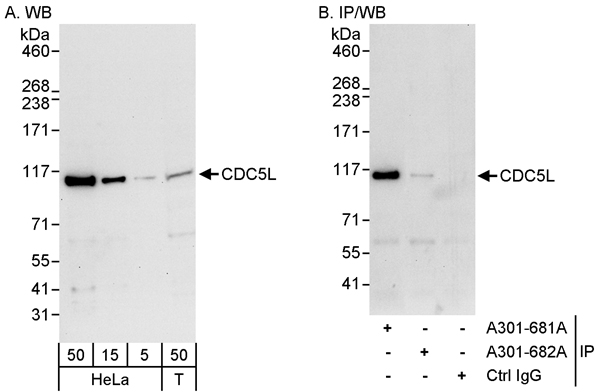 WB (Western Blot)
(Detection of human CDC5L by western blot and immunoprecipitation. Samples: Whole cell lysate from HeLa (5, 15 and 50 ug for WB; 1 mg for IP, 20% of IP loaded) and HEK293T (T; 50 ug) cells. Antibodies: Affinity purified rabbit anti-CDC5L antibody AAA211587 used for WB at 0.04 ug/ml (A) and 1 ug/ml (B) and used for IP at 3 ug/mg lysate. CDC5L was also immunoprecipitated by rabbit anti-CDC5L antibody which recognizes an upstream epitope. Detection: Chemiluminescence with exposure times of 30 seconds (A) and 10 seconds (B).)
WB (Western Blot)
(Detection of human CDC5L by western blot and immunoprecipitation. Samples: Whole cell lysate from HeLa (5, 15 and 50 ug for WB; 1 mg for IP, 20% of IP loaded) and HEK293T (T; 50 ug) cells. Antibodies: Affinity purified rabbit anti-CDC5L antibody AAA211587 used for WB at 0.04 ug/ml (A) and 1 ug/ml (B) and used for IP at 3 ug/mg lysate. CDC5L was also immunoprecipitated by rabbit anti-CDC5L antibody which recognizes an upstream epitope. Detection: Chemiluminescence with exposure times of 30 seconds (A) and 10 seconds (B).)
CDC5L, Polyclonal Antibody (Cat# AAA211587)
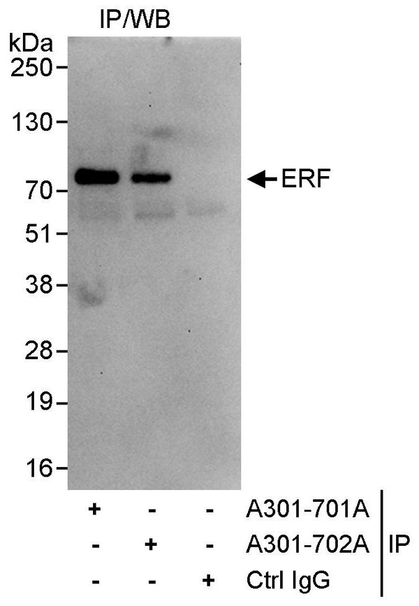 IP (Immunoprecipitation)
(Detection of human ERF by western blot of immunoprecipitates. Samples: Whole cell lysate (1 mg for IP, 20% of IP loaded) from HeLa cells. Antibodies: Affinity purified rabbit anti-ERF antibody AAA211595 used for IP at 3 ug/mg lysate. ERF was also immunoprecipitated by rabbit anti-ERF antibody which recognizes an upstream epitope. For blotting immunoprecipitated ERF, AAA211595 was used at 1 ug/ml. Detection: Chemiluminescence with an exposure time of 30 seconds.)
IP (Immunoprecipitation)
(Detection of human ERF by western blot of immunoprecipitates. Samples: Whole cell lysate (1 mg for IP, 20% of IP loaded) from HeLa cells. Antibodies: Affinity purified rabbit anti-ERF antibody AAA211595 used for IP at 3 ug/mg lysate. ERF was also immunoprecipitated by rabbit anti-ERF antibody which recognizes an upstream epitope. For blotting immunoprecipitated ERF, AAA211595 was used at 1 ug/ml. Detection: Chemiluminescence with an exposure time of 30 seconds.)
ERF, Polyclonal Antibody (Cat# AAA211595)
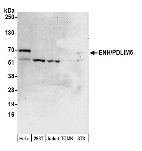 WB (Western Blot)
(Detection of human and mouse ENH/PDLIM5 by western blot. Samples: Whole cell lysate (50 ug) prepared using NETN buffer from HeLa, HEK293T, Jurkat, mouse TCMK-1, and mouse NIH 3T3 cells. Antibodies: Affinity purified rabbit anti-ENH/PDLIM5 antibody AAA211596 (lot AAA211596-2) used for WB at 0.1 ug/ml. Detection: Chemiluminescence with an exposure time of 75 seconds.)
WB (Western Blot)
(Detection of human and mouse ENH/PDLIM5 by western blot. Samples: Whole cell lysate (50 ug) prepared using NETN buffer from HeLa, HEK293T, Jurkat, mouse TCMK-1, and mouse NIH 3T3 cells. Antibodies: Affinity purified rabbit anti-ENH/PDLIM5 antibody AAA211596 (lot AAA211596-2) used for WB at 0.1 ug/ml. Detection: Chemiluminescence with an exposure time of 75 seconds.)
ENH/PDLIM5, Polyclonal Antibody (Cat# AAA211596)
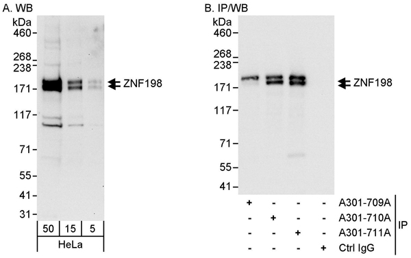 WB (Western Blot)
(Detection of human ZNF198 by western blot and immunoprecipitation. Samples: Whole cell lysate (5, 15 and 50 ug for WB; 1 mg for IP, 20% of IP loaded) from HeLa cells. Antibodies: Affinity purified rabbit anti-ZNF198 antibody AAA211597 used for WB at 0.04 ug/ml (A) and 1 ug/ml (B) and used for IP at 3 ug/mg lysate. ZNF198 was also immunoprecipitated by rabbit anti-ZNF198 antibodies and which recognize upstream epitopes. Detection: Chemiluminescence with exposure times of 30 seconds (A) and 1 second (B).)
WB (Western Blot)
(Detection of human ZNF198 by western blot and immunoprecipitation. Samples: Whole cell lysate (5, 15 and 50 ug for WB; 1 mg for IP, 20% of IP loaded) from HeLa cells. Antibodies: Affinity purified rabbit anti-ZNF198 antibody AAA211597 used for WB at 0.04 ug/ml (A) and 1 ug/ml (B) and used for IP at 3 ug/mg lysate. ZNF198 was also immunoprecipitated by rabbit anti-ZNF198 antibodies and which recognize upstream epitopes. Detection: Chemiluminescence with exposure times of 30 seconds (A) and 1 second (B).)
ZNF198, Polyclonal Antibody (Cat# AAA211597)
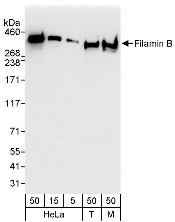 WB (Western Blot)
(Detection of human and mouse Filamin B by western blot. Samples: Whole cell lysate from HeLa (5, 15 and 50 ug), HEK293T (T; 50 ug), and mouse NIH 3T3 (M; 50 ug) cells. Antibodies: Affinity purified rabbit anti-Filamin B antibody AAA211604 used for WB at 0.04 ug/ml. Detection: Chemiluminescence with an exposure time of 3 seconds.)
WB (Western Blot)
(Detection of human and mouse Filamin B by western blot. Samples: Whole cell lysate from HeLa (5, 15 and 50 ug), HEK293T (T; 50 ug), and mouse NIH 3T3 (M; 50 ug) cells. Antibodies: Affinity purified rabbit anti-Filamin B antibody AAA211604 used for WB at 0.04 ug/ml. Detection: Chemiluminescence with an exposure time of 3 seconds.)
Filamin B, Polyclonal Antibody (Cat# AAA211604)
 WB (Western Blot)
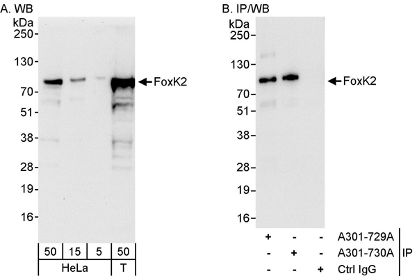
(Detection of human FOXK2 by western blot and immunoprecipitation. Samples: Whole cell lysate from HeLa (5, 15 and 50 ug for WB; 1 mg for IP, 20% of IP loaded) and HEK293T (T; 50 ug) cells. Antibodies: Affinity purified rabbit anti-FOXK2 antibody AAA211607 used for WB at 0.04 ug/ml (A) and 1 ug/ml (B) and used for IP at 3 ug/mg lysate. FOXK2 was also immunoprecipitated by rabbit anti-FOXK2 antibody which recognizes a downstream epitope. Detection: Chemiluminescence with exposure times of 30 seconds (A) and 3 seconds (B).)
WB (Western Blot)
(Detection of human FOXK2 by western blot and immunoprecipitation. Samples: Whole cell lysate from HeLa (5, 15 and 50 ug for WB; 1 mg for IP, 20% of IP loaded) and HEK293T (T; 50 ug) cells. Antibodies: Affinity purified rabbit anti-FOXK2 antibody AAA211607 used for WB at 0.04 ug/ml (A) and 1 ug/ml (B) and used for IP at 3 ug/mg lysate. FOXK2 was also immunoprecipitated by rabbit anti-FOXK2 antibody which recognizes a downstream epitope. Detection: Chemiluminescence with exposure times of 30 seconds (A) and 3 seconds (B).)
FOXK2, Polyclonal Antibody (Cat# AAA211607)
 WB (Western Blot)
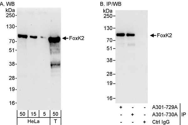
(Detection of human FOXK2 by western blot and immunoprecipitation. Samples: Whole cell lysate from HeLa (5, 15 and 50 ug for WB; 1 mg for IP, 20% of IP loaded) and HEK293T (T; 50 ug) cells. Antibodies: Affinity purified rabbit anti-FOXK2 antibody AAA211608 used for WB at 0.04 ug/ml (A) and 1 ug/ml (B) and used for IP at 3 ug/mg lysate. FOXK2 was also immunoprecipitated by rabbit anti-FOXK2 antibody which recognizes an upstream epitope. Detection: Chemiluminescence with exposure times of 30 seconds (A) and 3 seconds (B).)
WB (Western Blot)
(Detection of human FOXK2 by western blot and immunoprecipitation. Samples: Whole cell lysate from HeLa (5, 15 and 50 ug for WB; 1 mg for IP, 20% of IP loaded) and HEK293T (T; 50 ug) cells. Antibodies: Affinity purified rabbit anti-FOXK2 antibody AAA211608 used for WB at 0.04 ug/ml (A) and 1 ug/ml (B) and used for IP at 3 ug/mg lysate. FOXK2 was also immunoprecipitated by rabbit anti-FOXK2 antibody which recognizes an upstream epitope. Detection: Chemiluminescence with exposure times of 30 seconds (A) and 3 seconds (B).)
FOXK2, Polyclonal Antibody (Cat# AAA211608)
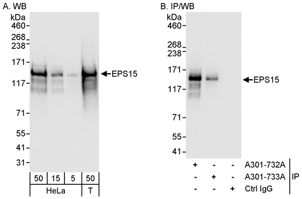 WB (Western Blot)
(Detection of human EPS15 by western blot and immunoprecipitation. Samples: Whole cell lysate from HeLa (5, 15 and 50 ug for WB; 1 mg for IP, 20% of IP loaded) and HEK293T (T; 50 ug) cells. Antibodies: Affinity purified rabbit anti-EPS15 antibody AAA211609 used for WB at 0.04 ug/ml (A) and 0.1 ug/ml (B) and used for IP at 3 ug/mg lysate. EPS15 was also immunoprecipitated by rabbit anti-EPS15 antibody which recognizes a downstream epitope. Detection: Chemiluminescence with exposure times of 1 second (A and B).)
WB (Western Blot)
(Detection of human EPS15 by western blot and immunoprecipitation. Samples: Whole cell lysate from HeLa (5, 15 and 50 ug for WB; 1 mg for IP, 20% of IP loaded) and HEK293T (T; 50 ug) cells. Antibodies: Affinity purified rabbit anti-EPS15 antibody AAA211609 used for WB at 0.04 ug/ml (A) and 0.1 ug/ml (B) and used for IP at 3 ug/mg lysate. EPS15 was also immunoprecipitated by rabbit anti-EPS15 antibody which recognizes a downstream epitope. Detection: Chemiluminescence with exposure times of 1 second (A and B).)
EPS15, Polyclonal Antibody (Cat# AAA211609)
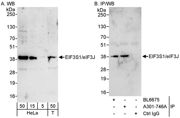 WB (Western Blot)
(Detection of human eIF3J/EIF3S1 by western blot and immunoprecipitation. Samples: Whole cell lysate from HeLa (5, 15 and 50 ug for WB; 1 mg for IP, 20% of IP loaded) and HEK293T (T; 50 ug) cells. Antibodies: Affinity purified rabbit anti-eIF3J/EIF3S1 antibody AAA211614 used for WB at 0.1 ug/ml (A) and 1 ug/ml (B) and used for IP at 3 ug/mg lysate. eIF3J/EIF3S1 was also immunoprecipitated by rabbit anti-eIF3J/EIF3S1 antibody BL6675, which recognizes an upstream epitope. Detection: Chemiluminescence with exposure times of 3 minutes (A) and 30 seconds (B).)
WB (Western Blot)
(Detection of human eIF3J/EIF3S1 by western blot and immunoprecipitation. Samples: Whole cell lysate from HeLa (5, 15 and 50 ug for WB; 1 mg for IP, 20% of IP loaded) and HEK293T (T; 50 ug) cells. Antibodies: Affinity purified rabbit anti-eIF3J/EIF3S1 antibody AAA211614 used for WB at 0.1 ug/ml (A) and 1 ug/ml (B) and used for IP at 3 ug/mg lysate. eIF3J/EIF3S1 was also immunoprecipitated by rabbit anti-eIF3J/EIF3S1 antibody BL6675, which recognizes an upstream epitope. Detection: Chemiluminescence with exposure times of 3 minutes (A) and 30 seconds (B).)
eIF3J/EIF3S1, Polyclonal Antibody (Cat# AAA211614)
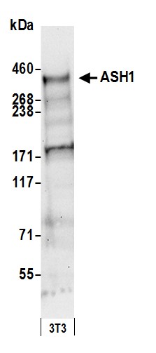
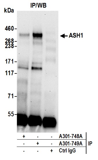
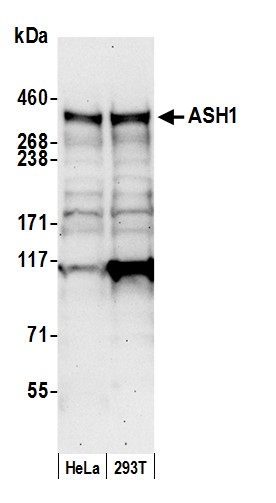 WB (Western Blot)
(Detection of human ASH1 by western blot. Samples: Whole cell lysate (50 ug) from HeLa and HEK293T cells prepared using NETN lysis buffer. Antibody: Affinity purified rabbit anti-ASH1 antibody AAA211616 (lot AAA211616-3) used for WB at 0.1 ug/ml. Detection: Chemiluminescence with an exposure time of 30 seconds.)
WB (Western Blot)
(Detection of human ASH1 by western blot. Samples: Whole cell lysate (50 ug) from HeLa and HEK293T cells prepared using NETN lysis buffer. Antibody: Affinity purified rabbit anti-ASH1 antibody AAA211616 (lot AAA211616-3) used for WB at 0.1 ug/ml. Detection: Chemiluminescence with an exposure time of 30 seconds.)
ASH1, Polyclonal Antibody (Cat# AAA211616)
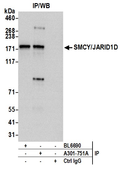
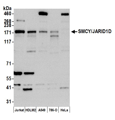 WB (Western Blot)
(Detection of human SMCY/JARID1D by western blot. Samples: Whole cell lysate (15 ug) from Jurkat, HDLM-2, A-549, 786-O, and HeLa cells prepared using NETN lysis buffer. Antibody: Affinity purified rabbit anti-SMCY/JARID1D antibody AAA211617 (lot AAA211617-4) used for WB at 0.1 ug/ml. Detection: Chemiluminescence with an exposure time of 3 minutes.)
WB (Western Blot)
(Detection of human SMCY/JARID1D by western blot. Samples: Whole cell lysate (15 ug) from Jurkat, HDLM-2, A-549, 786-O, and HeLa cells prepared using NETN lysis buffer. Antibody: Affinity purified rabbit anti-SMCY/JARID1D antibody AAA211617 (lot AAA211617-4) used for WB at 0.1 ug/ml. Detection: Chemiluminescence with an exposure time of 3 minutes.)
JARID1D/SMCY, Polyclonal Antibody (Cat# AAA211617)

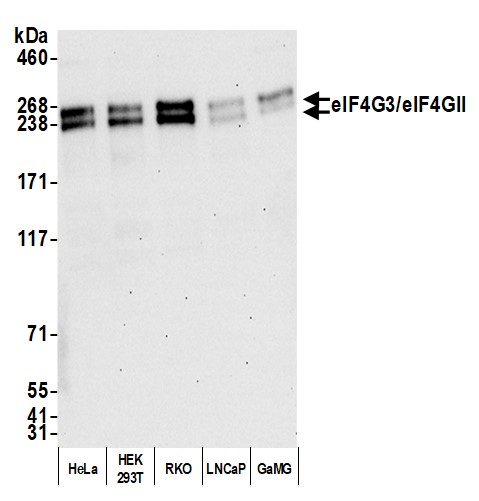 WB (Western Blot)
(Detection of human eIF4G3/eIF4GII by western blot. Samples: Whole cell lysate (10 ug) from HeLa, HEK293T, RKO, LNCaP, and GaMG cells prepared using NETN lysis buffer. Antibody: Affinity purified rabbit anti-eIF4G3/eIF4GII antibody AAA211622 (lot AAA211622-1) used for WB at 0.04 ug/ml. Detection: Chemiluminescence with an exposure time of 30 seconds.)
WB (Western Blot)
(Detection of human eIF4G3/eIF4GII by western blot. Samples: Whole cell lysate (10 ug) from HeLa, HEK293T, RKO, LNCaP, and GaMG cells prepared using NETN lysis buffer. Antibody: Affinity purified rabbit anti-eIF4G3/eIF4GII antibody AAA211622 (lot AAA211622-1) used for WB at 0.04 ug/ml. Detection: Chemiluminescence with an exposure time of 30 seconds.)
eIF4G3/eIF4GII, Polyclonal Antibody (Cat# AAA211622)
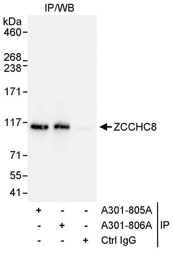 IP (Immunoprecipitation)
(Detection of human ZCCHC8 by western blot of immunoprecipitates. Samples: Whole cell lysate (1 mg for IP, 20% of IP loaded) from HeLa cells. Antibody: Affinity purified rabbit anti-ZCCHC8 antibody AAA211638 used for IP at 3 ug/mg lysate. ZCCHC8 was also immunoprecipitated by rabbit anti-ZCCHC8 antibody which recognizes a downstream epitope. For blotting immunoprecipitated ZCCHC8, was used at 0.1 ug/ml. Detection: Chemiluminescence with an exposure time of 5 seconds.)
IP (Immunoprecipitation)
(Detection of human ZCCHC8 by western blot of immunoprecipitates. Samples: Whole cell lysate (1 mg for IP, 20% of IP loaded) from HeLa cells. Antibody: Affinity purified rabbit anti-ZCCHC8 antibody AAA211638 used for IP at 3 ug/mg lysate. ZCCHC8 was also immunoprecipitated by rabbit anti-ZCCHC8 antibody which recognizes a downstream epitope. For blotting immunoprecipitated ZCCHC8, was used at 0.1 ug/ml. Detection: Chemiluminescence with an exposure time of 5 seconds.)
ZCCHC8, Polyclonal Antibody (Cat# AAA211638)
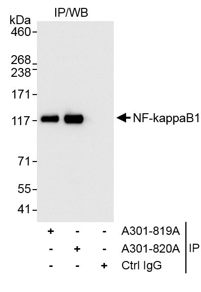
 WB (Western Blot)
(Detection of human NF-kappaB1 by western blot. Samples: Whole cell lysate from HeLa (H; 50 ug) and Jurkat (J; 50 ug) cells. Antibodies: Affinity purified rabbit anti-NF-kappaB1 antibody AAA211642 (lot AAA211642-1) used for WB at 0.04 ug/ml. Detection: Chemiluminescence with exposure time of 1 minute.)
WB (Western Blot)
(Detection of human NF-kappaB1 by western blot. Samples: Whole cell lysate from HeLa (H; 50 ug) and Jurkat (J; 50 ug) cells. Antibodies: Affinity purified rabbit anti-NF-kappaB1 antibody AAA211642 (lot AAA211642-1) used for WB at 0.04 ug/ml. Detection: Chemiluminescence with exposure time of 1 minute.)
NF-kappaB1, Polyclonal Antibody (Cat# AAA211642)
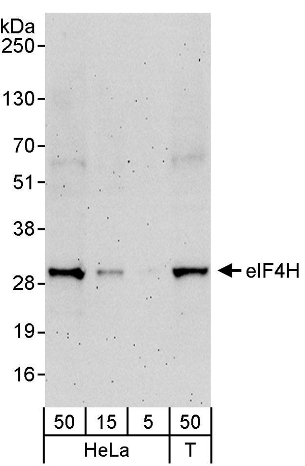 WB (Western Blot)
(Detection of human eIF4H by western blot. Samples: Whole cell lysate from HeLa (5, 15 and 50 ug) and HEK293T (T; 50 ug) cells. Antibodies: Affinity purified rabbit anti-eIF4H antibody AAA211653 used for WB at 0.04 ug/ml. Detection: Chemiluminescence with an exposure time of 3 minutes.)
WB (Western Blot)
(Detection of human eIF4H by western blot. Samples: Whole cell lysate from HeLa (5, 15 and 50 ug) and HEK293T (T; 50 ug) cells. Antibodies: Affinity purified rabbit anti-eIF4H antibody AAA211653 used for WB at 0.04 ug/ml. Detection: Chemiluminescence with an exposure time of 3 minutes.)
eIF4H, Polyclonal Antibody (Cat# AAA211653)
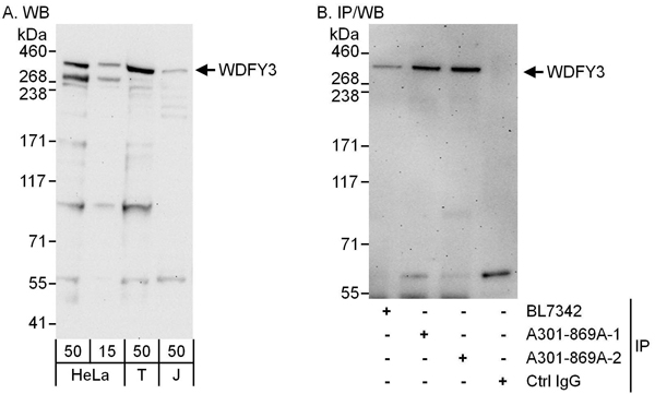 WB (Western Blot)
(Detection of human WDFY3 by western blot and immunoprecipitation. Samples: Whole cell lysate from HeLa (15 and 50 ug for WB; 1 mg for IP, 20% of IP loaded), HEK293T (T; 50 ug) and Jurkat (J; 50 ug) cells. Antibodies: Affinity purified rabbit anti-WDFY3 antibody AAA211658 (lot AAA211658-2) used for WB at 0.4 ug/ml (A) and 1 ug/ml (B) and used for IP at 6 ug/mg lysate. WDFY3 was also immunoprecipitated by a previous lot (lot AAA211658-1) of this antibody and by rabbit anti-WDFY3 antibody BL7342, which recognizes a downstream epitope. Detection: Chemiluminescence with exposure times of 3 minutes (A) and 30 seconds (B).)
WB (Western Blot)
(Detection of human WDFY3 by western blot and immunoprecipitation. Samples: Whole cell lysate from HeLa (15 and 50 ug for WB; 1 mg for IP, 20% of IP loaded), HEK293T (T; 50 ug) and Jurkat (J; 50 ug) cells. Antibodies: Affinity purified rabbit anti-WDFY3 antibody AAA211658 (lot AAA211658-2) used for WB at 0.4 ug/ml (A) and 1 ug/ml (B) and used for IP at 6 ug/mg lysate. WDFY3 was also immunoprecipitated by a previous lot (lot AAA211658-1) of this antibody and by rabbit anti-WDFY3 antibody BL7342, which recognizes a downstream epitope. Detection: Chemiluminescence with exposure times of 3 minutes (A) and 30 seconds (B).)
WDFY3, Polyclonal Antibody (Cat# AAA211658)
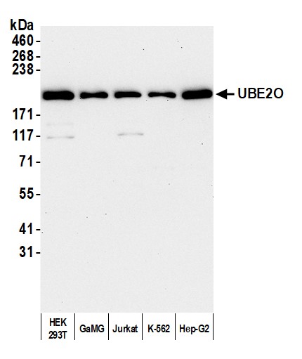
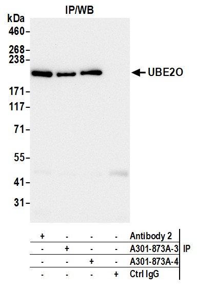
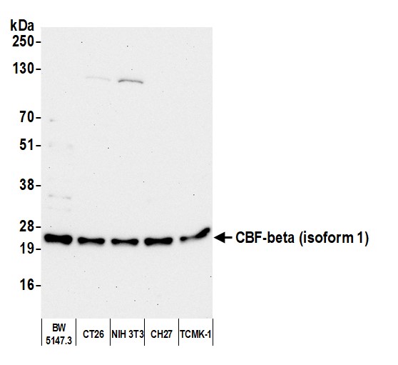
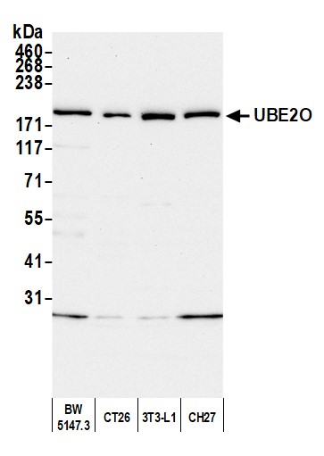 WB (Western Blot)
(Detection of mouse UBE2O by western blot. Samples: Whole cell lysate (50 ug) from BW5147.3, CT26, 3T3-L1, and CH27 cells prepared using NETN lysis buffer. Antibody: Affinity purified rabbit anti-UBE2O antibody (AAA211659 lot 4) used for WB at 0.1 ug/ml. Detection: Chemiluminescence with an exposure time of 30 seconds.)
WB (Western Blot)
(Detection of mouse UBE2O by western blot. Samples: Whole cell lysate (50 ug) from BW5147.3, CT26, 3T3-L1, and CH27 cells prepared using NETN lysis buffer. Antibody: Affinity purified rabbit anti-UBE2O antibody (AAA211659 lot 4) used for WB at 0.1 ug/ml. Detection: Chemiluminescence with an exposure time of 30 seconds.)
UBE2O, Polyclonal Antibody (Cat# AAA211659)
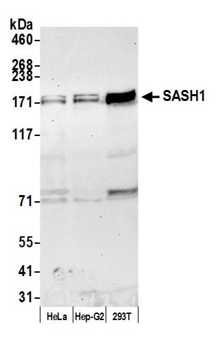 WB (Western Blot)
(Detection of human SASH1 by western blot. Samples: Whole cell lysate (50 ug) from HeLa, Hep-G2, and HEK293T cells prepared using NETN lysis buffer. Antibody: Affinity purified rabbit anti-SASH1 antibody AAA211822 (lot AAA211822-2) used for WB at 0.1 ug/ml. Detection: Chemiluminescence with an exposure time of 30 seconds.)
WB (Western Blot)
(Detection of human SASH1 by western blot. Samples: Whole cell lysate (50 ug) from HeLa, Hep-G2, and HEK293T cells prepared using NETN lysis buffer. Antibody: Affinity purified rabbit anti-SASH1 antibody AAA211822 (lot AAA211822-2) used for WB at 0.1 ug/ml. Detection: Chemiluminescence with an exposure time of 30 seconds.)
SASH1, Polyclonal Antibody (Cat# AAA211822)
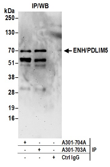
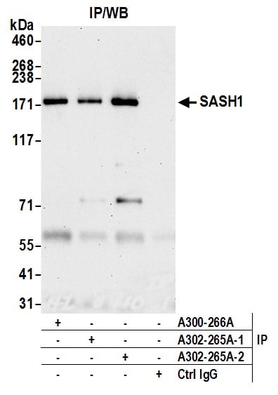
 IP (Immunoprecipitation)
(Detection of human SASH1 by western blot of immunoprecipitates. Samples: Whole cell lysate (1 mg for IP, 20% of IP loaded) from HeLa cells. Antibodies: Affinity purified rabbit anti-SASH1 antibody AAA211823 used for IP at 3 ug/mg lysate. SASH1 was also immunoprecipitated by rabbit anti-SASH1 antibody which recognizes an upstream epitope. For blotting immunoprecipitated SASH1, A320-265A was used at 1 ug/ml. Detection: Chemiluminescence with an exposure time of 30 seconds.)
IP (Immunoprecipitation)
(Detection of human SASH1 by western blot of immunoprecipitates. Samples: Whole cell lysate (1 mg for IP, 20% of IP loaded) from HeLa cells. Antibodies: Affinity purified rabbit anti-SASH1 antibody AAA211823 used for IP at 3 ug/mg lysate. SASH1 was also immunoprecipitated by rabbit anti-SASH1 antibody which recognizes an upstream epitope. For blotting immunoprecipitated SASH1, A320-265A was used at 1 ug/ml. Detection: Chemiluminescence with an exposure time of 30 seconds.)
SASH1, Polyclonal Antibody (Cat# AAA211823)
What are Polyclonal Antibodies?
Polyclonal antibodies are antibodies that come from multiple B cell clones of a host animal. The typical hosts used for the majority of polyclonal antibody production are rabbits, goats, sheep, and donkeys. These polyclonal antibodies, once having identified their target, will bind to different epitopes located at different regions or sequences on the same protein/antigen. As a result, they are ideal at locating and binding to the target, even if the target is in very low concentrations (due to many different antibodies being able to bind to the same target molecule, which allows for significant amplification of a downstream signal).
Polyclonal antibodies are typically produced by injecting an antigen into a host animal, which causes the animal’s immune system to attack the foreign antigen by mass generating antibodies against it. After a period of time, serum is collected from the animal and purified using physicochemical fractionation, class-specific affinity purification, and/or antigen-affinity purification.
Key Uses of Polyclonal Antibodies
- Western Blotting: This method is used to find specific proteins in biological samples after separating them by size.
- Immunohistochemistry: IHC helps visualize the location of proteins in tissue sections using various staining techniques.
- ELISA: (Enzyme-Linked Immunosorbent Assay) is typically used to identify specific protein quantities in a sample. ELISAs can be either “Quantitative” or “Qualitative”.
- Flow Cytometry: technique that identifies and measures the specific protein on the surface or inside the cells in a fluid suspension.
- Immunoprecipitation: IP isolates and studies a specific protein from a complex mixture using antibodies.
Why Buy Polyclonal Antibodies from AAA Biotech?
1. Ideal for Various Applications
Our antibodies are generally going to be validated for use in multiple types of assays, including ELISA, Western Blotting, Immunohistochemistry, Immunoprecipitation, amongst others. They are ideal for a wide range of research applications.
2. Rigorous Quality Control
All of the antibodies in our catalog undergo strict quality testing to ensure specificity, sensitivity, and consistent performance. We are confident in the ability of our antibodies to provide you with accurate results.
3. Wide Assortment of Antibodies
Antibodies in are catalog can be found for both common and exotic species, and these antibodies are also available in both conjugated and recombinant forms to suit many diverse experimental needs.
4. Highly Purified
Our antibodies are available in purified forms with over 85% purity, as confirmed by SDS-PAGE. They are also available with tags such as His, Flag, GST, or MBP. We cater to customers worldwide.
FAQ
1. How are polyclonal antibodies produced?
Traditionally, polyclonal antibodies are produced by injecting an antigen into a host animal (such as a rabbit or goat), which then triggers an immune response from the host animal. The animal’s B cells produce antibodies that will recognize different parts of the injected antigen. These antibodies are then collected from the animal’s blood and purified for use.
2. How do polyclonal antibodies differ from monoclonal antibodies?
Polyclonal antibodies are a mix of antibodies that bind to different locations (epitopes) of the same antigen, while monoclonal antibodies are identical and bind to just one specific epitope. This makes polyclonal antibodies more versatile and better at detecting proteins that may be present in low quantities or in altered/modified forms.
3. How should I store polyclonal antibodies?
Polyclonal antibodies should be stored at 4°C for short-term use (up to a few weeks) and at -20°C or -80°C for long-term storage. Avoid repeated freeze-thaw cycles by dividing them into small aliquots. Always check the datasheet for specific storage instructions.