Filters
▼Clonality
▼Type
▼Reactivity
▼Gene Name
▼Isotype
▼Host
▼Application
▼Clone
▼Polyclonal Antibodies
At AAA Biotech also known as AAA Bio or AAABio, we provide a broad range of purified polyclonal antibodies (pAbs) that are able to all be browsed online through our website. Due to their high specificity and strong binding affinity, these antibodies are ideal for wide swathes of research and experimental applications.
Our polyclonal antibodies can easily support your work, whether you use them for Western Blotting, Immunocytochemistry (with or without Immunofluorescence used in conjunction), Immunohistochemistry, Immunoprecipitation, and ELISA tests. We highly encourage you to browse our range of pAbs and choose the one that best suits your experimental model.
Viewing 4650-4700 of 96812 product results
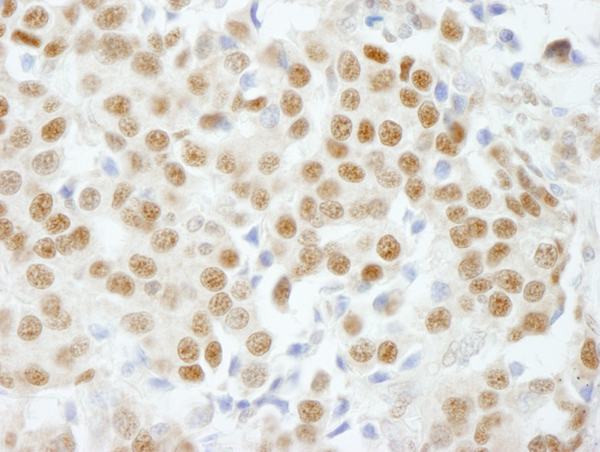
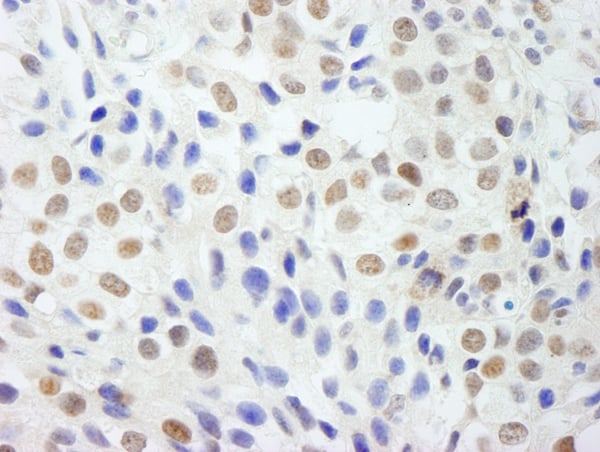
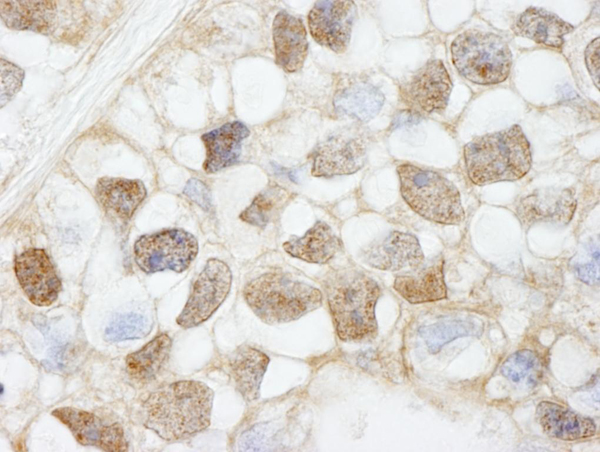
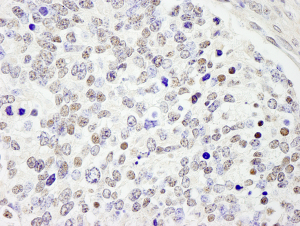
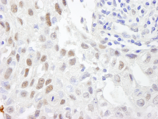
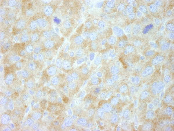
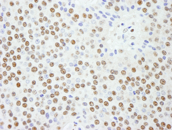
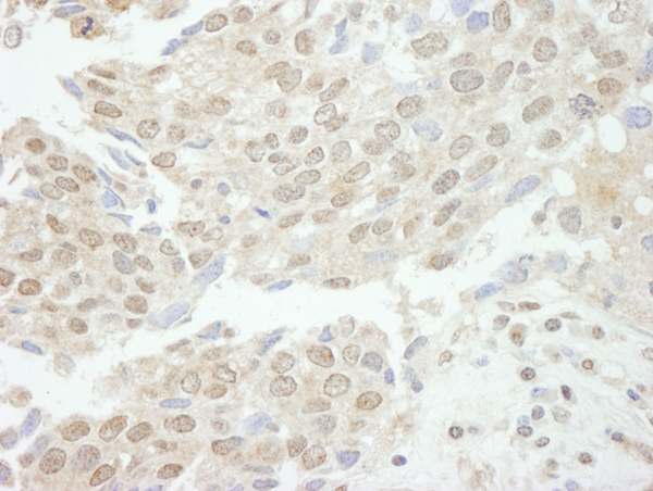
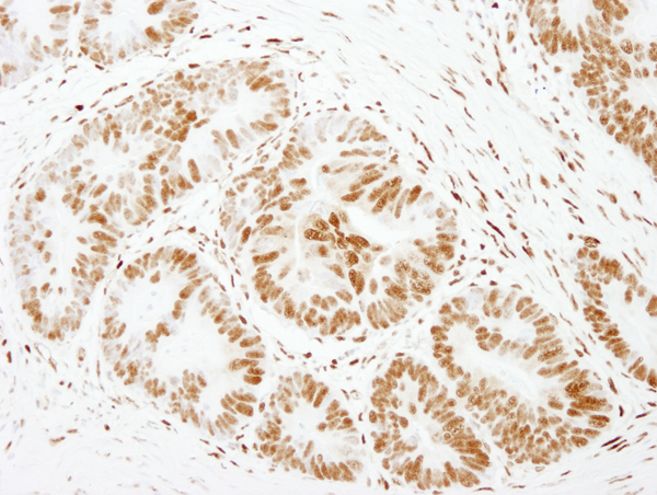
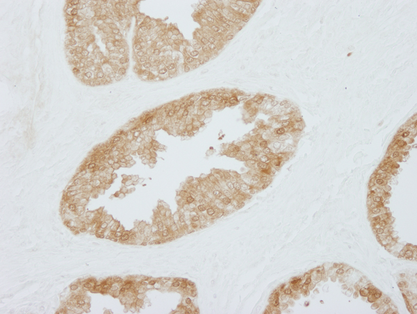
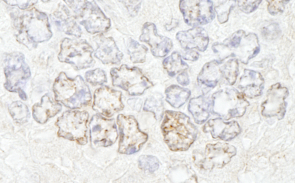
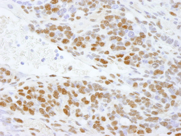
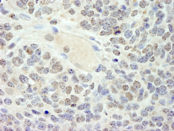
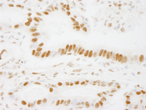
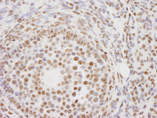
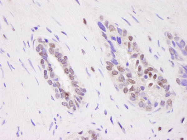
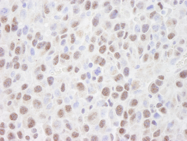
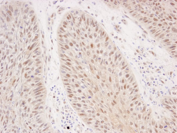
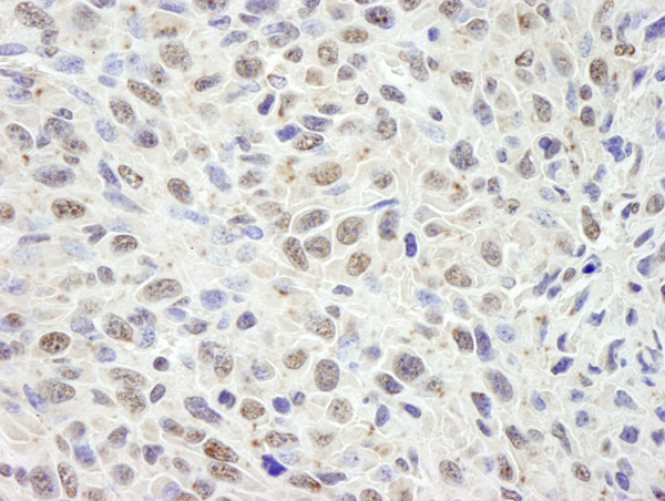
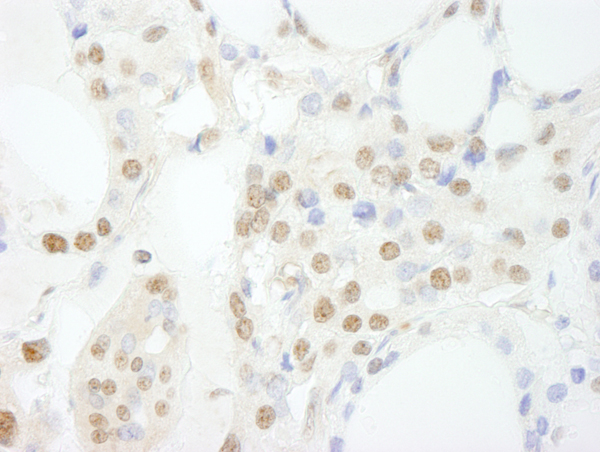 IHC (Immunohiostchemistry)
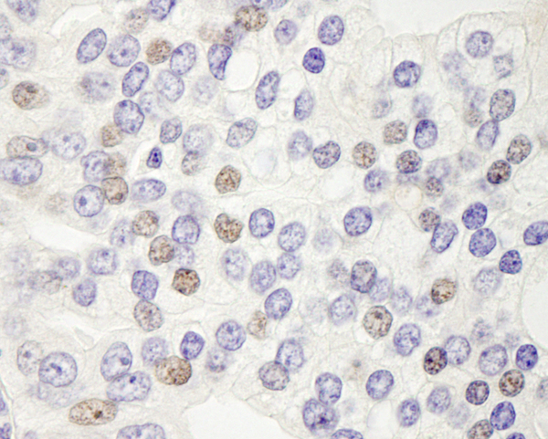
(Detection of human GTF3C3/TFIIIC102 by immunohistochemistry. Sample: FFPE section of human thyroid carcinoma. Antibody: Affinity purified rabbit anti-GTF3C3/TFIIIC102 (Cat. No. AAA213899) used at a dilution of 1:100. Detection: DAB)
IHC (Immunohiostchemistry)
(Detection of human GTF3C3/TFIIIC102 by immunohistochemistry. Sample: FFPE section of human thyroid carcinoma. Antibody: Affinity purified rabbit anti-GTF3C3/TFIIIC102 (Cat. No. AAA213899) used at a dilution of 1:100. Detection: DAB)
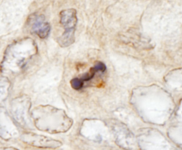
GTF3C3/TFIII1C102, Polyclonal Antibody (Cat# AAA213899)
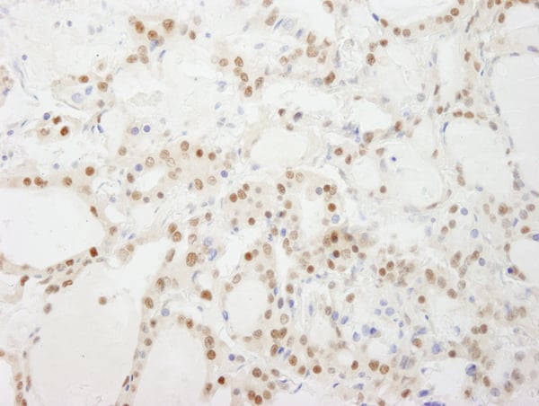
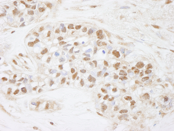
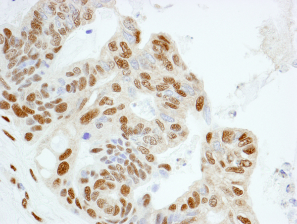
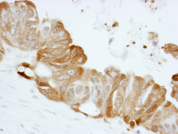
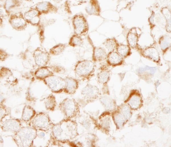
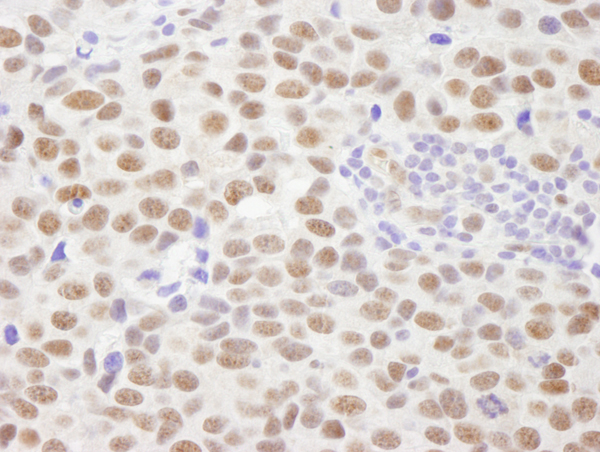
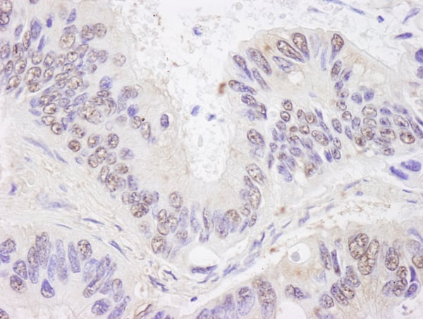
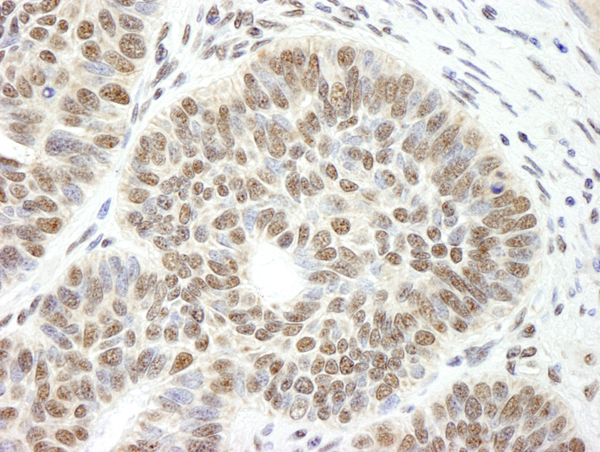
 IHC (Immunohiostchemistry)
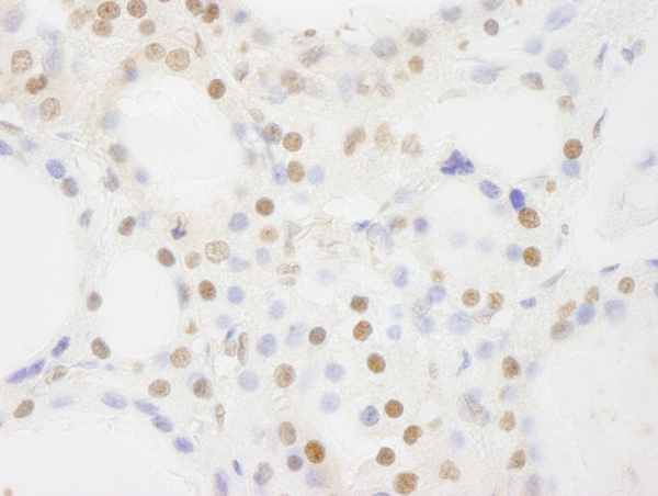
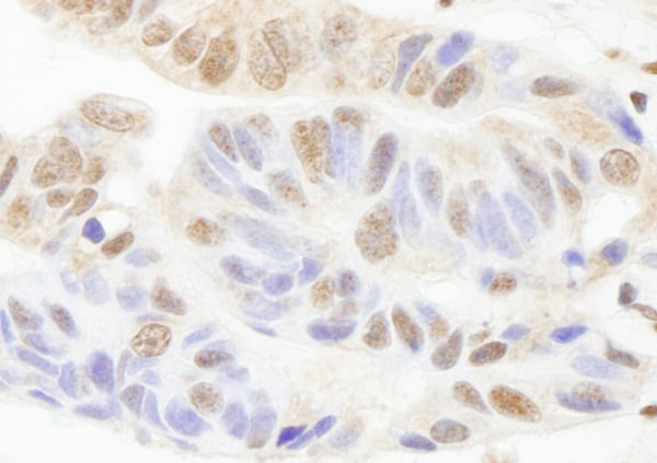
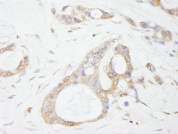
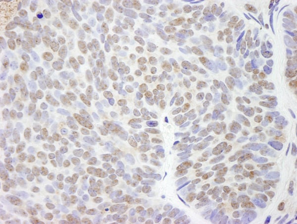
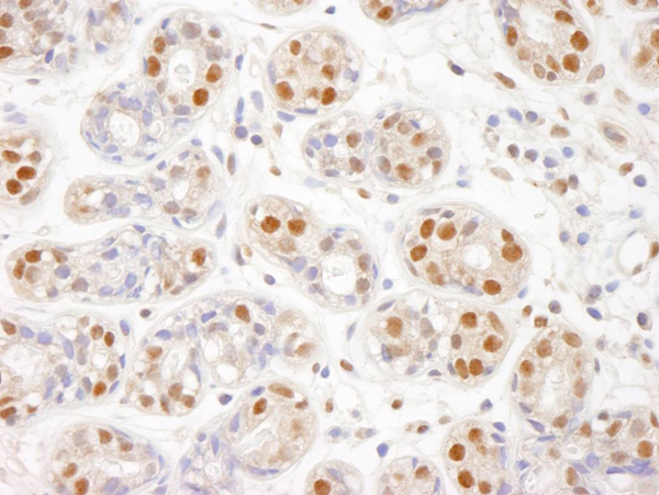
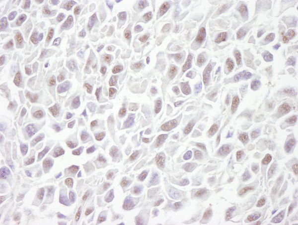
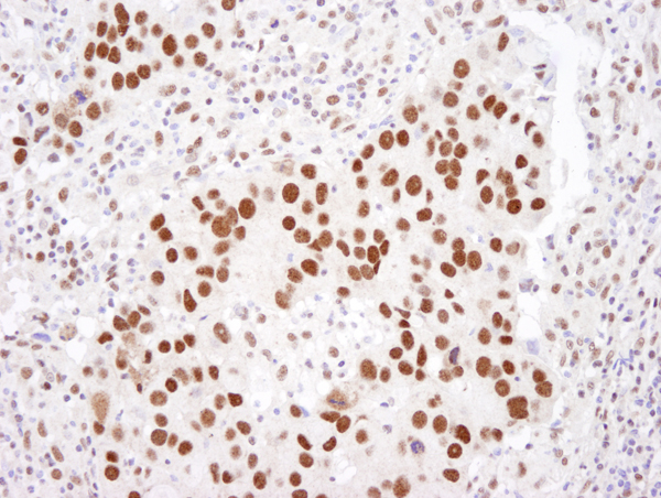
(Detection of human GTF3C4/TFIIIC90 by immunohistochemistry. Sample: FFPE section of human breast carcinoma. Antibody: Affinity purified rabbit anti-GTF3C4/TFIIIC90 (Cat. No. AAA213900) used at a dilution of 1:100. Detection: DAB)
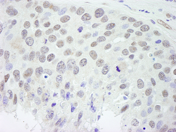
IHC (Immunohiostchemistry)
(Detection of human GTF3C4/TFIIIC90 by immunohistochemistry. Sample: FFPE section of human breast carcinoma. Antibody: Affinity purified rabbit anti-GTF3C4/TFIIIC90 (Cat. No. AAA213900) used at a dilution of 1:100. Detection: DAB)
GTF3C4/TFIII1C90, Polyclonal Antibody (Cat# AAA213900)
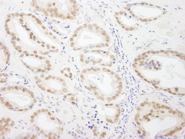
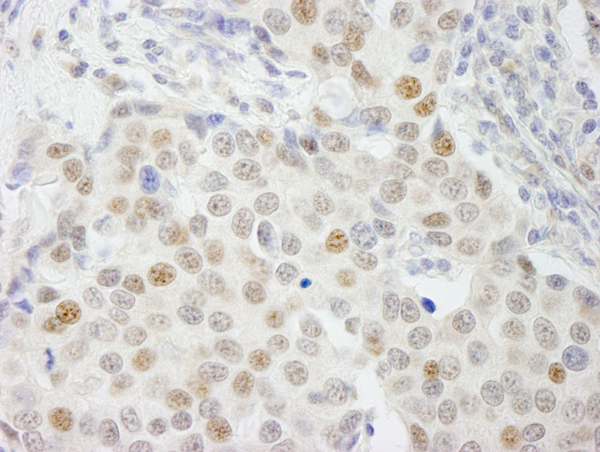
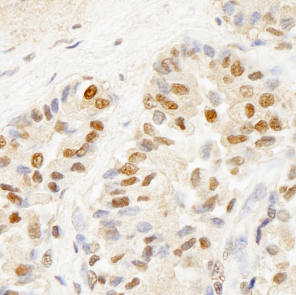
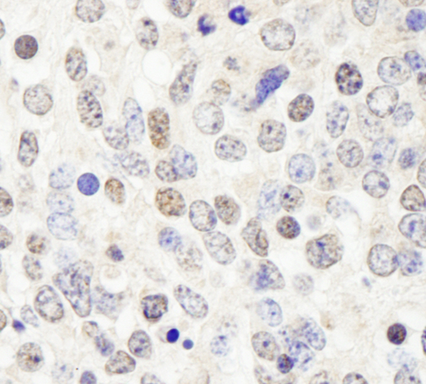
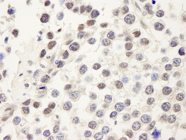
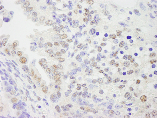
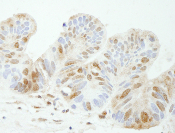
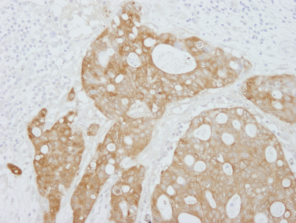
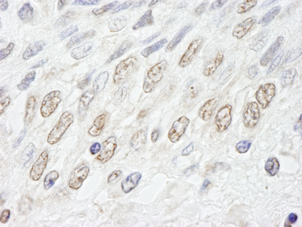
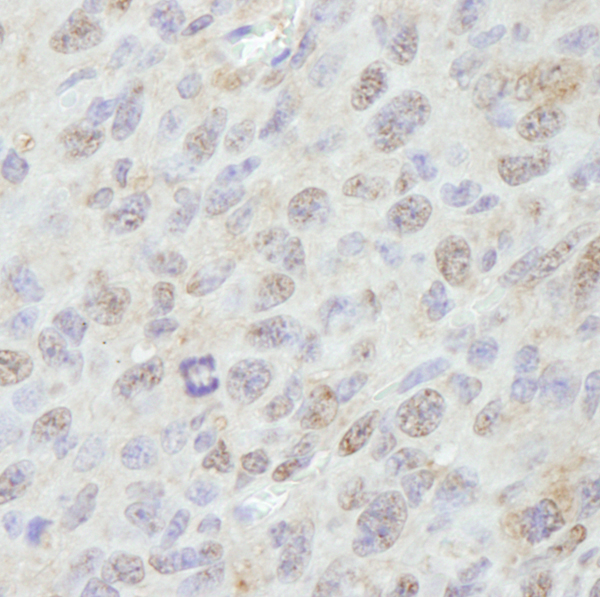
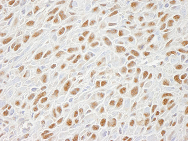
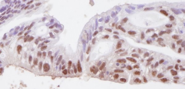
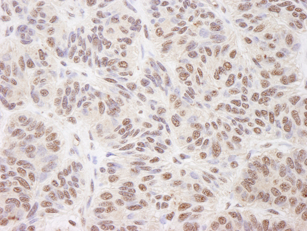
 IHC (Immunohistochemisry)
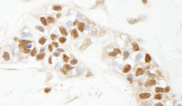
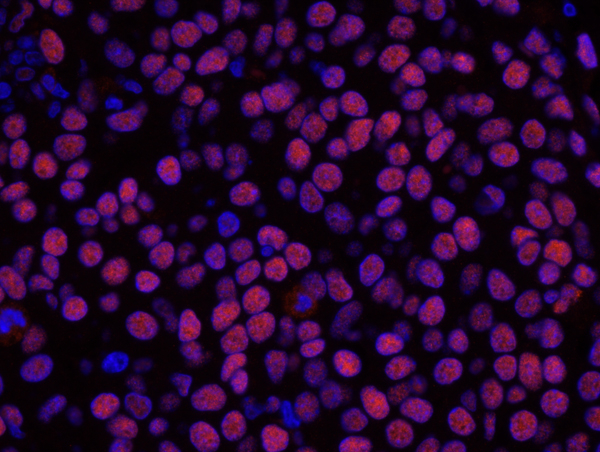
(Detection of human GTF3C5/TFIIIC63 by immunohistochemistry. Sample: FFPE section of human thyroid carcinoma. Antibody: Affinity purified rabbit anti-GTF3C5/TFIIIC63 (Cat. No. AAA213901) used at a dilution of 1:500. Detection: DAB)
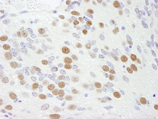
IHC (Immunohistochemisry)
(Detection of human GTF3C5/TFIIIC63 by immunohistochemistry. Sample: FFPE section of human thyroid carcinoma. Antibody: Affinity purified rabbit anti-GTF3C5/TFIIIC63 (Cat. No. AAA213901) used at a dilution of 1:500. Detection: DAB)
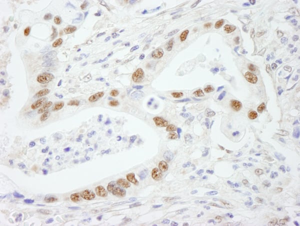
GTF3C5/TFIII1C63, Polyclonal Antibody (Cat# AAA213901)
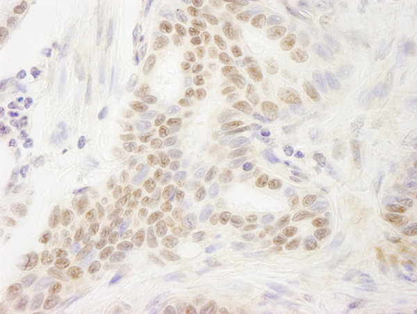
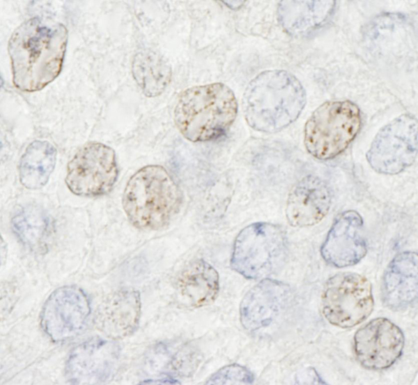
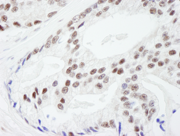
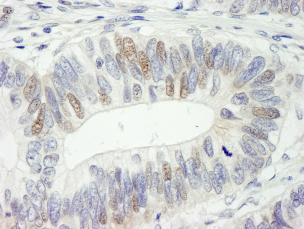
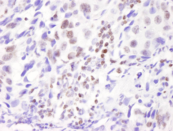
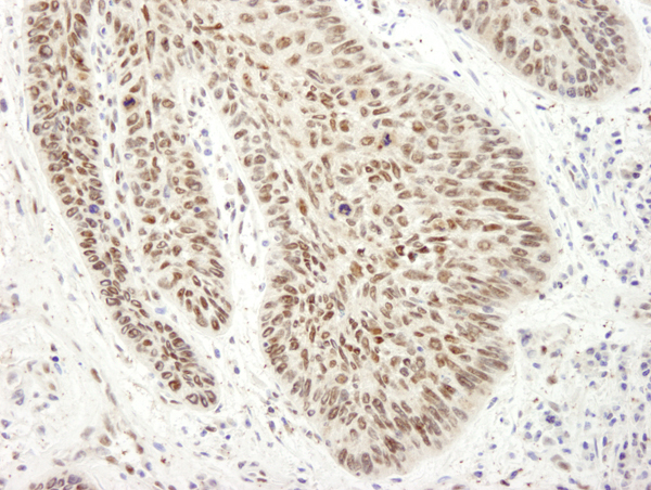
 IHC (Immunohiostchemistry)
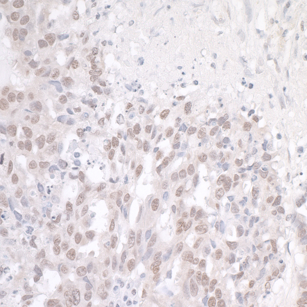
(Detection of human DDX54 by immunohistochemistry. Sample: FFPE section of human breast carcinoma. Antibody: Affinity purified rabbit anti-DDX54 (Cat. No. AAA213905) used at a dilution of 1:250. Detection: DAB)
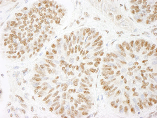
IHC (Immunohiostchemistry)
(Detection of human DDX54 by immunohistochemistry. Sample: FFPE section of human breast carcinoma. Antibody: Affinity purified rabbit anti-DDX54 (Cat. No. AAA213905) used at a dilution of 1:250. Detection: DAB)
DDX54, Polyclonal Antibody (Cat# AAA213905)
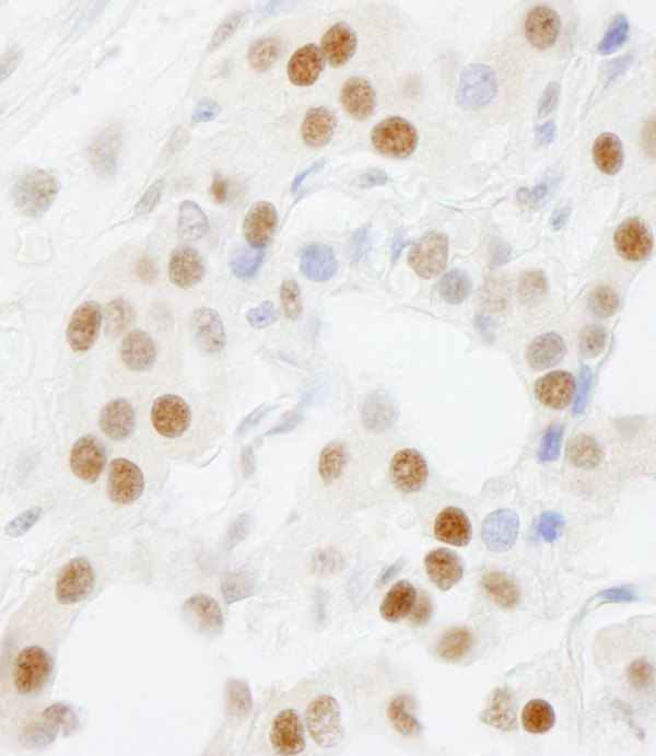
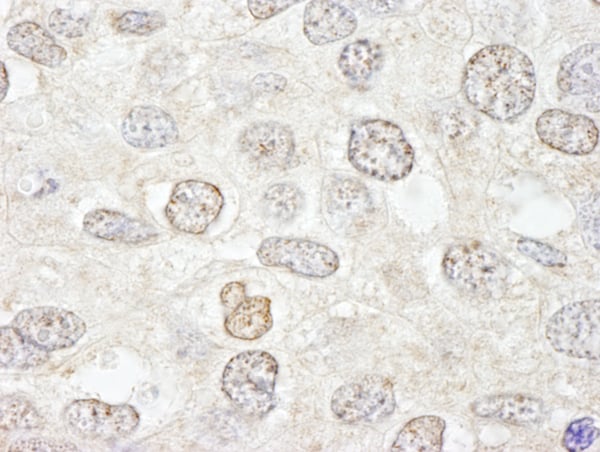
 IHC (Immunohiostchemistry)
(Detection of human BRF1 by immunohistochemistry. Sample: FFPE section of human prostate carcinoma. Antibody: Affinity purified rabbit anti-BRF1 (Cat. No. AAA213912) used at a dilution of 1:100. Detection: DAB)
IHC (Immunohiostchemistry)
(Detection of human BRF1 by immunohistochemistry. Sample: FFPE section of human prostate carcinoma. Antibody: Affinity purified rabbit anti-BRF1 (Cat. No. AAA213912) used at a dilution of 1:100. Detection: DAB)
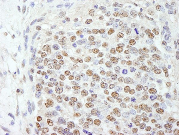
BRF1, Polyclonal Antibody (Cat# AAA213912)
 IHC (Immunohiostchemistry)
(Detection of human DEK by immunohistochemistry. Sample: FFPE section of human prostate carcinoma. Antibody: Affinity purified rabbit anti-DEK (Cat. No. AAA213916) used at a dilution of 1:250. Detection: DAB)
IHC (Immunohiostchemistry)
(Detection of human DEK by immunohistochemistry. Sample: FFPE section of human prostate carcinoma. Antibody: Affinity purified rabbit anti-DEK (Cat. No. AAA213916) used at a dilution of 1:250. Detection: DAB)
DEK, Polyclonal Antibody (Cat# AAA213916)
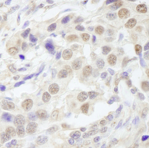
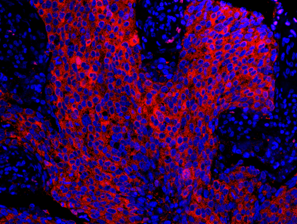
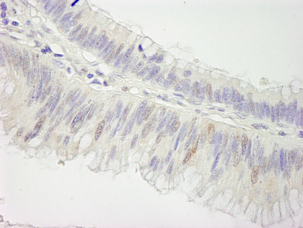
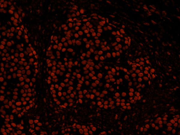
 IHC (Immunohistochemisry)
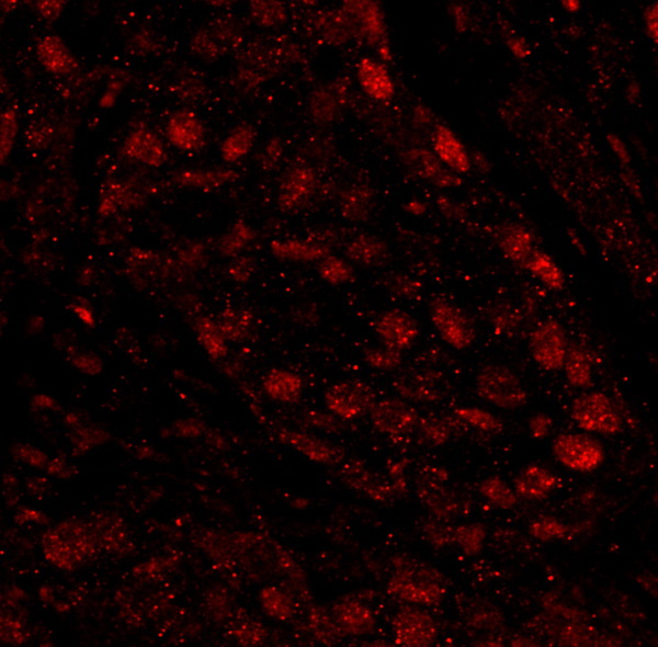
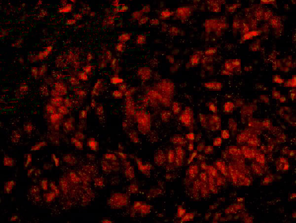
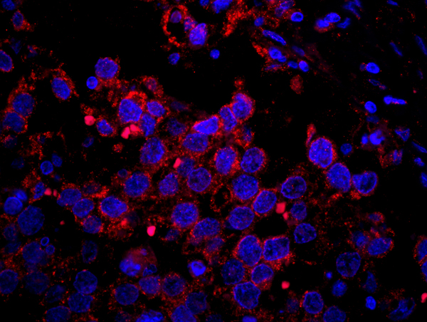
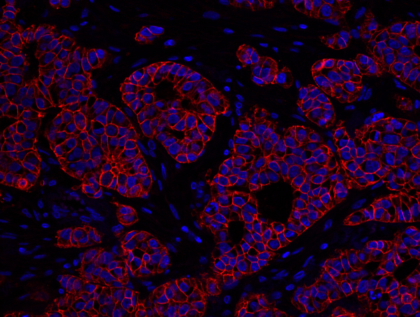
(Detection of human PARP1 by immunohistochemistry. Sample: FFPE section of human breast carcinoma. Antibody: Affinity purified rabbit anti-PARP1 (Cat. No. AAA213919) used at a dilution of 1:100. Detection: Red-fluorescent goat anti-rabbit IgG highly cross-adsorbed Antibody used at a dilution of 1:100.)
IHC (Immunohistochemisry)
(Detection of human PARP1 by immunohistochemistry. Sample: FFPE section of human breast carcinoma. Antibody: Affinity purified rabbit anti-PARP1 (Cat. No. AAA213919) used at a dilution of 1:100. Detection: Red-fluorescent goat anti-rabbit IgG highly cross-adsorbed Antibody used at a dilution of 1:100.)
PARP1, Polyclonal Antibody (Cat# AAA213919)
 IHC (Immunohistochemistry)
(Detection of human HDAC7 by immunohistochemistry. Sample: FFPE section of human ovarian carcinoma. Antibody: Affinity purified rabbit anti-HDAC7 (Cat. No. AAA213923) used at a dilution of 1:100. Detection: DAB)
IHC (Immunohistochemistry)
(Detection of human HDAC7 by immunohistochemistry. Sample: FFPE section of human ovarian carcinoma. Antibody: Affinity purified rabbit anti-HDAC7 (Cat. No. AAA213923) used at a dilution of 1:100. Detection: DAB)
HDAC7, Polyclonal Antibody (Cat# AAA213923)
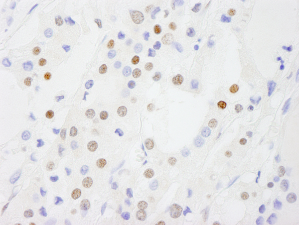
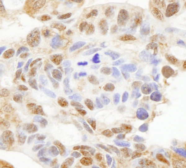
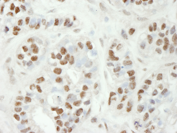
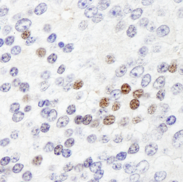
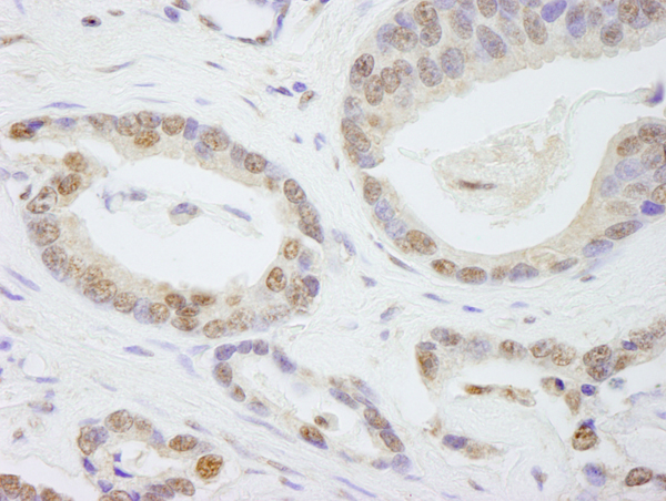
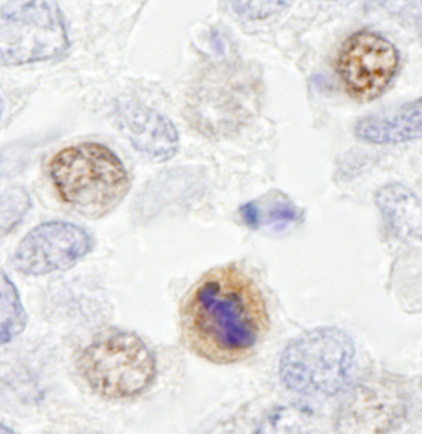
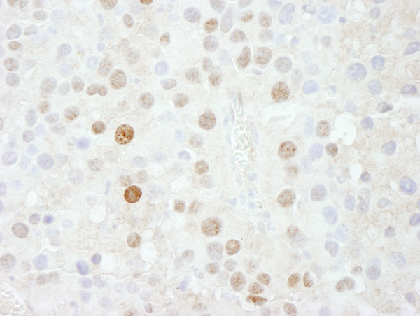
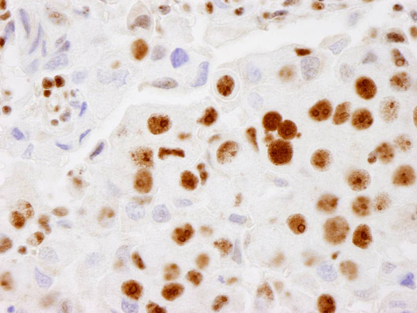
 IHC (Immunohistochemisry)
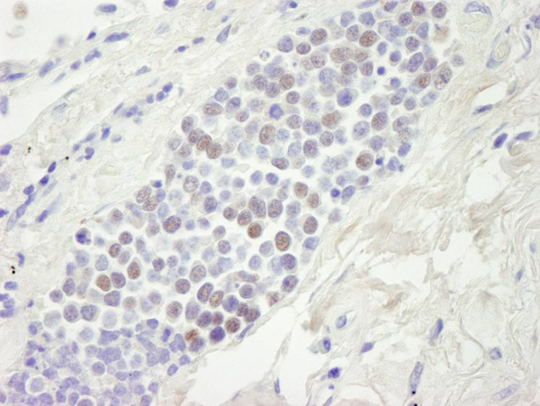
(Detection of human CDC25c by immunohistochemistry. Sample: FFPE section of human seminoma. Antibody: Affinity purified rabbit anti-CDC25c (Cat. No. AAA213926) used at a dilution of 1:250. Detection: DAB)
IHC (Immunohistochemisry)
(Detection of human CDC25c by immunohistochemistry. Sample: FFPE section of human seminoma. Antibody: Affinity purified rabbit anti-CDC25c (Cat. No. AAA213926) used at a dilution of 1:250. Detection: DAB)
CDC25c, Polyclonal Antibody (Cat# AAA213926)
 IHC (Immunohistochemisry)
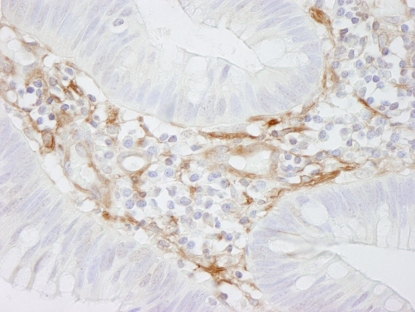
(Detection of human RanBP3 by immunohistochemistry. Sample: FFPE section of human colon carcinoma. Antibody: Affinity purified rabbit anti-RanBP3 (Cat. No. AAA213933) used at a dilution of 1:100. Detection: Red-fluorescent goat anti-rabbit IgG highly cross-adsorbed Antibody Hilyte Plus 555 used at a dilution of 1:100.)
IHC (Immunohistochemisry)
(Detection of human RanBP3 by immunohistochemistry. Sample: FFPE section of human colon carcinoma. Antibody: Affinity purified rabbit anti-RanBP3 (Cat. No. AAA213933) used at a dilution of 1:100. Detection: Red-fluorescent goat anti-rabbit IgG highly cross-adsorbed Antibody Hilyte Plus 555 used at a dilution of 1:100.)
RanBP3, Polyclonal Antibody (Cat# AAA213933)
 IHC (Immunohiostchemistry)
(Detection of human ZC3H11A by immunohistochemistry. Sample: FFPE section of human ovarian carcinoma. Antibody: Affinity purified rabbit anti-ZC3H11A (Cat. No. AAA213934) used at a dilution of 1:100. Detection: DAB)
IHC (Immunohiostchemistry)
(Detection of human ZC3H11A by immunohistochemistry. Sample: FFPE section of human ovarian carcinoma. Antibody: Affinity purified rabbit anti-ZC3H11A (Cat. No. AAA213934) used at a dilution of 1:100. Detection: DAB)
ZC3H11A, Polyclonal Antibody (Cat# AAA213934)
 IHC (Immunohistochemisry)
(Detection of human ZHX3 by immunohistochemistry. Sample: FFPE section of human prostate carcinoma. Antibody: Affinity purified rabbit anti-ZHX3 (Cat. No. AAA213935) used at a dilution of 1:100. Detection: Detection: Red-fluorescent goat anti-rabbit IgG highly cross-adsorbed Antibody used at a dilution of 1:100.)
IHC (Immunohistochemisry)
(Detection of human ZHX3 by immunohistochemistry. Sample: FFPE section of human prostate carcinoma. Antibody: Affinity purified rabbit anti-ZHX3 (Cat. No. AAA213935) used at a dilution of 1:100. Detection: Detection: Red-fluorescent goat anti-rabbit IgG highly cross-adsorbed Antibody used at a dilution of 1:100.)
ZHX3, Polyclonal Antibody (Cat# AAA213935)
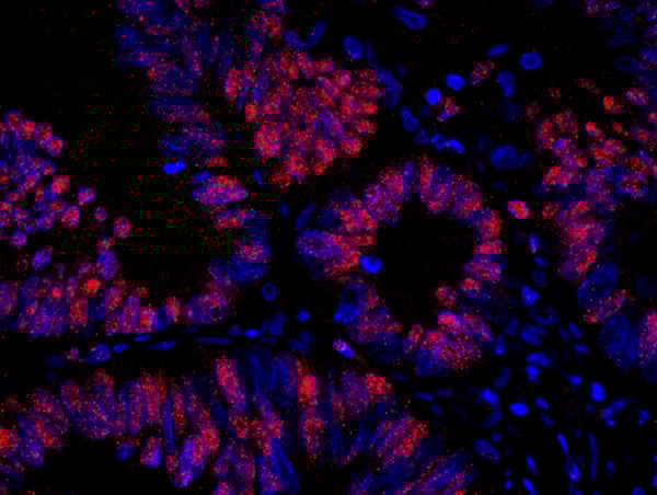
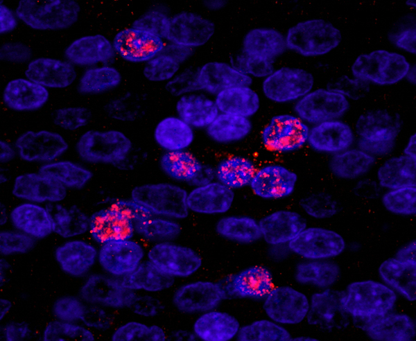
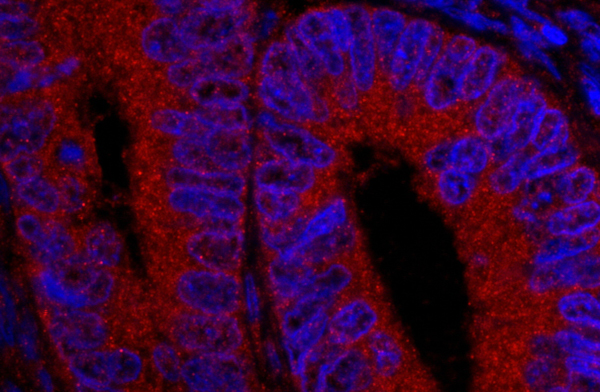
 IHC (Immunohistochemisry)
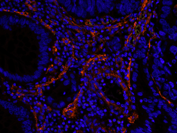
(Detection of human FASN by IHC-IF. Sample: FFPE section of human breast carcinoma. Antibody: Affinity purified rabbit anti-FASN (Cat. No. AAA213940) used at a dilution of 1:100. Detection: Red-fluorescent goat anti-rabbit IgG highly cross-adsorbed Antibody Hilyte Plus 555 used at a dilution of 1:100.)
IHC (Immunohistochemisry)
(Detection of human FASN by IHC-IF. Sample: FFPE section of human breast carcinoma. Antibody: Affinity purified rabbit anti-FASN (Cat. No. AAA213940) used at a dilution of 1:100. Detection: Red-fluorescent goat anti-rabbit IgG highly cross-adsorbed Antibody Hilyte Plus 555 used at a dilution of 1:100.)
FASN, Polyclonal Antibody (Cat# AAA213940)
 IHC (Immunohiostchemistry)
(Detection of human TAF6 by immunohistochemistry. Sample: FFPE section of human prostate carcinoma. Antibody: Affinity purified rabbit anti-TAF6 (Cat. No. AAA213944) used at a dilution of 1:250. Detection: DAB)
IHC (Immunohiostchemistry)
(Detection of human TAF6 by immunohistochemistry. Sample: FFPE section of human prostate carcinoma. Antibody: Affinity purified rabbit anti-TAF6 (Cat. No. AAA213944) used at a dilution of 1:250. Detection: DAB)
TAF6, Polyclonal Antibody (Cat# AAA213944)
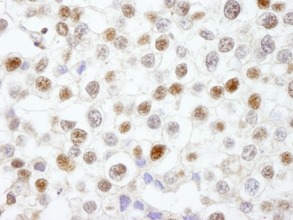
 IHC (Immunohistochemisry)
(Detection of human USP5/IsoT by IHC-IF. Sample: FFPE section of human colon carcinoma. Antibody: Affinity purified rabbit anti-USP5/IsoT (Cat. No. AAA213946) used at a dilution of 1:100. Detection: Red-fluorescent goat anti-rabbit IgG highly cross-adsorbed Antibody Hilyte Plus 555 used at a dilution of 1:100.)
IHC (Immunohistochemisry)
(Detection of human USP5/IsoT by IHC-IF. Sample: FFPE section of human colon carcinoma. Antibody: Affinity purified rabbit anti-USP5/IsoT (Cat. No. AAA213946) used at a dilution of 1:100. Detection: Red-fluorescent goat anti-rabbit IgG highly cross-adsorbed Antibody Hilyte Plus 555 used at a dilution of 1:100.)
USP5/IsoT, Polyclonal Antibody (Cat# AAA213946)
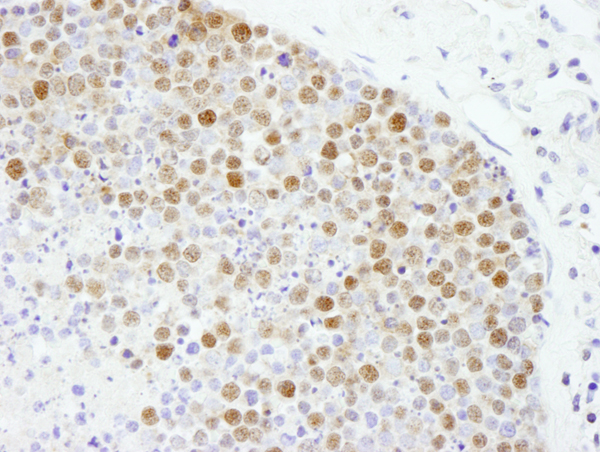
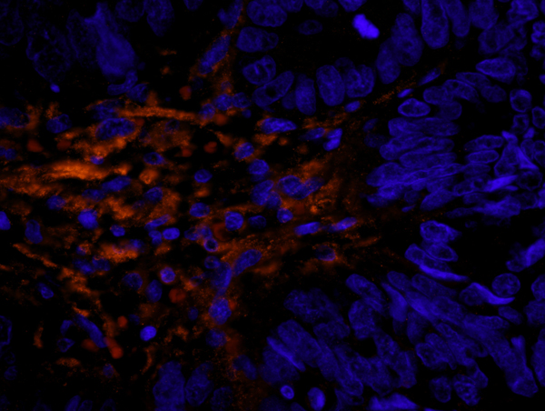
 IHC (Immunohistochemistry)
(Detection of human CENP-F/Mitosin by IHC-IF. Sample: FFPE section of human ewing sarcoma. Antibody: Affinity purified rabbit anti-CENP-F/Mitosin (Cat. No. AAA213947) used at a dilution of 1:100. Detection: Red-fluorescent goat anti-rabbit IgG highly cross-adsorbed Antibody Hilyte Plus 555 used at a dilution of 1:100.)
IHC (Immunohistochemistry)
(Detection of human CENP-F/Mitosin by IHC-IF. Sample: FFPE section of human ewing sarcoma. Antibody: Affinity purified rabbit anti-CENP-F/Mitosin (Cat. No. AAA213947) used at a dilution of 1:100. Detection: Red-fluorescent goat anti-rabbit IgG highly cross-adsorbed Antibody Hilyte Plus 555 used at a dilution of 1:100.)
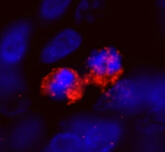
CENP-F/Mitosin, Polyclonal Antibody (Cat# AAA213947)
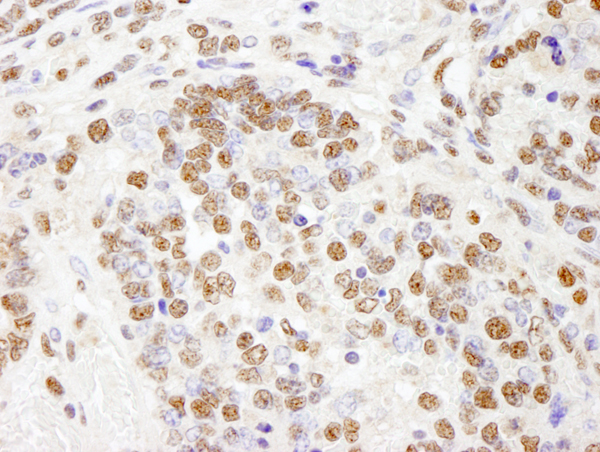
 IHC (Immunohistochemisry)
(Detection of human SF3b145/SAP145 by IHC-IF. Sample: FFPE section of human stomach carcinoma. Antibody: Affinity purified rabbit anti-SF3b145/SAP145 (Cat. No. AAA213949) used at a dilution of 1:100. Detection: Red-fluorescent goat anti-rabbit IgG highly cross-adsorbed Antibody used at a dilution of 1:100.)
IHC (Immunohistochemisry)
(Detection of human SF3b145/SAP145 by IHC-IF. Sample: FFPE section of human stomach carcinoma. Antibody: Affinity purified rabbit anti-SF3b145/SAP145 (Cat. No. AAA213949) used at a dilution of 1:100. Detection: Red-fluorescent goat anti-rabbit IgG highly cross-adsorbed Antibody used at a dilution of 1:100.)
SF3b145/SAP145, Polyclonal Antibody (Cat# AAA213949)
 IHC (Immunohistochemistry)
(Detection of human KPNA4 by IHC-IF. Sample: FFPE section of human ovarian tumor. Antibody: Affinity purified rabbit anti-KPNA4 (Cat. No. AAA213953) used at a dilution of 1:100. Detection: Red-fluorescent goat anti-rabbit IgG highly cross-adsorbed Antibody used at a dilution of 1:100.)
IHC (Immunohistochemistry)
(Detection of human KPNA4 by IHC-IF. Sample: FFPE section of human ovarian tumor. Antibody: Affinity purified rabbit anti-KPNA4 (Cat. No. AAA213953) used at a dilution of 1:100. Detection: Red-fluorescent goat anti-rabbit IgG highly cross-adsorbed Antibody used at a dilution of 1:100.)
KPNA4, Polyclonal Antibody (Cat# AAA213953)
 IHC (Immunohiostchemistry)
(Detection of human TDP1 by immunohistochemistry. Sample: FFPE section of human ovarian tumor. Antibody: Affinity purified rabbit anti-TDP1 (Cat. No. AAA213954) used at a dilution of 1:250. Detection: DAB)
IHC (Immunohiostchemistry)
(Detection of human TDP1 by immunohistochemistry. Sample: FFPE section of human ovarian tumor. Antibody: Affinity purified rabbit anti-TDP1 (Cat. No. AAA213954) used at a dilution of 1:250. Detection: DAB)
TDP1, Polyclonal Antibody (Cat# AAA213954)
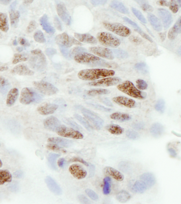
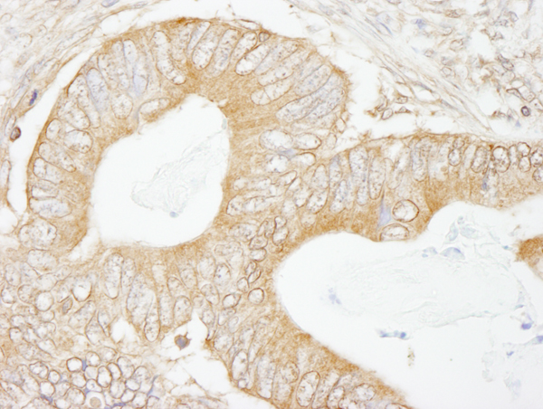
 IHC (Immunohiostchemistry)
(Detection of human FOXK1 by immunohistochemistry. Sample: FFPE section of human colon carcinoma. Antibody: Affinity purified rabbit anti-FOXK1 (Cat. No. AAA213961) used at a dilution of 1:500. Detection: DAB)
IHC (Immunohiostchemistry)
(Detection of human FOXK1 by immunohistochemistry. Sample: FFPE section of human colon carcinoma. Antibody: Affinity purified rabbit anti-FOXK1 (Cat. No. AAA213961) used at a dilution of 1:500. Detection: DAB)
FOXK1, Polyclonal Antibody (Cat# AAA213961)
 IHC (Immunohiostchemistry)
(Detection of human Fen1 by immunohistochemistry. Sample: FFPE section of human small cell lung cancer. Antibody: Affinity purified rabbit anti-Fen1 (Cat. No. AAA213962) used at a dilution of 1:250. Detection: DAB)
IHC (Immunohiostchemistry)
(Detection of human Fen1 by immunohistochemistry. Sample: FFPE section of human small cell lung cancer. Antibody: Affinity purified rabbit anti-Fen1 (Cat. No. AAA213962) used at a dilution of 1:250. Detection: DAB)
Fen1, Polyclonal Antibody (Cat# AAA213962)

 IHC (Immunohiostchemistry)
(Detection of mouse PHF8 by immunohistochemistry. Sample: FFPE section of mouse teratoma. Antibody: Affinity purified rabbit anti-PHF8 (Cat. No. AAA213967) used at a dilution of 1:100. Detection: DAB)
IHC (Immunohiostchemistry)
(Detection of mouse PHF8 by immunohistochemistry. Sample: FFPE section of mouse teratoma. Antibody: Affinity purified rabbit anti-PHF8 (Cat. No. AAA213967) used at a dilution of 1:100. Detection: DAB)
PHF8, Polyclonal Antibody (Cat# AAA213967)
 IHC (Immunohistochemistry)
(Detection of human 14-3-3 Sigma by IHC-IF. Sample: FFPE section of human breast carcinoma. Antibody: Affinity purified rabbit anti-14-3-3 Sigma (Cat. No. AAA213968) used at a dilution of 1:100. Detection: Red-fluorescent goat anti-rabbit IgG highly cross-adsorbed Antibody Hilyte Plus 555 used at a dilution of 1:100.)
IHC (Immunohistochemistry)
(Detection of human 14-3-3 Sigma by IHC-IF. Sample: FFPE section of human breast carcinoma. Antibody: Affinity purified rabbit anti-14-3-3 Sigma (Cat. No. AAA213968) used at a dilution of 1:100. Detection: Red-fluorescent goat anti-rabbit IgG highly cross-adsorbed Antibody Hilyte Plus 555 used at a dilution of 1:100.)
14-3-3 Sigma, Polyclonal Antibody (Cat# AAA213968)
 IHC (Immunohistochemisry)
(Detection of human NUP50 by immunohistochemistry. Sample: FFPE section of human breast carcinoma. Antibody: Affinity purified rabbit anti-NUP50 (Cat. No. AAA213970) used at a dilution of 1:500. Detection: DAB)
IHC (Immunohistochemisry)
(Detection of human NUP50 by immunohistochemistry. Sample: FFPE section of human breast carcinoma. Antibody: Affinity purified rabbit anti-NUP50 (Cat. No. AAA213970) used at a dilution of 1:500. Detection: DAB)
NUP50, Polyclonal Antibody (Cat# AAA213970)
 IHC (Immunohiostchemistry)
(Detection of human IRS2 by immunohistochemistry. Sample: FFPE section of human testicular seminoma. Antibody: Affinity purified rabbit anti-IRS2 (Cat. No. AAA213972) used at a dilution of 1:100. Detection: Red-fluorescent goat anti-rabbit IgG highly cross-adsorbed Antibody used at a dilution of 1:100.)
IHC (Immunohiostchemistry)
(Detection of human IRS2 by immunohistochemistry. Sample: FFPE section of human testicular seminoma. Antibody: Affinity purified rabbit anti-IRS2 (Cat. No. AAA213972) used at a dilution of 1:100. Detection: Red-fluorescent goat anti-rabbit IgG highly cross-adsorbed Antibody used at a dilution of 1:100.)
IRS2, Polyclonal Antibody (Cat# AAA213972)
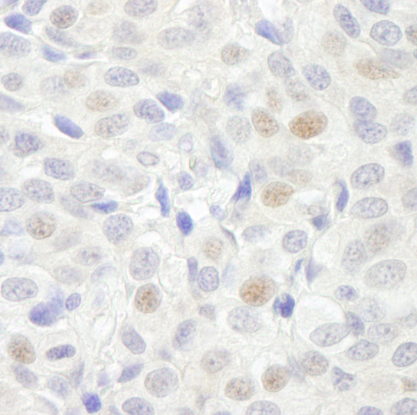
TIF1 Alpha/TRIM24, Polyclonal Antibody (Cat# AAA213826)
 IHC (Immunohiostchemistry)
(Detection of human UBCH7 by immunohistochemistry. Sample: FFPE section of human cervix. Antibody: Affinity purified rabbit anti-UBCH7 (Cat. No. AAA213830) used at a dilution of 1:500. Detection: DAB)
IHC (Immunohiostchemistry)
(Detection of human UBCH7 by immunohistochemistry. Sample: FFPE section of human cervix. Antibody: Affinity purified rabbit anti-UBCH7 (Cat. No. AAA213830) used at a dilution of 1:500. Detection: DAB)
UBCH7, Polyclonal Antibody (Cat# AAA213830)
 IHC (Immunohiostchemistry)
(Detection of mouse DDX5 by immunohistochemistry. Sample: FFPE section of mouse teratoma. Antibody: Affinity purified rabbit anti-DDX5 (Cat. No. AAA213836) used at a dilution of 1:100. Detection: DAB)
IHC (Immunohiostchemistry)
(Detection of mouse DDX5 by immunohistochemistry. Sample: FFPE section of mouse teratoma. Antibody: Affinity purified rabbit anti-DDX5 (Cat. No. AAA213836) used at a dilution of 1:100. Detection: DAB)
DDX5, Polyclonal Antibody (Cat# AAA213836)
 IHC (Immunohiostchemistry)
(Detection of mouse PPM1G by immunohistochemistry. Sample: FFPE section of mouse squamous cell carcinoma. Antibody: Affinity purified rabbit anti-PPM1G (Cat. No. AAA213840) used at a dilution of 1:100. Detection: DAB)
IHC (Immunohiostchemistry)
(Detection of mouse PPM1G by immunohistochemistry. Sample: FFPE section of mouse squamous cell carcinoma. Antibody: Affinity purified rabbit anti-PPM1G (Cat. No. AAA213840) used at a dilution of 1:100. Detection: DAB)
PPM1G, Polyclonal Antibody (Cat# AAA213840)
 IHC (Immunohiostchemistry)
(Detection of mouse HMG2a by immunohistochemistry. Sample: FFPE section of mouse teratoma. Antibody: Affinity purified rabbit anti-HMG2a (Cat. No. AAA213848) used at a dilution of 1:250. Detection: DAB)
IHC (Immunohiostchemistry)
(Detection of mouse HMG2a by immunohistochemistry. Sample: FFPE section of mouse teratoma. Antibody: Affinity purified rabbit anti-HMG2a (Cat. No. AAA213848) used at a dilution of 1:250. Detection: DAB)
HMG2a, Polyclonal Antibody (Cat# AAA213848)
 IHC (Immunohiostchemistry)
(Detection of mouse FOX2/RBM9 by immunohistochemistry. Sample: FFPE section of mouse teratoma. Antibody: Affinity purified rabbit anti-FOX2/RBM9 (Cat. No. AAA213860) used at a dilution of 1:250. Detection: DAB)
IHC (Immunohiostchemistry)
(Detection of mouse FOX2/RBM9 by immunohistochemistry. Sample: FFPE section of mouse teratoma. Antibody: Affinity purified rabbit anti-FOX2/RBM9 (Cat. No. AAA213860) used at a dilution of 1:250. Detection: DAB)
FOX2/RBM9, Polyclonal Antibody (Cat# AAA213860)
 IHC (Immunohiostchemistry)
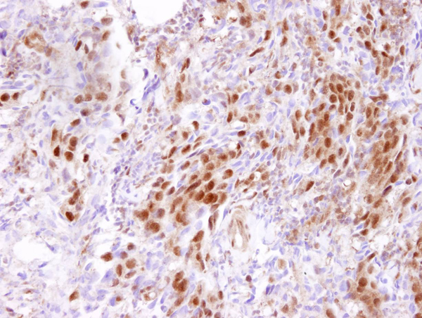
(Detection of human ACINUS/ACN1 by immunohistochemistry. Sample: FFPE section of human stomach adenocarcinoma. Antibody: Affinity purified rabbit anti-ACINUS/ACN1 (Cat. No. AAA213870) used at a dilution of 1:250. Detection: DAB)
IHC (Immunohiostchemistry)
(Detection of human ACINUS/ACN1 by immunohistochemistry. Sample: FFPE section of human stomach adenocarcinoma. Antibody: Affinity purified rabbit anti-ACINUS/ACN1 (Cat. No. AAA213870) used at a dilution of 1:250. Detection: DAB)
ACINUS/ACN1, Polyclonal Antibody (Cat# AAA213870)

 IHC (Immunohiostchemistry)
(Detection of human Cul3 by immunohistochemistry. Sample: FFPE section of human colon carcinoma. Antibody: Affinity purified rabbit anti-Cul3 (Cat. No. AAA213875) used at a dilution of 1:250. Detection: DAB)
IHC (Immunohiostchemistry)
(Detection of human Cul3 by immunohistochemistry. Sample: FFPE section of human colon carcinoma. Antibody: Affinity purified rabbit anti-Cul3 (Cat. No. AAA213875) used at a dilution of 1:250. Detection: DAB)
Cul3, Polyclonal Antibody (Cat# AAA213875)
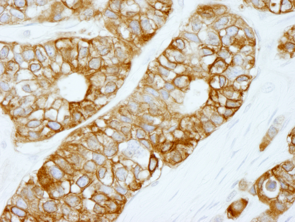
 IHC (Immunohistochemistry)
(Detection of human Filamin by immunohistochemistry. Sample: FFPE section of human colon carcinoma. Antibody: Affinity purified rabbit anti-Filamin (Cat. No. AAA213879) used at a dilution of 1:100. Detection: Red-fluorescent Alexa Fluor 555 goat anti-rabbit IgG (Invitrogen) used at a dilution of 1:500.)
IHC (Immunohistochemistry)
(Detection of human Filamin by immunohistochemistry. Sample: FFPE section of human colon carcinoma. Antibody: Affinity purified rabbit anti-Filamin (Cat. No. AAA213879) used at a dilution of 1:100. Detection: Red-fluorescent Alexa Fluor 555 goat anti-rabbit IgG (Invitrogen) used at a dilution of 1:500.)
Filamin A, Polyclonal Antibody (Cat# AAA213879)

 IHC (Immunohiostchemistry)
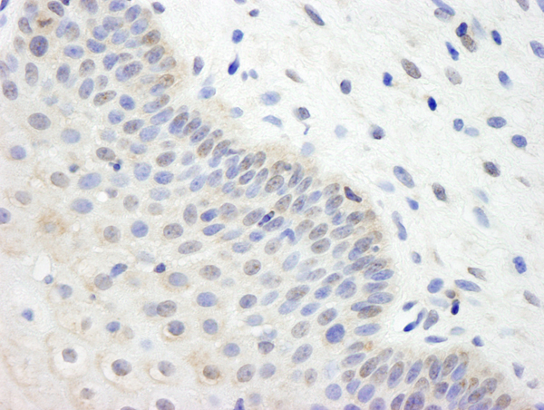
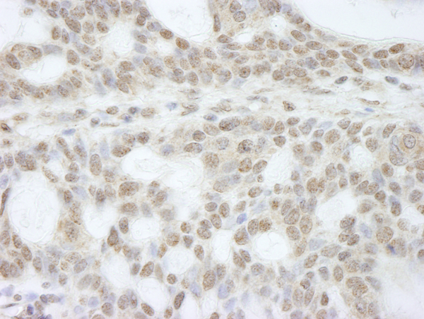
(Detection of human ATRX by immunohistochemistry. Sample: FFPE section of human basal cell carcinoma. Antibody: Affinity purified rabbit anti-ATRX (Cat. No. AAA213887) used at a dilution of 1:250. Detection: DAB)
IHC (Immunohiostchemistry)
(Detection of human ATRX by immunohistochemistry. Sample: FFPE section of human basal cell carcinoma. Antibody: Affinity purified rabbit anti-ATRX (Cat. No. AAA213887) used at a dilution of 1:250. Detection: DAB)
ATRX, Polyclonal Antibody (Cat# AAA213887)

 IHC (Immunohiostchemistry)
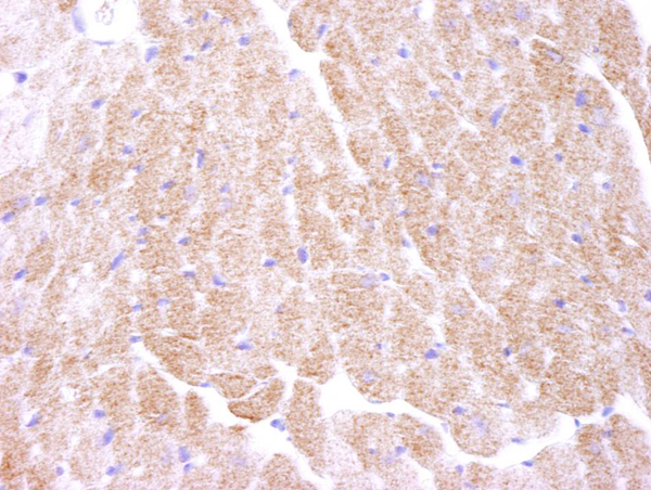
(Detection of mouse AMPK alpha 1 by immunohistochemistry. Samples: FFPE section of mouse heart. Antibody: Affinity purified rabbit anti-AMPK alpha 1 (Cat. No. AAA213720) used at a dilution of 1:250. Detection: DAB)
IHC (Immunohiostchemistry)
(Detection of mouse AMPK alpha 1 by immunohistochemistry. Samples: FFPE section of mouse heart. Antibody: Affinity purified rabbit anti-AMPK alpha 1 (Cat. No. AAA213720) used at a dilution of 1:250. Detection: DAB)
AMPK alpha 1, Polyclonal Antibody (Cat# AAA213720)
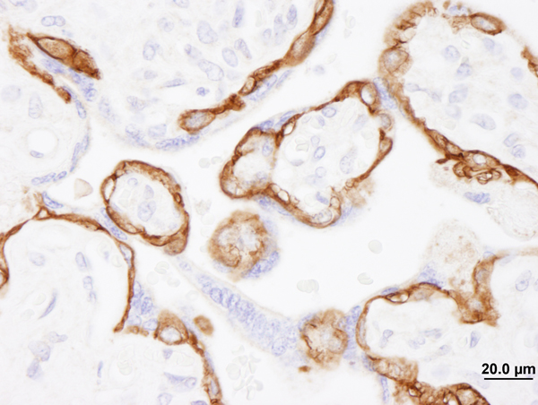
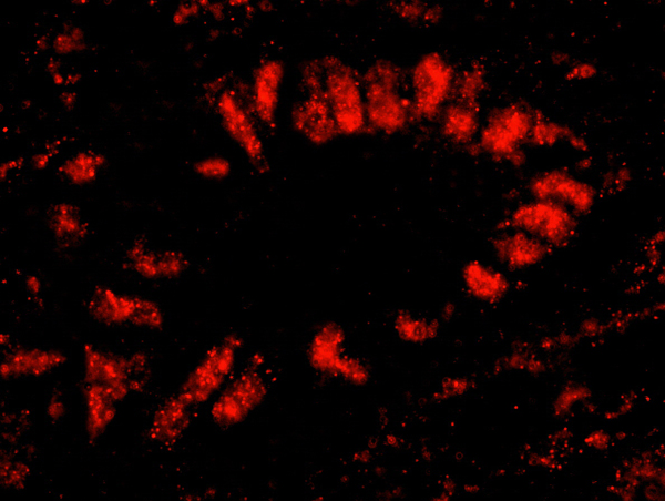
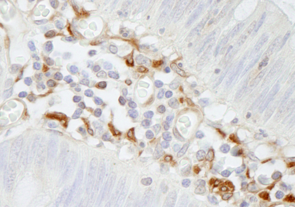
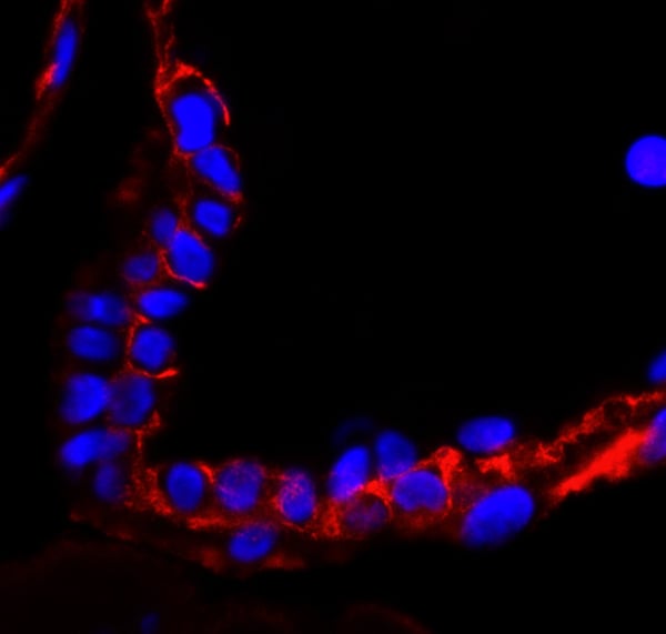 IHC (Immunohistochemisry)
(Detection of human EGFR by immunohistochemistry. Sample: FFPE section of human placenta. Antibody: Affinity purified rabbit anti-EGFR (Cat. No. AAA213723-T) used at a dilution of 1:100. Detection: Red-fluorescent goat anti-rabbit IgG highly cross-adsorbed Antibody)
IHC (Immunohistochemisry)
(Detection of human EGFR by immunohistochemistry. Sample: FFPE section of human placenta. Antibody: Affinity purified rabbit anti-EGFR (Cat. No. AAA213723-T) used at a dilution of 1:100. Detection: Red-fluorescent goat anti-rabbit IgG highly cross-adsorbed Antibody)
EGFR, Polyclonal Antibody (Cat# AAA213723)
 IHC (Immunohiostchemistry)
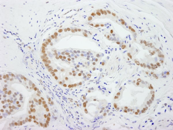
(Detection of mouse PELP1/MNAR by immunohistochemistry. Sample: FFPE section of mouse ovary. Antibody: Affinity purified rabbit anti-PELP1/MNAR (Cat. No. AAA213729) used at a dilution of 1:100. Detection: DAB)
IHC (Immunohiostchemistry)
(Detection of mouse PELP1/MNAR by immunohistochemistry. Sample: FFPE section of mouse ovary. Antibody: Affinity purified rabbit anti-PELP1/MNAR (Cat. No. AAA213729) used at a dilution of 1:100. Detection: DAB)
PELP1/MNAR, Polyclonal Antibody (Cat# AAA213729)
 IHC (Immunohiostchemistry)
(Detection of human SMC1 by immunohistochemistry. Sample: FFPE section of human breast. Antibody: Affinity purified rabbit anti-SMC1 (Cat. No. AAA213733) used at a dilution of 1:500. Detection: DAB)
IHC (Immunohiostchemistry)
(Detection of human SMC1 by immunohistochemistry. Sample: FFPE section of human breast. Antibody: Affinity purified rabbit anti-SMC1 (Cat. No. AAA213733) used at a dilution of 1:500. Detection: DAB)
SMC1, Polyclonal Antibody (Cat# AAA213733)
 IHC (Immunohiostchemistry)
(Detection of human ASC2 by immunohistochemistry. Sample: FFPE section of human stomach adenocarcinoma. Antibody: Affinity purified rabbit anti-ASC2 (Cat. No. AAA213735) used at a dilution of 1:100. Detection: DAB)
IHC (Immunohiostchemistry)
(Detection of human ASC2 by immunohistochemistry. Sample: FFPE section of human stomach adenocarcinoma. Antibody: Affinity purified rabbit anti-ASC2 (Cat. No. AAA213735) used at a dilution of 1:100. Detection: DAB)
ASC2, Polyclonal Antibody (Cat# AAA213735)
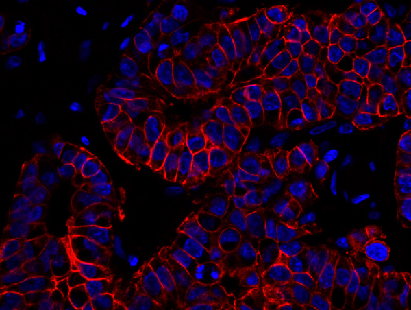
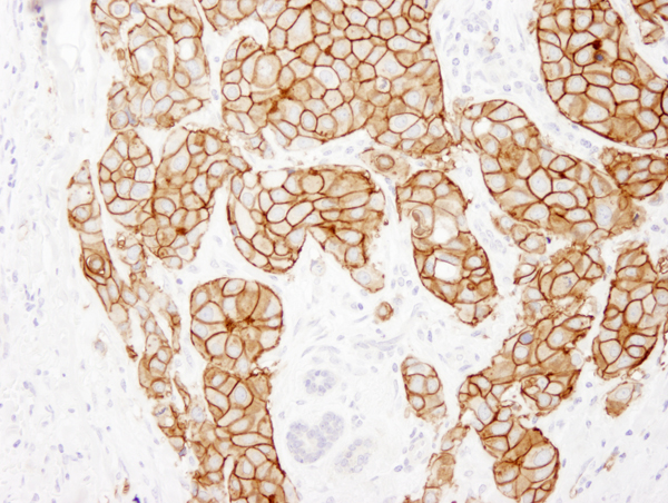
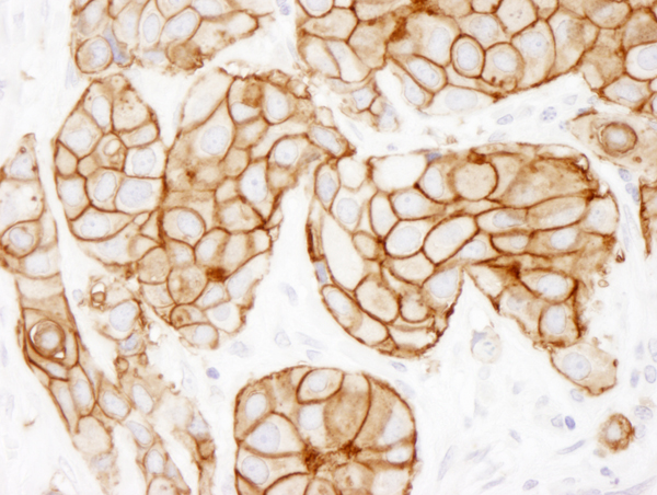
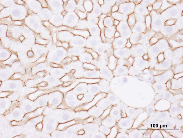
 IHC (Immunohistochemistry)
(Detection of human ErbB2 by immunohistochemistry. Sample: FFPE section of human breast carcinoma. Antibody: Affinity purified rabbit anti-ErbB2 (Cat. No. AAA213744) used at a dilution of 1:100. Detection: Red-fluorescent goat anti-rabbit IgG highly cross-adsorbed Antibody.)
IHC (Immunohistochemistry)
(Detection of human ErbB2 by immunohistochemistry. Sample: FFPE section of human breast carcinoma. Antibody: Affinity purified rabbit anti-ErbB2 (Cat. No. AAA213744) used at a dilution of 1:100. Detection: Red-fluorescent goat anti-rabbit IgG highly cross-adsorbed Antibody.)
ErbB2, Polyclonal Antibody (Cat# AAA213744)
 IHC (Immunohiostchemistry)
(Detection of human MCM4 by immunohistochemistry. Sample: FFPE section of human small cell lung carcinoma. Antibody: Affinity purified goat anti-MCM4 (Cat. No. AAA213748) used at a dilution of 1:250. Detection: DAB)
IHC (Immunohiostchemistry)
(Detection of human MCM4 by immunohistochemistry. Sample: FFPE section of human small cell lung carcinoma. Antibody: Affinity purified goat anti-MCM4 (Cat. No. AAA213748) used at a dilution of 1:250. Detection: DAB)
MCM4, Polyclonal Antibody (Cat# AAA213748)
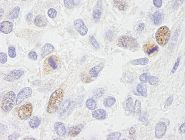
 IHC (Immunohiostchemistry)
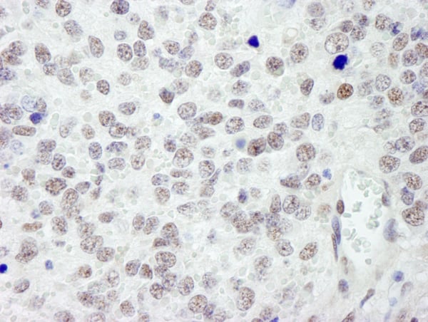
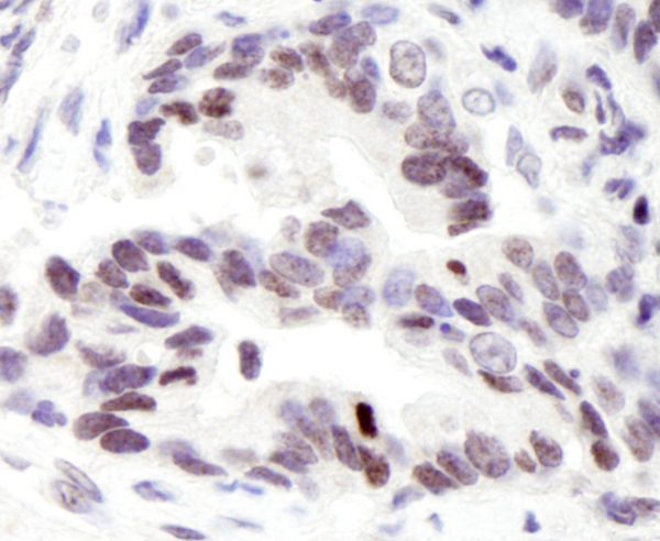
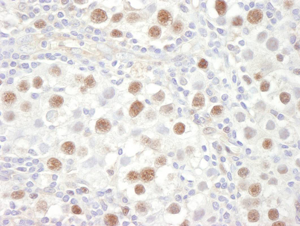
(Detection of human CARM1 by immunohistochemistry. Sample: FFPE section of prostate adenocarcinoma used at a dilution of 1:500. Antibody: Affinity purified rabbit anti-CARM1 (Cat. No. AAA213753). Detection: DAB)
IHC (Immunohiostchemistry)
(Detection of human CARM1 by immunohistochemistry. Sample: FFPE section of prostate adenocarcinoma used at a dilution of 1:500. Antibody: Affinity purified rabbit anti-CARM1 (Cat. No. AAA213753). Detection: DAB)
CARM1, Polyclonal Antibody (Cat# AAA213753)
 IHC (Immunohiostchemistry)
(Detection of mouse SMC3 by immunohistochemistry. Sample: FFPE section of mouse squamous cell carcinoma. Antibody: Affinity purified rabbit anti-SMC3 (Cat. No. AAA213757) used at a dilution of 1:250. Detection: DAB)
IHC (Immunohiostchemistry)
(Detection of mouse SMC3 by immunohistochemistry. Sample: FFPE section of mouse squamous cell carcinoma. Antibody: Affinity purified rabbit anti-SMC3 (Cat. No. AAA213757) used at a dilution of 1:250. Detection: DAB)
SMC3, Polyclonal Antibody (Cat# AAA213757)
 IHC (Immunohistochemistry)
(Detection of human HDAC2 by immunohistochemistry. Sample: FFPE section of human breast carcinoma. Antibody: Affinity purified rabbit anti-HDAC2 (Cat. No. AAA213759) used at a dilution of 1:100. Detection: Red-fluorescent Alexa Fluor 555 goat anti-rabbit IgG (Invitrogen) used at a dilution of 1:500.)
IHC (Immunohistochemistry)
(Detection of human HDAC2 by immunohistochemistry. Sample: FFPE section of human breast carcinoma. Antibody: Affinity purified rabbit anti-HDAC2 (Cat. No. AAA213759) used at a dilution of 1:100. Detection: Red-fluorescent Alexa Fluor 555 goat anti-rabbit IgG (Invitrogen) used at a dilution of 1:500.)
HDAC2, Polyclonal Antibody (Cat# AAA213759)
 IHC (Immunohiostchemistry)
(Detection of mouse FUS by immunohistochemistry. Sample: FFPE section of mouse renal cell carcinoma. Antibody: Affinity purified rabbit anti-FUS (Cat. No. AAA213772-2) used at a dilution of 1:100. Detection: DAB)
IHC (Immunohiostchemistry)
(Detection of mouse FUS by immunohistochemistry. Sample: FFPE section of mouse renal cell carcinoma. Antibody: Affinity purified rabbit anti-FUS (Cat. No. AAA213772-2) used at a dilution of 1:100. Detection: DAB)
FUS, Polyclonal Antibody (Cat# AAA213772)
 IHC (Immunohiostchemistry)
(Detection of mouse Rad50 by immunohistochemistry. Sample: FFPE section of mouse squamous cell carcinoma. Antibody: Affinity purified rabbit anti-Rad50 (Cat. No. AAA213774) used at a dilution of 1:250. Detection: DAB)
IHC (Immunohiostchemistry)
(Detection of mouse Rad50 by immunohistochemistry. Sample: FFPE section of mouse squamous cell carcinoma. Antibody: Affinity purified rabbit anti-Rad50 (Cat. No. AAA213774) used at a dilution of 1:250. Detection: DAB)
Rad50, Polyclonal Antibody (Cat# AAA213774)
 IHC (Immunohiostchemistry)
(Detection of human BTF by immunohistochemistry. Sample: FFPE section of human skin basal cell carcinoma. Antibody: Affinity purified rabbit anti-BTF (Cat. No. AAA213778) used at a dilution of 1:250. Detection: DAB staining using anti-rabbit IHC antibody (Cat. No. at a dilution of 1:100.)
IHC (Immunohiostchemistry)
(Detection of human BTF by immunohistochemistry. Sample: FFPE section of human skin basal cell carcinoma. Antibody: Affinity purified rabbit anti-BTF (Cat. No. AAA213778) used at a dilution of 1:250. Detection: DAB staining using anti-rabbit IHC antibody (Cat. No. at a dilution of 1:100.)
BTF, Polyclonal Antibody (Cat# AAA213778)
 IHC (Immunohiostchemistry)
(Detection of human EWS by immunohistochemistry. Sample: FFPE section of human breast adenocarcinoma. Antibody: Affinity purified rabbit anti-EWS (Cat. No. AAA213783) used at a dilution of 1:250. Detection: DAB staining using anti-rabbit IHC antibody (Cat. No. at a dilution of 1:100.)
IHC (Immunohiostchemistry)
(Detection of human EWS by immunohistochemistry. Sample: FFPE section of human breast adenocarcinoma. Antibody: Affinity purified rabbit anti-EWS (Cat. No. AAA213783) used at a dilution of 1:250. Detection: DAB staining using anti-rabbit IHC antibody (Cat. No. at a dilution of 1:100.)
EWS, Polyclonal Antibody (Cat# AAA213783)
 IHC (Immunohiostchemistry)
(Detection of human CCAR1 by immunohistochemistry. Sample: FFPE section of human skin basal cell carcinoma. Antibody: Affinity purified rabbit anti-CCAR1 (Cat. No. AAA213787) used at a dilution of 1:250. Detection: DAB staining using anti-rabbit IHC antibody (Cat. No. at a dilution of 1:100.)
IHC (Immunohiostchemistry)
(Detection of human CCAR1 by immunohistochemistry. Sample: FFPE section of human skin basal cell carcinoma. Antibody: Affinity purified rabbit anti-CCAR1 (Cat. No. AAA213787) used at a dilution of 1:250. Detection: DAB staining using anti-rabbit IHC antibody (Cat. No. at a dilution of 1:100.)
CCAR1, Polyclonal Antibody (Cat# AAA213787)
What are Polyclonal Antibodies?
Polyclonal antibodies are antibodies that come from multiple B cell clones of a host animal. The typical hosts used for the majority of polyclonal antibody production are rabbits, goats, sheep, and donkeys. These polyclonal antibodies, once having identified their target, will bind to different epitopes located at different regions or sequences on the same protein/antigen. As a result, they are ideal at locating and binding to the target, even if the target is in very low concentrations (due to many different antibodies being able to bind to the same target molecule, which allows for significant amplification of a downstream signal).
Polyclonal antibodies are typically produced by injecting an antigen into a host animal, which causes the animal’s immune system to attack the foreign antigen by mass generating antibodies against it. After a period of time, serum is collected from the animal and purified using physicochemical fractionation, class-specific affinity purification, and/or antigen-affinity purification.
Key Uses of Polyclonal Antibodies
- Western Blotting: This method is used to find specific proteins in biological samples after separating them by size.
- Immunohistochemistry: IHC helps visualize the location of proteins in tissue sections using various staining techniques.
- ELISA: (Enzyme-Linked Immunosorbent Assay) is typically used to identify specific protein quantities in a sample. ELISAs can be either “Quantitative” or “Qualitative”.
- Flow Cytometry: technique that identifies and measures the specific protein on the surface or inside the cells in a fluid suspension.
- Immunoprecipitation: IP isolates and studies a specific protein from a complex mixture using antibodies.
Why Buy Polyclonal Antibodies from AAA Biotech?
1. Ideal for Various Applications
Our antibodies are generally going to be validated for use in multiple types of assays, including ELISA, Western Blotting, Immunohistochemistry, Immunoprecipitation, amongst others. They are ideal for a wide range of research applications.
2. Rigorous Quality Control
All of the antibodies in our catalog undergo strict quality testing to ensure specificity, sensitivity, and consistent performance. We are confident in the ability of our antibodies to provide you with accurate results.
3. Wide Assortment of Antibodies
Antibodies in are catalog can be found for both common and exotic species, and these antibodies are also available in both conjugated and recombinant forms to suit many diverse experimental needs.
4. Highly Purified
Our antibodies are available in purified forms with over 85% purity, as confirmed by SDS-PAGE. They are also available with tags such as His, Flag, GST, or MBP. We cater to customers worldwide.
FAQ
1. How are polyclonal antibodies produced?
Traditionally, polyclonal antibodies are produced by injecting an antigen into a host animal (such as a rabbit or goat), which then triggers an immune response from the host animal. The animal’s B cells produce antibodies that will recognize different parts of the injected antigen. These antibodies are then collected from the animal’s blood and purified for use.
2. How do polyclonal antibodies differ from monoclonal antibodies?
Polyclonal antibodies are a mix of antibodies that bind to different locations (epitopes) of the same antigen, while monoclonal antibodies are identical and bind to just one specific epitope. This makes polyclonal antibodies more versatile and better at detecting proteins that may be present in low quantities or in altered/modified forms.
3. How should I store polyclonal antibodies?
Polyclonal antibodies should be stored at 4°C for short-term use (up to a few weeks) and at -20°C or -80°C for long-term storage. Avoid repeated freeze-thaw cycles by dividing them into small aliquots. Always check the datasheet for specific storage instructions.