Filters
▼Clonality
▼Type
▼Reactivity
▼Gene Name
▼Isotype
▼Host
▼Application
▼Clone
▼Polyclonal Antibodies
At AAA Biotech also known as AAA Bio or AAABio, we provide a broad range of purified polyclonal antibodies (pAbs) that are able to all be browsed online through our website. Due to their high specificity and strong binding affinity, these antibodies are ideal for wide swathes of research and experimental applications.
Our polyclonal antibodies can easily support your work, whether you use them for Western Blotting, Immunocytochemistry (with or without Immunofluorescence used in conjunction), Immunohistochemistry, Immunoprecipitation, and ELISA tests. We highly encourage you to browse our range of pAbs and choose the one that best suits your experimental model.
Viewing 4700-4750 of 96812 product results
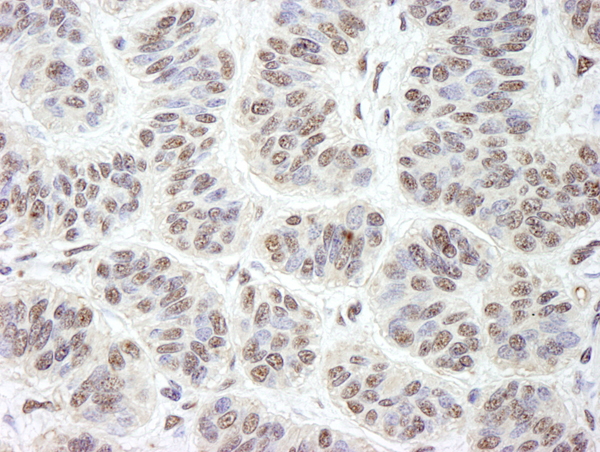
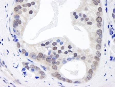
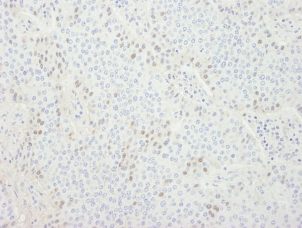
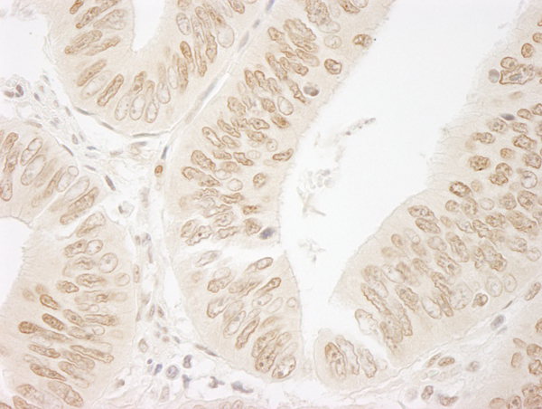
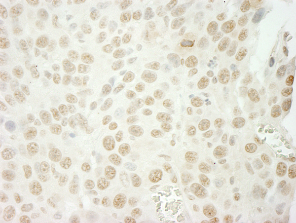
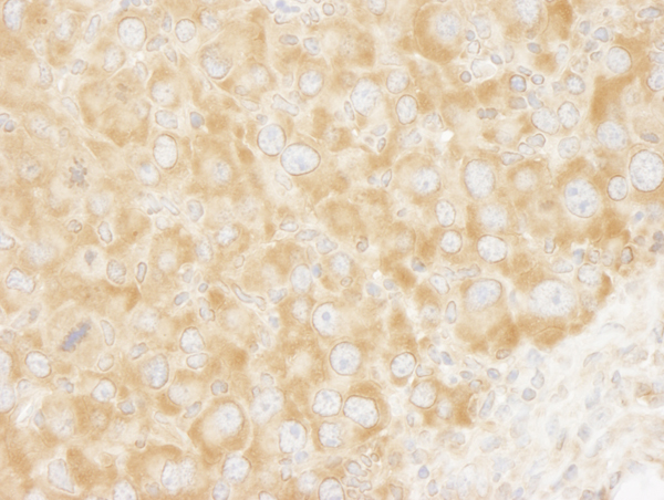
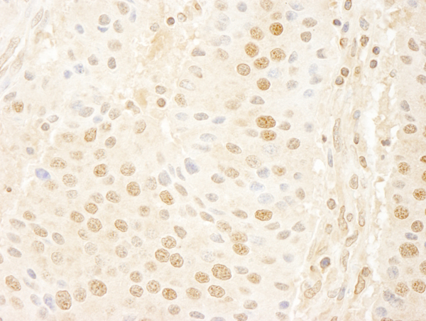
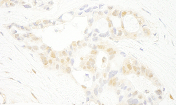
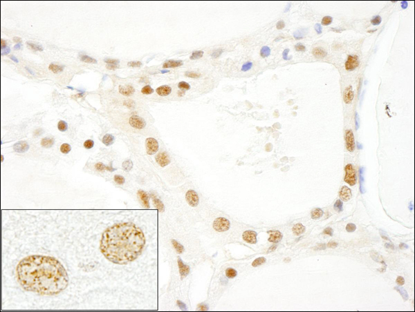
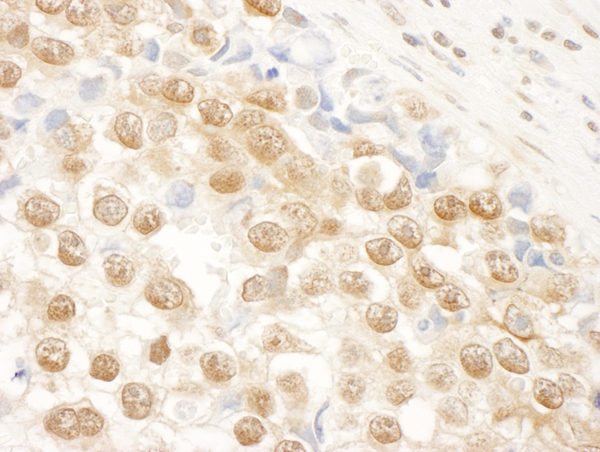
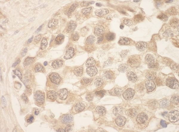
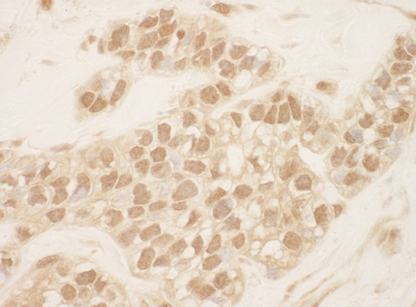
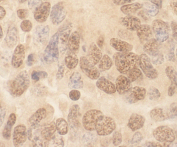
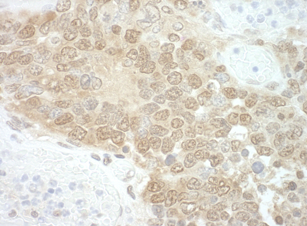
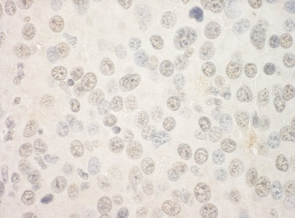
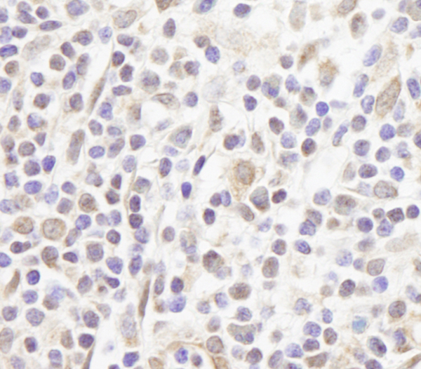
 IHC (Immunohiostchemistry)
(Detection of mouse GNL3 by immunohistochemistry. Sample: FFPE section of mouse squamous cell carcinoma. Antibody: Affinity purified rabbit anti-GNL3 (Cat. No. AAA213788) used at a dilution of 1:100. Detection: DAB)
IHC (Immunohiostchemistry)
(Detection of mouse GNL3 by immunohistochemistry. Sample: FFPE section of mouse squamous cell carcinoma. Antibody: Affinity purified rabbit anti-GNL3 (Cat. No. AAA213788) used at a dilution of 1:100. Detection: DAB)
GNL3, Polyclonal Antibody (Cat# AAA213788)
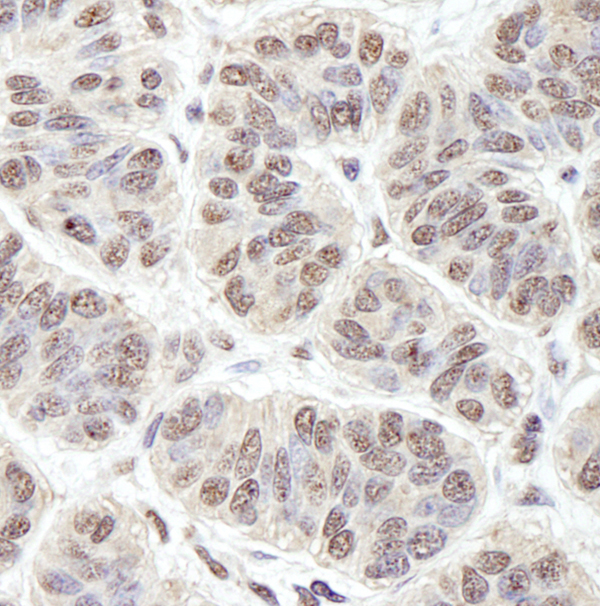
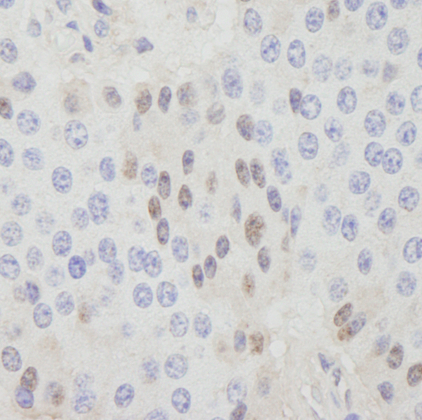
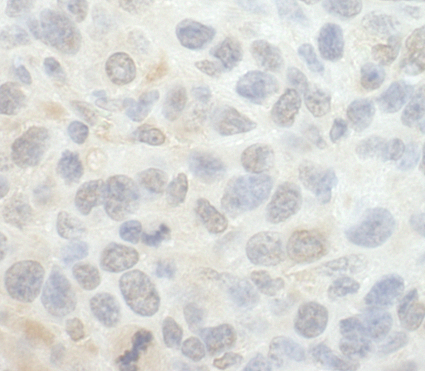
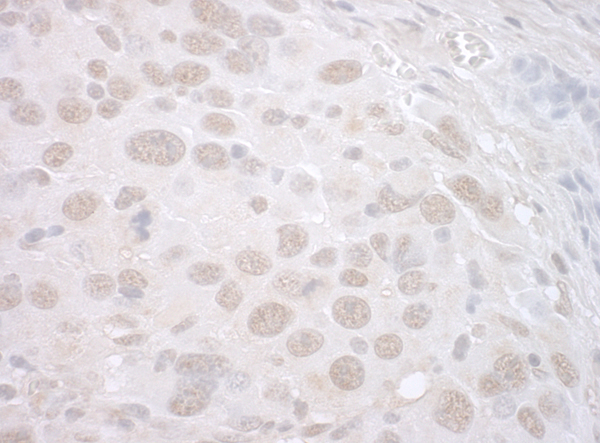
 IHC (Immunohiostchemistry)
(Detection of mouse PRKRIR by immunohistochemistry. Sample: FFPE section of mouse squamous cell carcinoma. Antibody: Affinity purified rabbit anti-PRKRIR (Cat. No. AAA213789) used at a dilution of 1:250. Detection: DAB)
IHC (Immunohiostchemistry)
(Detection of mouse PRKRIR by immunohistochemistry. Sample: FFPE section of mouse squamous cell carcinoma. Antibody: Affinity purified rabbit anti-PRKRIR (Cat. No. AAA213789) used at a dilution of 1:250. Detection: DAB)
PRKRIR, Polyclonal Antibody (Cat# AAA213789)
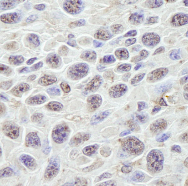
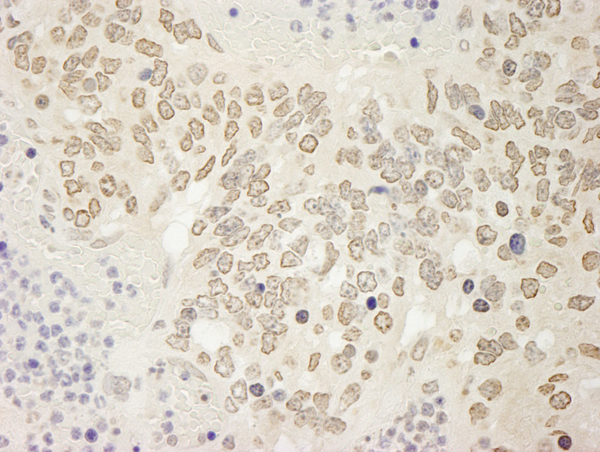
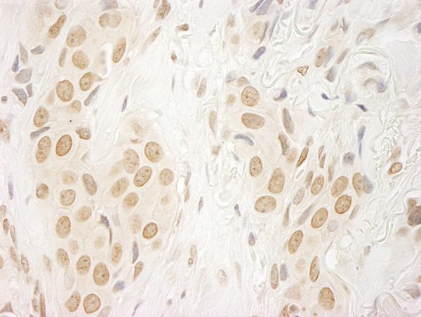
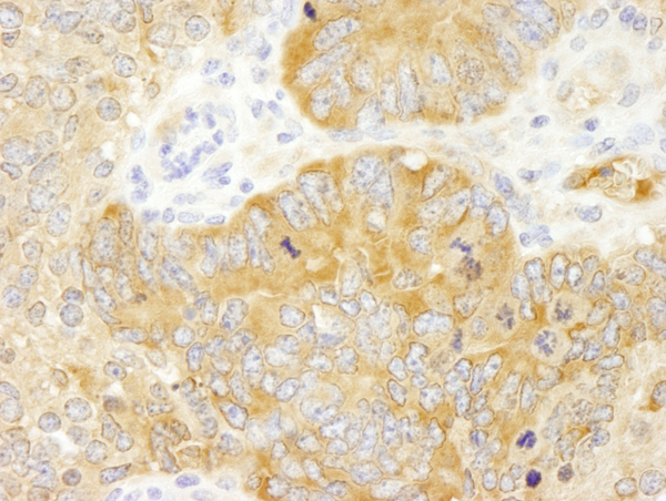
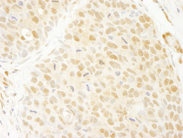
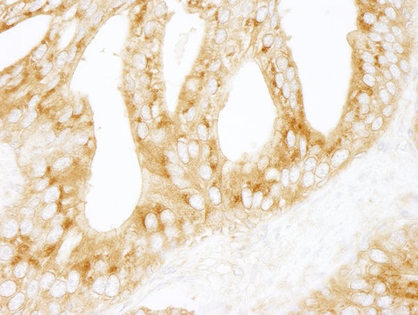
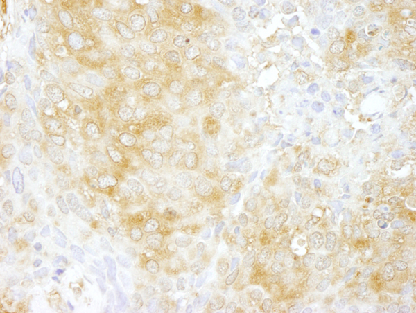
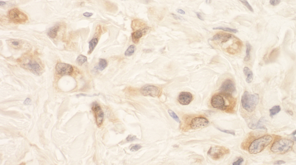
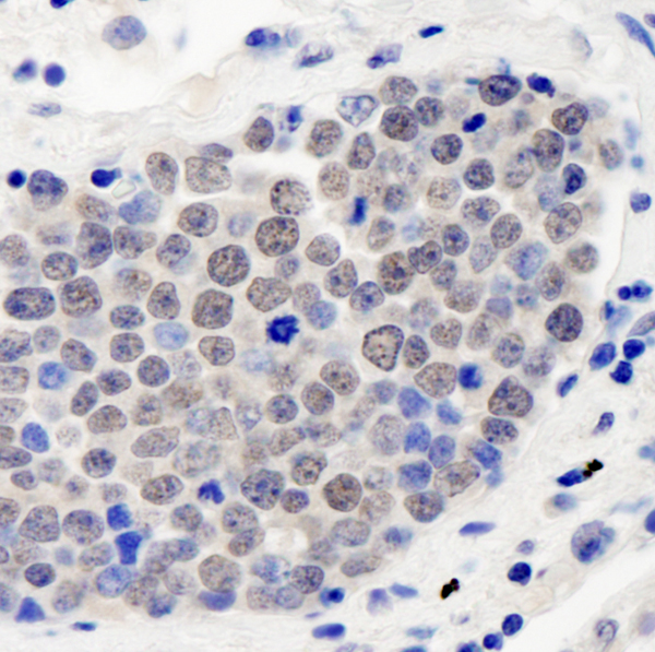 IHC (Immunohiostchemistry)
(Detection of human CSN3 by immunohistochemistry. Sample: FFPE section of human small cell lung cancer. Antibody: Affinity purified rabbit anti-CSN3 (Cat. No. AAA213800) used at a dilution of 1:250. Detection: DAB)
IHC (Immunohiostchemistry)
(Detection of human CSN3 by immunohistochemistry. Sample: FFPE section of human small cell lung cancer. Antibody: Affinity purified rabbit anti-CSN3 (Cat. No. AAA213800) used at a dilution of 1:250. Detection: DAB)
CSN3, Polyclonal Antibody (Cat# AAA213800)
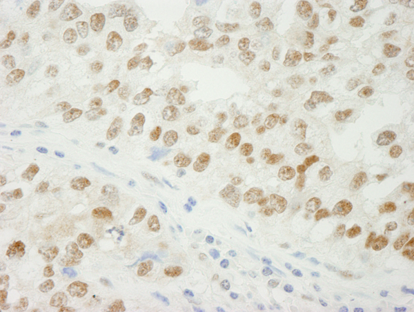
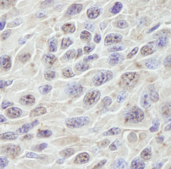
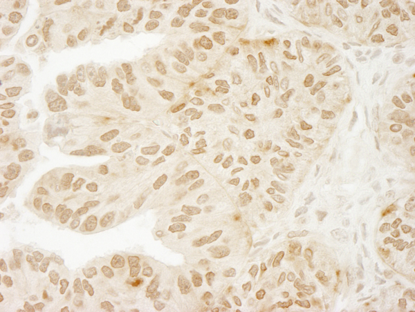
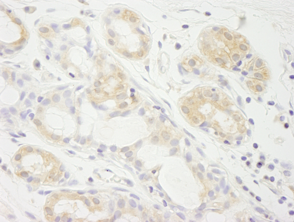
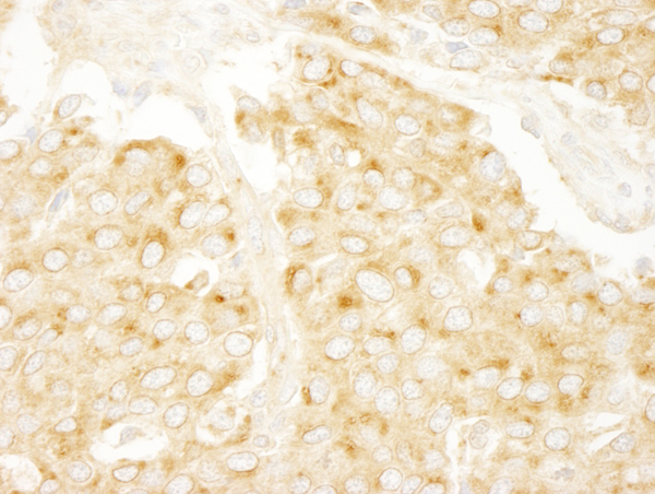
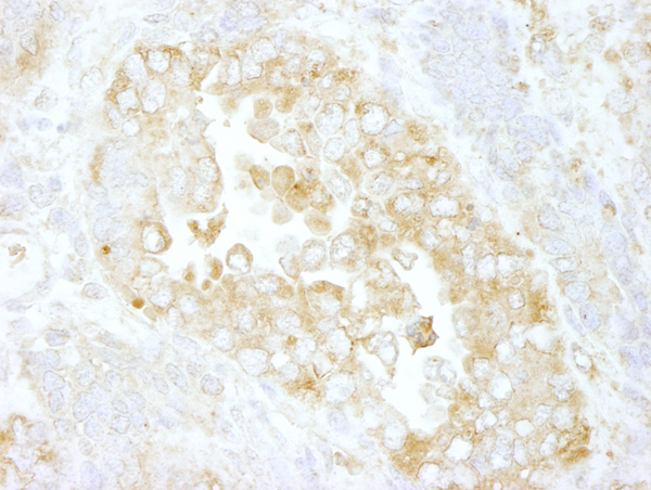
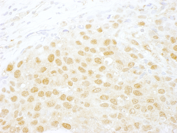
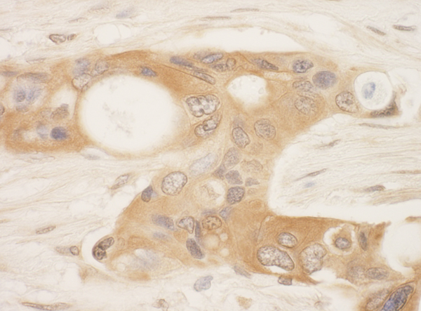
 IHC (Immunohiostchemistry)
(Detection of human ADNP by immunohistochemistry. Sample: FFPE section of human metastatic bone marrow. Antibody: Affinity purified rabbit anti-ADNP (Cat. No. AAA213805) used at a dilution of 1:250. Detection: DAB)
IHC (Immunohiostchemistry)
(Detection of human ADNP by immunohistochemistry. Sample: FFPE section of human metastatic bone marrow. Antibody: Affinity purified rabbit anti-ADNP (Cat. No. AAA213805) used at a dilution of 1:250. Detection: DAB)
ADNP, Polyclonal Antibody (Cat# AAA213805)
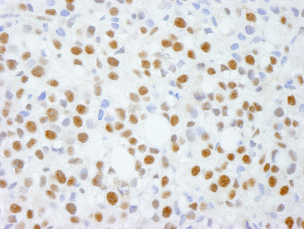
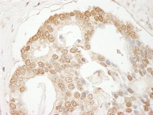
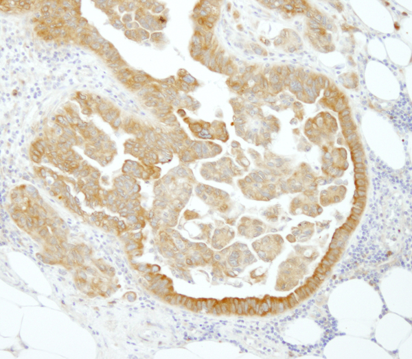
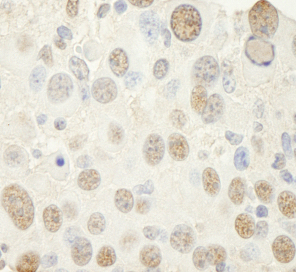
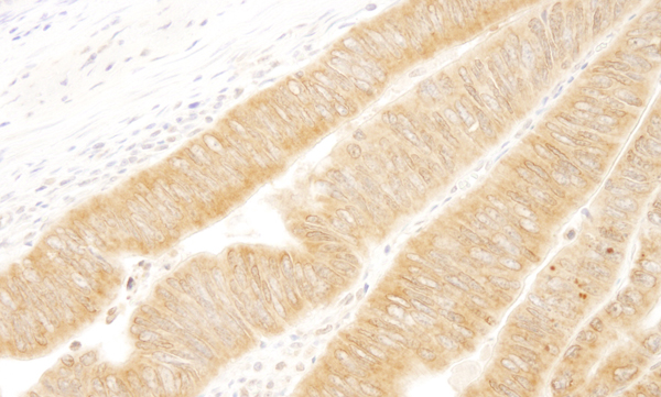
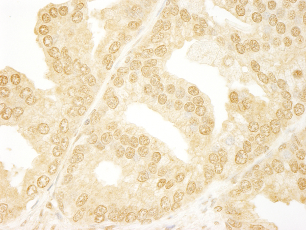
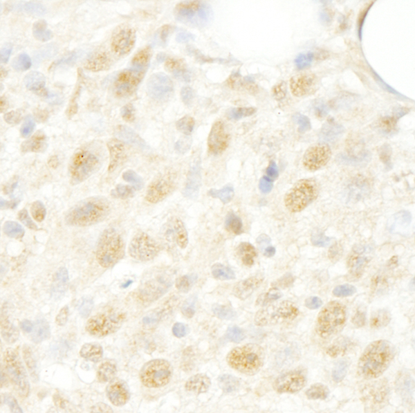
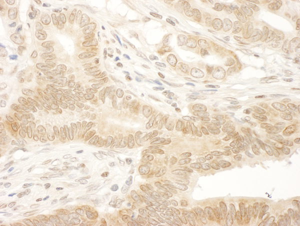
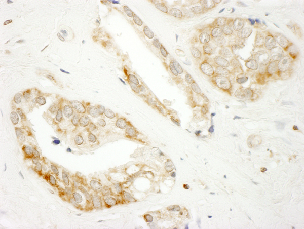
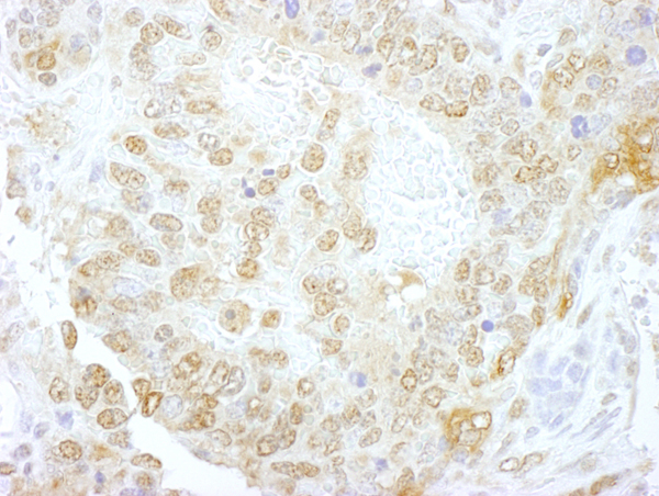
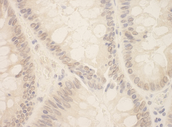
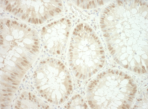
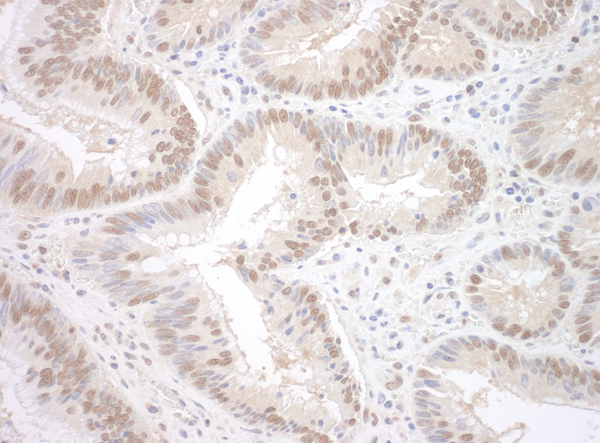
 IHC (Immunohiostchemistry)
(Detection of human CDT1 by immunohistochemistry. Sample: FFPE section of human pancreatic islet cell tumor. Antibody: Affinity purified rabbit anti-CDT1 (Cat. No. AAA213812) used at a dilution of 1:250. Detection: DAB)
IHC (Immunohiostchemistry)
(Detection of human CDT1 by immunohistochemistry. Sample: FFPE section of human pancreatic islet cell tumor. Antibody: Affinity purified rabbit anti-CDT1 (Cat. No. AAA213812) used at a dilution of 1:250. Detection: DAB)
CDT1, Polyclonal Antibody (Cat# AAA213812)
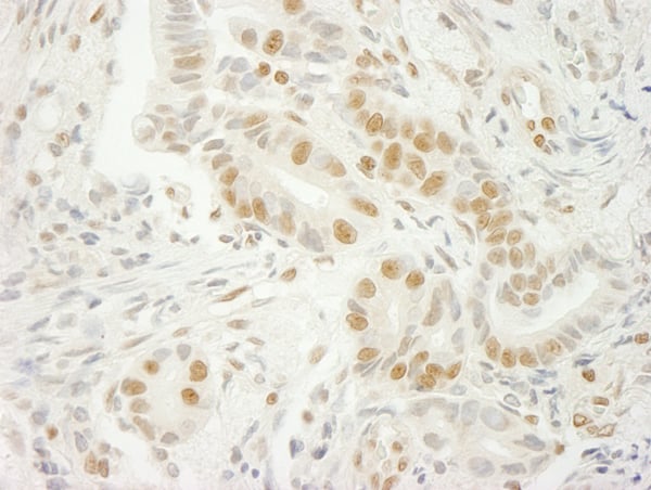
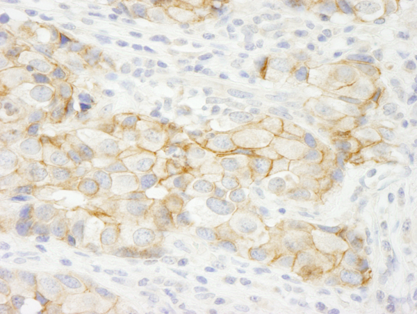
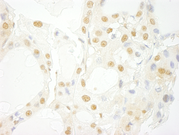
 IHC (Immunohiostchemistry)
(Detection of human USP15 by immunohistochemistry. Sample: FFPE section of human ovarian carcinoma. Antibody: Affinity purified rabbit anti-USP15 (Cat. No. AAA214059) used at a dilution of 1:100. Detection: DAB)
IHC (Immunohiostchemistry)
(Detection of human USP15 by immunohistochemistry. Sample: FFPE section of human ovarian carcinoma. Antibody: Affinity purified rabbit anti-USP15 (Cat. No. AAA214059) used at a dilution of 1:100. Detection: DAB)
USP15, Polyclonal Antibody (Cat# AAA214059)
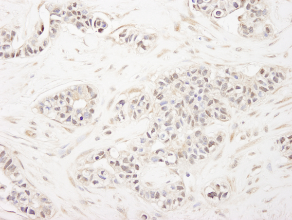
 IHC (Immunohiostchemistry)
(Detection of human FALZ/BPTF immunohistochemistry. Sample: FFPE section of human breast carcinoma. Antibody: Affinity purified rabbit anti-FALZ/BPTF (Cat. No. AAA214066) used at a dilution of 1:250. Detection: DAB)
IHC (Immunohiostchemistry)
(Detection of human FALZ/BPTF immunohistochemistry. Sample: FFPE section of human breast carcinoma. Antibody: Affinity purified rabbit anti-FALZ/BPTF (Cat. No. AAA214066) used at a dilution of 1:250. Detection: DAB)
FALZ/BPTF, Polyclonal Antibody (Cat# AAA214066)

 IHC (Immunohiostchemistry)
(Detection of human C1orf55 immunohistochemistry. Sample: FFPE section of human prostate carcinoma. Antibody: Affinity purified rabbit anti-C1orf55 (Cat. No. AAA214067) used at a dilution of 1:100. Detection: DAB)
IHC (Immunohiostchemistry)
(Detection of human C1orf55 immunohistochemistry. Sample: FFPE section of human prostate carcinoma. Antibody: Affinity purified rabbit anti-C1orf55 (Cat. No. AAA214067) used at a dilution of 1:100. Detection: DAB)
C1orf55, Polyclonal Antibody (Cat# AAA214067)
 IHC (Immunohiostchemistry)
(Detection of mouse U2AF35 by immunohistochemistry. Sample: FFPE section of mouse colon carcinoma. Antibody: Affinity purified rabbit anti-U2AF35 (Cat. No. AAA214072) used at a dilution of 1:250. Detection: DAB)
IHC (Immunohiostchemistry)
(Detection of mouse U2AF35 by immunohistochemistry. Sample: FFPE section of mouse colon carcinoma. Antibody: Affinity purified rabbit anti-U2AF35 (Cat. No. AAA214072) used at a dilution of 1:250. Detection: DAB)
U2AF35, Polyclonal Antibody (Cat# AAA214072)
 IHC (Immunohiostchemistry)
(Detection of human PABP4 by immunohistochemistry. Sample: FFPE section of human ovarian carcinoma. Antibody: Affinity purified rabbit anti-PABP4 (Cat. No. AAA214076) used at a dilution of 1:250. Detection: DAB)
IHC (Immunohiostchemistry)
(Detection of human PABP4 by immunohistochemistry. Sample: FFPE section of human ovarian carcinoma. Antibody: Affinity purified rabbit anti-PABP4 (Cat. No. AAA214076) used at a dilution of 1:250. Detection: DAB)
PABP4, Polyclonal Antibody (Cat# AAA214076)
 IHC (Immunohiostchemistry)
(Detection of human KIAA0528 by immunohistochemistry. Sample: FFPE section of human prostate carcinoma. Antibody: Affinity purified rabbit anti-KIAA0528 (Cat. No. AAA214077) used at a dilution of 1:250. Detection: DAB)
IHC (Immunohiostchemistry)
(Detection of human KIAA0528 by immunohistochemistry. Sample: FFPE section of human prostate carcinoma. Antibody: Affinity purified rabbit anti-KIAA0528 (Cat. No. AAA214077) used at a dilution of 1:250. Detection: DAB)
KIAA0528, Polyclonal Antibody (Cat# AAA214077)
 IHC (Immunohiostchemistry)
(Detection of human Leo1 by immunohistochemistry. Sample: FFPE section of human stomach adenocarcinoma. Antibody: Affinity purified rabbit anti-Leo1 (Cat. No. AAA214082) used at a dilution of 1:100. Detection: DAB)
IHC (Immunohiostchemistry)
(Detection of human Leo1 by immunohistochemistry. Sample: FFPE section of human stomach adenocarcinoma. Antibody: Affinity purified rabbit anti-Leo1 (Cat. No. AAA214082) used at a dilution of 1:100. Detection: DAB)
Leo1, Polyclonal Antibody (Cat# AAA214082)
 IHC (Immunohiostchemistry)
(Detection of mouse NF-YC by immunohistochemistry. Sample: FFPE section of mouse renal cell carcinoma. Antibody: Affinity purified rabbit anti-NF-YC (Cat. No. AAA214085) used at a dilution of 1:250. Detection: DAB)
IHC (Immunohiostchemistry)
(Detection of mouse NF-YC by immunohistochemistry. Sample: FFPE section of mouse renal cell carcinoma. Antibody: Affinity purified rabbit anti-NF-YC (Cat. No. AAA214085) used at a dilution of 1:250. Detection: DAB)
NF-YC, Polyclonal Antibody (Cat# AAA214085)
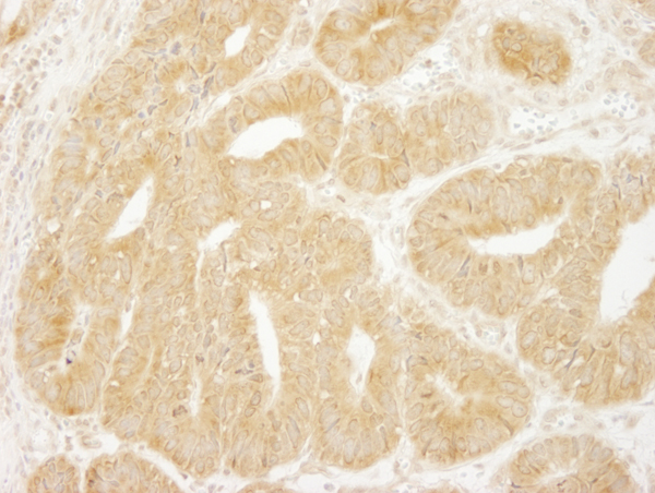 IHC (Immunohiostchemistry)
(Detection of human eIF2A by immunohistochemistry. Sample: FFPE section of human colon carcinoma. Antibody: Affinity purified rabbit anti-eIF2A (Cat. No. AAA214087) used at a dilution of 1:250. Detection: DAB)
IHC (Immunohiostchemistry)
(Detection of human eIF2A by immunohistochemistry. Sample: FFPE section of human colon carcinoma. Antibody: Affinity purified rabbit anti-eIF2A (Cat. No. AAA214087) used at a dilution of 1:250. Detection: DAB)
eIF2A, Polyclonal Antibody (Cat# AAA214087)
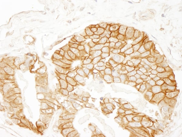
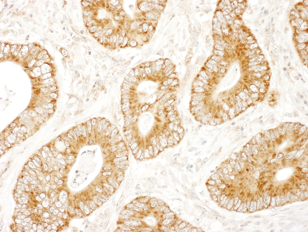
 IHC (Immunohistochemistry)
(Detection of human beta Catenin by immunohistochemistry. Sample: FFPE section of human colon carcinoma. Antibody: Affinity purified rabbit anti-beta Catenin (Cat. No. AAA214090) used at a dilution of 1:250. Detection: DAB)
IHC (Immunohistochemistry)
(Detection of human beta Catenin by immunohistochemistry. Sample: FFPE section of human colon carcinoma. Antibody: Affinity purified rabbit anti-beta Catenin (Cat. No. AAA214090) used at a dilution of 1:250. Detection: DAB)
beta catenin, Polyclonal Antibody (Cat# AAA214090)
 IHC (Immunohiostchemistry)
(Detection of mouse eIF3A/eIF3S10 by immunohistochemistry. Sample: FFPE section of mouse renal cell carcinoma. Antibody: Affinity purified rabbit anti-eIF3A/eIF3S10 (Cat. No. AAA214091) used at a dilution of 1:250. Detection: DAB)
IHC (Immunohiostchemistry)
(Detection of mouse eIF3A/eIF3S10 by immunohistochemistry. Sample: FFPE section of mouse renal cell carcinoma. Antibody: Affinity purified rabbit anti-eIF3A/eIF3S10 (Cat. No. AAA214091) used at a dilution of 1:250. Detection: DAB)
eIF3A/eIF3S10, Polyclonal Antibody (Cat# AAA214091)
 IHC (Immunohiostchemistry)
(Detection of mouse UVRAG by immunohistochemistry. Sample: FFPE section of mouse renal cell carcinoma. Antibody: Affinity purified rabbit anti-UVRAG (Cat. No. AAA214092) used at a dilution of 1:250. Detection: DAB)
IHC (Immunohiostchemistry)
(Detection of mouse UVRAG by immunohistochemistry. Sample: FFPE section of mouse renal cell carcinoma. Antibody: Affinity purified rabbit anti-UVRAG (Cat. No. AAA214092) used at a dilution of 1:250. Detection: DAB)
UVRAG, Polyclonal Antibody (Cat# AAA214092)
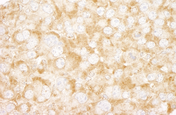
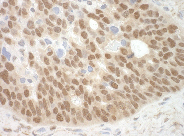
 IHC (Immunohiostchemistry)
(Detection of human Cyclin C by immunohistochemistry. Sample: FFPE section of human pancreatic islet cell tumor. Antibody: Affinity purified rabbit anti-Cyclin C (Cat. No. AAA214094) used at a dilution of 1:100. Detection: DAB)
IHC (Immunohiostchemistry)
(Detection of human Cyclin C by immunohistochemistry. Sample: FFPE section of human pancreatic islet cell tumor. Antibody: Affinity purified rabbit anti-Cyclin C (Cat. No. AAA214094) used at a dilution of 1:100. Detection: DAB)
Cyclin C, Polyclonal Antibody (Cat# AAA214094)
 IHC (Immunohiostchemistry)
(Detection of human ANKRD17 by immunohistochemistry. Sample: FFPE section of human prostate carcinoma. Antibody: Affinity purified rabbit anti-ANKRD17 (Cat. No. AAA214096) used at a dilution of 1:250. Detection: DAB)
IHC (Immunohiostchemistry)
(Detection of human ANKRD17 by immunohistochemistry. Sample: FFPE section of human prostate carcinoma. Antibody: Affinity purified rabbit anti-ANKRD17 (Cat. No. AAA214096) used at a dilution of 1:250. Detection: DAB)
ANKRD17, Polyclonal Antibody (Cat# AAA214096)
 IHC (Immunohiostchemistry)
(Detection of human PRPF4B by immunohistochemistry. Sample: FFPE section of human ovarian carcinoma. Antibody: Affinity purified rabbit anti-PRPF4B (Cat. No. AAA214097) used at a dilution of 1:250. Detection: DAB)
IHC (Immunohiostchemistry)
(Detection of human PRPF4B by immunohistochemistry. Sample: FFPE section of human ovarian carcinoma. Antibody: Affinity purified rabbit anti-PRPF4B (Cat. No. AAA214097) used at a dilution of 1:250. Detection: DAB)
PRPF4B, Polyclonal Antibody (Cat# AAA214097)
 IHC (Immunohiostchemistry)
(Detection of human PRKAR2A/PKA-RIIalpha by immunohistochemistry. Sample: FFPE section of human breast carcinoma. Antibody: Affinity purified rabbit anti-PRKAR2A/PKA-RIIalpha (Cat. No. AAA214099) used at a dilution of 1:100. Detection: DAB)
IHC (Immunohiostchemistry)
(Detection of human PRKAR2A/PKA-RIIalpha by immunohistochemistry. Sample: FFPE section of human breast carcinoma. Antibody: Affinity purified rabbit anti-PRKAR2A/PKA-RIIalpha (Cat. No. AAA214099) used at a dilution of 1:100. Detection: DAB)
PRKAR2A/PKA-RIIalpha, Polyclonal Antibody (Cat# AAA214099)
 IHC (Immunohiostchemistry)
(Detection of human CROP/Luc7A by immunohistochemistry. Sample: FFPE section of human thyroid carcinoma. Antibody: Affinity purified rabbit anti-CROP/Luc7A (Cat. No. AAA214100) used at a dilution of 1:250. Detection: DAB)
IHC (Immunohiostchemistry)
(Detection of human CROP/Luc7A by immunohistochemistry. Sample: FFPE section of human thyroid carcinoma. Antibody: Affinity purified rabbit anti-CROP/Luc7A (Cat. No. AAA214100) used at a dilution of 1:250. Detection: DAB)
CROP/Luc7A, Polyclonal Antibody (Cat# AAA214100)
 IHC (Immunohiostchemistry)
(Detection of mouse Cdc42GAP by immunohistochemistry. Sample: FFPE section of mouse teratoma. Antibody: Affinity purified rabbit anti-Cdc42GAP (Cat. No. AAA214105) used at a dilution of 1:100. Detection: DAB)
IHC (Immunohiostchemistry)
(Detection of mouse Cdc42GAP by immunohistochemistry. Sample: FFPE section of mouse teratoma. Antibody: Affinity purified rabbit anti-Cdc42GAP (Cat. No. AAA214105) used at a dilution of 1:100. Detection: DAB)
Cdc42GAP, Polyclonal Antibody (Cat# AAA214105)
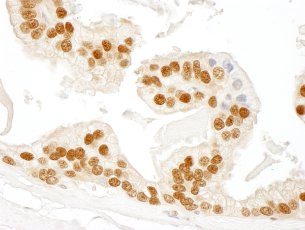
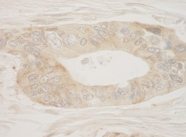
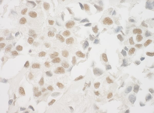
 IHC (Immunohiostchemistry)
(Detection of human NARG1 by immunohistochemistry. Sample: FFPE section of human testicular seminoma. Antibody: Affinity purified rabbit anti-NARG1 (Cat. No. AAA214111) used at a dilution of 1:250. Detection: DAB)
IHC (Immunohiostchemistry)
(Detection of human NARG1 by immunohistochemistry. Sample: FFPE section of human testicular seminoma. Antibody: Affinity purified rabbit anti-NARG1 (Cat. No. AAA214111) used at a dilution of 1:250. Detection: DAB)
NARG1, Polyclonal Antibody (Cat# AAA214111)
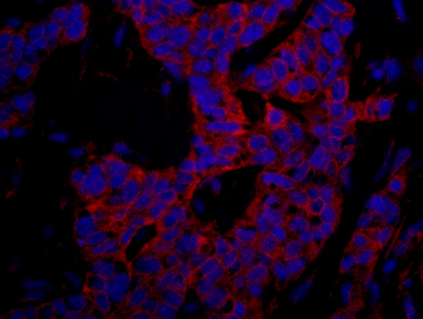
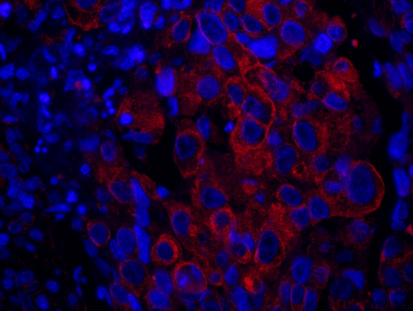
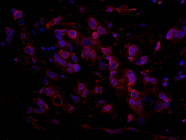
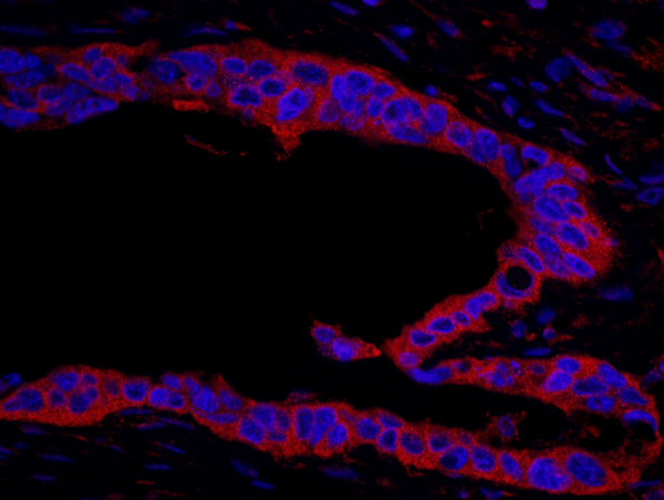
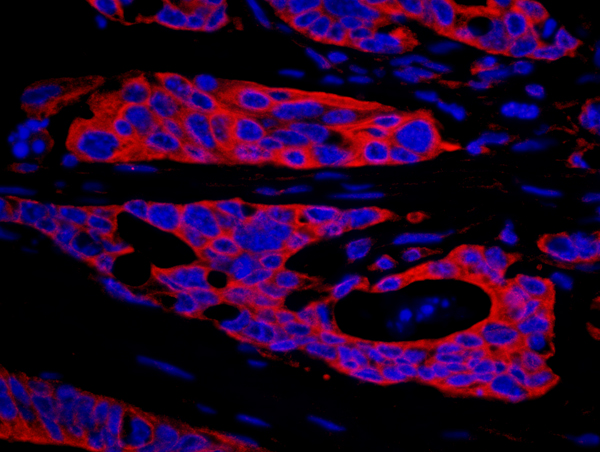
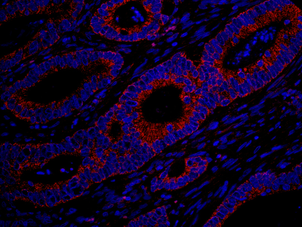 IHC (Immunohiostchemistry)
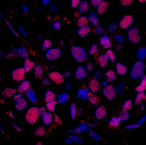
(Detection of human MADD by immunohistochemistry. Sample: FFPE section of human colon carcinoma. Antibody: Affinity purified rabbit anti-MADD (Cat. No. AAA214112) used at a dilution of 1:100. Detection: Red-fluorescent goat anti-rabbit IgG-heavy and light chain cross-adsorbed Antibody DyLight 594 Conjugated used at a dilution of 1:100.)
IHC (Immunohiostchemistry)
(Detection of human MADD by immunohistochemistry. Sample: FFPE section of human colon carcinoma. Antibody: Affinity purified rabbit anti-MADD (Cat. No. AAA214112) used at a dilution of 1:100. Detection: Red-fluorescent goat anti-rabbit IgG-heavy and light chain cross-adsorbed Antibody DyLight 594 Conjugated used at a dilution of 1:100.)
MADD, Polyclonal Antibody (Cat# AAA214112)
 IHC (Immunohiostchemistry)
(Detection of human p600 by immunohistochemistry. Sample: FFPE section of human prostate carcinoma. Antibody: Affinity purified rabbit anti-p600 (Cat. No. AAA214120) used at a dilution of 1:100. Detection: Red-fluorescent goat anti-rabbit IgG-heavy and light chain cross-adsorbed Antibody DyLight 594 Conjugated used at a dilution of 1:100.)
IHC (Immunohiostchemistry)
(Detection of human p600 by immunohistochemistry. Sample: FFPE section of human prostate carcinoma. Antibody: Affinity purified rabbit anti-p600 (Cat. No. AAA214120) used at a dilution of 1:100. Detection: Red-fluorescent goat anti-rabbit IgG-heavy and light chain cross-adsorbed Antibody DyLight 594 Conjugated used at a dilution of 1:100.)
p600, Polyclonal Antibody (Cat# AAA214120)
 IHC (Immunohiostchemistry)
(Detection of human FUSIP1 by immunohistochemistry. Sample: FFPE section of human breast carcinoma. Antibody: Affinity purified rabbit anti-FUSIP1 (Cat. No. AAA214122) used at a dilution of 1:100. Detection: Red-fluorescent goat anti-rabbit IgG-heavy and light chain cross-adsorbed Antibody DyLight 594 Conjugated used at a dilution of 1:100.)
IHC (Immunohiostchemistry)
(Detection of human FUSIP1 by immunohistochemistry. Sample: FFPE section of human breast carcinoma. Antibody: Affinity purified rabbit anti-FUSIP1 (Cat. No. AAA214122) used at a dilution of 1:100. Detection: Red-fluorescent goat anti-rabbit IgG-heavy and light chain cross-adsorbed Antibody DyLight 594 Conjugated used at a dilution of 1:100.)
FUSIP1, Polyclonal Antibody (Cat# AAA214122)
 IHC (Immunohiostchemistry)
(Detection of human SASH1 by immunohistochemistry. Sample: FFPE section of human breast carcinoma. Antibody: Affinity purified rabbit anti-SASH1 (Cat. No. AAA214124) used at a dilution of 1:100. Detection: Red-fluorescent goat anti-rabbit IgG-heavy and light chain cross-adsorbed Antibody DyLight 594 Conjugated used at a dilution of 1:100.)
IHC (Immunohiostchemistry)
(Detection of human SASH1 by immunohistochemistry. Sample: FFPE section of human breast carcinoma. Antibody: Affinity purified rabbit anti-SASH1 (Cat. No. AAA214124) used at a dilution of 1:100. Detection: Red-fluorescent goat anti-rabbit IgG-heavy and light chain cross-adsorbed Antibody DyLight 594 Conjugated used at a dilution of 1:100.)
SASH1, Polyclonal Antibody (Cat# AAA214124)
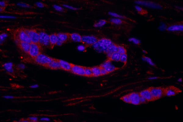
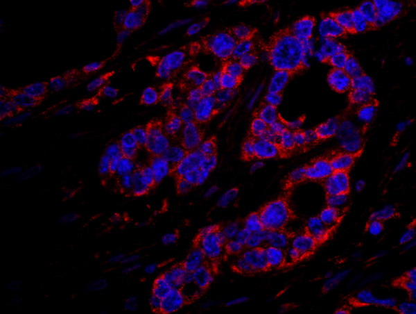 IHC (Immunohiostchemistry)
(Detection of human DOCK4 by immunohistochemistry. Sample: FFPE section of human ovarian carcinoma. Antibody: Affinity purified rabbit anti-DOCK4 (Cat. No. AAA214125) used at a dilution of 1:100. Detection: Red-fluorescent goat anti-rabbit IgG-heavy and light chain cross-adsorbed Antibody DyLight 594 Conjugated used at a dilution of 1:100.)
IHC (Immunohiostchemistry)
(Detection of human DOCK4 by immunohistochemistry. Sample: FFPE section of human ovarian carcinoma. Antibody: Affinity purified rabbit anti-DOCK4 (Cat. No. AAA214125) used at a dilution of 1:100. Detection: Red-fluorescent goat anti-rabbit IgG-heavy and light chain cross-adsorbed Antibody DyLight 594 Conjugated used at a dilution of 1:100.)
DOCK4, Polyclonal Antibody (Cat# AAA214125)
 IHC (Immunohiostchemistry)
(Detection of human ARID4B by immunohistochemistry. Sample: FFPE section of human ovarian carcinoma. Antibody: Affinity purified rabbit anti-ARID4B (Cat. No. AAA214130) used at a dilution of 1:100. Detection: Red-fluorescent goat anti-rabbit IgG-heavy and light chain cross-adsorbed Antibody DyLight 594 Conjugated used at a dilution of 1:100.)
IHC (Immunohiostchemistry)
(Detection of human ARID4B by immunohistochemistry. Sample: FFPE section of human ovarian carcinoma. Antibody: Affinity purified rabbit anti-ARID4B (Cat. No. AAA214130) used at a dilution of 1:100. Detection: Red-fluorescent goat anti-rabbit IgG-heavy and light chain cross-adsorbed Antibody DyLight 594 Conjugated used at a dilution of 1:100.)
ARID4B, Polyclonal Antibody (Cat# AAA214130)
 IHC (Immunohiostchemistry)
(Detection of mouse NCBP1/CBP80 by immunohistochemistry. Sample: FFPE section of mouse teratoma. Antibody: Affinity purified rabbit anti-NCBP1/CBP80 (Cat. No. AAA214132) used at a dilution of 1:250. Detection: DAB)
IHC (Immunohiostchemistry)
(Detection of mouse NCBP1/CBP80 by immunohistochemistry. Sample: FFPE section of mouse teratoma. Antibody: Affinity purified rabbit anti-NCBP1/CBP80 (Cat. No. AAA214132) used at a dilution of 1:250. Detection: DAB)
NCBP1/CBP80, Polyclonal Antibody (Cat# AAA214132)
 IHC (Immunohiostchemistry)
(Detection of human RAD23B by immunohistochemistry. Sample: FFPE section of human breast carcinoma. Antibody: Affinity purified rabbit anti-RAD23B (Cat. No. AAA214133) used at a dilution of 1:100. Detection: Red-fluorescent goat anti-rabbit IgG-heavy and light chain cross-adsorbed Antibody DyLight 594 Conjugated used at a dilution of 1:100.)
IHC (Immunohiostchemistry)
(Detection of human RAD23B by immunohistochemistry. Sample: FFPE section of human breast carcinoma. Antibody: Affinity purified rabbit anti-RAD23B (Cat. No. AAA214133) used at a dilution of 1:100. Detection: Red-fluorescent goat anti-rabbit IgG-heavy and light chain cross-adsorbed Antibody DyLight 594 Conjugated used at a dilution of 1:100.)
RAD23B, Polyclonal Antibody (Cat# AAA214133)
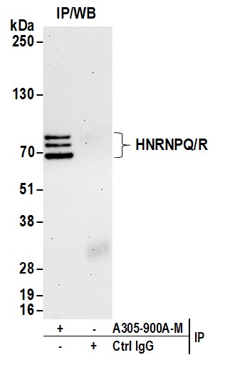
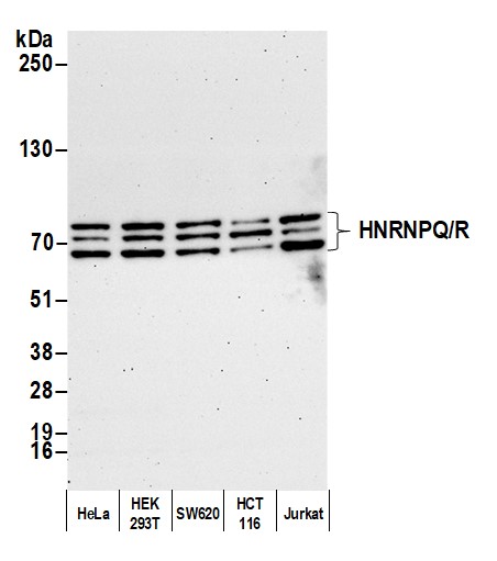 WB (Western Blot)
(Detection of human HNRNPQ/R by western blot. Samples: Whole cell lysate (10 ug) from HeLa, HEK293T, SW620, HCT 116, and Jurkat cells prepared using NETN lysis buffer. Antibody: Affinity purified rabbit anti-HNRNPQ/R antibody (AAA213489 lot 1) used for WB at 0.1 mg/ml. Detection: Chemiluminescence with an exposure time of 75 seconds.)
WB (Western Blot)
(Detection of human HNRNPQ/R by western blot. Samples: Whole cell lysate (10 ug) from HeLa, HEK293T, SW620, HCT 116, and Jurkat cells prepared using NETN lysis buffer. Antibody: Affinity purified rabbit anti-HNRNPQ/R antibody (AAA213489 lot 1) used for WB at 0.1 mg/ml. Detection: Chemiluminescence with an exposure time of 75 seconds.)
HNRNPQ/R, Polyclonal Antibody (Cat# AAA213489)
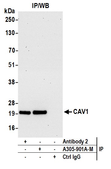
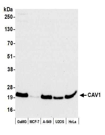 WB (Western Blot)
(Detection of human CAV1 by western blot. Samples: Whole cell lysate (50 ug) from GaMG, MCF-7, A-549, U2OS, and HeLa cells prepared using NETN lysis buffer. Antibody: Affinity purified rabbit anti-CAV1 antibody (lot AAA213490 lot 1) used for WB at 0.04 mg/ml. Detection: Chemiluminescence with an exposure time of 10 seconds.)
WB (Western Blot)
(Detection of human CAV1 by western blot. Samples: Whole cell lysate (50 ug) from GaMG, MCF-7, A-549, U2OS, and HeLa cells prepared using NETN lysis buffer. Antibody: Affinity purified rabbit anti-CAV1 antibody (lot AAA213490 lot 1) used for WB at 0.04 mg/ml. Detection: Chemiluminescence with an exposure time of 10 seconds.)
CAV1, Polyclonal Antibody (Cat# AAA213490)
 IHC (Immunohiostchemistry)
(Detection of human DAP5 by immunohistochemistry. Sample: FFPE section of human ovarian carcinoma. Antibody: Affinity purified rabbit anti-DAP5 (Cat. No. AAA214139) used at a dilution of 1:100. Detection: Red-fluorescent goat anti-rabbit IgG-heavy and light chain cross-adsorbed Antibody DyLight 594 Conjugated used at a dilution of 1:100.)
IHC (Immunohiostchemistry)
(Detection of human DAP5 by immunohistochemistry. Sample: FFPE section of human ovarian carcinoma. Antibody: Affinity purified rabbit anti-DAP5 (Cat. No. AAA214139) used at a dilution of 1:100. Detection: Red-fluorescent goat anti-rabbit IgG-heavy and light chain cross-adsorbed Antibody DyLight 594 Conjugated used at a dilution of 1:100.)
DAP5, Polyclonal Antibody (Cat# AAA214139)
 IHC (Immunohiostchemistry)
(Detection of mouse WDR5 by immunohistochemistry. Sample: FFPE section of mouse renal cell carcinoma. Antibody: Affinity purified rabbit anti-WDR5 (Cat. No. AAA214145) used at a dilution of 1:250 Detection: DAB)
IHC (Immunohiostchemistry)
(Detection of mouse WDR5 by immunohistochemistry. Sample: FFPE section of mouse renal cell carcinoma. Antibody: Affinity purified rabbit anti-WDR5 (Cat. No. AAA214145) used at a dilution of 1:250 Detection: DAB)
WDR5, Polyclonal Antibody (Cat# AAA214145)

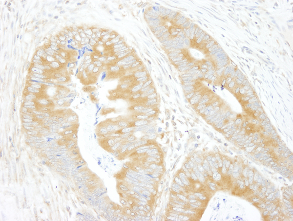
 IHC (Immunohiostchemistry)
(Detection of human RSK1 by immunohistochemistry. Sample: FFPE section of human ovarian carcinoma. Antibody: Affinity purified rabbit anti-RSK1 (Cat. No. AAA214146) used at a dilution of 1:100. Detection: Red-fluorescent goat anti-rabbit IgG-heavy and light chain cross-adsorbed Antibody DyLight 594 Conjugated used at a dilution of 1:100.)
IHC (Immunohiostchemistry)
(Detection of human RSK1 by immunohistochemistry. Sample: FFPE section of human ovarian carcinoma. Antibody: Affinity purified rabbit anti-RSK1 (Cat. No. AAA214146) used at a dilution of 1:100. Detection: Red-fluorescent goat anti-rabbit IgG-heavy and light chain cross-adsorbed Antibody DyLight 594 Conjugated used at a dilution of 1:100.)
RSK1, Polyclonal Antibody (Cat# AAA214146)
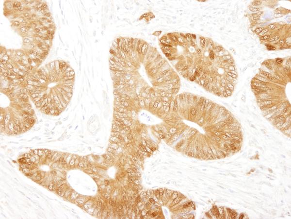
 IHC (Immunohiostchemistry)
(Detection of human RSK2 by immunohistochemistry. Sample: FFPE section of human ovarian carcinoma. Antibody: Affinity purified rabbit anti-RSK2 (Cat. No. AAA214147) used at a dilution of 1:100. Detection: Red-fluorescent goat anti-rabbit IgG-heavy and light chain cross-adsorbed Antibody DyLight 594 Conjugated used at a dilution of 1:100.)
IHC (Immunohiostchemistry)
(Detection of human RSK2 by immunohistochemistry. Sample: FFPE section of human ovarian carcinoma. Antibody: Affinity purified rabbit anti-RSK2 (Cat. No. AAA214147) used at a dilution of 1:100. Detection: Red-fluorescent goat anti-rabbit IgG-heavy and light chain cross-adsorbed Antibody DyLight 594 Conjugated used at a dilution of 1:100.)
RSK2, Polyclonal Antibody (Cat# AAA214147)
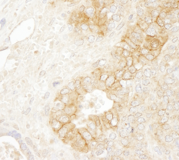
 IHC (Immunohiostchemistry)
(Detection of mouse SRPK1 by immunohistochemistry. Sample: FFPE section of mouse CT26 tumor. Antibody: Affinity purified rabbit anti-SRPK1 (Cat. No. AAA214148) used at a dilution of 1:250. Detection: DAB)
IHC (Immunohiostchemistry)
(Detection of mouse SRPK1 by immunohistochemistry. Sample: FFPE section of mouse CT26 tumor. Antibody: Affinity purified rabbit anti-SRPK1 (Cat. No. AAA214148) used at a dilution of 1:250. Detection: DAB)
SRPK1, Polyclonal Antibody (Cat# AAA214148)
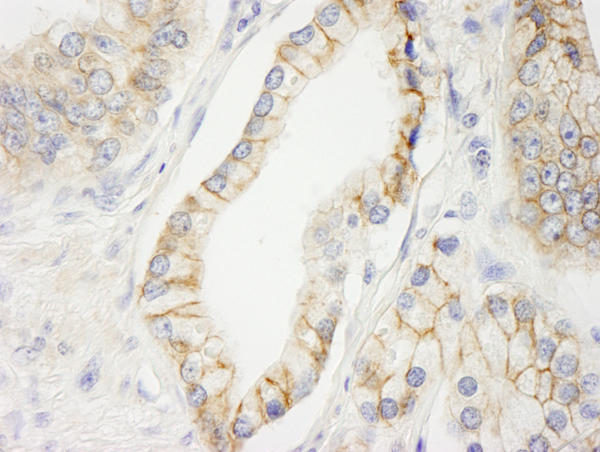
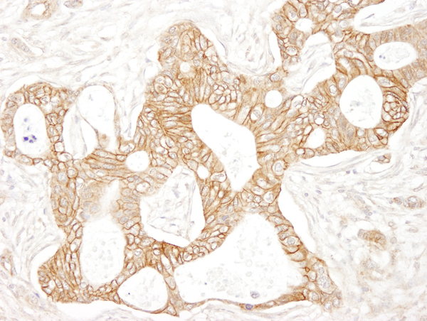
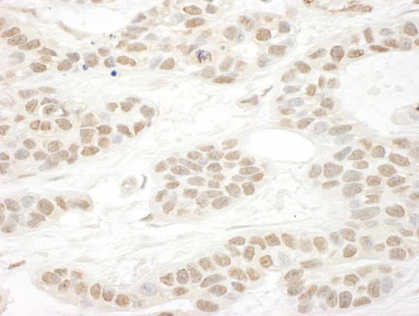
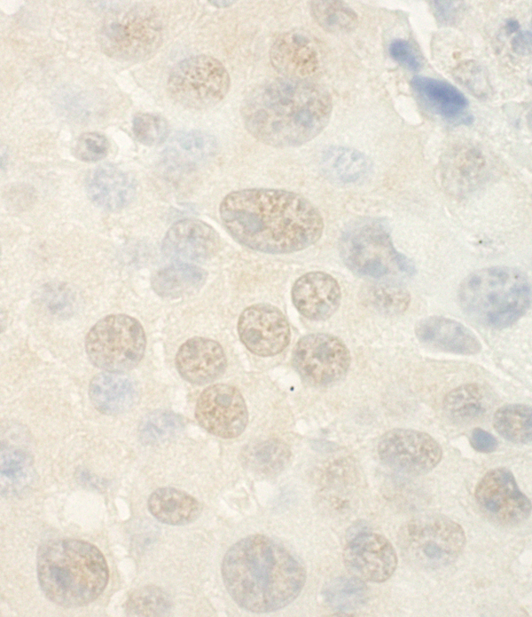
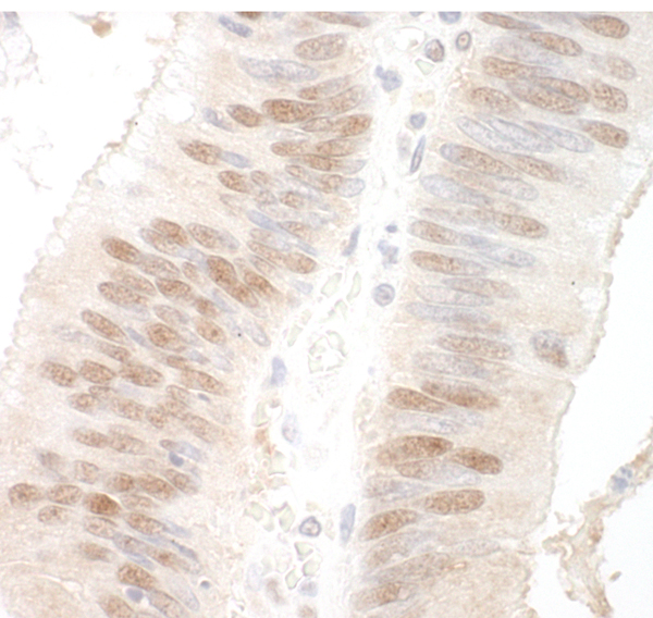
 IHC (Immunohistochemisry)
(Detection of human SPAK by immunohistochemistry. Sample: FFPE section of human ovarian carcinoma. Antibody: Affinity purified rabbit anti-SPAK (Cat. No. AAA214149) used at a dilution of 1:100. Detection: Red-fluorescent goat anti-rabbit IgG-heavy and light chain cross-adsorbed Antibody DyLight 594 Conjugated used at a dilution of 1:100.)
IHC (Immunohistochemisry)
(Detection of human SPAK by immunohistochemistry. Sample: FFPE section of human ovarian carcinoma. Antibody: Affinity purified rabbit anti-SPAK (Cat. No. AAA214149) used at a dilution of 1:100. Detection: Red-fluorescent goat anti-rabbit IgG-heavy and light chain cross-adsorbed Antibody DyLight 594 Conjugated used at a dilution of 1:100.)
SPAK, Polyclonal Antibody (Cat# AAA214149)
 IHC (Immunohiostchemistry)
(Detection of mouse SIP1 by immunohistochemistry. Sample: FFPE section of mouse teratoma. Antibody: Affinity purified rabbit anti-ZEB2/SIP (Cat. No. AAA214152) used at a dilution of 1:250 Detection: DAB)
IHC (Immunohiostchemistry)
(Detection of mouse SIP1 by immunohistochemistry. Sample: FFPE section of mouse teratoma. Antibody: Affinity purified rabbit anti-ZEB2/SIP (Cat. No. AAA214152) used at a dilution of 1:250 Detection: DAB)
ZEB2/SIP, Polyclonal Antibody (Cat# AAA214152)
 IHC (Immunohiostchemistry)
(Detection of mouse Cul2 by immunohistochemistry. Sample: FFPE section of mouse teratoma. Antibody: Affinity purified rabbit anti-Cul2 (Cat. No. AAA214153) used at a dilution of 1:100 Detection: DAB)
IHC (Immunohiostchemistry)
(Detection of mouse Cul2 by immunohistochemistry. Sample: FFPE section of mouse teratoma. Antibody: Affinity purified rabbit anti-Cul2 (Cat. No. AAA214153) used at a dilution of 1:100 Detection: DAB)
Cul2, Polyclonal Antibody (Cat# AAA214153)
 IHC (Immunohiostchemistry)
(Detection of human MBD3 by immunohistochemistry. Sample: FFPE section of human ovarian carcinoma. Antibody: Affinity purified rabbit anti-MBD3 (Cat. No. AAA214154) used at a dilution of 1:250 Detection: DAB)
IHC (Immunohiostchemistry)
(Detection of human MBD3 by immunohistochemistry. Sample: FFPE section of human ovarian carcinoma. Antibody: Affinity purified rabbit anti-MBD3 (Cat. No. AAA214154) used at a dilution of 1:250 Detection: DAB)
MBD3, Polyclonal Antibody (Cat# AAA214154)
 IHC (Immunohiostchemistry)
(Detection of mouse RuvBL2 by immunohistochemistry. Sample: FFPE section of mouse teratoma. Antibody: Affinity purified rabbit anti-RuvBL2 (Cat. No. AAA214155) used at a dilution of 1:250 Detection: DAB)
IHC (Immunohiostchemistry)
(Detection of mouse RuvBL2 by immunohistochemistry. Sample: FFPE section of mouse teratoma. Antibody: Affinity purified rabbit anti-RuvBL2 (Cat. No. AAA214155) used at a dilution of 1:250 Detection: DAB)
RuvBL2, Polyclonal Antibody (Cat# AAA214155)
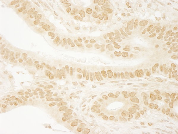
 IHC (Immunohiostchemistry)
(Detection of mouse SPF45 by immunohistochemistry. Sample: FFPE section of mouse teratoma. Antibody: Affinity purified rabbit anti-SPF45 (Cat. No. AAA214156) used at a dilution of 1:250 Detection: DAB)
IHC (Immunohiostchemistry)
(Detection of mouse SPF45 by immunohistochemistry. Sample: FFPE section of mouse teratoma. Antibody: Affinity purified rabbit anti-SPF45 (Cat. No. AAA214156) used at a dilution of 1:250 Detection: DAB)
SPF45, Polyclonal Antibody (Cat# AAA214156)
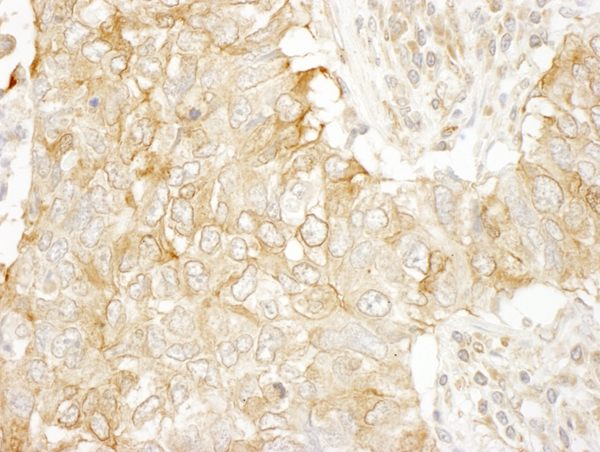
 IHC (Immunohiostchemistry)
(Detection of human Ku70 by immunohistochemistry. Sample: FFPE section of human colon carcinoma. Antibody: Affinity purified rabbit anti-Ku70 (Cat. No. AAA214166) used at a dilution of 1:100 Detection: DAB)
IHC (Immunohiostchemistry)
(Detection of human Ku70 by immunohistochemistry. Sample: FFPE section of human colon carcinoma. Antibody: Affinity purified rabbit anti-Ku70 (Cat. No. AAA214166) used at a dilution of 1:100 Detection: DAB)
Ku70, Polyclonal Antibody (Cat# AAA214166)
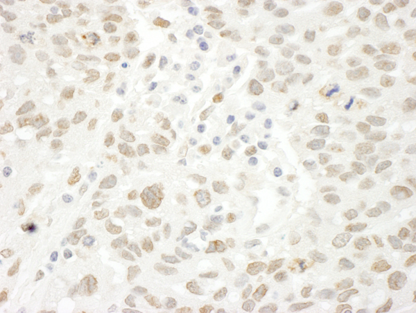
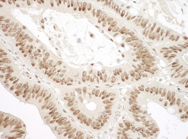
 IHC (Immunohiostchemistry)
(Detection of human FOXP1 by immunohistochemistry. Sample: FFPE section of human breast carcinoma. Antibody: Affinity purified rabbit anti-FOXP1 (Cat. No. AAA214167) used at a dilution of 1:250 Detection: DAB)
IHC (Immunohiostchemistry)
(Detection of human FOXP1 by immunohistochemistry. Sample: FFPE section of human breast carcinoma. Antibody: Affinity purified rabbit anti-FOXP1 (Cat. No. AAA214167) used at a dilution of 1:250 Detection: DAB)
FOXP1, Polyclonal Antibody (Cat# AAA214167)
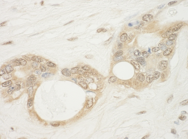
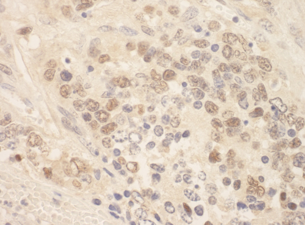
 IHC (Immunohiostchemistry)
(Detection of mouse CTNNBL1 by immunohistochemistry. Sample: FFPE section of mouse hybridoma tumor. Antibody: Affinity purified rabbit anti-CTNNBL1 (Cat. No. AAA214169) used at a dilution of 1:250 Detection: DAB)
IHC (Immunohiostchemistry)
(Detection of mouse CTNNBL1 by immunohistochemistry. Sample: FFPE section of mouse hybridoma tumor. Antibody: Affinity purified rabbit anti-CTNNBL1 (Cat. No. AAA214169) used at a dilution of 1:250 Detection: DAB)
CTNNBL1, Polyclonal Antibody (Cat# AAA214169)
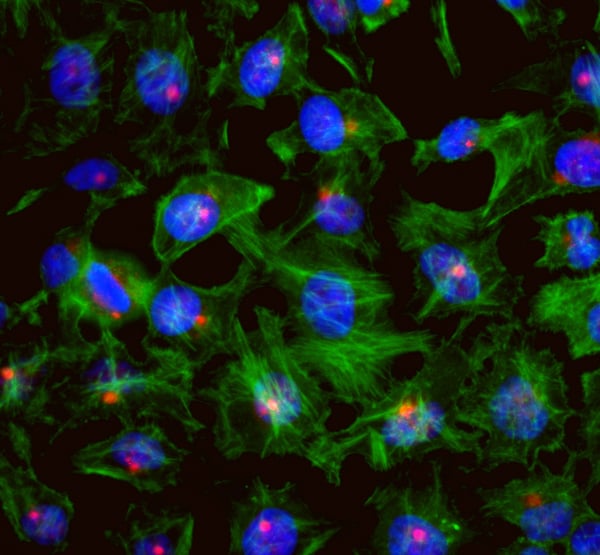
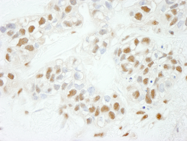
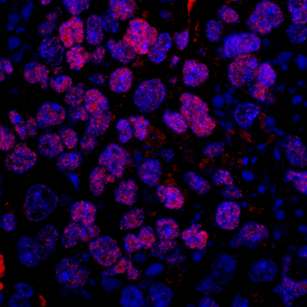
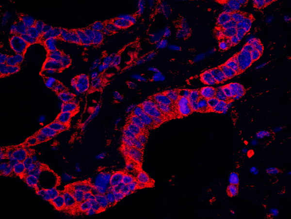
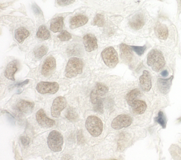
 ICC (Immunocytochemistry)
(Detection of human CEP131/AZ1 by immunocytochemistry. Sample: NBF-fixed asynchronous HeLa cells. Antibody: Affinity purified rabbit anti-CEP131/AZ1 (Cat. No. AAA213977) used at a dilution of 1:250. Detection: Red-fluorescent goat anti-rabbit IgG highly cross-adsorbed Antibody used at a dilution of 1:100.)
ICC (Immunocytochemistry)
(Detection of human CEP131/AZ1 by immunocytochemistry. Sample: NBF-fixed asynchronous HeLa cells. Antibody: Affinity purified rabbit anti-CEP131/AZ1 (Cat. No. AAA213977) used at a dilution of 1:250. Detection: Red-fluorescent goat anti-rabbit IgG highly cross-adsorbed Antibody used at a dilution of 1:100.)
CEP131/AZ1, Polyclonal Antibody (Cat# AAA213977)
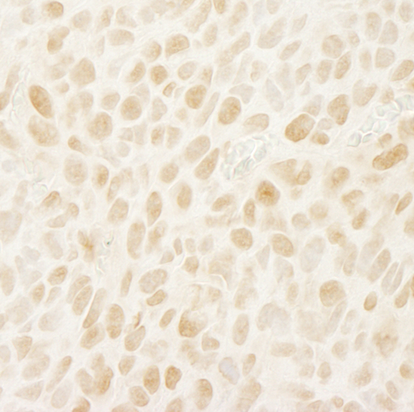
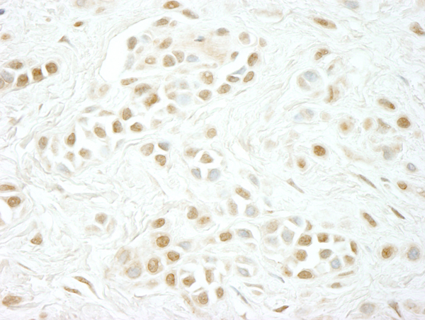
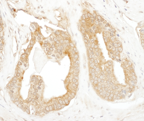
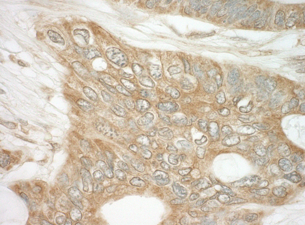
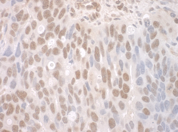
 IHC (Immunohiostchemistry)
(Detection of human FOG1/ZFPM1 by immunohistochemistry. Sample: FFPE section of human breast carcinoma. Antibody: Affinity purified rabbit anti-FOG1/ZFPM1 (Cat. No. AAA213980) used at a dilution of 1:250. Detection: DAB)
IHC (Immunohiostchemistry)
(Detection of human FOG1/ZFPM1 by immunohistochemistry. Sample: FFPE section of human breast carcinoma. Antibody: Affinity purified rabbit anti-FOG1/ZFPM1 (Cat. No. AAA213980) used at a dilution of 1:250. Detection: DAB)
FOG1/ZFPM1, Polyclonal Antibody (Cat# AAA213980)
What are Polyclonal Antibodies?
Polyclonal antibodies are antibodies that come from multiple B cell clones of a host animal. The typical hosts used for the majority of polyclonal antibody production are rabbits, goats, sheep, and donkeys. These polyclonal antibodies, once having identified their target, will bind to different epitopes located at different regions or sequences on the same protein/antigen. As a result, they are ideal at locating and binding to the target, even if the target is in very low concentrations (due to many different antibodies being able to bind to the same target molecule, which allows for significant amplification of a downstream signal).
Polyclonal antibodies are typically produced by injecting an antigen into a host animal, which causes the animal’s immune system to attack the foreign antigen by mass generating antibodies against it. After a period of time, serum is collected from the animal and purified using physicochemical fractionation, class-specific affinity purification, and/or antigen-affinity purification.
Key Uses of Polyclonal Antibodies
- Western Blotting: This method is used to find specific proteins in biological samples after separating them by size.
- Immunohistochemistry: IHC helps visualize the location of proteins in tissue sections using various staining techniques.
- ELISA: (Enzyme-Linked Immunosorbent Assay) is typically used to identify specific protein quantities in a sample. ELISAs can be either “Quantitative” or “Qualitative”.
- Flow Cytometry: technique that identifies and measures the specific protein on the surface or inside the cells in a fluid suspension.
- Immunoprecipitation: IP isolates and studies a specific protein from a complex mixture using antibodies.
Why Buy Polyclonal Antibodies from AAA Biotech?
1. Ideal for Various Applications
Our antibodies are generally going to be validated for use in multiple types of assays, including ELISA, Western Blotting, Immunohistochemistry, Immunoprecipitation, amongst others. They are ideal for a wide range of research applications.
2. Rigorous Quality Control
All of the antibodies in our catalog undergo strict quality testing to ensure specificity, sensitivity, and consistent performance. We are confident in the ability of our antibodies to provide you with accurate results.
3. Wide Assortment of Antibodies
Antibodies in are catalog can be found for both common and exotic species, and these antibodies are also available in both conjugated and recombinant forms to suit many diverse experimental needs.
4. Highly Purified
Our antibodies are available in purified forms with over 85% purity, as confirmed by SDS-PAGE. They are also available with tags such as His, Flag, GST, or MBP. We cater to customers worldwide.
FAQ
1. How are polyclonal antibodies produced?
Traditionally, polyclonal antibodies are produced by injecting an antigen into a host animal (such as a rabbit or goat), which then triggers an immune response from the host animal. The animal’s B cells produce antibodies that will recognize different parts of the injected antigen. These antibodies are then collected from the animal’s blood and purified for use.
2. How do polyclonal antibodies differ from monoclonal antibodies?
Polyclonal antibodies are a mix of antibodies that bind to different locations (epitopes) of the same antigen, while monoclonal antibodies are identical and bind to just one specific epitope. This makes polyclonal antibodies more versatile and better at detecting proteins that may be present in low quantities or in altered/modified forms.
3. How should I store polyclonal antibodies?
Polyclonal antibodies should be stored at 4°C for short-term use (up to a few weeks) and at -20°C or -80°C for long-term storage. Avoid repeated freeze-thaw cycles by dividing them into small aliquots. Always check the datasheet for specific storage instructions.