Filters
▼Clonality
▼Type
▼Reactivity
▼Gene Name
▼Isotype
▼Host
▼Application
▼Clone
▼Polyclonal Antibodies
At AAA Biotech also known as AAA Bio or AAABio, we provide a broad range of purified polyclonal antibodies (pAbs) that are able to all be browsed online through our website. Due to their high specificity and strong binding affinity, these antibodies are ideal for wide swathes of research and experimental applications.
Our polyclonal antibodies can easily support your work, whether you use them for Western Blotting, Immunocytochemistry (with or without Immunofluorescence used in conjunction), Immunohistochemistry, Immunoprecipitation, and ELISA tests. We highly encourage you to browse our range of pAbs and choose the one that best suits your experimental model.
Viewing 4850-4900 of 96812 product results
 WB (Western Blot)
(Detection of human APOL2 by western blot. Samples: Whole cell lysate (50 ug) from NCI-NCI-H226, HeLa, HEK293T, and Jurkat cells prepared using NETN lysis buffer. Antibody: Affinity purified rabbit anti-APOL2 antibody (AAA213354 lot 1) used for WB at 1 mg/ml. Detection: Chemiluminescence with an exposure time of 3 minutes.)
WB (Western Blot)
(Detection of human APOL2 by western blot. Samples: Whole cell lysate (50 ug) from NCI-NCI-H226, HeLa, HEK293T, and Jurkat cells prepared using NETN lysis buffer. Antibody: Affinity purified rabbit anti-APOL2 antibody (AAA213354 lot 1) used for WB at 1 mg/ml. Detection: Chemiluminescence with an exposure time of 3 minutes.)
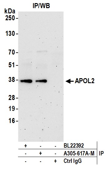
APOL2, Polyclonal Antibody (Cat# AAA213354)
 WB (Western Blot)
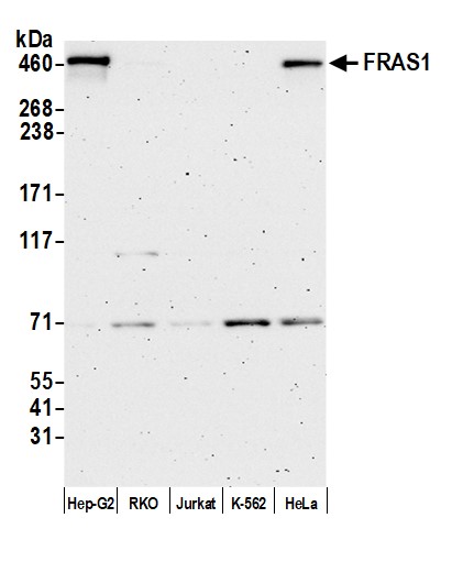
(Detection of human FRAS1 by western blot. Samples: Whole cell lysate (50 ug) from Hep-G2, RKO, Jurkat, K-562, and HeLa cells prepared using NETN lysis buffer. Antibody: Affinity purified rabbit anti-FRAS1 antibody (AAA213356 lot 2) used for WB at 0.1 mg/ml. Detection: Chemiluminescence with an exposure time of 3 minutes.)
WB (Western Blot)
(Detection of human FRAS1 by western blot. Samples: Whole cell lysate (50 ug) from Hep-G2, RKO, Jurkat, K-562, and HeLa cells prepared using NETN lysis buffer. Antibody: Affinity purified rabbit anti-FRAS1 antibody (AAA213356 lot 2) used for WB at 0.1 mg/ml. Detection: Chemiluminescence with an exposure time of 3 minutes.)
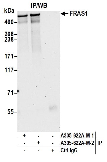
FRAS1, Polyclonal Antibody (Cat# AAA213356)
 WB (Western Blot)
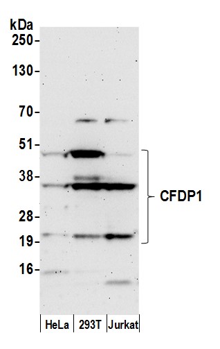
(Detection of human CFDP1 by western blot. Samples: Whole cell lysate (50 ug) from HeLa, HEK293T, and Jurkat cells prepared using NETN lysis buffer. Antibody: Affinity purified rabbit anti-CFDP1 antibody (AAA213358 lot 1) used for WB at 0.1 mg/ml. Detection: Chemiluminescence with an exposure time of 3 minutes.)
WB (Western Blot)
(Detection of human CFDP1 by western blot. Samples: Whole cell lysate (50 ug) from HeLa, HEK293T, and Jurkat cells prepared using NETN lysis buffer. Antibody: Affinity purified rabbit anti-CFDP1 antibody (AAA213358 lot 1) used for WB at 0.1 mg/ml. Detection: Chemiluminescence with an exposure time of 3 minutes.)
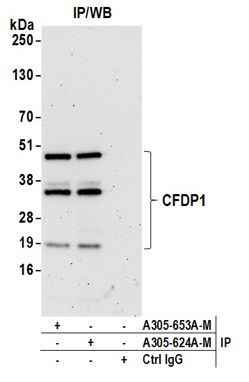
CFDP1, Polyclonal Antibody (Cat# AAA213358)
 WB (Western Blot)
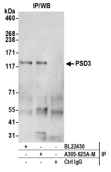
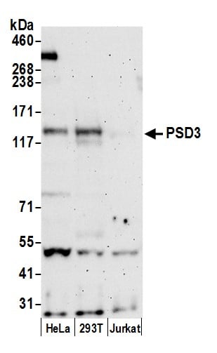
(Detection of human PSD3 by western blot. Samples: Whole cell lysate (50 ug) from HeLa, HEK293T, and Jurkat cells prepared using NETN lysis buffer. Antibody: Affinity purified rabbit anti-PSD3 antibody (AAA213359 lot 1) used for WB at 0.1 mg/ml. Detection: Chemiluminescence with an exposure time of 3 minutes.)
WB (Western Blot)
(Detection of human PSD3 by western blot. Samples: Whole cell lysate (50 ug) from HeLa, HEK293T, and Jurkat cells prepared using NETN lysis buffer. Antibody: Affinity purified rabbit anti-PSD3 antibody (AAA213359 lot 1) used for WB at 0.1 mg/ml. Detection: Chemiluminescence with an exposure time of 3 minutes.)
PSD3, Polyclonal Antibody (Cat# AAA213359)
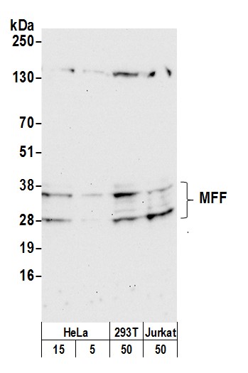 WB (Western Blot)
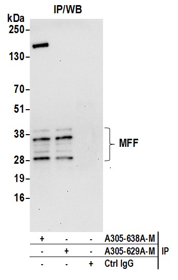
(Detection of human and mouse MFF by western blot. Samples: Whole cell lysate from HeLa (5 and 15 ug) 293T, (50ug), and Jurkat (50ug) cells prepared using NETN lysis buffer. Antibody: Affinity purified rabbit anti-MFF antibody (AAA213365 lot 1) used for WB at 0.1 mg/ml. Detection: Chemiluminescence with an exposure time of 30 seconds.)
WB (Western Blot)
(Detection of human and mouse MFF by western blot. Samples: Whole cell lysate from HeLa (5 and 15 ug) 293T, (50ug), and Jurkat (50ug) cells prepared using NETN lysis buffer. Antibody: Affinity purified rabbit anti-MFF antibody (AAA213365 lot 1) used for WB at 0.1 mg/ml. Detection: Chemiluminescence with an exposure time of 30 seconds.)
MFF, Polyclonal Antibody (Cat# AAA213365)
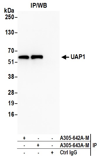
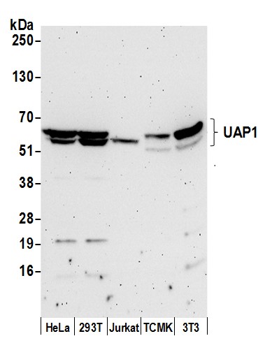 WB (Western Blot)
(Detection of human and mouse UAP1 by western blot. Samples: Whole cell lysate (50 ug) from HeLa, HEK293T, Jurkat, mouse TCMK-1, and mouse NIH 3T3 cells prepared using NETN lysis buffer. Antibody: Affinity purified rabbit anti-UAP1 antibody (AAA213368 lot 1) used for WB at 0.1 mg/ml. Detection: Chemiluminescence with an exposure time of 3 minutes.)
WB (Western Blot)
(Detection of human and mouse UAP1 by western blot. Samples: Whole cell lysate (50 ug) from HeLa, HEK293T, Jurkat, mouse TCMK-1, and mouse NIH 3T3 cells prepared using NETN lysis buffer. Antibody: Affinity purified rabbit anti-UAP1 antibody (AAA213368 lot 1) used for WB at 0.1 mg/ml. Detection: Chemiluminescence with an exposure time of 3 minutes.)
UAP1, Polyclonal Antibody (Cat# AAA213368)
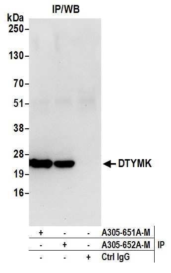
 WB (Western Blot)
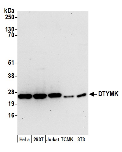
(Detection of human and mouse DTYMK by western blot. Samples: Whole cell lysate (15 ug) from HeLa, HEK293T, Jurkat, mouse TCMK-1, and mouse NIH 3T3 cells prepared using NETN lysis buffer. Antibody: Affinity purified rabbit anti-DTYMK antibody (AAA213370 lot 1) used for WB at 0.1 mg/ml. Detection: Chemiluminescence with an exposure time of 30 seconds.)
WB (Western Blot)
(Detection of human and mouse DTYMK by western blot. Samples: Whole cell lysate (15 ug) from HeLa, HEK293T, Jurkat, mouse TCMK-1, and mouse NIH 3T3 cells prepared using NETN lysis buffer. Antibody: Affinity purified rabbit anti-DTYMK antibody (AAA213370 lot 1) used for WB at 0.1 mg/ml. Detection: Chemiluminescence with an exposure time of 30 seconds.)
DTYMK, Polyclonal Antibody (Cat# AAA213370)
 WB (Western Blot)
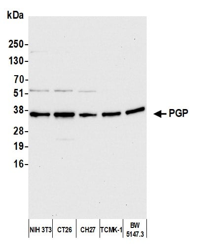
(Detection of human PGP by western blot. Samples: Whole cell lysate (10 ug) from HeLa, MCF-7, HEK293T, Hep-G2, and Jurkat cells prepared using NETN lysis buffer. Antibody: Affinity purified rabbit anti-PGP antibody (AAA213376 lot 2) used for WB at 0.4 mg/ml. Detection: Chemiluminescence with an exposure time of 10 seconds.)
WB (Western Blot)
(Detection of human PGP by western blot. Samples: Whole cell lysate (10 ug) from HeLa, MCF-7, HEK293T, Hep-G2, and Jurkat cells prepared using NETN lysis buffer. Antibody: Affinity purified rabbit anti-PGP antibody (AAA213376 lot 2) used for WB at 0.4 mg/ml. Detection: Chemiluminescence with an exposure time of 10 seconds.)
PGP, Polyclonal Antibody (Cat# AAA213376)
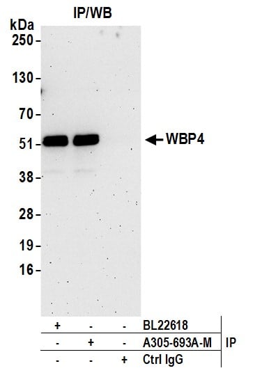
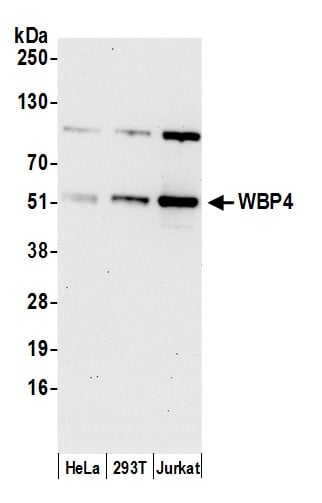 WB (Western Blot)
(Detection of human WBP4 by western blot. Samples: Whole cell lysate (15 ug) from HeLa, HEK293T, and Jurkat cells prepared using NETN lysis buffer. Antibody: Affinity purified rabbit anti-WBP4 antibody (AAA213385 lot 1) used for WB at 0.4 mg/ml. Detection: Chemiluminescence with an exposure time of 30 seconds.)
WB (Western Blot)
(Detection of human WBP4 by western blot. Samples: Whole cell lysate (15 ug) from HeLa, HEK293T, and Jurkat cells prepared using NETN lysis buffer. Antibody: Affinity purified rabbit anti-WBP4 antibody (AAA213385 lot 1) used for WB at 0.4 mg/ml. Detection: Chemiluminescence with an exposure time of 30 seconds.)
WBP4, Polyclonal Antibody (Cat# AAA213385)
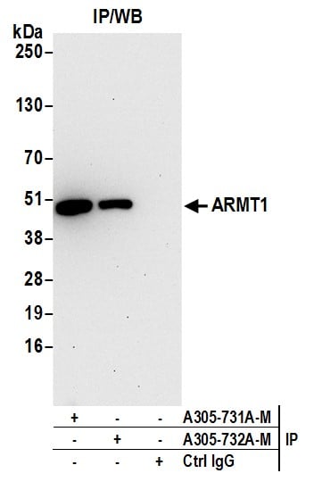
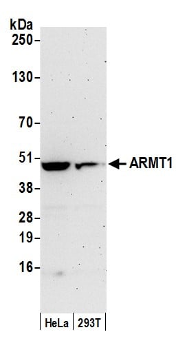 WB (Western Blot)
(Detection of human ARMT1 by western blot. Samples: Whole cell lysate (50 ug) from HeLa and 293T cells prepared using NETN lysis buffer. Antibody: Affinity purified rabbit anti-ARMT1 antibody (AAA213398 lot 1) used for WB at 0.1 mg/ml. Detection: Chemiluminescence with an exposure time of 3 minutes.)
WB (Western Blot)
(Detection of human ARMT1 by western blot. Samples: Whole cell lysate (50 ug) from HeLa and 293T cells prepared using NETN lysis buffer. Antibody: Affinity purified rabbit anti-ARMT1 antibody (AAA213398 lot 1) used for WB at 0.1 mg/ml. Detection: Chemiluminescence with an exposure time of 3 minutes.)
ARMT1, Polyclonal Antibody (Cat# AAA213398)
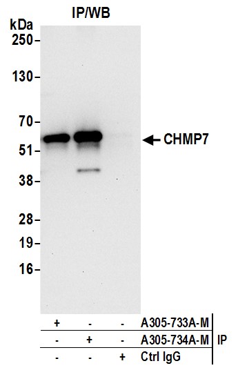
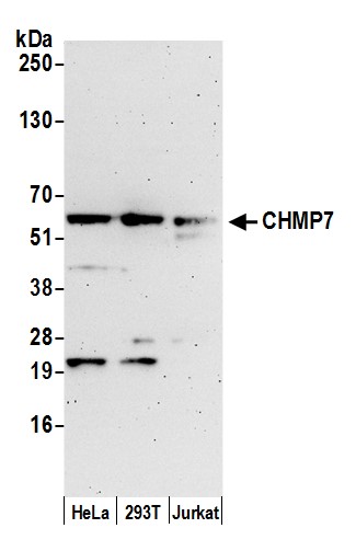 WB (Western Blot)
(Detection of human CHMP7 by western blot. Samples: Whole cell lysate (15 ug) from HeLa, HEK293T, and Jurkat cells prepared using NETN lysis buffer. Antibody: Affinity purified rabbit anti-CHMP7 antibody (AAA213400 lot 1) used for WB at 0.1 mg/ml. Detection: Chemiluminescence with an exposure time of 3 minutes.)
WB (Western Blot)
(Detection of human CHMP7 by western blot. Samples: Whole cell lysate (15 ug) from HeLa, HEK293T, and Jurkat cells prepared using NETN lysis buffer. Antibody: Affinity purified rabbit anti-CHMP7 antibody (AAA213400 lot 1) used for WB at 0.1 mg/ml. Detection: Chemiluminescence with an exposure time of 3 minutes.)
CHMP7, Polyclonal Antibody (Cat# AAA213400)
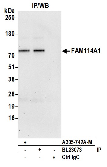
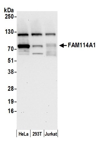 WB (Western Blot)
(Detection of human FAM114A1 by western blot. Samples: Whole cell lysate (15 ug) from HeLa, HEK293T, and Jurkat cells prepared using NETN lysis buffer. Antibody: Affinity purified rabbit anti-FAM114A1 antibody (AAA213402 lot 1) used for WB at 0.1 mg/ml. Detection: Chemiluminescence with an exposure time of 30 seconds.)
WB (Western Blot)
(Detection of human FAM114A1 by western blot. Samples: Whole cell lysate (15 ug) from HeLa, HEK293T, and Jurkat cells prepared using NETN lysis buffer. Antibody: Affinity purified rabbit anti-FAM114A1 antibody (AAA213402 lot 1) used for WB at 0.1 mg/ml. Detection: Chemiluminescence with an exposure time of 30 seconds.)
FAM114A1, Polyclonal Antibody (Cat# AAA213402)
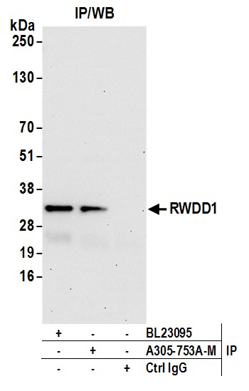
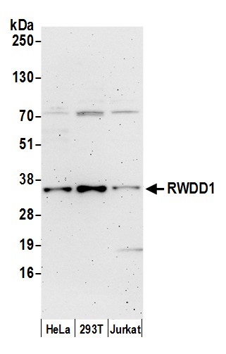 WB (Western Blot)
(Detection of human RWDD1 by western blot. Samples: Whole cell lysate (15 ug) from HeLa, HEK293T, and Jurkat cells prepared using NETN lysis buffer. Antibody: Affinity purified rabbit anti-RWDD1 antibody (AAA213407 lot 1) used for WB at 0.4 mg/ml. Detection: Chemiluminescence with an exposure time of 3 minutes.)
WB (Western Blot)
(Detection of human RWDD1 by western blot. Samples: Whole cell lysate (15 ug) from HeLa, HEK293T, and Jurkat cells prepared using NETN lysis buffer. Antibody: Affinity purified rabbit anti-RWDD1 antibody (AAA213407 lot 1) used for WB at 0.4 mg/ml. Detection: Chemiluminescence with an exposure time of 3 minutes.)
RWDD1, Polyclonal Antibody (Cat# AAA213407)
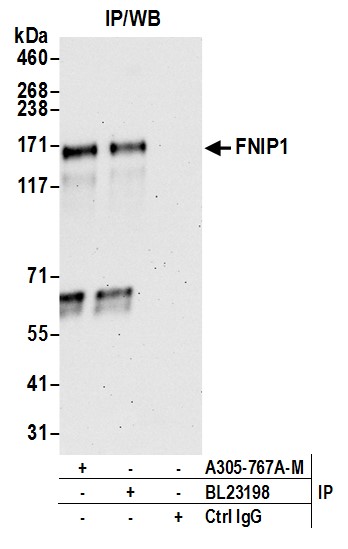
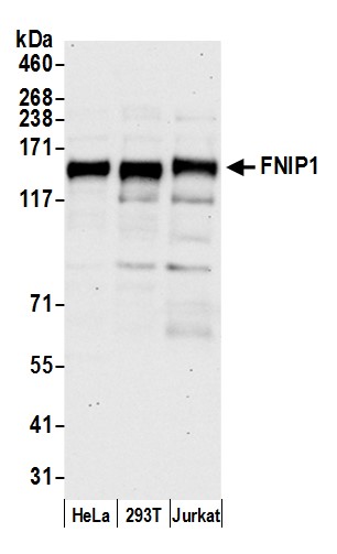 WB (Western Blot)
(Detection of human FNIP1 by western blot. Samples: Whole cell lysate (50 ug) from HeLa, HEK293T, and Jurkat cells prepared using NETN lysis buffer. Antibody: Affinity purified rabbit anti-FNIP1 antibody (AAA213412 lot 1) used for WB at 0.1 mg/ml. Detection: Chemiluminescence with an exposure time of 30 seconds.)
WB (Western Blot)
(Detection of human FNIP1 by western blot. Samples: Whole cell lysate (50 ug) from HeLa, HEK293T, and Jurkat cells prepared using NETN lysis buffer. Antibody: Affinity purified rabbit anti-FNIP1 antibody (AAA213412 lot 1) used for WB at 0.1 mg/ml. Detection: Chemiluminescence with an exposure time of 30 seconds.)
FNIP1, Polyclonal Antibody (Cat# AAA213412)
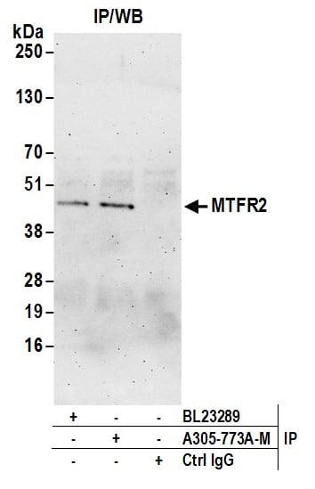
 WB (Western Blot)
(Detection of human MTFR2 by western blot. Samples: Whole cell lysate (50 ug) from HeLa, HEK293T, and Jurkat cells prepared using NETN lysis buffer. Antibody: Affinity purified rabbit anti-MTFR2 antibody (AAA213413 lot 1) used for WB at 0.4 mg/ml. Detection: Chemiluminescence with an exposure time of 3 minutes.)
WB (Western Blot)
(Detection of human MTFR2 by western blot. Samples: Whole cell lysate (50 ug) from HeLa, HEK293T, and Jurkat cells prepared using NETN lysis buffer. Antibody: Affinity purified rabbit anti-MTFR2 antibody (AAA213413 lot 1) used for WB at 0.4 mg/ml. Detection: Chemiluminescence with an exposure time of 3 minutes.)
MTFR2, Polyclonal Antibody (Cat# AAA213413)
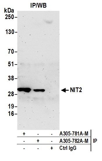
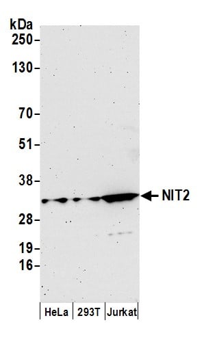 WB (Western Blot)
(Detection of human NIT2 by western blot. Samples: Whole cell lysate (15 ug) from HeLa, HEK293T, and Jurkat cells prepared using NETN lysis buffer. Antibody: Affinity purified rabbit anti-NIT2 antibody (AAA213414 lot 1) used for WB at 0.1 mg/ml. Detection: Chemiluminescence with an exposure time of 3 minutes.)
WB (Western Blot)
(Detection of human NIT2 by western blot. Samples: Whole cell lysate (15 ug) from HeLa, HEK293T, and Jurkat cells prepared using NETN lysis buffer. Antibody: Affinity purified rabbit anti-NIT2 antibody (AAA213414 lot 1) used for WB at 0.1 mg/ml. Detection: Chemiluminescence with an exposure time of 3 minutes.)
NIT2, Polyclonal Antibody (Cat# AAA213414)
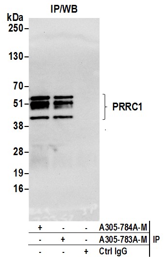
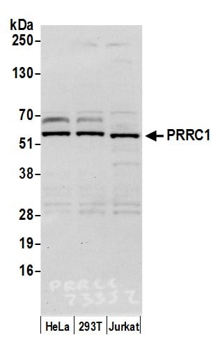 WB (Western Blot)
(Detection of human PRRC1 by western blot. Samples: Whole cell lysate (15 ug) from HeLa, HEK293T, and Jurkat cells prepared using NETN lysis buffer. Antibody: Affinity purified rabbit anti-PRRC1 antibody (AAA213415 lot 1) used for WB at 0.1 mg/ml. Detection: Chemiluminescence with an exposure time of 10 seconds.)
WB (Western Blot)
(Detection of human PRRC1 by western blot. Samples: Whole cell lysate (15 ug) from HeLa, HEK293T, and Jurkat cells prepared using NETN lysis buffer. Antibody: Affinity purified rabbit anti-PRRC1 antibody (AAA213415 lot 1) used for WB at 0.1 mg/ml. Detection: Chemiluminescence with an exposure time of 10 seconds.)
PRRC1, Polyclonal Antibody (Cat# AAA213415)
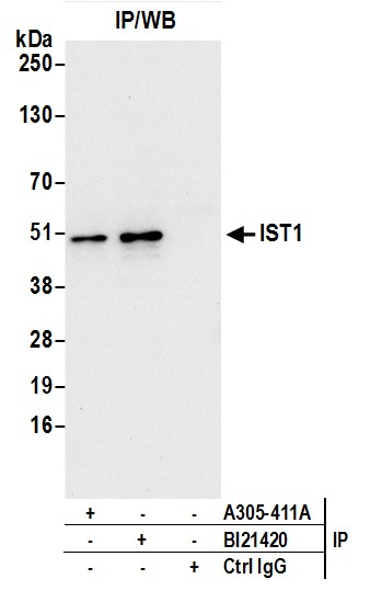
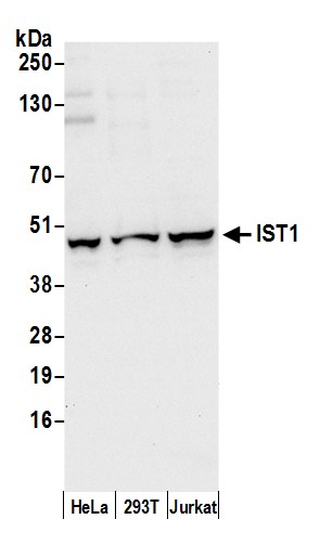 WB (Western Blot)
(Detection of human IST1 by western blot. Samples: Whole cell lysate (50 ug) from HeLa, HEK293T, and Jurkat cells prepared using NETN lysis buffer. Antibody: Affinity purified rabbit anti-IST1 antibody AAA213251 (lot AAA213251-1) used for WB at 0.1 ug/ml. Detection: Chemiluminescence with an exposure time of 10 seconds.)
WB (Western Blot)
(Detection of human IST1 by western blot. Samples: Whole cell lysate (50 ug) from HeLa, HEK293T, and Jurkat cells prepared using NETN lysis buffer. Antibody: Affinity purified rabbit anti-IST1 antibody AAA213251 (lot AAA213251-1) used for WB at 0.1 ug/ml. Detection: Chemiluminescence with an exposure time of 10 seconds.)
IST1/OLC1, Polyclonal Antibody (Cat# AAA213251)
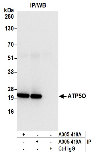
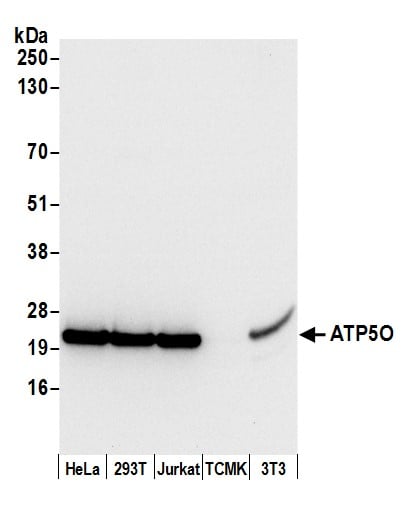 WB (Western Blot)
(Detection of human and mouse ATP5O by western blot. Samples: Whole cell lysate (50 ug) from HeLa, HEK293T, Jurkat, mouse TCMK-1, and mouse NIH 3T3 cells prepared using NETN lysis buffer. Antibody: Affinity purified rabbit anti-ATP5O antibody AAA213258 (lot AAA213258-1) used for WB at 0.1 ug/ml. Detection: Chemiluminescence with an exposure time of 3 seconds.)
WB (Western Blot)
(Detection of human and mouse ATP5O by western blot. Samples: Whole cell lysate (50 ug) from HeLa, HEK293T, Jurkat, mouse TCMK-1, and mouse NIH 3T3 cells prepared using NETN lysis buffer. Antibody: Affinity purified rabbit anti-ATP5O antibody AAA213258 (lot AAA213258-1) used for WB at 0.1 ug/ml. Detection: Chemiluminescence with an exposure time of 3 seconds.)
ATP5O, Polyclonal Antibody (Cat# AAA213258)

 WB (Western Blot)
(Detection of human and mouse IMPA1 by western blot. Samples: Whole cell lysate (50 ug) from HeLa, HEK293T, Jurkat, mouse TCMK-1, and mouse NIH 3T3 cells prepared using NETN lysis buffer. Antibody: Affinity purified rabbit anti-IMPA1 antibody AAA213262 (lot AAA213262-1) used for WB at 0.1 ug/ml. Detection: Chemiluminescence with an exposure time of 30 seconds.)
WB (Western Blot)
(Detection of human and mouse IMPA1 by western blot. Samples: Whole cell lysate (50 ug) from HeLa, HEK293T, Jurkat, mouse TCMK-1, and mouse NIH 3T3 cells prepared using NETN lysis buffer. Antibody: Affinity purified rabbit anti-IMPA1 antibody AAA213262 (lot AAA213262-1) used for WB at 0.1 ug/ml. Detection: Chemiluminescence with an exposure time of 30 seconds.)
IMPA1, Polyclonal Antibody (Cat# AAA213262)
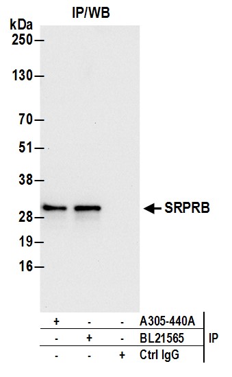
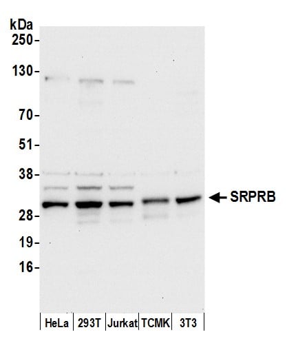 WB (Western Blot)
(Detection of human and mouse SRPRB by western blot. Samples: Whole cell lysate (50 ug) from HeLa, HEK293T, Jurkat, mouse TCMK-1, and mouse NIH 3T3 cells prepared using NETN lysis buffer. Antibody: Affinity purified rabbit anti-SRPRB antibody AAA213267 (lot AAA213267-1) used for WB at 0.1 ug/ml. Detection: Chemiluminescence with an exposure time of 10 seconds.)
WB (Western Blot)
(Detection of human and mouse SRPRB by western blot. Samples: Whole cell lysate (50 ug) from HeLa, HEK293T, Jurkat, mouse TCMK-1, and mouse NIH 3T3 cells prepared using NETN lysis buffer. Antibody: Affinity purified rabbit anti-SRPRB antibody AAA213267 (lot AAA213267-1) used for WB at 0.1 ug/ml. Detection: Chemiluminescence with an exposure time of 10 seconds.)
SRPRB, Polyclonal Antibody (Cat# AAA213267)
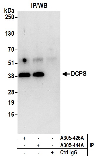
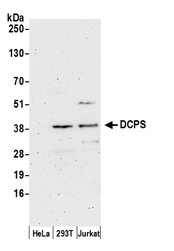 WB (Western Blot)
(Detection of human DCPS by western blot. Samples: Whole cell lysate (50 ug) from HeLa, HEK293T, and Jurkat cells prepared using NETN lysis buffer. Antibody: Affinity purified rabbit anti-DCPS antibody AAA213269 (lot AAA213269-1) used for WB at 0.1 ug/ml. Detection: Chemiluminescence with an exposure time of 3 minutes.)
WB (Western Blot)
(Detection of human DCPS by western blot. Samples: Whole cell lysate (50 ug) from HeLa, HEK293T, and Jurkat cells prepared using NETN lysis buffer. Antibody: Affinity purified rabbit anti-DCPS antibody AAA213269 (lot AAA213269-1) used for WB at 0.1 ug/ml. Detection: Chemiluminescence with an exposure time of 3 minutes.)
DCPS, Polyclonal Antibody (Cat# AAA213269)

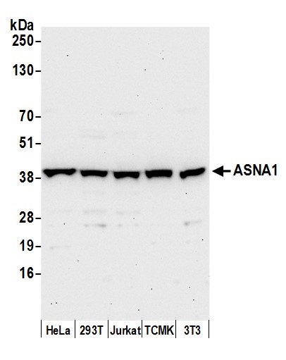 WB (Western Blot)
(Detection of human and mouse ASNA1 by western blot. Samples: Whole cell lysate (50 ug) from HeLa, HEK293T, Jurkat, mouse TCMK-1, and mouse NIH 3T3 cells prepared using NETN lysis buffer. Antibody: Affinity purified rabbit anti-ASNA1 antibody AAA213270 (lot AAA213270-1) used for WB at 0.1 ug/ml. Detection: Chemiluminescence with an exposure time of 30 seconds.)
WB (Western Blot)
(Detection of human and mouse ASNA1 by western blot. Samples: Whole cell lysate (50 ug) from HeLa, HEK293T, Jurkat, mouse TCMK-1, and mouse NIH 3T3 cells prepared using NETN lysis buffer. Antibody: Affinity purified rabbit anti-ASNA1 antibody AAA213270 (lot AAA213270-1) used for WB at 0.1 ug/ml. Detection: Chemiluminescence with an exposure time of 30 seconds.)
ASNA1, Polyclonal Antibody (Cat# AAA213270)
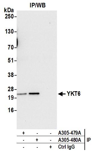
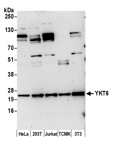 WB (Western Blot)
(Detection of human and mouse YKT6 by western blot. Samples: Whole cell lysate (15 ug) from HeLa, HEK293T, Jurkat, mouse TCMK-1, and mouse NIH 3T3 cells prepared using NETN lysis buffer. Antibody: Affinity purified rabbit anti-YKT6 antibody AAA213280 (lot AAA213280-1) used for WB at 0.1 ug/ml. Detection: Chemiluminescence with an exposure time of 3 minutes.)
WB (Western Blot)
(Detection of human and mouse YKT6 by western blot. Samples: Whole cell lysate (15 ug) from HeLa, HEK293T, Jurkat, mouse TCMK-1, and mouse NIH 3T3 cells prepared using NETN lysis buffer. Antibody: Affinity purified rabbit anti-YKT6 antibody AAA213280 (lot AAA213280-1) used for WB at 0.1 ug/ml. Detection: Chemiluminescence with an exposure time of 3 minutes.)
YKT6, Polyclonal Antibody (Cat# AAA213280)
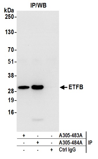
 WB (Western Blot)
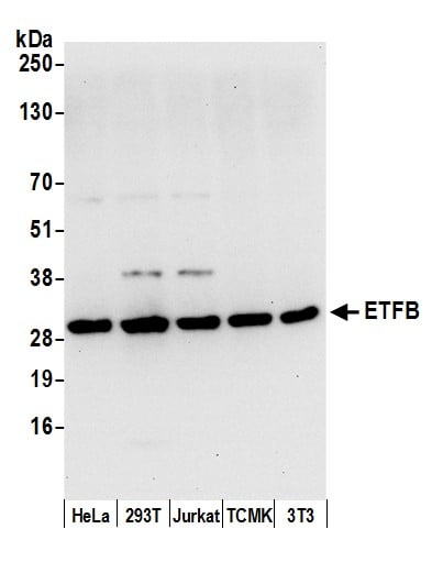
(Detection of human and mouse ETFB by western blot. Samples: Whole cell lysate (50 ug) from HeLa, HEK293T, Jurkat, mouse TCMK-1, and mouse NIH 3T3 cells prepared using NETN lysis buffer. Antibody: Affinity purified rabbit anti-ETFB antibody AAA213283 (lot AAA213283-1) used for WB at 0.1 ug/ml. Detection: Chemiluminescence with an exposure time of 10 seconds.)
WB (Western Blot)
(Detection of human and mouse ETFB by western blot. Samples: Whole cell lysate (50 ug) from HeLa, HEK293T, Jurkat, mouse TCMK-1, and mouse NIH 3T3 cells prepared using NETN lysis buffer. Antibody: Affinity purified rabbit anti-ETFB antibody AAA213283 (lot AAA213283-1) used for WB at 0.1 ug/ml. Detection: Chemiluminescence with an exposure time of 10 seconds.)
ETFB, Polyclonal Antibody (Cat# AAA213283)
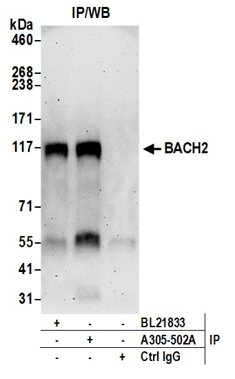
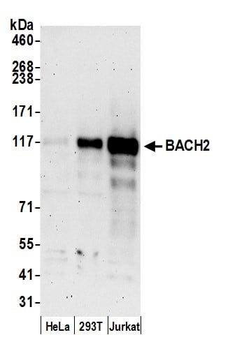 WB (Western Blot)
(Detection of human BACH2 by western blot. Samples: Whole cell lysate (15 ug) from HeLa, HEK293T, and Jurkat cells prepared using NETN lysis buffer. Antibody: Affinity purified rabbit anti-BACH2 antibody AAA213291 (lot AAA213291-1) used for WB at 0.1 ug/ml. Detection: Chemiluminescence with an exposure time of 3 minutes.)
WB (Western Blot)
(Detection of human BACH2 by western blot. Samples: Whole cell lysate (15 ug) from HeLa, HEK293T, and Jurkat cells prepared using NETN lysis buffer. Antibody: Affinity purified rabbit anti-BACH2 antibody AAA213291 (lot AAA213291-1) used for WB at 0.1 ug/ml. Detection: Chemiluminescence with an exposure time of 3 minutes.)
BACH2, Polyclonal Antibody (Cat# AAA213291)
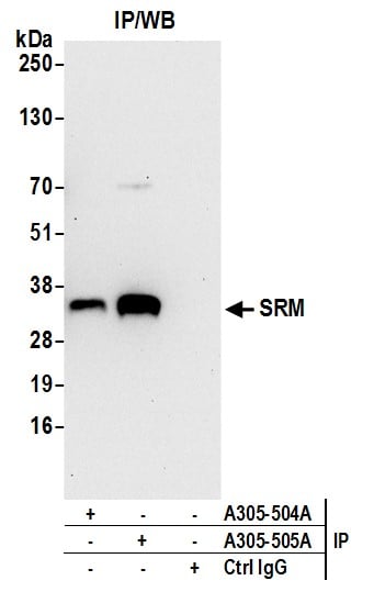
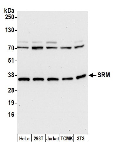 WB (Western Blot)
(Detection of human and mouse SRM by western blot. Samples: Whole cell lysate (15 ug) from HeLa, HEK293T, Jurkat, mouse TCMK-1, and mouse NIH 3T3 cells prepared using NETN lysis buffer. Antibody: Affinity purified rabbit anti-SRM antibody AAA213294 (lot AAA213294-1) used for WB at 0.1 ug/ml. Detection: Chemiluminescence with an exposure time of 30 seconds.)
WB (Western Blot)
(Detection of human and mouse SRM by western blot. Samples: Whole cell lysate (15 ug) from HeLa, HEK293T, Jurkat, mouse TCMK-1, and mouse NIH 3T3 cells prepared using NETN lysis buffer. Antibody: Affinity purified rabbit anti-SRM antibody AAA213294 (lot AAA213294-1) used for WB at 0.1 ug/ml. Detection: Chemiluminescence with an exposure time of 30 seconds.)
SRM, Polyclonal Antibody (Cat# AAA213294)
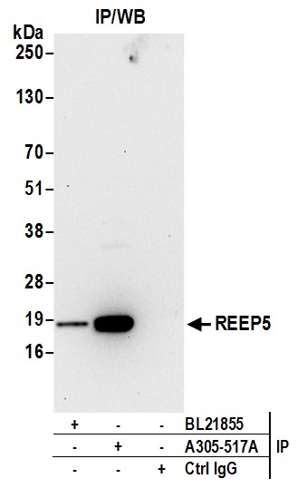
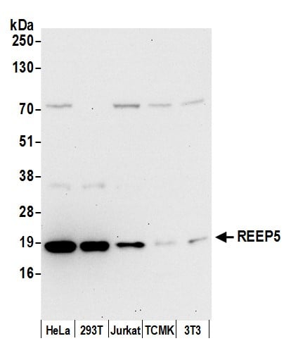 WB (Western Blot)
(Detection of human and mouse REEP5 by western blot. Samples: Whole cell lysate (15 ug) from HeLa, HEK293T, Jurkat, mouse TCMK-1, and mouse NIH 3T3 cells prepared using NETN lysis buffer. Antibody: Affinity purified rabbit anti-REEP5 antibody AAA213297 (lot AAA213297-1) used for WB at 0.04 ug/ml. Detection: Chemiluminescence with an exposure time of 30 seconds.)
WB (Western Blot)
(Detection of human and mouse REEP5 by western blot. Samples: Whole cell lysate (15 ug) from HeLa, HEK293T, Jurkat, mouse TCMK-1, and mouse NIH 3T3 cells prepared using NETN lysis buffer. Antibody: Affinity purified rabbit anti-REEP5 antibody AAA213297 (lot AAA213297-1) used for WB at 0.04 ug/ml. Detection: Chemiluminescence with an exposure time of 30 seconds.)
REEP5, Polyclonal Antibody (Cat# AAA213297)
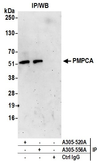
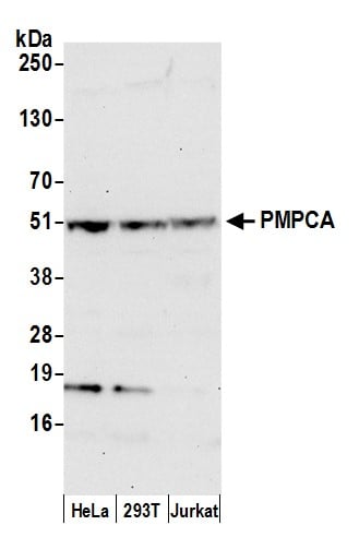 WB (Western Blot)
(Detection of human PMPCA by western blot. Samples: Whole cell lysate (50 ug) from HeLa, HEK293T, and Jurkat cells prepared using NETN lysis buffer. Antibody: Affinity purified rabbit anti-PMPCA antibody AAA213299 (lot AAA213299-1) used for WB at 0.1 ug/ml. Detection: Chemiluminescence with an exposure time of 30 seconds.)
WB (Western Blot)
(Detection of human PMPCA by western blot. Samples: Whole cell lysate (50 ug) from HeLa, HEK293T, and Jurkat cells prepared using NETN lysis buffer. Antibody: Affinity purified rabbit anti-PMPCA antibody AAA213299 (lot AAA213299-1) used for WB at 0.1 ug/ml. Detection: Chemiluminescence with an exposure time of 30 seconds.)
PMPCA, Polyclonal Antibody (Cat# AAA213299)
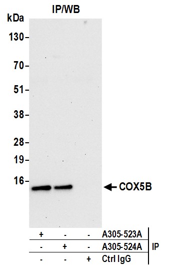
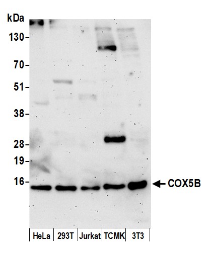 WB (Western Blot)
(Detection of human and mouse COX5B by western blot. Samples: Whole cell lysate (15 ug) from HeLa, HEK293T, Jurkat, mouse TCMK-1, and mouse NIH 3T3 cells prepared using NETN lysis buffer. Antibody: Affinity purified rabbit anti-COX5B antibody AAA213302 (lot AAA213302-1) used for WB at 0.1 ug/ml. Detection: Chemiluminescence with an exposure time of 3 minutes.)
WB (Western Blot)
(Detection of human and mouse COX5B by western blot. Samples: Whole cell lysate (15 ug) from HeLa, HEK293T, Jurkat, mouse TCMK-1, and mouse NIH 3T3 cells prepared using NETN lysis buffer. Antibody: Affinity purified rabbit anti-COX5B antibody AAA213302 (lot AAA213302-1) used for WB at 0.1 ug/ml. Detection: Chemiluminescence with an exposure time of 3 minutes.)
COX5B, Polyclonal Antibody (Cat# AAA213302)
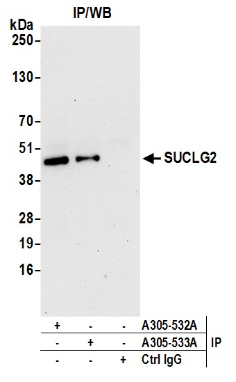
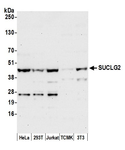 WB (Western Blot)
(Detection of human and mouse SUCLG2 by western blot. Samples: Whole cell lysate (50 ug) from HeLa, HEK293T, Jurkat, mouse TCMK-1, and mouse NIH 3T3 cells prepared using NETN lysis buffer. Antibody: Affinity purified rabbit anti-SUCLG2 antibody AAA213306 (lot AAA213306-1) used for WB at 0.4 ug/ml. Detection: Chemiluminescence with an exposure time of 30 seconds.)
WB (Western Blot)
(Detection of human and mouse SUCLG2 by western blot. Samples: Whole cell lysate (50 ug) from HeLa, HEK293T, Jurkat, mouse TCMK-1, and mouse NIH 3T3 cells prepared using NETN lysis buffer. Antibody: Affinity purified rabbit anti-SUCLG2 antibody AAA213306 (lot AAA213306-1) used for WB at 0.4 ug/ml. Detection: Chemiluminescence with an exposure time of 30 seconds.)
SUCLG2, Polyclonal Antibody (Cat# AAA213306)
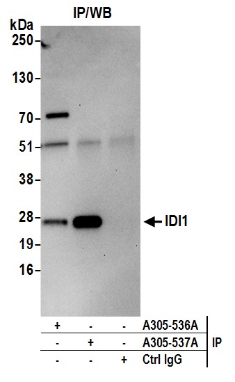
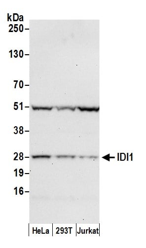 WB (Western Blot)
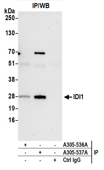
(Detection of human IDI1 by western blot. Samples: Whole cell lysate (50 ug) from HeLa, HEK293T, and Jurkat cells prepared using NETN lysis buffer. Antibody: Affinity purified rabbit anti-IDI1 antibody AAA213309 (lot AAA213309-1) used for WB at 0.1 ug/ml. Detection: Chemiluminescence with an exposure time of 30 seconds.)
WB (Western Blot)
(Detection of human IDI1 by western blot. Samples: Whole cell lysate (50 ug) from HeLa, HEK293T, and Jurkat cells prepared using NETN lysis buffer. Antibody: Affinity purified rabbit anti-IDI1 antibody AAA213309 (lot AAA213309-1) used for WB at 0.1 ug/ml. Detection: Chemiluminescence with an exposure time of 30 seconds.)
IDI1, Polyclonal Antibody (Cat# AAA213309)
 WB (Western Blot)
(Detection of human IDI1 by western blot. Samples: Whole cell lysate (50 ug) from HeLa, HEK293T, and Jurkat cells prepared using NETN lysis buffer. Antibody: Affinity purified rabbit anti-IDI1 antibody AAA213310 (lot AAA213310-1) used for WB at 0.4 ug/ml. Detection: Chemiluminescence with an exposure time of 30 seconds.)
WB (Western Blot)
(Detection of human IDI1 by western blot. Samples: Whole cell lysate (50 ug) from HeLa, HEK293T, and Jurkat cells prepared using NETN lysis buffer. Antibody: Affinity purified rabbit anti-IDI1 antibody AAA213310 (lot AAA213310-1) used for WB at 0.4 ug/ml. Detection: Chemiluminescence with an exposure time of 30 seconds.)
IDI1, Polyclonal Antibody (Cat# AAA213310)
 WB (Western Blot)
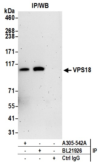
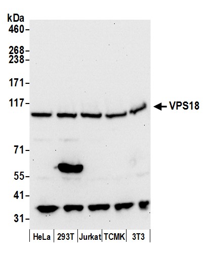
(Detection of human and mouse VPS18 by western blot. Samples: Whole cell lysate (50 ug) from HeLa, HEK293T, Jurkat, mouse TCMK-1, and mouse NIH 3T3 cells prepared using NETN lysis buffer. Antibody: Affinity purified rabbit anti-VPS18 antibody AAA213314 (lot AAA213314-1) used for WB at 0.1 ug/ml. Detection: Chemiluminescence with an exposure time of 30 seconds.)
WB (Western Blot)
(Detection of human and mouse VPS18 by western blot. Samples: Whole cell lysate (50 ug) from HeLa, HEK293T, Jurkat, mouse TCMK-1, and mouse NIH 3T3 cells prepared using NETN lysis buffer. Antibody: Affinity purified rabbit anti-VPS18 antibody AAA213314 (lot AAA213314-1) used for WB at 0.1 ug/ml. Detection: Chemiluminescence with an exposure time of 30 seconds.)
VPS18, Polyclonal Antibody (Cat# AAA213314)
 WB (Western Blot)
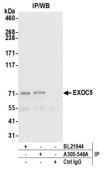
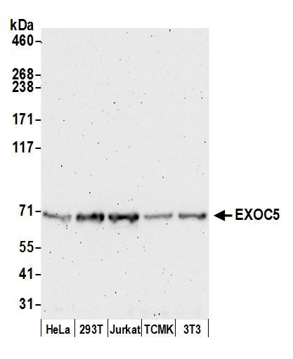
(Detection of human and mouse EXOC5 by western blot. Samples: Whole cell lysate (50 ug) from HeLa, HEK293T, Jurkat, mouse TCMK-1, and mouse NIH 3T3 cells prepared using NETN lysis buffer. Antibody: Affinity purified rabbit anti-EXOC5 antibody AAA213318 (lot AAA213318-1) used for WB at 0.1 ug/ml. Detection: Chemiluminescence with an exposure time of 3 minutes.)
WB (Western Blot)
(Detection of human and mouse EXOC5 by western blot. Samples: Whole cell lysate (50 ug) from HeLa, HEK293T, Jurkat, mouse TCMK-1, and mouse NIH 3T3 cells prepared using NETN lysis buffer. Antibody: Affinity purified rabbit anti-EXOC5 antibody AAA213318 (lot AAA213318-1) used for WB at 0.1 ug/ml. Detection: Chemiluminescence with an exposure time of 3 minutes.)
EXOC5, Polyclonal Antibody (Cat# AAA213318)
 WB (Western Blot)
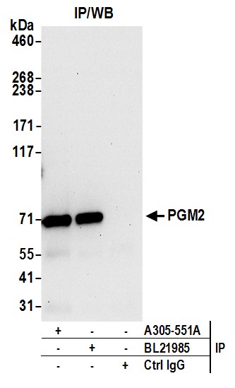
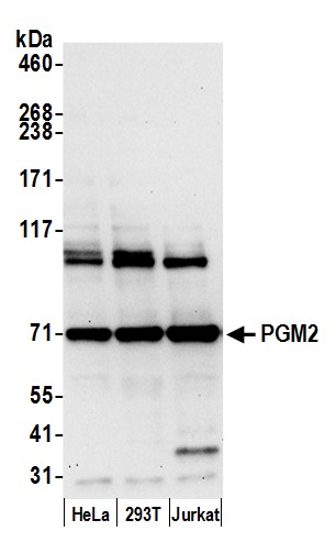
(Detection of human PGM2 by western blot. Samples: Whole cell lysate (15 ug) from HeLa, HEK293T, and Jurkat cells prepared using NETN lysis buffer. Antibody: Affinity purified rabbit anti-PGM2 antibody AAA213322 (lot AAA213322-1) used for WB at 0.1 ug/ml. Detection: Chemiluminescence with an exposure time of 30 seconds.)
WB (Western Blot)
(Detection of human PGM2 by western blot. Samples: Whole cell lysate (15 ug) from HeLa, HEK293T, and Jurkat cells prepared using NETN lysis buffer. Antibody: Affinity purified rabbit anti-PGM2 antibody AAA213322 (lot AAA213322-1) used for WB at 0.1 ug/ml. Detection: Chemiluminescence with an exposure time of 30 seconds.)
PGM2, Polyclonal Antibody (Cat# AAA213322)
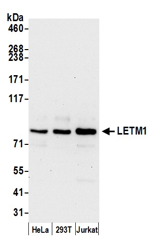 WB (Western Blot)
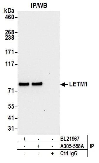
(Detection of human LETM1 by western blot. Samples: Whole cell lysate (15 ug) from HeLa, HEK293T, and Jurkat cells prepared using NETN lysis buffer. Antibody: Affinity purified rabbit anti-LETM1 antibody AAA213324 (lot AAA213324-1) used for WB at 0.1 ug/ml. Detection: Chemiluminescence with an exposure time of 30 seconds.)
WB (Western Blot)
(Detection of human LETM1 by western blot. Samples: Whole cell lysate (15 ug) from HeLa, HEK293T, and Jurkat cells prepared using NETN lysis buffer. Antibody: Affinity purified rabbit anti-LETM1 antibody AAA213324 (lot AAA213324-1) used for WB at 0.1 ug/ml. Detection: Chemiluminescence with an exposure time of 30 seconds.)
LETM1, Polyclonal Antibody (Cat# AAA213324)
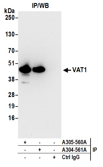
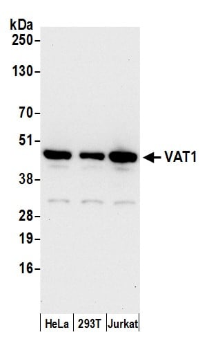 WB (Western Blot)
(Detection of human VAT1 by western blot. Samples: Whole cell lysate (15 ug) from HeLa, HEK293T, and Jurkat cells prepared using NETN lysis buffer. Antibody: Affinity purified rabbit anti-VAT1 antibody AAA213325 (lot AAA213325-1) used for WB at 0.1 ug/ml. Detection: Chemiluminescence with an exposure time of 10 seconds.)
WB (Western Blot)
(Detection of human VAT1 by western blot. Samples: Whole cell lysate (15 ug) from HeLa, HEK293T, and Jurkat cells prepared using NETN lysis buffer. Antibody: Affinity purified rabbit anti-VAT1 antibody AAA213325 (lot AAA213325-1) used for WB at 0.1 ug/ml. Detection: Chemiluminescence with an exposure time of 10 seconds.)
VAT1, Polyclonal Antibody (Cat# AAA213325)
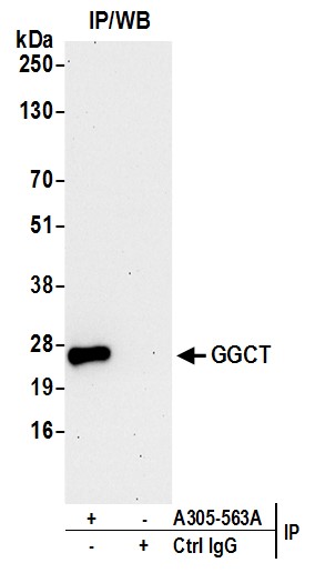
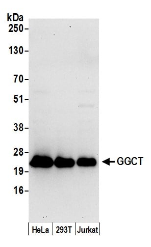 WB (Western Blot)
(Detection of human GGCT by western blot. Samples: Nuclear extract (50 ug) from HeLa, HEK293T, and Jurkat cells prepared using NETN lysis buffer. Antibody: Affinity purified rabbit anti-GGCT antibody AAA213326 (lot AAA213326-1) used for WB at 0.4 ug/ml. Detection: Chemiluminescence with an exposure time of 30 seconds.)
WB (Western Blot)
(Detection of human GGCT by western blot. Samples: Nuclear extract (50 ug) from HeLa, HEK293T, and Jurkat cells prepared using NETN lysis buffer. Antibody: Affinity purified rabbit anti-GGCT antibody AAA213326 (lot AAA213326-1) used for WB at 0.4 ug/ml. Detection: Chemiluminescence with an exposure time of 30 seconds.)
GGCT, Polyclonal Antibody (Cat# AAA213326)
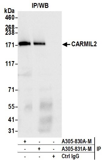
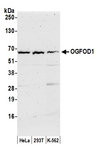 WB (Western Blot)
(Detection of human OGFOD1 by western blot. Samples: Whole cell lysate (50 ug) from HeLa, HEK293T, and K-562 cells prepared using NETN lysis buffer. Antibody: Affinity purified rabbit anti-OGFOD1 antibody (AAA213436 lot 1) used for WB at 0.04 mg/ml. Detection: Chemiluminescence with an exposure time of 3 minutes.)
WB (Western Blot)
(Detection of human OGFOD1 by western blot. Samples: Whole cell lysate (50 ug) from HeLa, HEK293T, and K-562 cells prepared using NETN lysis buffer. Antibody: Affinity purified rabbit anti-OGFOD1 antibody (AAA213436 lot 1) used for WB at 0.04 mg/ml. Detection: Chemiluminescence with an exposure time of 3 minutes.)
OGFOD1, Polyclonal Antibody (Cat# AAA213436)
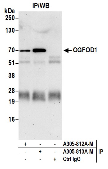
 WB (Western Blot)
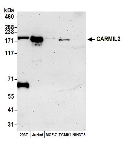
(Detection of human and mouse CARMIL2 by western blot. Samples: Whole cell lysate (50 ug) from HEK293T, Jurkat, MCF-7, mouse TCMK-1, and mouse NIH 3T3 cells prepared using NETN lysis buffer. Antibody: Affinity purified rabbit anti-CARMIL2 antibody (AAA213445 lot 1) used for WB at 0.04 mg/ml. Detection: Chemiluminescence with an exposure time of 3 minutes.)
WB (Western Blot)
(Detection of human and mouse CARMIL2 by western blot. Samples: Whole cell lysate (50 ug) from HEK293T, Jurkat, MCF-7, mouse TCMK-1, and mouse NIH 3T3 cells prepared using NETN lysis buffer. Antibody: Affinity purified rabbit anti-CARMIL2 antibody (AAA213445 lot 1) used for WB at 0.04 mg/ml. Detection: Chemiluminescence with an exposure time of 3 minutes.)
CARMIL2, Polyclonal Antibody (Cat# AAA213445)
 WB (Western Blot)
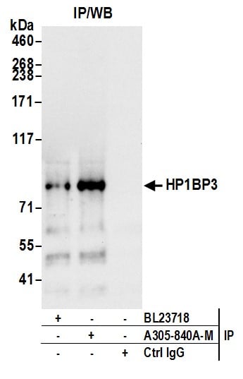
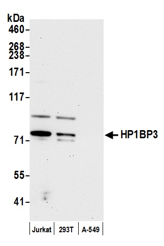
(Detection of human HP1BP3 by western blot. Samples: Whole cell lysate (15 ug) from Jurkat, HEK293T, and A-549 cells prepared using NETN lysis buffer. Antibody: Affinity purified rabbit anti-HP1BP3 antibody (AAA213450 lot 1) used for WB at 0.04 mg/ml. Detection: Chemiluminescence with an exposure time of 30 seconds.)
WB (Western Blot)
(Detection of human HP1BP3 by western blot. Samples: Whole cell lysate (15 ug) from Jurkat, HEK293T, and A-549 cells prepared using NETN lysis buffer. Antibody: Affinity purified rabbit anti-HP1BP3 antibody (AAA213450 lot 1) used for WB at 0.04 mg/ml. Detection: Chemiluminescence with an exposure time of 30 seconds.)
HP1BP3, Polyclonal Antibody (Cat# AAA213450)
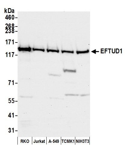
 WB (Western Blot)
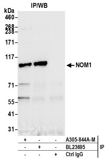
(Detection of human NOM1 by western blot. Samples: Whole cell lysate (50 ug) from HEK293T, RKO, and K-562 cells prepared using NETN lysis buffer. Antibody: Affinity purified rabbit anti-NOM1 antibody (AAA213451 lot 1) used for WB at 0.04 mg/ml. Detection: Chemiluminescence with an exposure time of 30 seconds.)
WB (Western Blot)
(Detection of human NOM1 by western blot. Samples: Whole cell lysate (50 ug) from HEK293T, RKO, and K-562 cells prepared using NETN lysis buffer. Antibody: Affinity purified rabbit anti-NOM1 antibody (AAA213451 lot 1) used for WB at 0.04 mg/ml. Detection: Chemiluminescence with an exposure time of 30 seconds.)
NOM1, Polyclonal Antibody (Cat# AAA213451)
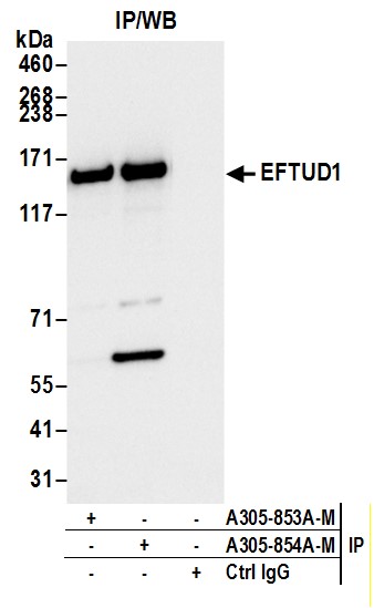
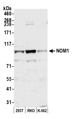
 WB (Western Blot)
(Detection of human and mouse EFTUD1 by western blot. Samples: Whole cell lysate (50 ug) from RKO, Jurkat, A-549, mouse TCMK-1, and mouse NIH 3T3 cells prepared using NETN lysis buffer. Antibody: Affinity purified rabbit anti-EFTUD1 antibody (AAA213455 lot 1) used for WB at 0.04 mg/ml. Detection: Chemiluminescence with an exposure time of 30 seconds.)
WB (Western Blot)
(Detection of human and mouse EFTUD1 by western blot. Samples: Whole cell lysate (50 ug) from RKO, Jurkat, A-549, mouse TCMK-1, and mouse NIH 3T3 cells prepared using NETN lysis buffer. Antibody: Affinity purified rabbit anti-EFTUD1 antibody (AAA213455 lot 1) used for WB at 0.04 mg/ml. Detection: Chemiluminescence with an exposure time of 30 seconds.)
EFTUD1, Polyclonal Antibody (Cat# AAA213455)
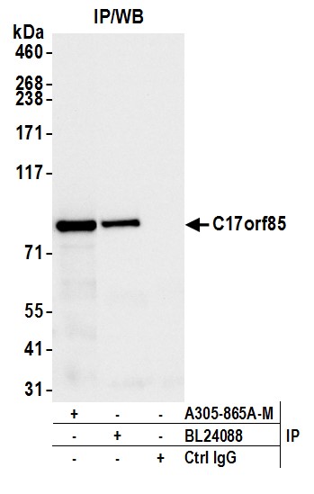
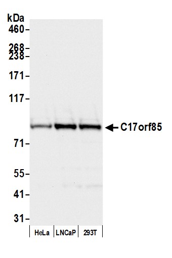 WB (Western Blot)
(Detection of human C17orf85 by western blot. Samples: Whole cell lysate (50 ug) from HeLa, LNCaP, and HEK293T cells prepared using NETN lysis buffer. Antibody: Affinity purified Rabbit anti-C17orf85 antibody (AAA213461 lot 1) used for WB at 0.04 mg/ml. Secondary: HRP-conjugated goat anti-rabbit IgG . Detection: Chemiluminescence with an exposure time of 3 seconds.)
WB (Western Blot)
(Detection of human C17orf85 by western blot. Samples: Whole cell lysate (50 ug) from HeLa, LNCaP, and HEK293T cells prepared using NETN lysis buffer. Antibody: Affinity purified Rabbit anti-C17orf85 antibody (AAA213461 lot 1) used for WB at 0.04 mg/ml. Secondary: HRP-conjugated goat anti-rabbit IgG . Detection: Chemiluminescence with an exposure time of 3 seconds.)
C17orf85, Polyclonal Antibody (Cat# AAA213461)
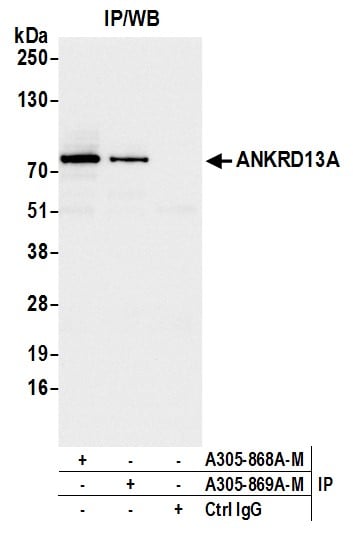
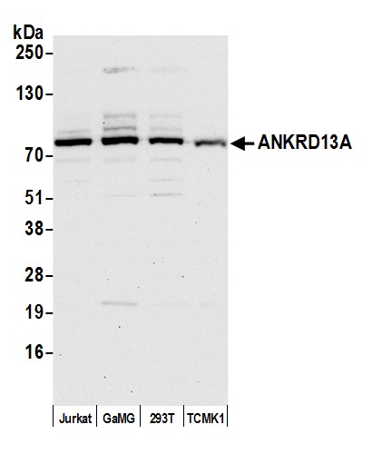 WB (Western Blot)
(Detection of human and mouse ANKRD13A by western blot. Samples: Whole cell lysate (15 ug) from Jurkat, GaMG, HEK293T, and TCMK-1 cells prepared using NETN lysis buffer. Antibody: Affinity purified Rabbit anti-ANKRD13A antibody (AAA213463 lot 1) used for WB at 0.04 mg/ml. Secondary: HRP-conjugated goat anti-rabbit IgG . Detection: Chemiluminescence with an exposure time of 30 seconds.)
WB (Western Blot)
(Detection of human and mouse ANKRD13A by western blot. Samples: Whole cell lysate (15 ug) from Jurkat, GaMG, HEK293T, and TCMK-1 cells prepared using NETN lysis buffer. Antibody: Affinity purified Rabbit anti-ANKRD13A antibody (AAA213463 lot 1) used for WB at 0.04 mg/ml. Secondary: HRP-conjugated goat anti-rabbit IgG . Detection: Chemiluminescence with an exposure time of 30 seconds.)
ANKRD13A, Polyclonal Antibody (Cat# AAA213463)
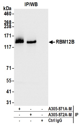
 WB (Western Blot)
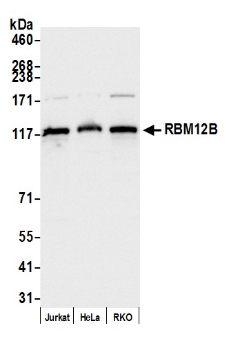
(Detection of human RBM12B by western blot. Samples: Whole cell lysate (10 ug) from Jurkat, HeLa, and RKO cells prepared using NETN lysis buffer. Antibody: Affinity purified rabbit anti-RBM12B antibody (AAA213465 lot 1) used for WB at 0.04 mg/ml. Detection: Chemiluminescence with an exposure time of 1 second.)
WB (Western Blot)
(Detection of human RBM12B by western blot. Samples: Whole cell lysate (10 ug) from Jurkat, HeLa, and RKO cells prepared using NETN lysis buffer. Antibody: Affinity purified rabbit anti-RBM12B antibody (AAA213465 lot 1) used for WB at 0.04 mg/ml. Detection: Chemiluminescence with an exposure time of 1 second.)
RBM12B, Polyclonal Antibody (Cat# AAA213465)
 WB (Western Blot)
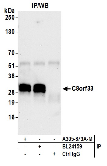
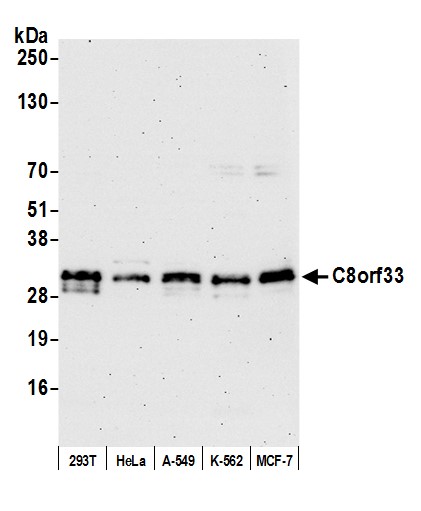
(Detection of human C8orf33 by western blot. Samples: Whole cell lysate (15 ug) from HEK293T, HeLa, A-549, K-562, and MCF-7 cells prepared using NETN lysis buffer. Antibody: Affinity purified rabbit anti-C8orf33 antibody (AAA213467 lot 1) used for WB at 0.04 mg/ml. Detection: Chemiluminescence with an exposure time of 75 seconds.)
WB (Western Blot)
(Detection of human C8orf33 by western blot. Samples: Whole cell lysate (15 ug) from HEK293T, HeLa, A-549, K-562, and MCF-7 cells prepared using NETN lysis buffer. Antibody: Affinity purified rabbit anti-C8orf33 antibody (AAA213467 lot 1) used for WB at 0.04 mg/ml. Detection: Chemiluminescence with an exposure time of 75 seconds.)
C8orf33, Polyclonal Antibody (Cat# AAA213467)
 WB (Western Blot)
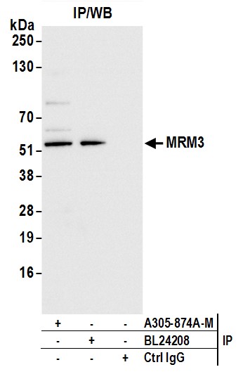
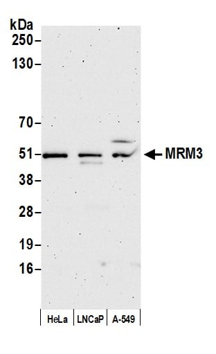
(Detection of human MRM3 by western blot. Samples: Whole cell lysate (5 ug) from HeLa, LNCaP, and A-549 cells prepared using NETN lysis buffer. Antibody: Affinity purified Rabbit anti-MRM3 antibody (AAA213468 lot 1) used for WB at 0.04 mg/ml. Detection: Chemiluminescence with an exposure time of 3 minutes.)
WB (Western Blot)
(Detection of human MRM3 by western blot. Samples: Whole cell lysate (5 ug) from HeLa, LNCaP, and A-549 cells prepared using NETN lysis buffer. Antibody: Affinity purified Rabbit anti-MRM3 antibody (AAA213468 lot 1) used for WB at 0.04 mg/ml. Detection: Chemiluminescence with an exposure time of 3 minutes.)
MRM3, Polyclonal Antibody (Cat# AAA213468)
 WB (Western Blot)
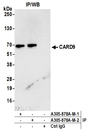
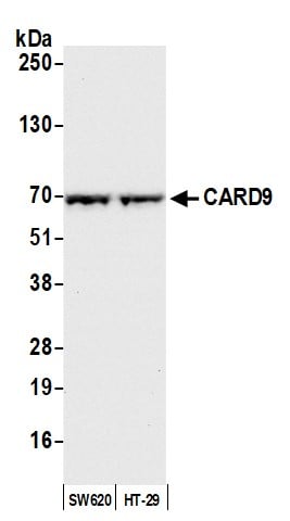
(Detection of human CARD9 by western blot. Samples: Whole cell lysate (50 ug) from SW620 and HT-29 cells prepared using NETN lysis buffer. Antibody: Affinity purified rabbit anti-CARD9 antibody (AAA213472 lot 2) used for WB at 0.04 mg/ml. Detection: Chemiluminescence with an exposure time of 10 seconds.)
WB (Western Blot)
(Detection of human CARD9 by western blot. Samples: Whole cell lysate (50 ug) from SW620 and HT-29 cells prepared using NETN lysis buffer. Antibody: Affinity purified rabbit anti-CARD9 antibody (AAA213472 lot 2) used for WB at 0.04 mg/ml. Detection: Chemiluminescence with an exposure time of 10 seconds.)
CARD9, Polyclonal Antibody (Cat# AAA213472)
What are Polyclonal Antibodies?
Polyclonal antibodies are antibodies that come from multiple B cell clones of a host animal. The typical hosts used for the majority of polyclonal antibody production are rabbits, goats, sheep, and donkeys. These polyclonal antibodies, once having identified their target, will bind to different epitopes located at different regions or sequences on the same protein/antigen. As a result, they are ideal at locating and binding to the target, even if the target is in very low concentrations (due to many different antibodies being able to bind to the same target molecule, which allows for significant amplification of a downstream signal).
Polyclonal antibodies are typically produced by injecting an antigen into a host animal, which causes the animal’s immune system to attack the foreign antigen by mass generating antibodies against it. After a period of time, serum is collected from the animal and purified using physicochemical fractionation, class-specific affinity purification, and/or antigen-affinity purification.
Key Uses of Polyclonal Antibodies
- Western Blotting: This method is used to find specific proteins in biological samples after separating them by size.
- Immunohistochemistry: IHC helps visualize the location of proteins in tissue sections using various staining techniques.
- ELISA: (Enzyme-Linked Immunosorbent Assay) is typically used to identify specific protein quantities in a sample. ELISAs can be either “Quantitative” or “Qualitative”.
- Flow Cytometry: technique that identifies and measures the specific protein on the surface or inside the cells in a fluid suspension.
- Immunoprecipitation: IP isolates and studies a specific protein from a complex mixture using antibodies.
Why Buy Polyclonal Antibodies from AAA Biotech?
1. Ideal for Various Applications
Our antibodies are generally going to be validated for use in multiple types of assays, including ELISA, Western Blotting, Immunohistochemistry, Immunoprecipitation, amongst others. They are ideal for a wide range of research applications.
2. Rigorous Quality Control
All of the antibodies in our catalog undergo strict quality testing to ensure specificity, sensitivity, and consistent performance. We are confident in the ability of our antibodies to provide you with accurate results.
3. Wide Assortment of Antibodies
Antibodies in are catalog can be found for both common and exotic species, and these antibodies are also available in both conjugated and recombinant forms to suit many diverse experimental needs.
4. Highly Purified
Our antibodies are available in purified forms with over 85% purity, as confirmed by SDS-PAGE. They are also available with tags such as His, Flag, GST, or MBP. We cater to customers worldwide.
FAQ
1. How are polyclonal antibodies produced?
Traditionally, polyclonal antibodies are produced by injecting an antigen into a host animal (such as a rabbit or goat), which then triggers an immune response from the host animal. The animal’s B cells produce antibodies that will recognize different parts of the injected antigen. These antibodies are then collected from the animal’s blood and purified for use.
2. How do polyclonal antibodies differ from monoclonal antibodies?
Polyclonal antibodies are a mix of antibodies that bind to different locations (epitopes) of the same antigen, while monoclonal antibodies are identical and bind to just one specific epitope. This makes polyclonal antibodies more versatile and better at detecting proteins that may be present in low quantities or in altered/modified forms.
3. How should I store polyclonal antibodies?
Polyclonal antibodies should be stored at 4°C for short-term use (up to a few weeks) and at -20°C or -80°C for long-term storage. Avoid repeated freeze-thaw cycles by dividing them into small aliquots. Always check the datasheet for specific storage instructions.