Filters
▼Clonality
▼Type
▼Reactivity
▼Gene Name
▼Isotype
▼Host
▼Application
▼Clone
▼Polyclonal Antibodies
At AAA Biotech also known as AAA Bio or AAABio, we provide a broad range of purified polyclonal antibodies (pAbs) that are able to all be browsed online through our website. Due to their high specificity and strong binding affinity, these antibodies are ideal for wide swathes of research and experimental applications.
Our polyclonal antibodies can easily support your work, whether you use them for Western Blotting, Immunocytochemistry (with or without Immunofluorescence used in conjunction), Immunohistochemistry, Immunoprecipitation, and ELISA tests. We highly encourage you to browse our range of pAbs and choose the one that best suits your experimental model.
Viewing 5000-5050 of 96812 product results
 WB (Western Blot)
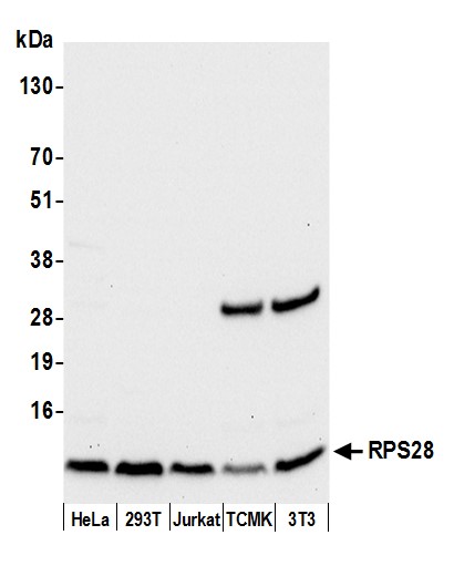
(Detection of human and mouse RPS28 by western blot. Samples: Whole cell lysate (50 ug) from HeLa, HEK293T, Jurkat, mouse TCMK-1, and mouse NIH 3T3 cells prepared using NETN lysis buffer. Antibody: Affinity purified rabbit anti-RPS28 antibody AAA213098 (lot AAA213098-1) used for WB at 0.04 ug/ml. Detection: Chemiluminescence with an exposure time of 10 seconds.)
WB (Western Blot)
(Detection of human and mouse RPS28 by western blot. Samples: Whole cell lysate (50 ug) from HeLa, HEK293T, Jurkat, mouse TCMK-1, and mouse NIH 3T3 cells prepared using NETN lysis buffer. Antibody: Affinity purified rabbit anti-RPS28 antibody AAA213098 (lot AAA213098-1) used for WB at 0.04 ug/ml. Detection: Chemiluminescence with an exposure time of 10 seconds.)
RPS28/Ribosomal Protein S28, Polyclonal Antibody (Cat# AAA213098)
 WB (Western Blot)
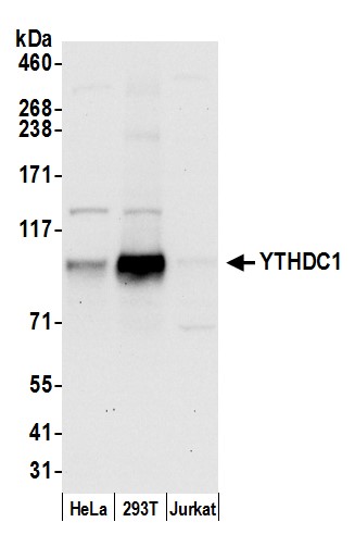
(Detection of human YTHDC1 by western blot. Samples: Whole cell lysate (50 ug) from HeLa, HEK293T, and Jurkat cells prepared using NETN lysis buffer. Antibody: Affinity purified rabbit anti-YTHDC1 antibody AAA213099 (lot AAA213099-1) used for WB at 0.1 ug/ml. Detection: Chemiluminescence with an exposure time of 30 seconds.)
WB (Western Blot)
(Detection of human YTHDC1 by western blot. Samples: Whole cell lysate (50 ug) from HeLa, HEK293T, and Jurkat cells prepared using NETN lysis buffer. Antibody: Affinity purified rabbit anti-YTHDC1 antibody AAA213099 (lot AAA213099-1) used for WB at 0.1 ug/ml. Detection: Chemiluminescence with an exposure time of 30 seconds.)
YTHDC1/YT521, Polyclonal Antibody (Cat# AAA213099)
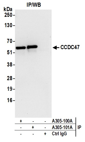
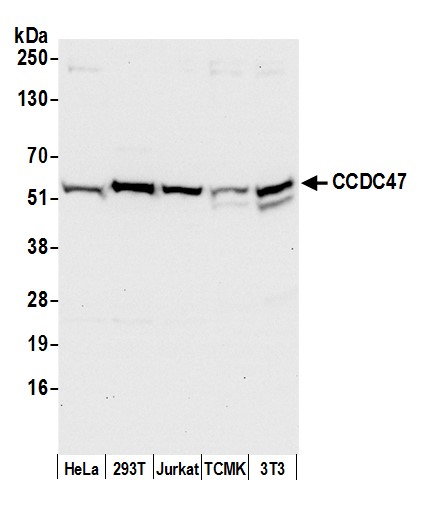 WB (Western Blot)
(Detection of human and mouse CCDC47 by western blot. Samples: Whole cell lysate (50 ug) from HeLa, HEK293T, Jurkat, mouse TCMK-1, and mouse NIH 3T3 cells prepared using NETN lysis buffer. Antibody: Affinity purified rabbit anti-CCDC47 antibody AAA213103 (lot AAA213103-1) used for WB at 0.1 ug/ml. Detection: Chemiluminescence with an exposure time of 10 seconds.)
WB (Western Blot)
(Detection of human and mouse CCDC47 by western blot. Samples: Whole cell lysate (50 ug) from HeLa, HEK293T, Jurkat, mouse TCMK-1, and mouse NIH 3T3 cells prepared using NETN lysis buffer. Antibody: Affinity purified rabbit anti-CCDC47 antibody AAA213103 (lot AAA213103-1) used for WB at 0.1 ug/ml. Detection: Chemiluminescence with an exposure time of 10 seconds.)
CCDC47, Polyclonal Antibody (Cat# AAA213103)
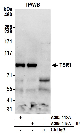
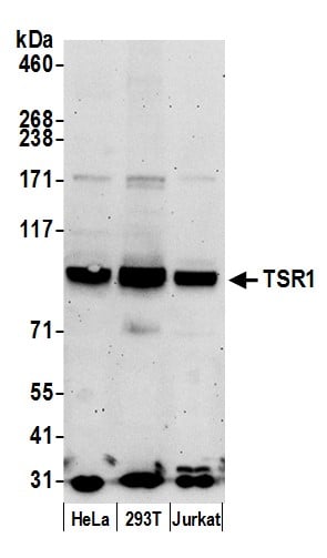 WB (Western Blot)
(Detection of human TSR1 by western blot. Samples: Whole cell lysate (50 ug) from HeLa, HEK293T, and Jurkat cells prepared using NETN lysis buffer. Antibody: Affinity purified rabbit anti-TSR1 antibody AAA213109 (lot AAA213109-1) used for WB at 0.4 ug/ml. Detection: Chemiluminescence with an exposure time of 3 minutes.)
WB (Western Blot)
(Detection of human TSR1 by western blot. Samples: Whole cell lysate (50 ug) from HeLa, HEK293T, and Jurkat cells prepared using NETN lysis buffer. Antibody: Affinity purified rabbit anti-TSR1 antibody AAA213109 (lot AAA213109-1) used for WB at 0.4 ug/ml. Detection: Chemiluminescence with an exposure time of 3 minutes.)
TSR1, Polyclonal Antibody (Cat# AAA213109)
 WB (Western Blot)
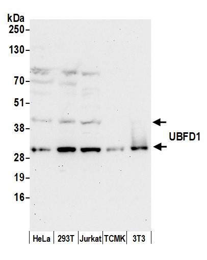
(Detection of human and mouse UBFD1 by western blot. Samples: Whole cell lysate (50 ug) from HeLa, HEK293T, Jurkat, mouse TCMK-1, and mouse NIH 3T3 cells prepared using NETN lysis buffer. Antibody: Affinity purified rabbit anti-UBFD1 antibody AAA213124 (lot AAA213124-1) used for WB at 0.1 ug/ml. Detection: Chemiluminescence with an exposure time of 30 seconds.)
WB (Western Blot)
(Detection of human and mouse UBFD1 by western blot. Samples: Whole cell lysate (50 ug) from HeLa, HEK293T, Jurkat, mouse TCMK-1, and mouse NIH 3T3 cells prepared using NETN lysis buffer. Antibody: Affinity purified rabbit anti-UBFD1 antibody AAA213124 (lot AAA213124-1) used for WB at 0.1 ug/ml. Detection: Chemiluminescence with an exposure time of 30 seconds.)
UBFD1, Polyclonal Antibody (Cat# AAA213124)
 WB (Western Blot)
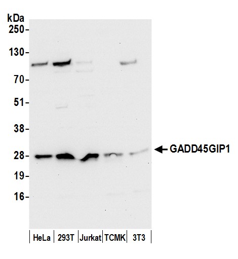
(Detection of human and mouse GADD45GIP1 by western blot. Samples: Whole cell lysate (50 ug) from HeLa, HEK293T, Jurkat, mouse TCMK-1, and mouse NIH 3T3 cells prepared using NETN lysis buffer. Antibody: Affinity purified rabbit anti-GADD45GIP1 antibody AAA213127 (lot AAA213127-1) used for WB at 0.1 ug/ml. Detection: Chemiluminescence with an exposure time of 10 seconds.)
WB (Western Blot)
(Detection of human and mouse GADD45GIP1 by western blot. Samples: Whole cell lysate (50 ug) from HeLa, HEK293T, Jurkat, mouse TCMK-1, and mouse NIH 3T3 cells prepared using NETN lysis buffer. Antibody: Affinity purified rabbit anti-GADD45GIP1 antibody AAA213127 (lot AAA213127-1) used for WB at 0.1 ug/ml. Detection: Chemiluminescence with an exposure time of 10 seconds.)
GADD45GIP1/CRIF1, Polyclonal Antibody (Cat# AAA213127)
 WB (Western Blot)
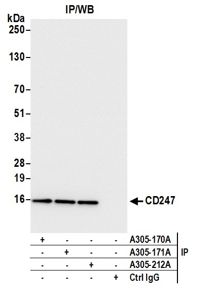
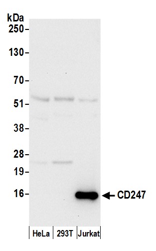
(Detection of human CD247 by western blot. Samples: Whole cell lysate (15 ug) from HeLa, HEK293T, and Jurkat cells prepared using NETN lysis buffer. Antibody: Affinity purified rabbit anti-CD247 antibody AAA213131 (lot AAA213131-1) used for WB at 0.1 ug/ml. Detection: Chemiluminescence with an exposure time of 10 seconds.)
WB (Western Blot)
(Detection of human CD247 by western blot. Samples: Whole cell lysate (15 ug) from HeLa, HEK293T, and Jurkat cells prepared using NETN lysis buffer. Antibody: Affinity purified rabbit anti-CD247 antibody AAA213131 (lot AAA213131-1) used for WB at 0.1 ug/ml. Detection: Chemiluminescence with an exposure time of 10 seconds.)
CD247/CD3Z, Polyclonal Antibody (Cat# AAA213131)
 WB (Western Blot)
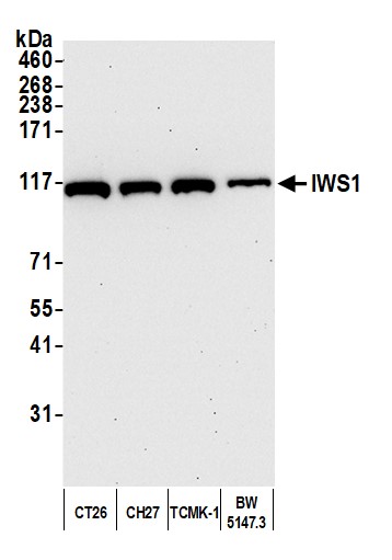
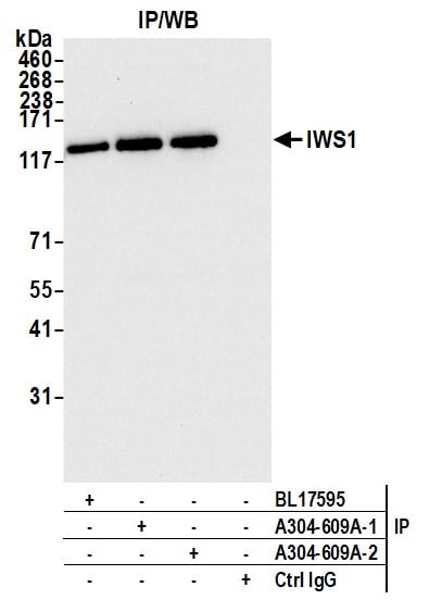
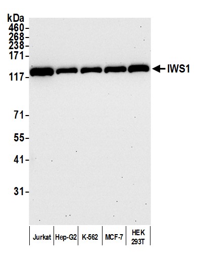
(Detection of human IWS1 by western blot. Samples: Whole cell lysate (10 ug) from Jurkat, Hep-G2, K-562, MCF-7, and HEK293T cells prepared using NETN lysis buffer. Antibody: Affinity purified rabbit anti-IWS1 antibody (AAA212807 lot 2) used for WB at 0.1 ug/ml. Detection: Chemiluminescence with an exposure time of 30 seconds.)
WB (Western Blot)
(Detection of human IWS1 by western blot. Samples: Whole cell lysate (10 ug) from Jurkat, Hep-G2, K-562, MCF-7, and HEK293T cells prepared using NETN lysis buffer. Antibody: Affinity purified rabbit anti-IWS1 antibody (AAA212807 lot 2) used for WB at 0.1 ug/ml. Detection: Chemiluminescence with an exposure time of 30 seconds.)
IWS1, Polyclonal Antibody (Cat# AAA212807)
 WB (Western Blot)
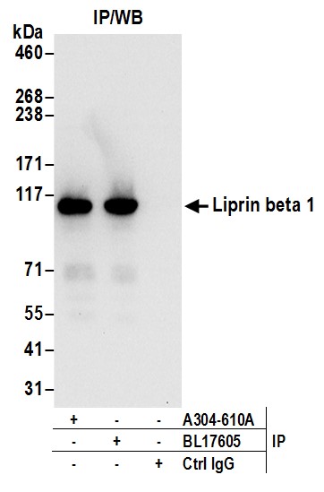
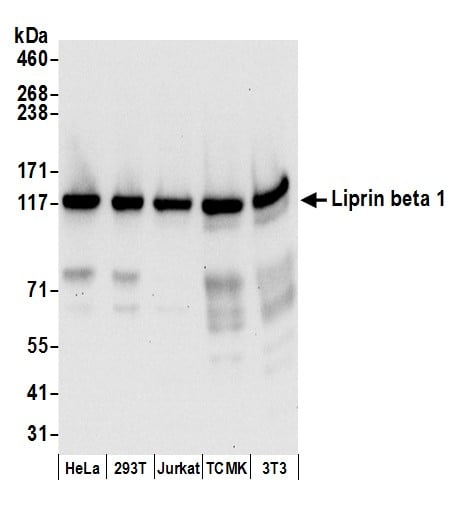
(Detection of human and mouse Liprin beta 1 by western blot. Samples: Whole cell lysate (50 ug) from HeLa, HEK293T, Jurkat, mouse TCMK-1, and mouse NIH 3T3 cells prepared using NETN lysis buffer. Antibodies: Affinity purified rabbit anti-Liprin beta 1 antibody AAA212808 (lot AAA212808-1) used for WB at 0.1 ug/ml. Detection: Chemiluminescence with an exposure time of 10 seconds.)
WB (Western Blot)
(Detection of human and mouse Liprin beta 1 by western blot. Samples: Whole cell lysate (50 ug) from HeLa, HEK293T, Jurkat, mouse TCMK-1, and mouse NIH 3T3 cells prepared using NETN lysis buffer. Antibodies: Affinity purified rabbit anti-Liprin beta 1 antibody AAA212808 (lot AAA212808-1) used for WB at 0.1 ug/ml. Detection: Chemiluminescence with an exposure time of 10 seconds.)
Liprin beta 1, Polyclonal Antibody (Cat# AAA212808)
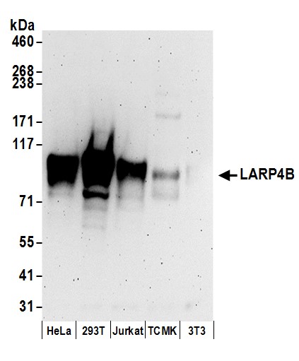 WB (Western Blot)
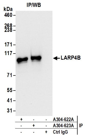
(Detection of human and mouse LARP4B by western blot. Samples: Whole cell lysate (50 ug) from HeLa, HEK293T, Jurkat, mouse TCMK-1, and mouse NIH 3T3 cells prepared using NETN lysis buffer. Antibodies: Affinity purified rabbit anti-LARP4B antibody AAA212817 (lot AAA212817-1) used for WB at 0.1 ug/ml. Detection: Chemiluminescence with an exposure time of 3 minutes.)
WB (Western Blot)
(Detection of human and mouse LARP4B by western blot. Samples: Whole cell lysate (50 ug) from HeLa, HEK293T, Jurkat, mouse TCMK-1, and mouse NIH 3T3 cells prepared using NETN lysis buffer. Antibodies: Affinity purified rabbit anti-LARP4B antibody AAA212817 (lot AAA212817-1) used for WB at 0.1 ug/ml. Detection: Chemiluminescence with an exposure time of 3 minutes.)
LARP4B, Polyclonal Antibody (Cat# AAA212817)
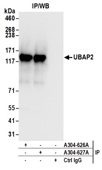
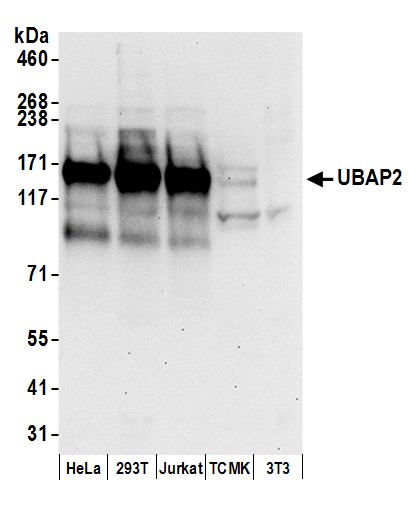 WB (Western Blot)
(Detection of human and mouse UBAP2 by western blot. Samples: Whole cell lysate (50 ug) from HeLa, HEK293T, Jurkat, mouse TCMK-1, and mouse NIH 3T3 cells prepared using NETN lysis buffer. Antibodies: Affinity purified rabbit anti-UBAP2 antibody AAA212819 (lot AAA212819-1) used for WB at 0.1 ug/ml. Detection: Chemiluminescence with an exposure time of 30 seconds.)
WB (Western Blot)
(Detection of human and mouse UBAP2 by western blot. Samples: Whole cell lysate (50 ug) from HeLa, HEK293T, Jurkat, mouse TCMK-1, and mouse NIH 3T3 cells prepared using NETN lysis buffer. Antibodies: Affinity purified rabbit anti-UBAP2 antibody AAA212819 (lot AAA212819-1) used for WB at 0.1 ug/ml. Detection: Chemiluminescence with an exposure time of 30 seconds.)
UBAP2, Polyclonal Antibody (Cat# AAA212819)
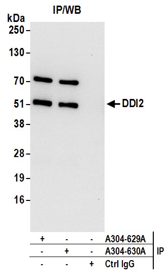
 WB (Western Blot)
(Detection of human and mouse DDI2 by western blot. Samples: Whole cell lysate (50 ug) from HeLa, HEK293T, Jurkat, mouse TCMK-1, and mouse NIH 3T3 cells prepared using NETN lysis buffer. Antibodies: Affinity purified rabbit anti-DDI2 antibody AAA212823 (lot AAA212823-1) used for WB at 0.1 ug/ml. Detection: Chemiluminescence with an exposure time of 3 minutes.)
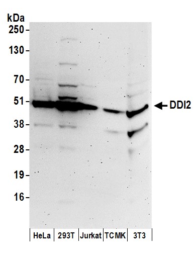
WB (Western Blot)
(Detection of human and mouse DDI2 by western blot. Samples: Whole cell lysate (50 ug) from HeLa, HEK293T, Jurkat, mouse TCMK-1, and mouse NIH 3T3 cells prepared using NETN lysis buffer. Antibodies: Affinity purified rabbit anti-DDI2 antibody AAA212823 (lot AAA212823-1) used for WB at 0.1 ug/ml. Detection: Chemiluminescence with an exposure time of 3 minutes.)
DDI2, Polyclonal Antibody (Cat# AAA212823)
 WB (Western Blot)
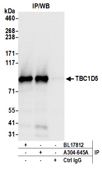
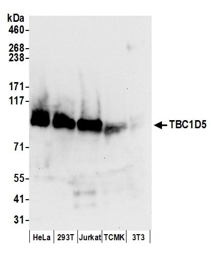
(Detection of human and mouse TBC1D5 by western blot. Samples: Whole cell lysate (50 ug) from HeLa, HEK293T, Jurkat, mouse TCMK-1, and mouse NIH 3T3 cells prepared using NETN lysis buffer. Antibodies: Affinity purified rabbit anti-TBC1D5 antibody AAA212835 (lot AAA212835-1) used for WB at 0.1 ug/ml. Detection: Chemiluminescence with an exposure time of 10 seconds.)
WB (Western Blot)
(Detection of human and mouse TBC1D5 by western blot. Samples: Whole cell lysate (50 ug) from HeLa, HEK293T, Jurkat, mouse TCMK-1, and mouse NIH 3T3 cells prepared using NETN lysis buffer. Antibodies: Affinity purified rabbit anti-TBC1D5 antibody AAA212835 (lot AAA212835-1) used for WB at 0.1 ug/ml. Detection: Chemiluminescence with an exposure time of 10 seconds.)
TBC1D5, Polyclonal Antibody (Cat# AAA212835)
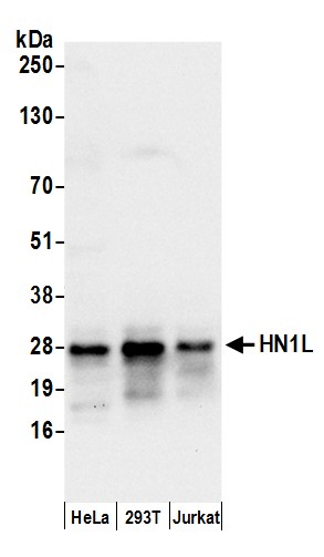 WB (Western Blot)
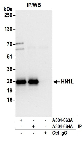
(Detection of human HN1L by western blot. Samples: Whole cell lysate (50 ug) from HeLa, HEK293T, and Jurkat cells prepared using NETN lysis buffer. Antibodies: Affinity purified rabbit anti-HN1L antibody AAA212846 (lot AAA212846-1) used for WB at 0.1 ug/ml. Detection: Chemiluminescence with an exposure time of 10 seconds.)
WB (Western Blot)
(Detection of human HN1L by western blot. Samples: Whole cell lysate (50 ug) from HeLa, HEK293T, and Jurkat cells prepared using NETN lysis buffer. Antibodies: Affinity purified rabbit anti-HN1L antibody AAA212846 (lot AAA212846-1) used for WB at 0.1 ug/ml. Detection: Chemiluminescence with an exposure time of 10 seconds.)
HN1L, Polyclonal Antibody (Cat# AAA212846)
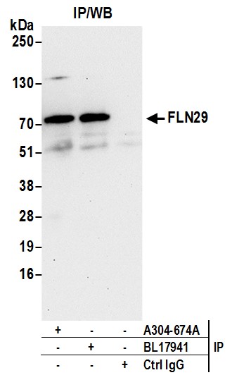
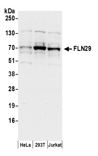 WB (Western Blot)
(Detection of human FLN29 by western blot. Samples: Whole cell lysate (50 ug) from HeLa, HEK293T, and Jurkat cells prepared using NETN lysis buffer. Antibodies: Affinity purified rabbit anti-FLN29 antibody AAA212854 (lot AAA212854-1) used for WB at 0.1 ug/ml. Detection: Chemiluminescence with an exposure time of 30 seconds.)
WB (Western Blot)
(Detection of human FLN29 by western blot. Samples: Whole cell lysate (50 ug) from HeLa, HEK293T, and Jurkat cells prepared using NETN lysis buffer. Antibodies: Affinity purified rabbit anti-FLN29 antibody AAA212854 (lot AAA212854-1) used for WB at 0.1 ug/ml. Detection: Chemiluminescence with an exposure time of 30 seconds.)
FLN29, Polyclonal Antibody (Cat# AAA212854)
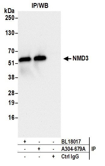
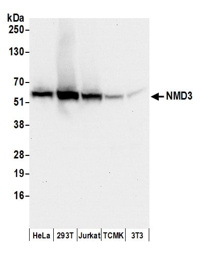 WB (Western Blot)
(Detection of human and mouse NMD3 by western blot. Samples: Whole cell lysate (50 ug) from HeLa, HEK293T, Jurkat, mouse TCMK-1, and mouse NIH 3T3 cells prepared using NETN lysis buffer. Antibodies: Affinity purified rabbit anti-NMD3 antibody AAA212858 (lot AAA212858-1) used for WB at 0.1 ug/ml. Detection: Chemiluminescence with an exposure time of 10 seconds.)
WB (Western Blot)
(Detection of human and mouse NMD3 by western blot. Samples: Whole cell lysate (50 ug) from HeLa, HEK293T, Jurkat, mouse TCMK-1, and mouse NIH 3T3 cells prepared using NETN lysis buffer. Antibodies: Affinity purified rabbit anti-NMD3 antibody AAA212858 (lot AAA212858-1) used for WB at 0.1 ug/ml. Detection: Chemiluminescence with an exposure time of 10 seconds.)
NMD3, Polyclonal Antibody (Cat# AAA212858)
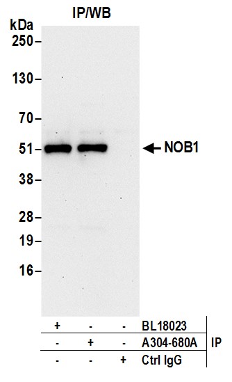
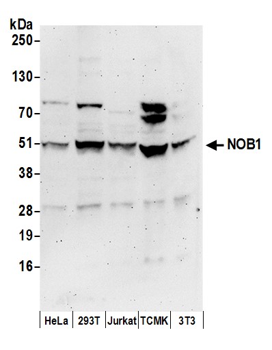 WB (Western Blot)
(Detection of human and mouse NOB1 by western blot. Samples: Whole cell lysate (50 ug) from HeLa, HEK293T, Jurkat, mouse TCMK-1, and mouse NIH 3T3 cells prepared using NETN lysis buffer. Antibodies: Affinity purified rabbit anti-NOB1 antibody AAA212859 (lot AAA212859-1) used for WB at 0.1 ug/ml. Detection: Chemiluminescence with an exposure time of 3 minutes.)
WB (Western Blot)
(Detection of human and mouse NOB1 by western blot. Samples: Whole cell lysate (50 ug) from HeLa, HEK293T, Jurkat, mouse TCMK-1, and mouse NIH 3T3 cells prepared using NETN lysis buffer. Antibodies: Affinity purified rabbit anti-NOB1 antibody AAA212859 (lot AAA212859-1) used for WB at 0.1 ug/ml. Detection: Chemiluminescence with an exposure time of 3 minutes.)
NOB1, Polyclonal Antibody (Cat# AAA212859)
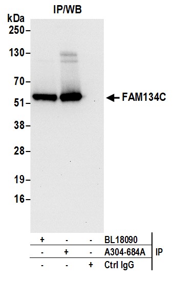
 WB (Western Blot)
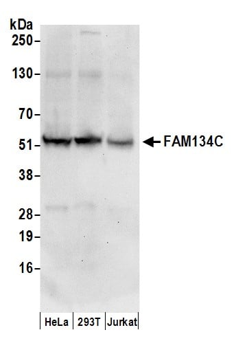
(Detection of human FAM134C by western blot. Samples: Whole cell lysate (50 ug) from HeLa, HEK293T, and Jurkat cells prepared using NETN lysis buffer. Antibodies: Affinity purified rabbit anti-FAM134C antibody AAA212862 (lot AAA212862-1) used for WB at 0.1 ug/ml. Detection: Chemiluminescence with an exposure time of 30 seconds.)
WB (Western Blot)
(Detection of human FAM134C by western blot. Samples: Whole cell lysate (50 ug) from HeLa, HEK293T, and Jurkat cells prepared using NETN lysis buffer. Antibodies: Affinity purified rabbit anti-FAM134C antibody AAA212862 (lot AAA212862-1) used for WB at 0.1 ug/ml. Detection: Chemiluminescence with an exposure time of 30 seconds.)
FAM134C, Polyclonal Antibody (Cat# AAA212862)
 WB (Western Blot)
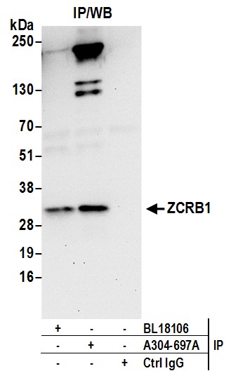
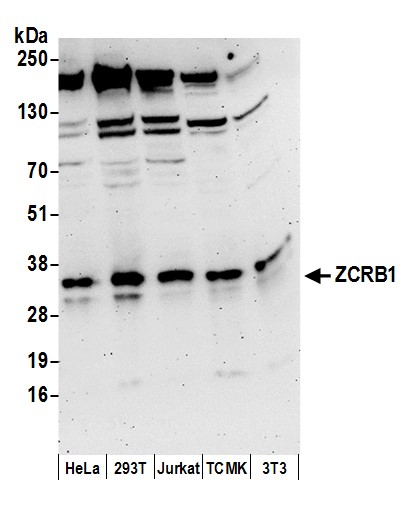
(Detection of human and mouse ZCRB1 by western blot. Samples: Whole cell lysate (50 ug) from HeLa, HEK293T, Jurkat, mouse TCMK-1, and mouse NIH 3T3 cells prepared using NETN lysis buffer. Antibodies: Affinity purified rabbit anti-ZCRB1 antibody AAA212868 (lot AAA212868-1) used for WB at 0.1 ug/ml. Detection: Chemiluminescence with an exposure time of 3 minutes.)
WB (Western Blot)
(Detection of human and mouse ZCRB1 by western blot. Samples: Whole cell lysate (50 ug) from HeLa, HEK293T, Jurkat, mouse TCMK-1, and mouse NIH 3T3 cells prepared using NETN lysis buffer. Antibodies: Affinity purified rabbit anti-ZCRB1 antibody AAA212868 (lot AAA212868-1) used for WB at 0.1 ug/ml. Detection: Chemiluminescence with an exposure time of 3 minutes.)
ZCRB1, Polyclonal Antibody (Cat# AAA212868)
 WB (Western Blot)
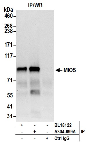
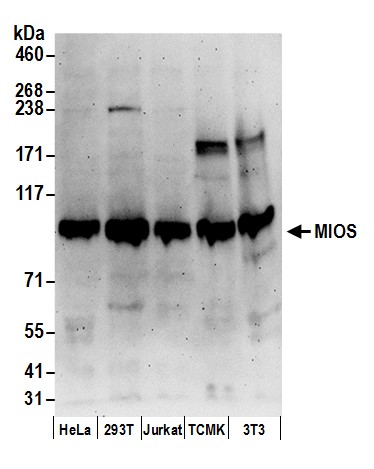
(Detection of human and mouse MIOS by western blot. Samples: Whole cell lysate (50 ug) from HeLa, HEK293T, Jurkat, mouse TCMK-1, and mouse NIH 3T3 cells prepared using NETN lysis buffer. Antibodies: Affinity purified rabbit anti-MIOS antibody AAA212870 (lot AAA212870-1) used for WB at 0.1 ug/ml. Detection: Chemiluminescence with an exposure time of 3 minutes.)
WB (Western Blot)
(Detection of human and mouse MIOS by western blot. Samples: Whole cell lysate (50 ug) from HeLa, HEK293T, Jurkat, mouse TCMK-1, and mouse NIH 3T3 cells prepared using NETN lysis buffer. Antibodies: Affinity purified rabbit anti-MIOS antibody AAA212870 (lot AAA212870-1) used for WB at 0.1 ug/ml. Detection: Chemiluminescence with an exposure time of 3 minutes.)
MIOS, Polyclonal Antibody (Cat# AAA212870)
 WB (Western Blot)
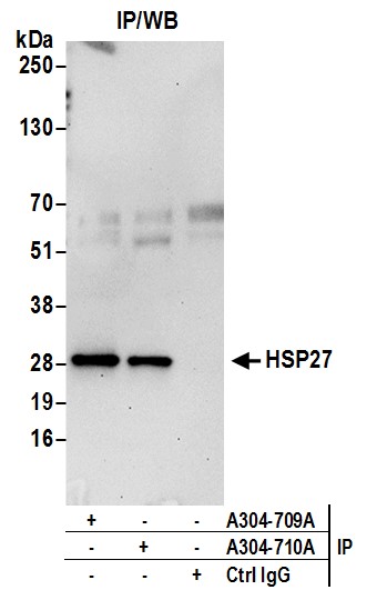
(Detection of human HSP27 by western blot. Samples: Whole cell lysate (50 ug) from HeLa and 293T cells prepared using NETN lysis buffer. Antibodies: Affinity purified rabbit anti-HSP27 antibody AAA212876 (lot AAA212876-1) used for WB at 0.4 ug/ml. Detection: Chemiluminescence with an exposure time of 10 seconds.)
WB (Western Blot)
(Detection of human HSP27 by western blot. Samples: Whole cell lysate (50 ug) from HeLa and 293T cells prepared using NETN lysis buffer. Antibodies: Affinity purified rabbit anti-HSP27 antibody AAA212876 (lot AAA212876-1) used for WB at 0.4 ug/ml. Detection: Chemiluminescence with an exposure time of 10 seconds.)
HSP27, Polyclonal Antibody (Cat# AAA212876)
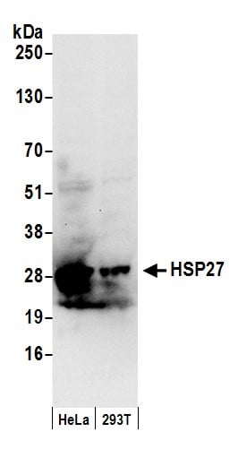 WB (Western Blot)
(Detection of human HSP27 by western blot. Samples: Whole cell lysate (50 ug) from HeLa and 293T cells prepared using NETN lysis buffer. Antibodies: Affinity purified rabbit anti-HSP27 antibody AAA212877 (lot AAA212877-1) used for WB at 0.1 ug/ml. Detection: Chemiluminescence with an exposure time of 10 seconds.)
WB (Western Blot)
(Detection of human HSP27 by western blot. Samples: Whole cell lysate (50 ug) from HeLa and 293T cells prepared using NETN lysis buffer. Antibodies: Affinity purified rabbit anti-HSP27 antibody AAA212877 (lot AAA212877-1) used for WB at 0.1 ug/ml. Detection: Chemiluminescence with an exposure time of 10 seconds.)
HSP27, Polyclonal Antibody (Cat# AAA212877)
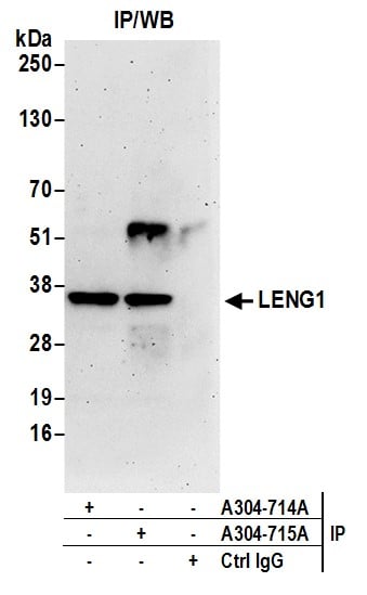
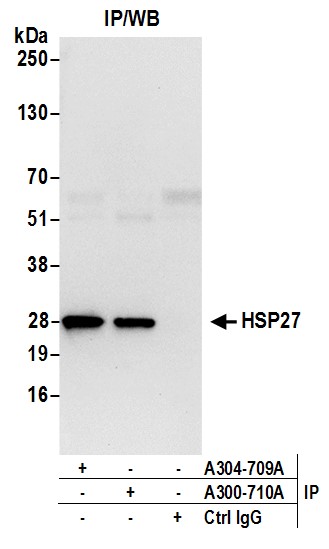
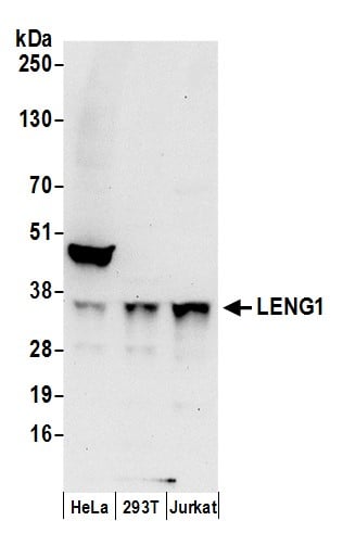 WB (Western Blot)
(Detection of human LENG1 by western blot. Samples: Whole cell lysate (50 ug) from HeLa, HEK293T, and Jurkat cells prepared using NETN lysis buffer. Antibodies: Affinity purified rabbit anti-LENG1 antibody AAA212880 (lot AAA212880-1) used for WB at 1 ug/ml. Detection: Chemiluminescence with an exposure time of 30 seconds.)
WB (Western Blot)
(Detection of human LENG1 by western blot. Samples: Whole cell lysate (50 ug) from HeLa, HEK293T, and Jurkat cells prepared using NETN lysis buffer. Antibodies: Affinity purified rabbit anti-LENG1 antibody AAA212880 (lot AAA212880-1) used for WB at 1 ug/ml. Detection: Chemiluminescence with an exposure time of 30 seconds.)
LENG1, Polyclonal Antibody (Cat# AAA212880)
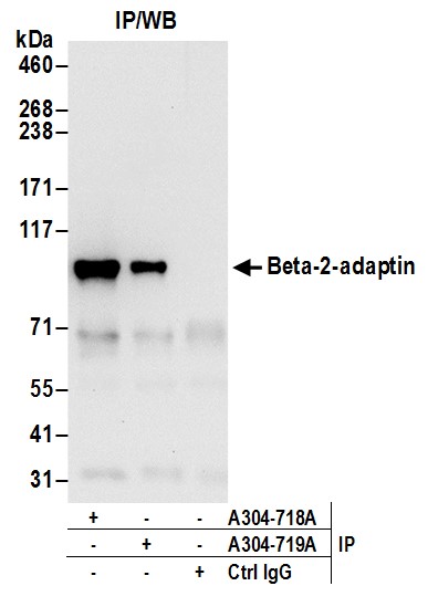
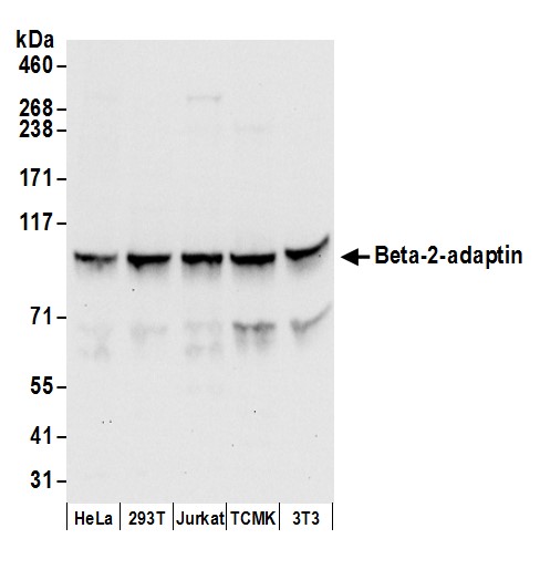 WB (Western Blot)
(Detection of human and mouse Beta-2-adaptin by western blot. Samples: Whole cell lysate (50 ug) from HeLa, HEK293T, Jurkat, mouse TCMK-1, and mouse NIH 3T3 cells prepared using NETN lysis buffer. Antibodies: Affinity purified rabbit anti-Beta-2-adaptin antibody AAA212882 (lot AAA212882-1) used for WB at 0.1 ug/ml. Detection: Chemiluminescence with an exposure time of 10 seconds.)
WB (Western Blot)
(Detection of human and mouse Beta-2-adaptin by western blot. Samples: Whole cell lysate (50 ug) from HeLa, HEK293T, Jurkat, mouse TCMK-1, and mouse NIH 3T3 cells prepared using NETN lysis buffer. Antibodies: Affinity purified rabbit anti-Beta-2-adaptin antibody AAA212882 (lot AAA212882-1) used for WB at 0.1 ug/ml. Detection: Chemiluminescence with an exposure time of 10 seconds.)
Beta-2-adaptin, Polyclonal Antibody (Cat# AAA212882)
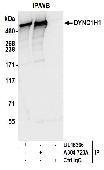
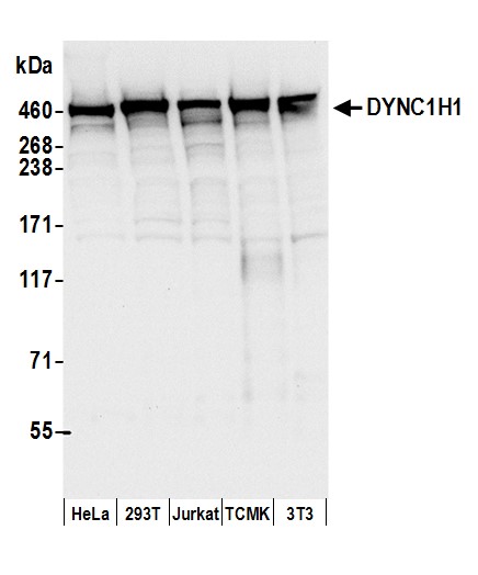 WB (Western Blot)
(Detection of human and mouse DYNC1H1 by western blot. Samples: Whole cell lysate (50 ug) from HeLa, HEK293T, Jurkat, mouse TCMK-1, and mouse NIH 3T3 cells prepared using NETN lysis buffer. Antibodies: Affinity purified rabbit anti-DYNC1H1 antibody AAA212884 (lot AAA212884-1) used for WB at 0.1 ug/ml. Detection: Chemiluminescence with an exposure time of 3 seconds.)
WB (Western Blot)
(Detection of human and mouse DYNC1H1 by western blot. Samples: Whole cell lysate (50 ug) from HeLa, HEK293T, Jurkat, mouse TCMK-1, and mouse NIH 3T3 cells prepared using NETN lysis buffer. Antibodies: Affinity purified rabbit anti-DYNC1H1 antibody AAA212884 (lot AAA212884-1) used for WB at 0.1 ug/ml. Detection: Chemiluminescence with an exposure time of 3 seconds.)
DYNC1H1, Polyclonal Antibody (Cat# AAA212884)
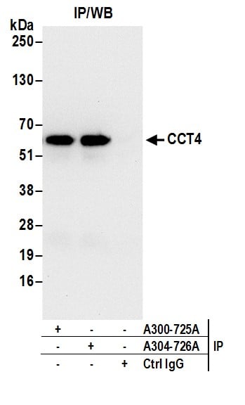
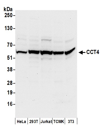 WB (Western Blot)
(Detection of human and mouse CCT4 by western blot. Samples: Whole cell lysate (50 ug) from HeLa, HEK293T, Jurkat, mouse TCMK-1, and mouse NIH 3T3 cells prepared using NETN lysis buffer. Antibodies: Affinity purified rabbit anti-CCT4 antibody AAA212889 (lot AAA212889-1) used for WB at 0.4 ug/ml. Detection: Chemiluminescence with an exposure time of 10 seconds.)
WB (Western Blot)
(Detection of human and mouse CCT4 by western blot. Samples: Whole cell lysate (50 ug) from HeLa, HEK293T, Jurkat, mouse TCMK-1, and mouse NIH 3T3 cells prepared using NETN lysis buffer. Antibodies: Affinity purified rabbit anti-CCT4 antibody AAA212889 (lot AAA212889-1) used for WB at 0.4 ug/ml. Detection: Chemiluminescence with an exposure time of 10 seconds.)
CCT4, Polyclonal Antibody (Cat# AAA212889)
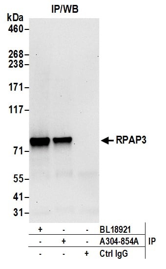
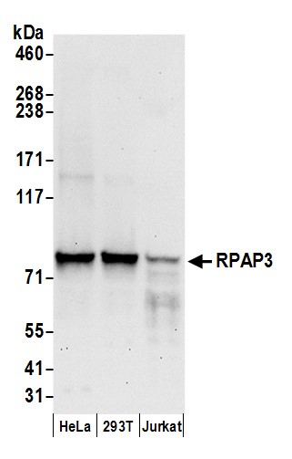 WB (Western Blot)
(Detection of human RPAP3 by western blot. Samples: Whole cell lysate (50 ug) from HeLa, HEK293T, and Jurkat cells prepared using NETN lysis buffer. Antibody: Affinity purified rabbit anti-RPAP3 antibody AAA212978 (lot AAA212978-1) used for WB at 0.1 ug/ml. Detection: Chemiluminescence with an exposure time of 30 seconds.)
WB (Western Blot)
(Detection of human RPAP3 by western blot. Samples: Whole cell lysate (50 ug) from HeLa, HEK293T, and Jurkat cells prepared using NETN lysis buffer. Antibody: Affinity purified rabbit anti-RPAP3 antibody AAA212978 (lot AAA212978-1) used for WB at 0.1 ug/ml. Detection: Chemiluminescence with an exposure time of 30 seconds.)
RPAP3, Polyclonal Antibody (Cat# AAA212978)
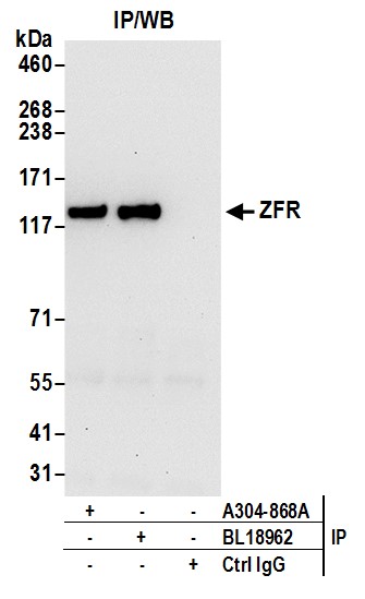
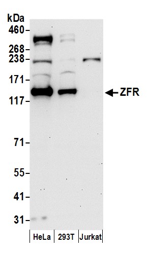 WB (Western Blot)
(Detection of human ZFR by western blot. Samples: Whole cell lysate (50 ug) from HeLa, HEK293T, and Jurkat cells prepared using NETN lysis buffer. Antibody: Affinity purified rabbit anti-ZFR antibody AAA212985 (lot AAA212985-1) used for WB at 0.4 ug/ml. Detection: Chemiluminescence with an exposure time of 30 seconds.)
WB (Western Blot)
(Detection of human ZFR by western blot. Samples: Whole cell lysate (50 ug) from HeLa, HEK293T, and Jurkat cells prepared using NETN lysis buffer. Antibody: Affinity purified rabbit anti-ZFR antibody AAA212985 (lot AAA212985-1) used for WB at 0.4 ug/ml. Detection: Chemiluminescence with an exposure time of 30 seconds.)
ZFR, Polyclonal Antibody (Cat# AAA212985)
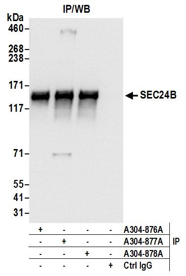
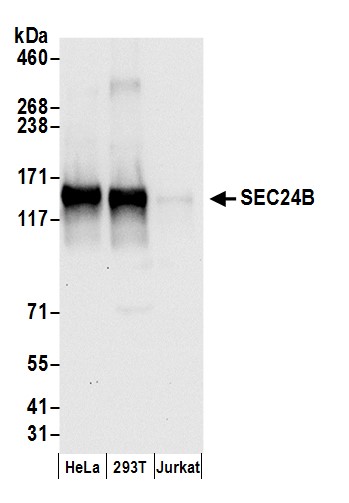 WB (Western Blot)
(Detection of human SEC24B by western blot. Samples: Whole cell lysate (50 ug) from HeLa, HEK293T, and Jurkat cells prepared using NETN lysis buffer. Antibody: Affinity purified rabbit anti-SEC24B antibody AAA212990 (lot AAA212990-1) used for WB at 0.1 ug/ml. Detection: Chemiluminescence with an exposure time of 10 seconds.)
WB (Western Blot)
(Detection of human SEC24B by western blot. Samples: Whole cell lysate (50 ug) from HeLa, HEK293T, and Jurkat cells prepared using NETN lysis buffer. Antibody: Affinity purified rabbit anti-SEC24B antibody AAA212990 (lot AAA212990-1) used for WB at 0.1 ug/ml. Detection: Chemiluminescence with an exposure time of 10 seconds.)
SEC24B, Polyclonal Antibody (Cat# AAA212990)
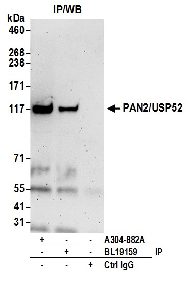
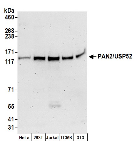 WB (Western Blot)
(Detection of human and mouse PAN2/USP52 by western blot. Samples: Whole cell lysate (50 ug) from HeLa, HEK293T, Jurkat, mouse TCMK-1, and mouse NIH 3T3 cells prepared using NETN lysis buffer. Antibody: Affinity purified rabbit anti-PAN2/USP52 antibody AAA212991 (lot AAA212991-1) used for WB at 0.1 ug/ml. Detection: Chemiluminescence with an exposure time of 3 minutes.)
WB (Western Blot)
(Detection of human and mouse PAN2/USP52 by western blot. Samples: Whole cell lysate (50 ug) from HeLa, HEK293T, Jurkat, mouse TCMK-1, and mouse NIH 3T3 cells prepared using NETN lysis buffer. Antibody: Affinity purified rabbit anti-PAN2/USP52 antibody AAA212991 (lot AAA212991-1) used for WB at 0.1 ug/ml. Detection: Chemiluminescence with an exposure time of 3 minutes.)
PAN2/USP52, Polyclonal Antibody (Cat# AAA212991)
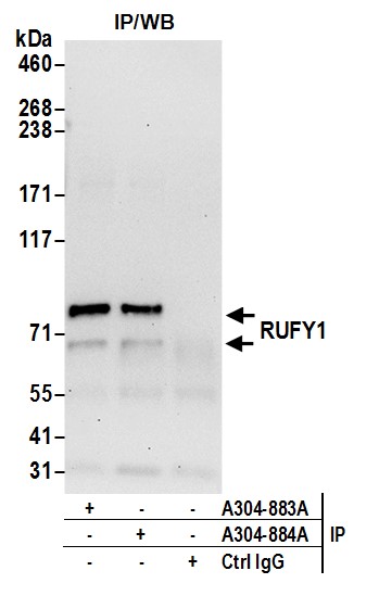
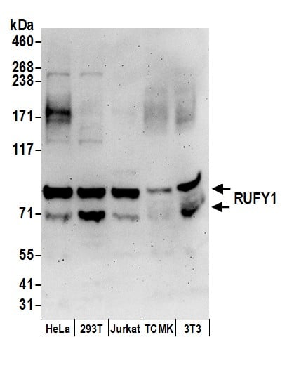 WB (Western Blot)
(Detection of human and mouse RUFY1 by western blot. Samples: Whole cell lysate (50 ug) from HeLa, HEK293T, Jurkat, mouse TCMK-1, and mouse NIH 3T3 cells prepared using NETN lysis buffer. Antibody: Affinity purified rabbit anti-RUFY1 antibody AAA212992 (lot AAA212992-1) used for WB at 0.1 ug/ml. Detection: Chemiluminescence with an exposure time of 3 minutes.)
WB (Western Blot)
(Detection of human and mouse RUFY1 by western blot. Samples: Whole cell lysate (50 ug) from HeLa, HEK293T, Jurkat, mouse TCMK-1, and mouse NIH 3T3 cells prepared using NETN lysis buffer. Antibody: Affinity purified rabbit anti-RUFY1 antibody AAA212992 (lot AAA212992-1) used for WB at 0.1 ug/ml. Detection: Chemiluminescence with an exposure time of 3 minutes.)
RUFY1, Polyclonal Antibody (Cat# AAA212992)
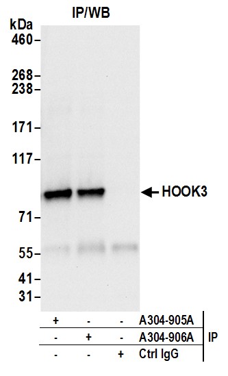
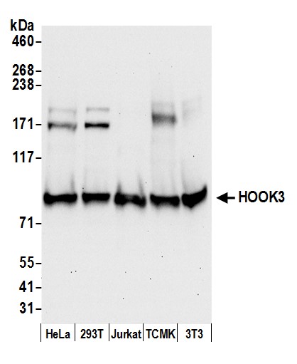 WB (Western Blot)
(Detection of human and mouse HOOK3 by western blot. Samples: Whole cell lysate (50 ug) from HeLa, HEK293T, Jurkat, mouse TCMK-1, and mouse NIH 3T3 cells prepared using NETN lysis buffer. Antibody: Affinity purified rabbit anti-HOOK3 antibody AAA213002 (lot AAA213002-1) used for WB at 0.1 ug/ml. Detection: Chemiluminescence with an exposure time of 30 seconds.)
WB (Western Blot)
(Detection of human and mouse HOOK3 by western blot. Samples: Whole cell lysate (50 ug) from HeLa, HEK293T, Jurkat, mouse TCMK-1, and mouse NIH 3T3 cells prepared using NETN lysis buffer. Antibody: Affinity purified rabbit anti-HOOK3 antibody AAA213002 (lot AAA213002-1) used for WB at 0.1 ug/ml. Detection: Chemiluminescence with an exposure time of 30 seconds.)
HOOK3, Polyclonal Antibody (Cat# AAA213002)
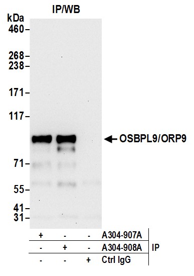
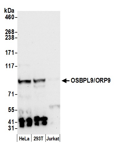 WB (Western Blot)
(Detection of human OSBPL9/ORP9 by western blot. Samples: Whole cell lysate (50 ug) from HeLa, HEK293T, and Jurkat cells prepared using NETN lysis buffer. Antibody: Affinity purified rabbit anti-OSBPL9/ORP9 antibody AAA213004 (lot AAA213004-1) used for WB at 0.4 ug/ml. Detection: Chemiluminescence with an exposure time of 30 seconds.)
WB (Western Blot)
(Detection of human OSBPL9/ORP9 by western blot. Samples: Whole cell lysate (50 ug) from HeLa, HEK293T, and Jurkat cells prepared using NETN lysis buffer. Antibody: Affinity purified rabbit anti-OSBPL9/ORP9 antibody AAA213004 (lot AAA213004-1) used for WB at 0.4 ug/ml. Detection: Chemiluminescence with an exposure time of 30 seconds.)
OSBPL9/ORP9, Polyclonal Antibody (Cat# AAA213004)
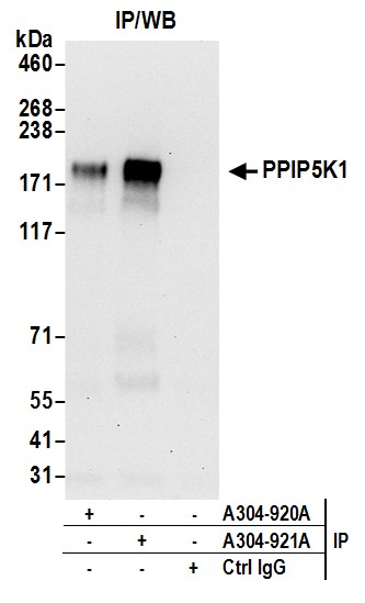
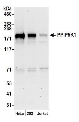 WB (Western Blot)
(Detection of human PPIP5K1 by western blot. Samples: Whole cell lysate (50 ug) from HeLa, HEK293T, and Jurkat cells prepared using NETN lysis buffer. Antibody: Affinity purified rabbit anti-PPIP5K1 antibody AAA213010 (lot AAA213010-1) used for WB at 0.1 ug/ml. Detection: Chemiluminescence with an exposure time of 30 seconds.)
WB (Western Blot)
(Detection of human PPIP5K1 by western blot. Samples: Whole cell lysate (50 ug) from HeLa, HEK293T, and Jurkat cells prepared using NETN lysis buffer. Antibody: Affinity purified rabbit anti-PPIP5K1 antibody AAA213010 (lot AAA213010-1) used for WB at 0.1 ug/ml. Detection: Chemiluminescence with an exposure time of 30 seconds.)
PPIP5K1/HISPPD2A, Polyclonal Antibody (Cat# AAA213010)
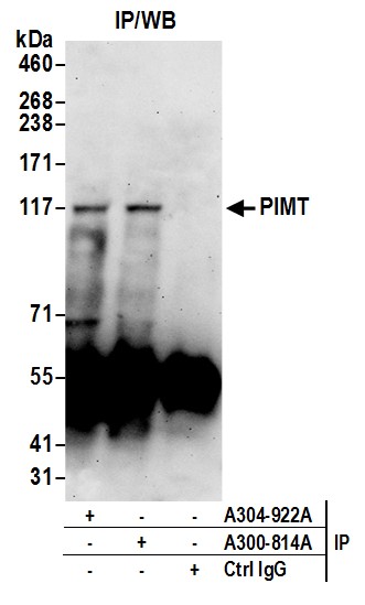
 WB (Western Blot)
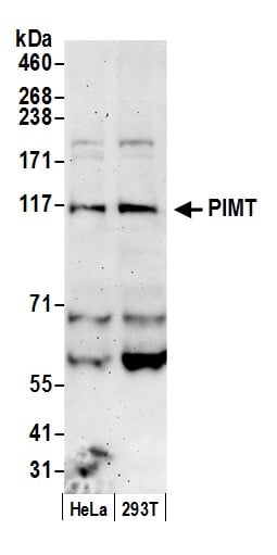
(Detection of human PIMT by western blot. Samples: Whole cell lysate (50 ug) from HeLa and 293T cells prepared using NETN lysis buffer. Antibody: Affinity purified rabbit anti-PIMT antibody AAA213011 (lot AAA213011-1) used for WB at 0.4 ug/ml. Detection: Chemiluminescence with an exposure time of 3 minutes.)
WB (Western Blot)
(Detection of human PIMT by western blot. Samples: Whole cell lysate (50 ug) from HeLa and 293T cells prepared using NETN lysis buffer. Antibody: Affinity purified rabbit anti-PIMT antibody AAA213011 (lot AAA213011-1) used for WB at 0.4 ug/ml. Detection: Chemiluminescence with an exposure time of 3 minutes.)
PIMT, Polyclonal Antibody (Cat# AAA213011)
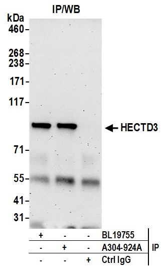
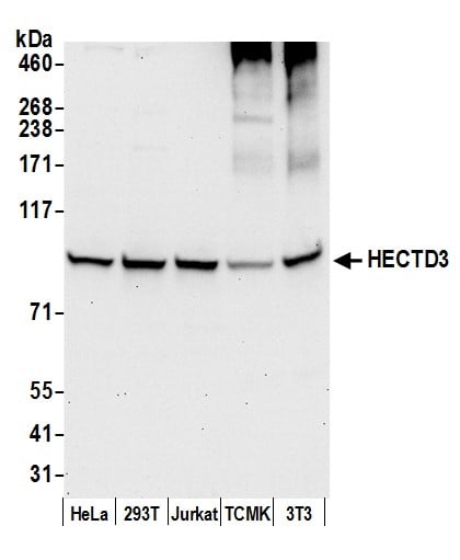 WB (Western Blot)
(Detection of human and mouse HECTD3 by western blot. Samples: Whole cell lysate (50 ug) from HeLa, HEK293T, Jurkat, mouse TCMK-1, and mouse NIH 3T3 cells prepared using NETN lysis buffer. Antibody: Affinity purified rabbit anti-HECTD3 antibody AAA213012 (lot AAA213012-1) used for WB at 1 ug/ml. Detection: Chemiluminescence with an exposure time of 30 seconds.)
WB (Western Blot)
(Detection of human and mouse HECTD3 by western blot. Samples: Whole cell lysate (50 ug) from HeLa, HEK293T, Jurkat, mouse TCMK-1, and mouse NIH 3T3 cells prepared using NETN lysis buffer. Antibody: Affinity purified rabbit anti-HECTD3 antibody AAA213012 (lot AAA213012-1) used for WB at 1 ug/ml. Detection: Chemiluminescence with an exposure time of 30 seconds.)
HECTD3, Polyclonal Antibody (Cat# AAA213012)
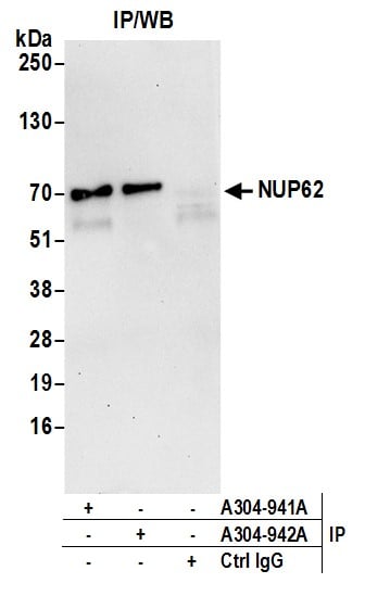
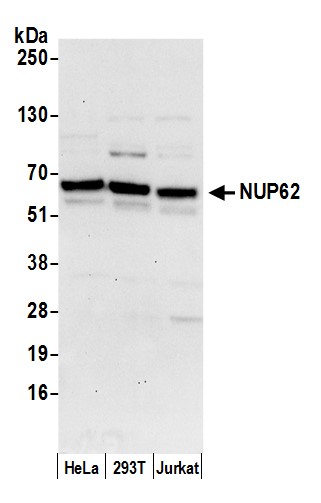 WB (Western Blot)
(Detection of human NUP62 by western blot. Samples: Whole cell lysate (50 ug) from HeLa, HEK293T, and Jurkat cells prepared using NETN lysis buffer. Antibody: Affinity purified rabbit anti-NUP62 antibody AAA213019 (lot AAA213019-1) used for WB at 0.1 ug/ml. Detection: Chemiluminescence with an exposure time of 30 seconds.)
WB (Western Blot)
(Detection of human NUP62 by western blot. Samples: Whole cell lysate (50 ug) from HeLa, HEK293T, and Jurkat cells prepared using NETN lysis buffer. Antibody: Affinity purified rabbit anti-NUP62 antibody AAA213019 (lot AAA213019-1) used for WB at 0.1 ug/ml. Detection: Chemiluminescence with an exposure time of 30 seconds.)
Nucleoporin p62/NUP62, Polyclonal Antibody (Cat# AAA213019)
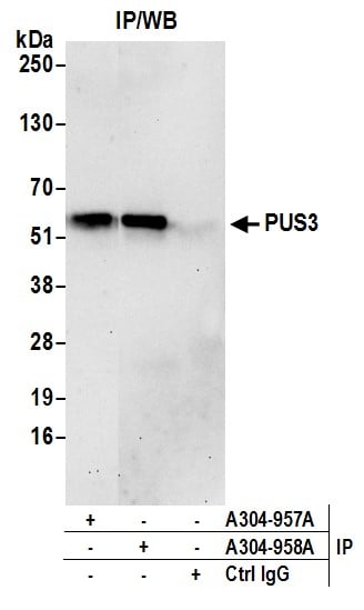
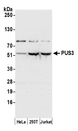 WB (Western Blot)
(Detection of human PUS3 by western blot. Samples: Whole cell lysate (50 ug) from HeLa, HEK293T, and Jurkat cells prepared using NETN lysis buffer. Antibody: Affinity purified rabbit anti-PUS3 antibody AAA213026 (lot AAA213026-1) used for WB at 0.1 ug/ml. Detection: Chemiluminescence with an exposure time of 30 seconds.)
WB (Western Blot)
(Detection of human PUS3 by western blot. Samples: Whole cell lysate (50 ug) from HeLa, HEK293T, and Jurkat cells prepared using NETN lysis buffer. Antibody: Affinity purified rabbit anti-PUS3 antibody AAA213026 (lot AAA213026-1) used for WB at 0.1 ug/ml. Detection: Chemiluminescence with an exposure time of 30 seconds.)
PUS3, Polyclonal Antibody (Cat# AAA213026)
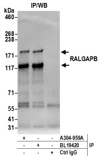
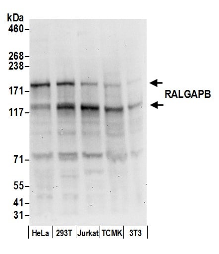 WB (Western Blot)
(Detection of human and mouse RALGAPB by western blot. Samples: Whole cell lysate (50 ug) from HeLa, HEK293T, Jurkat, mouse TCMK-1, and mouse NIH 3T3 cells prepared using NETN lysis buffer. Antibody: Affinity purified rabbit anti-RALGAPB antibody AAA213027 (lot AAA213027-1) used for WB at 0.1 ug/ml. Detection: Chemiluminescence with an exposure time of 30 seconds.)
WB (Western Blot)
(Detection of human and mouse RALGAPB by western blot. Samples: Whole cell lysate (50 ug) from HeLa, HEK293T, Jurkat, mouse TCMK-1, and mouse NIH 3T3 cells prepared using NETN lysis buffer. Antibody: Affinity purified rabbit anti-RALGAPB antibody AAA213027 (lot AAA213027-1) used for WB at 0.1 ug/ml. Detection: Chemiluminescence with an exposure time of 30 seconds.)
RALGAPB, Polyclonal Antibody (Cat# AAA213027)
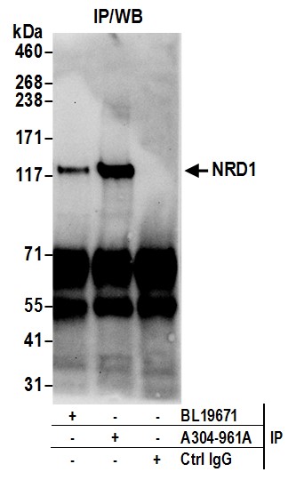
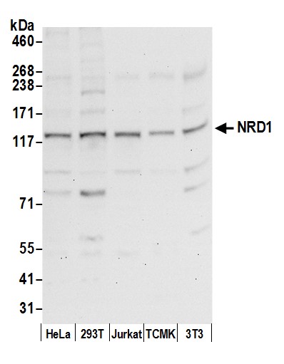 WB (Western Blot)
(Detection of human and mouse NRD1 by western blot. Samples: Whole cell lysate (50 ug) from HeLa, HEK293T, Jurkat, mouse TCMK-1, and mouse NIH 3T3 cells prepared using NETN lysis buffer. Antibody: Affinity purified rabbit anti-NRD1 antibody AAA213029 (lot AAA213029-1) used for WB at 0.1 ug/ml. Detection: Chemiluminescence with an exposure time of 30 seconds.)
WB (Western Blot)
(Detection of human and mouse NRD1 by western blot. Samples: Whole cell lysate (50 ug) from HeLa, HEK293T, Jurkat, mouse TCMK-1, and mouse NIH 3T3 cells prepared using NETN lysis buffer. Antibody: Affinity purified rabbit anti-NRD1 antibody AAA213029 (lot AAA213029-1) used for WB at 0.1 ug/ml. Detection: Chemiluminescence with an exposure time of 30 seconds.)
NRD1/Nardilysin, Polyclonal Antibody (Cat# AAA213029)
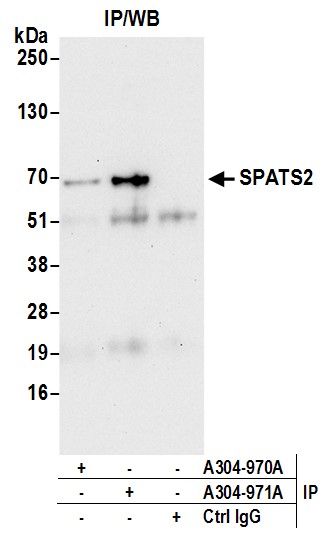
 WB (Western Blot)
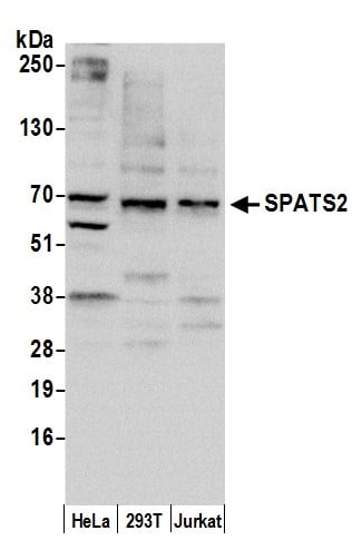
(Detection of human SPATS2 by western blot. Samples: Whole cell lysate (10 ug) from HeLa, HEK293T, and Jurkat cells prepared using NETN lysis buffer. Antibody: Affinity purified rabbit anti-SPATS2 antibody AAA213035 (lot AAA213035-1) used for WB at 0.4 ug/ml. Detection: Chemiluminescence with an exposure time of 30 seconds.)
WB (Western Blot)
(Detection of human SPATS2 by western blot. Samples: Whole cell lysate (10 ug) from HeLa, HEK293T, and Jurkat cells prepared using NETN lysis buffer. Antibody: Affinity purified rabbit anti-SPATS2 antibody AAA213035 (lot AAA213035-1) used for WB at 0.4 ug/ml. Detection: Chemiluminescence with an exposure time of 30 seconds.)
SPATS2/SPATA10, Polyclonal Antibody (Cat# AAA213035)
 WB (Western Blot)
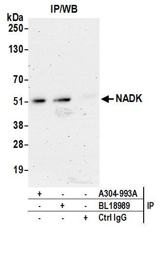
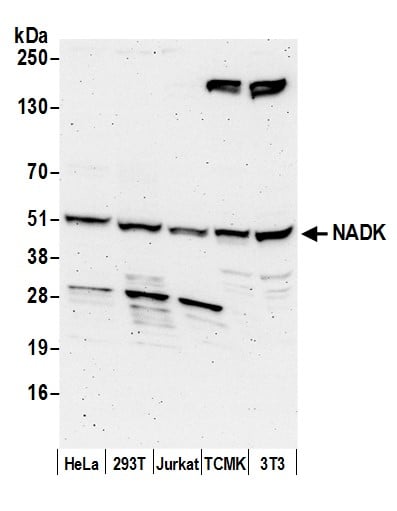
(Detection of human and mouse NADK by western blot. Samples: Whole cell lysate (50 ug) from HeLa, HEK293T, Jurkat, mouse TCMK-1, and mouse NIH 3T3 cells prepared using NETN lysis buffer. Antibody: Affinity purified rabbit anti-NADK antibody AAA213043 (lot AAA213043-1) used for WB at 1 ug/ml. Detection: Chemiluminescence with an exposure time of 3 minutes.)
WB (Western Blot)
(Detection of human and mouse NADK by western blot. Samples: Whole cell lysate (50 ug) from HeLa, HEK293T, Jurkat, mouse TCMK-1, and mouse NIH 3T3 cells prepared using NETN lysis buffer. Antibody: Affinity purified rabbit anti-NADK antibody AAA213043 (lot AAA213043-1) used for WB at 1 ug/ml. Detection: Chemiluminescence with an exposure time of 3 minutes.)
NADK, Polyclonal Antibody (Cat# AAA213043)
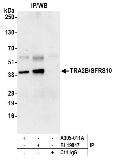
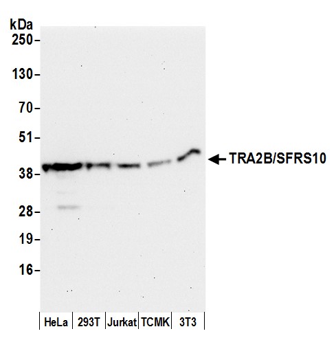 WB (Western Blot)
(Detection of human and mouse TRA2B/SFRS10 by western blot. Samples: Whole cell lysate (50 ug) from HeLa, HEK293T, Jurkat, mouse TCMK-1, and mouse NIH 3T3 cells prepared using NETN lysis buffer. Antibody: Affinity purified rabbit anti-TRA2B/SFRS10 antibody AAA213056 (lot AAA213056-1) used for WB at 0.1 ug/ml. Detection: Chemiluminescence with an exposure time of 30 seconds.)
WB (Western Blot)
(Detection of human and mouse TRA2B/SFRS10 by western blot. Samples: Whole cell lysate (50 ug) from HeLa, HEK293T, Jurkat, mouse TCMK-1, and mouse NIH 3T3 cells prepared using NETN lysis buffer. Antibody: Affinity purified rabbit anti-TRA2B/SFRS10 antibody AAA213056 (lot AAA213056-1) used for WB at 0.1 ug/ml. Detection: Chemiluminescence with an exposure time of 30 seconds.)
TRA2B/SFRS10, Polyclonal Antibody (Cat# AAA213056)
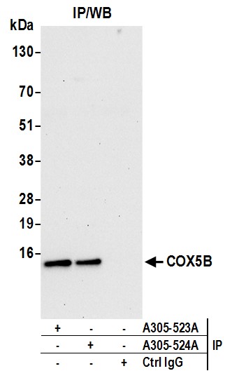
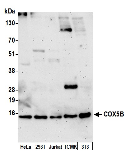 WB (Western Blot)
(Detection of human and mouse COX5B by western blot. Samples: Whole cell lysate (15 ug) from HeLa, HEK293T, Jurkat, mouse TCMK-1, and mouse NIH 3T3 cells prepared using NETN lysis buffer. Antibody: Affinity purified rabbit anti-COX5B antibody AAA213302 (lot AAA213302-1) used for WB at 0.1 ug/ml. Detection: Chemiluminescence with an exposure time of 3 minutes.)
WB (Western Blot)
(Detection of human and mouse COX5B by western blot. Samples: Whole cell lysate (15 ug) from HeLa, HEK293T, Jurkat, mouse TCMK-1, and mouse NIH 3T3 cells prepared using NETN lysis buffer. Antibody: Affinity purified rabbit anti-COX5B antibody AAA213302 (lot AAA213302-1) used for WB at 0.1 ug/ml. Detection: Chemiluminescence with an exposure time of 3 minutes.)
COX5B, Polyclonal Antibody (Cat# AAA213302)
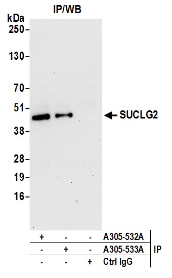
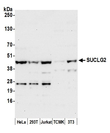 WB (Western Blot)
(Detection of human and mouse SUCLG2 by western blot. Samples: Whole cell lysate (50 ug) from HeLa, HEK293T, Jurkat, mouse TCMK-1, and mouse NIH 3T3 cells prepared using NETN lysis buffer. Antibody: Affinity purified rabbit anti-SUCLG2 antibody AAA213306 (lot AAA213306-1) used for WB at 0.4 ug/ml. Detection: Chemiluminescence with an exposure time of 30 seconds.)
WB (Western Blot)
(Detection of human and mouse SUCLG2 by western blot. Samples: Whole cell lysate (50 ug) from HeLa, HEK293T, Jurkat, mouse TCMK-1, and mouse NIH 3T3 cells prepared using NETN lysis buffer. Antibody: Affinity purified rabbit anti-SUCLG2 antibody AAA213306 (lot AAA213306-1) used for WB at 0.4 ug/ml. Detection: Chemiluminescence with an exposure time of 30 seconds.)
SUCLG2, Polyclonal Antibody (Cat# AAA213306)
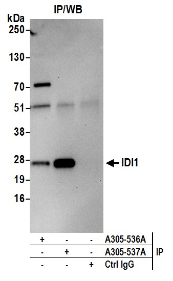
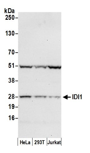 WB (Western Blot)
(Detection of human IDI1 by western blot. Samples: Whole cell lysate (50 ug) from HeLa, HEK293T, and Jurkat cells prepared using NETN lysis buffer. Antibody: Affinity purified rabbit anti-IDI1 antibody AAA213309 (lot AAA213309-1) used for WB at 0.1 ug/ml. Detection: Chemiluminescence with an exposure time of 30 seconds.)
WB (Western Blot)
(Detection of human IDI1 by western blot. Samples: Whole cell lysate (50 ug) from HeLa, HEK293T, and Jurkat cells prepared using NETN lysis buffer. Antibody: Affinity purified rabbit anti-IDI1 antibody AAA213309 (lot AAA213309-1) used for WB at 0.1 ug/ml. Detection: Chemiluminescence with an exposure time of 30 seconds.)
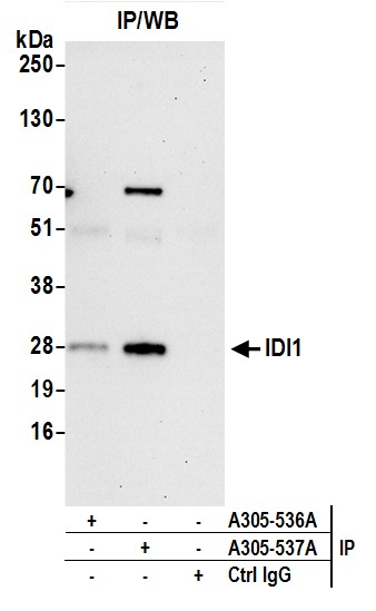
IDI1, Polyclonal Antibody (Cat# AAA213309)
 WB (Western Blot)
(Detection of human IDI1 by western blot. Samples: Whole cell lysate (50 ug) from HeLa, HEK293T, and Jurkat cells prepared using NETN lysis buffer. Antibody: Affinity purified rabbit anti-IDI1 antibody AAA213310 (lot AAA213310-1) used for WB at 0.4 ug/ml. Detection: Chemiluminescence with an exposure time of 30 seconds.)
WB (Western Blot)
(Detection of human IDI1 by western blot. Samples: Whole cell lysate (50 ug) from HeLa, HEK293T, and Jurkat cells prepared using NETN lysis buffer. Antibody: Affinity purified rabbit anti-IDI1 antibody AAA213310 (lot AAA213310-1) used for WB at 0.4 ug/ml. Detection: Chemiluminescence with an exposure time of 30 seconds.)
IDI1, Polyclonal Antibody (Cat# AAA213310)
 WB (Western Blot)
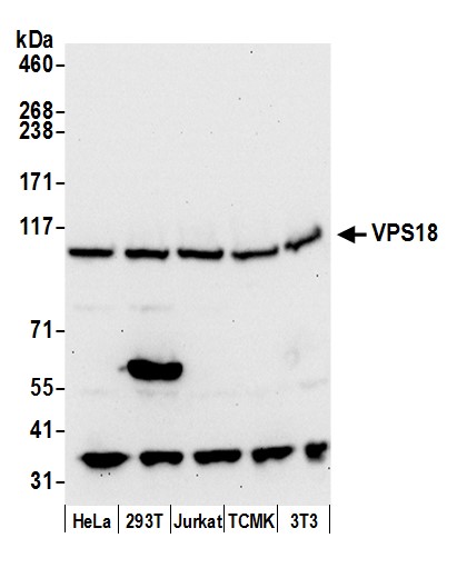
(Detection of human and mouse VPS18 by western blot. Samples: Whole cell lysate (50 ug) from HeLa, HEK293T, Jurkat, mouse TCMK-1, and mouse NIH 3T3 cells prepared using NETN lysis buffer. Antibody: Affinity purified rabbit anti-VPS18 antibody AAA213314 (lot AAA213314-1) used for WB at 0.1 ug/ml. Detection: Chemiluminescence with an exposure time of 30 seconds.)
WB (Western Blot)
(Detection of human and mouse VPS18 by western blot. Samples: Whole cell lysate (50 ug) from HeLa, HEK293T, Jurkat, mouse TCMK-1, and mouse NIH 3T3 cells prepared using NETN lysis buffer. Antibody: Affinity purified rabbit anti-VPS18 antibody AAA213314 (lot AAA213314-1) used for WB at 0.1 ug/ml. Detection: Chemiluminescence with an exposure time of 30 seconds.)
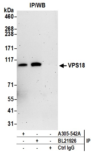
VPS18, Polyclonal Antibody (Cat# AAA213314)
 WB (Western Blot)
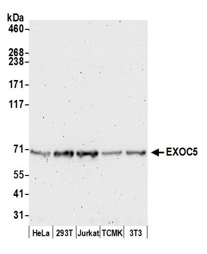
(Detection of human and mouse EXOC5 by western blot. Samples: Whole cell lysate (50 ug) from HeLa, HEK293T, Jurkat, mouse TCMK-1, and mouse NIH 3T3 cells prepared using NETN lysis buffer. Antibody: Affinity purified rabbit anti-EXOC5 antibody AAA213318 (lot AAA213318-1) used for WB at 0.1 ug/ml. Detection: Chemiluminescence with an exposure time of 3 minutes.)
WB (Western Blot)
(Detection of human and mouse EXOC5 by western blot. Samples: Whole cell lysate (50 ug) from HeLa, HEK293T, Jurkat, mouse TCMK-1, and mouse NIH 3T3 cells prepared using NETN lysis buffer. Antibody: Affinity purified rabbit anti-EXOC5 antibody AAA213318 (lot AAA213318-1) used for WB at 0.1 ug/ml. Detection: Chemiluminescence with an exposure time of 3 minutes.)
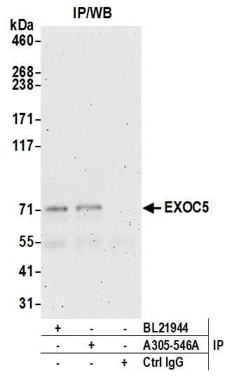
EXOC5, Polyclonal Antibody (Cat# AAA213318)
 WB (Western Blot)
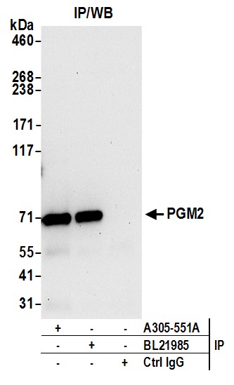
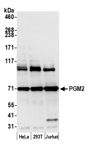
(Detection of human PGM2 by western blot. Samples: Whole cell lysate (15 ug) from HeLa, HEK293T, and Jurkat cells prepared using NETN lysis buffer. Antibody: Affinity purified rabbit anti-PGM2 antibody AAA213322 (lot AAA213322-1) used for WB at 0.1 ug/ml. Detection: Chemiluminescence with an exposure time of 30 seconds.)
WB (Western Blot)
(Detection of human PGM2 by western blot. Samples: Whole cell lysate (15 ug) from HeLa, HEK293T, and Jurkat cells prepared using NETN lysis buffer. Antibody: Affinity purified rabbit anti-PGM2 antibody AAA213322 (lot AAA213322-1) used for WB at 0.1 ug/ml. Detection: Chemiluminescence with an exposure time of 30 seconds.)
PGM2, Polyclonal Antibody (Cat# AAA213322)
What are Polyclonal Antibodies?
Polyclonal antibodies are antibodies that come from multiple B cell clones of a host animal. The typical hosts used for the majority of polyclonal antibody production are rabbits, goats, sheep, and donkeys. These polyclonal antibodies, once having identified their target, will bind to different epitopes located at different regions or sequences on the same protein/antigen. As a result, they are ideal at locating and binding to the target, even if the target is in very low concentrations (due to many different antibodies being able to bind to the same target molecule, which allows for significant amplification of a downstream signal).
Polyclonal antibodies are typically produced by injecting an antigen into a host animal, which causes the animal’s immune system to attack the foreign antigen by mass generating antibodies against it. After a period of time, serum is collected from the animal and purified using physicochemical fractionation, class-specific affinity purification, and/or antigen-affinity purification.
Key Uses of Polyclonal Antibodies
- Western Blotting: This method is used to find specific proteins in biological samples after separating them by size.
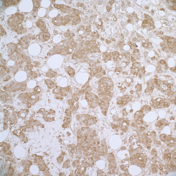
- Immunohistochemistry: IHC helps visualize the location of proteins in tissue sections using various staining techniques.
- ELISA: (Enzyme-Linked Immunosorbent Assay) is typically used to identify specific protein quantities in a sample. ELISAs can be either “Quantitative” or “Qualitative”.
- Flow Cytometry: technique that identifies and measures the specific protein on the surface or inside the cells in a fluid suspension.
- Immunoprecipitation: IP isolates and studies a specific protein from a complex mixture using antibodies.
Why Buy Polyclonal Antibodies from AAA Biotech?
1. Ideal for Various Applications
Our antibodies are generally going to be validated for use in multiple types of assays, including ELISA, Western Blotting, Immunohistochemistry, Immunoprecipitation, amongst others. They are ideal for a wide range of research applications.
2. Rigorous Quality Control
All of the antibodies in our catalog undergo strict quality testing to ensure specificity, sensitivity, and consistent performance. We are confident in the ability of our antibodies to provide you with accurate results.
3. Wide Assortment of Antibodies
Antibodies in are catalog can be found for both common and exotic species, and these antibodies are also available in both conjugated and recombinant forms to suit many diverse experimental needs.
4. Highly Purified
Our antibodies are available in purified forms with over 85% purity, as confirmed by SDS-PAGE. They are also available with tags such as His, Flag, GST, or MBP. We cater to customers worldwide.
FAQ
1. How are polyclonal antibodies produced?
Traditionally, polyclonal antibodies are produced by injecting an antigen into a host animal (such as a rabbit or goat), which then triggers an immune response from the host animal. The animal’s B cells produce antibodies that will recognize different parts of the injected antigen. These antibodies are then collected from the animal’s blood and purified for use.
2. How do polyclonal antibodies differ from monoclonal antibodies?
Polyclonal antibodies are a mix of antibodies that bind to different locations (epitopes) of the same antigen, while monoclonal antibodies are identical and bind to just one specific epitope. This makes polyclonal antibodies more versatile and better at detecting proteins that may be present in low quantities or in altered/modified forms.
3. How should I store polyclonal antibodies?
Polyclonal antibodies should be stored at 4°C for short-term use (up to a few weeks) and at -20°C or -80°C for long-term storage. Avoid repeated freeze-thaw cycles by dividing them into small aliquots. Always check the datasheet for specific storage instructions.