Filters
▼Clonality
▼Type
▼Reactivity
▼Gene Name
▼Isotype
▼Host
▼Application
▼Clone
▼Polyclonal Antibodies
At AAA Biotech also known as AAA Bio or AAABio, we provide a broad range of purified polyclonal antibodies (pAbs) that are able to all be browsed online through our website. Due to their high specificity and strong binding affinity, these antibodies are ideal for wide swathes of research and experimental applications.
Our polyclonal antibodies can easily support your work, whether you use them for Western Blotting, Immunocytochemistry (with or without Immunofluorescence used in conjunction), Immunohistochemistry, Immunoprecipitation, and ELISA tests. We highly encourage you to browse our range of pAbs and choose the one that best suits your experimental model.
Viewing 4950-5000 of 96812 product results
 WB (Western Blot)
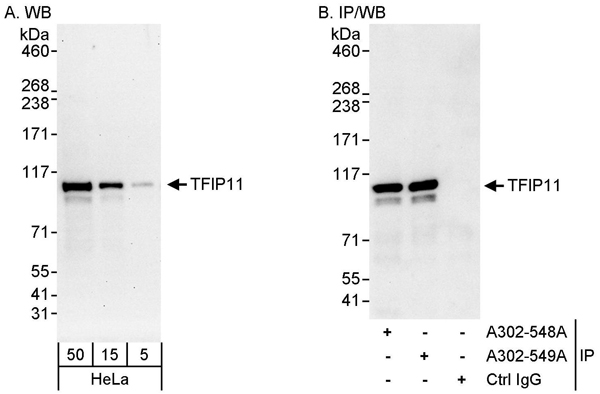
(Detection of human TFIP11 by western blot and immunoprecipitation. Samples: Whole cell lysate (5, 15 and 50 ug for WB; 1 mg for IP, 20% of IP loaded) from HeLa cells. Antibodies: Affinity purified rabbit anti-TFIP11 antibody AAA211919 used for WB at 0.04 ug/ml (A) and 1 ug/ml (B) and used for IP at 3 ug/mg lysate. TFIP11 was also immunoprecipitated by rabbit anti-TFIP11 antibody which recognizes a downstream epitope. Detection: Chemiluminescence with exposure times of 30 seconds (A) and 10 seconds (B).)
WB (Western Blot)
(Detection of human TFIP11 by western blot and immunoprecipitation. Samples: Whole cell lysate (5, 15 and 50 ug for WB; 1 mg for IP, 20% of IP loaded) from HeLa cells. Antibodies: Affinity purified rabbit anti-TFIP11 antibody AAA211919 used for WB at 0.04 ug/ml (A) and 1 ug/ml (B) and used for IP at 3 ug/mg lysate. TFIP11 was also immunoprecipitated by rabbit anti-TFIP11 antibody which recognizes a downstream epitope. Detection: Chemiluminescence with exposure times of 30 seconds (A) and 10 seconds (B).)
TFIP11, Polyclonal Antibody (Cat# AAA211919)
 IP (Immunoprecipitation)
(Detection of human APC7 by western blot of immunoprecipitates. Samples: Whole cell lysate (1 mg for IP, 20% of IP loaded) from HeLa cells. Antibodies: Affinity purified rabbit anti-APC7 antibody AAA211920 used for IP at 3 ug/mg lysate. APC7 was also immunoprecipitated by rabbit anti-APC7 antibody which recognizes a downstream epitope. For blotting immunoprecipitated APC7, was used at 1 ug/ml. Detection: Chemiluminescence with an exposure time of 3 seconds.)
IP (Immunoprecipitation)
(Detection of human APC7 by western blot of immunoprecipitates. Samples: Whole cell lysate (1 mg for IP, 20% of IP loaded) from HeLa cells. Antibodies: Affinity purified rabbit anti-APC7 antibody AAA211920 used for IP at 3 ug/mg lysate. APC7 was also immunoprecipitated by rabbit anti-APC7 antibody which recognizes a downstream epitope. For blotting immunoprecipitated APC7, was used at 1 ug/ml. Detection: Chemiluminescence with an exposure time of 3 seconds.)
APC7, Polyclonal Antibody (Cat# AAA211920)
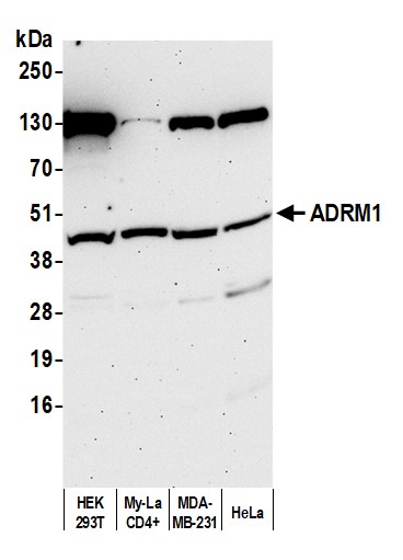 WB (Western Blot)
(Detection of human ADRM1 by western blot. Samples: Whole cell lysate (50 ug) from HEK293T, My-La CD4+, MDA-MB-231, and HeLa cells prepared using NETN lysis buffer. Antibody: Affinity purified rabbit anti-ADRM1 antibody (AAA211924 lot 3) used for WB at 1 ug/ml. Detection: Chemiluminescence with an exposure time of 75 seconds.)
WB (Western Blot)
(Detection of human ADRM1 by western blot. Samples: Whole cell lysate (50 ug) from HEK293T, My-La CD4+, MDA-MB-231, and HeLa cells prepared using NETN lysis buffer. Antibody: Affinity purified rabbit anti-ADRM1 antibody (AAA211924 lot 3) used for WB at 1 ug/ml. Detection: Chemiluminescence with an exposure time of 75 seconds.)
ADRM1, Polyclonal Antibody (Cat# AAA211924)
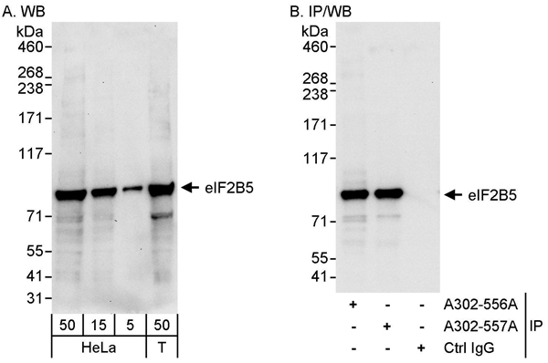 WB (Western Blot)
(Detection of human eIF2B5 by western blot and immunoprecipitation. Samples: Whole cell lysate from HeLa (5, 15 and 50 ug for WB; 1 mg for IP, 20% of IP loaded) and HEK293T (T; 50 ug) cells. Antibodies: Affinity purified rabbit anti-eIF2B5 antibody AAA211925 used for WB at 0.1 ug/ml (A) and 1 ug/ml (B) and used for IP at 10 ug/mg lysate. eIF2B5 was also immunoprecipitated by rabbit anti-eIF2B5 antibody which recognizes a downstream epitope. Detection: Chemiluminescence with exposure times of 30 seconds (A) and 10 seconds (B).)
WB (Western Blot)
(Detection of human eIF2B5 by western blot and immunoprecipitation. Samples: Whole cell lysate from HeLa (5, 15 and 50 ug for WB; 1 mg for IP, 20% of IP loaded) and HEK293T (T; 50 ug) cells. Antibodies: Affinity purified rabbit anti-eIF2B5 antibody AAA211925 used for WB at 0.1 ug/ml (A) and 1 ug/ml (B) and used for IP at 10 ug/mg lysate. eIF2B5 was also immunoprecipitated by rabbit anti-eIF2B5 antibody which recognizes a downstream epitope. Detection: Chemiluminescence with exposure times of 30 seconds (A) and 10 seconds (B).)
eIF2B5, Polyclonal Antibody (Cat# AAA211925)
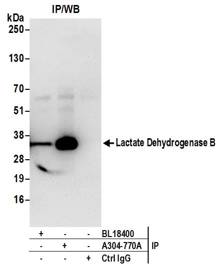
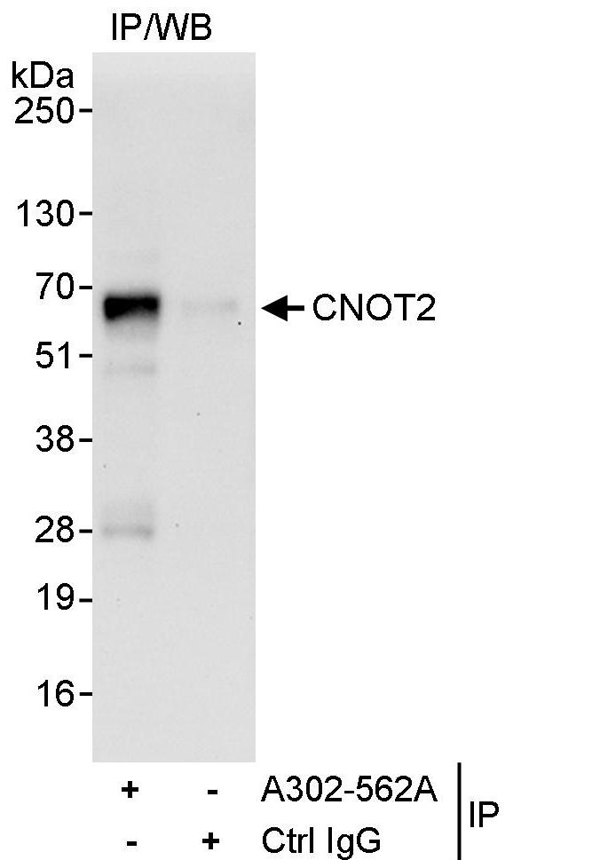 IP (Immunoprecipitation)
(Detection of human CNOT2 by western blot of immunoprecipitates. Samples: Whole cell lysate (1 mg for IP, 20% of IP loaded) from HeLa cells. Antibodies: Affinity purified rabbit anti-CNOT2 antibody AAA211927 used for IP at 10 ug/mg lysate. For blotting immunoprecipitated CNOT2, AAA211927 was used at 1 ug/ml. Detection: Chemiluminescence with an exposure time of 30 seconds.)
IP (Immunoprecipitation)
(Detection of human CNOT2 by western blot of immunoprecipitates. Samples: Whole cell lysate (1 mg for IP, 20% of IP loaded) from HeLa cells. Antibodies: Affinity purified rabbit anti-CNOT2 antibody AAA211927 used for IP at 10 ug/mg lysate. For blotting immunoprecipitated CNOT2, AAA211927 was used at 1 ug/ml. Detection: Chemiluminescence with an exposure time of 30 seconds.)
CNOT2, Polyclonal Antibody (Cat# AAA211927)
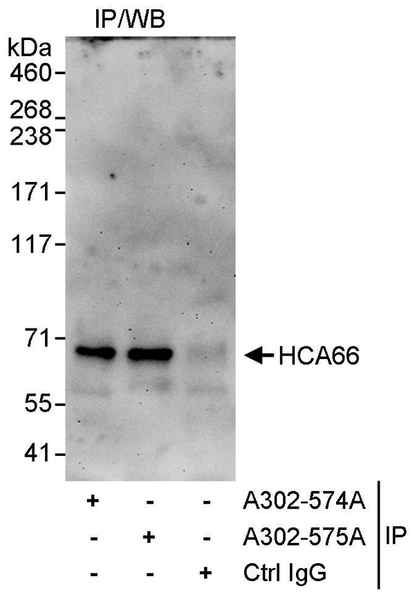 IP (Immunoprecipitation)
(Detection of human HCA66 by western blot of immunoprecipitates. Samples: Whole cell lysate (1 mg for IP, 20% of IP loaded) from HeLa cells. Antibodies: Affinity purified rabbit anti-HCA66 antibody AAA211932 used for IP at 3 ug/mg lysate. HCA66 was also immunoprecipitated by rabbit anti-HCA66 antibody which recognizes an upstream epitope. For blotting immunoprecipitated HCA66, AAA211932 was used at 1 ug/ml. Detection: Chemiluminescence with an exposure time of 3 minutes.)
IP (Immunoprecipitation)
(Detection of human HCA66 by western blot of immunoprecipitates. Samples: Whole cell lysate (1 mg for IP, 20% of IP loaded) from HeLa cells. Antibodies: Affinity purified rabbit anti-HCA66 antibody AAA211932 used for IP at 3 ug/mg lysate. HCA66 was also immunoprecipitated by rabbit anti-HCA66 antibody which recognizes an upstream epitope. For blotting immunoprecipitated HCA66, AAA211932 was used at 1 ug/ml. Detection: Chemiluminescence with an exposure time of 3 minutes.)
HCA66, Polyclonal Antibody (Cat# AAA211932)
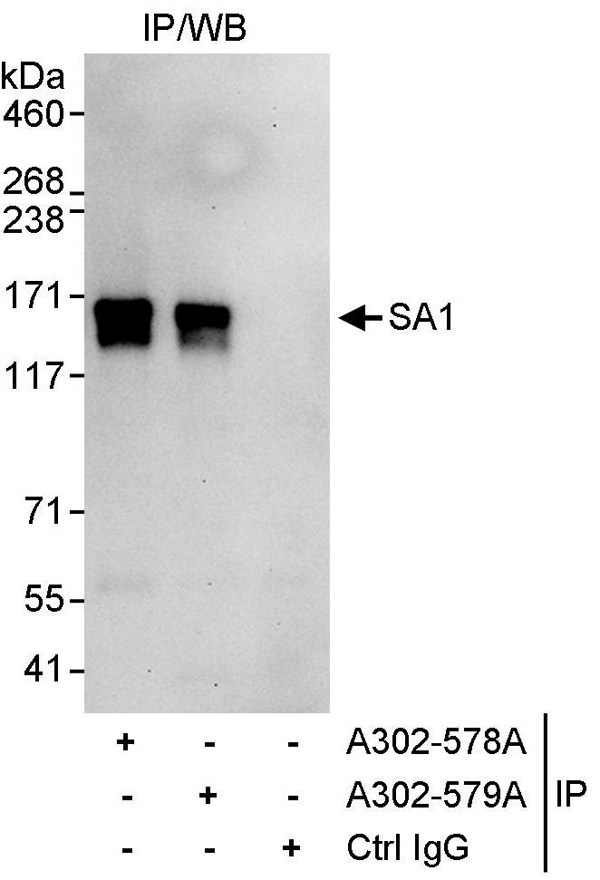 IP (Immunoprecipitation)
(Detection of human SA1 by western blot of immunoprecipitates. Samples: Whole cell lysate (1 mg for IP, 20% of IP loaded) from HeLa cells. Antibodies: Affinity purified rabbit anti-SA1 antibody AAA211933 used for IP at 3 ug/mg lysate. SA1 was also immunoprecipitated by rabbit anti-SA1 antibody which recognizes a downstream epitope. For blotting immunoprecipitated SA1, was used at 1 ug/ml. Detection: Chemiluminescence with an exposure time of 30 seconds.)
IP (Immunoprecipitation)
(Detection of human SA1 by western blot of immunoprecipitates. Samples: Whole cell lysate (1 mg for IP, 20% of IP loaded) from HeLa cells. Antibodies: Affinity purified rabbit anti-SA1 antibody AAA211933 used for IP at 3 ug/mg lysate. SA1 was also immunoprecipitated by rabbit anti-SA1 antibody which recognizes a downstream epitope. For blotting immunoprecipitated SA1, was used at 1 ug/ml. Detection: Chemiluminescence with an exposure time of 30 seconds.)
SA1, Polyclonal Antibody (Cat# AAA211933)
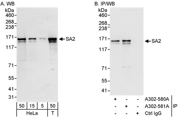 WB (Western Blot)
(Detection of human SA2 by western blot and immunoprecipitation. Samples: Whole cell lysate from HeLa (5, 15 and 50 ug for WB; 1 mg for IP, 20% of IP loaded) and HEK293T (T; 50 ug) cells. Antibodies: Affinity purified rabbit anti-SA2 antibody AAA211936 used for WB at 0.04 ug/ml (A) and 0.4 ug/ml (B) and used for IP at 3 ug/mg lysate. SA2 was also immunoprecipitated by rabbit anti-SA2 antibody which recognizes an upstream epitope. Detection: Chemiluminescence with exposure times of 10 seconds (A) and 1 second (B).)
WB (Western Blot)
(Detection of human SA2 by western blot and immunoprecipitation. Samples: Whole cell lysate from HeLa (5, 15 and 50 ug for WB; 1 mg for IP, 20% of IP loaded) and HEK293T (T; 50 ug) cells. Antibodies: Affinity purified rabbit anti-SA2 antibody AAA211936 used for WB at 0.04 ug/ml (A) and 0.4 ug/ml (B) and used for IP at 3 ug/mg lysate. SA2 was also immunoprecipitated by rabbit anti-SA2 antibody which recognizes an upstream epitope. Detection: Chemiluminescence with exposure times of 10 seconds (A) and 1 second (B).)
SA2, Polyclonal Antibody (Cat# AAA211936)
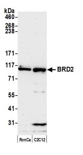
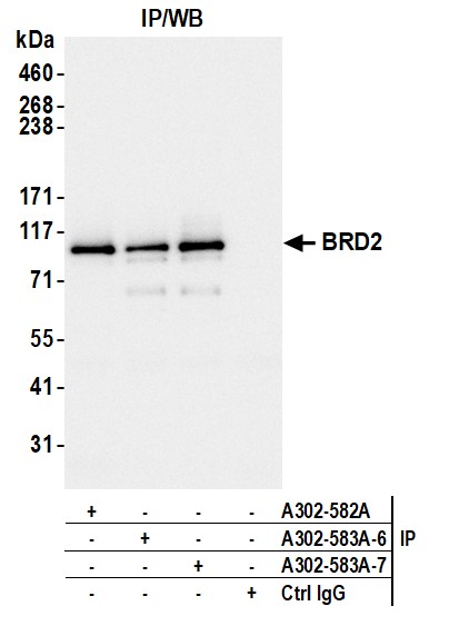
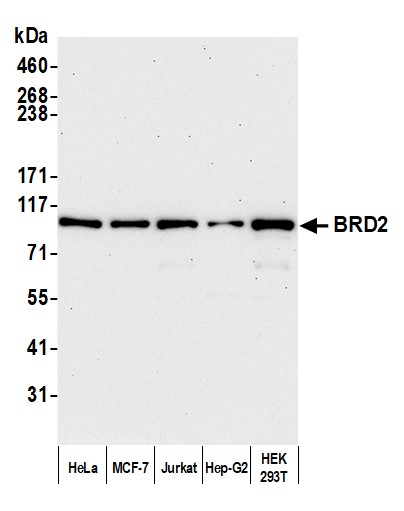 WB (Western Blot)
(Detection of human BRD2 by western blot. Samples: Whole cell lysate (10 ug) from HeLa, MCF-7, Jurkat, Hep-G2, and HEK293T cells prepared using NETN lysis buffer. Antibody: Affinity purified rabbit anti-BRD2 antibody (AAA211938 lot 7) used for WB at 0.04 ug/ml. Detection: Chemiluminescence with an exposure time of 3 minutes.)
WB (Western Blot)
(Detection of human BRD2 by western blot. Samples: Whole cell lysate (10 ug) from HeLa, MCF-7, Jurkat, Hep-G2, and HEK293T cells prepared using NETN lysis buffer. Antibody: Affinity purified rabbit anti-BRD2 antibody (AAA211938 lot 7) used for WB at 0.04 ug/ml. Detection: Chemiluminescence with an exposure time of 3 minutes.)
BRD2, Polyclonal Antibody (Cat# AAA211938)
 WB (Western Blot)
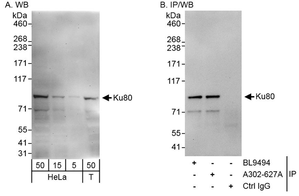
(Detection of human Ku80 by western blot and immunoprecipitation. Samples: Whole cell lysate from HeLa (5, 15 and 50 ug for WB; 1 mg for IP, 20% of IP loaded) and HEK293T (T; 50 ug) cells. Antibodies: Affinity purified rabbit anti-Ku80 antibody AAA211947 used for WB at 0.04 ug/ml (A) and 0.4 ug/ml (B) and used for IP at 3 ug/mg lysate. Ku80 was also immunoprecipitated by rabbit anti-Ku80 antibody BL9494, which recognizes an upstream epitope. Detection: Chemiluminescence with exposure times of 3 minutes (A) and 30 seconds (B).)
WB (Western Blot)
(Detection of human Ku80 by western blot and immunoprecipitation. Samples: Whole cell lysate from HeLa (5, 15 and 50 ug for WB; 1 mg for IP, 20% of IP loaded) and HEK293T (T; 50 ug) cells. Antibodies: Affinity purified rabbit anti-Ku80 antibody AAA211947 used for WB at 0.04 ug/ml (A) and 0.4 ug/ml (B) and used for IP at 3 ug/mg lysate. Ku80 was also immunoprecipitated by rabbit anti-Ku80 antibody BL9494, which recognizes an upstream epitope. Detection: Chemiluminescence with exposure times of 3 minutes (A) and 30 seconds (B).)
Ku80, Polyclonal Antibody (Cat# AAA211947)
 WB (Western Blot)
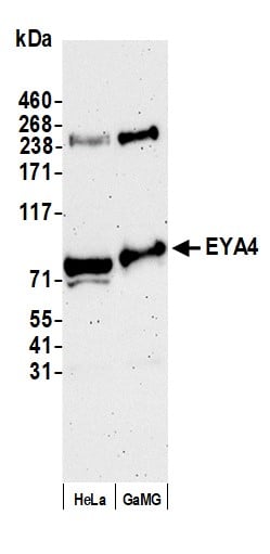
(Detection of human EYA4 by western blot. Samples: Whole cell lysate (50 ug) from HeLa and GaMG cells prepared using NETN lysis buffer. Antibody: Affinity purified rabbit anti-EYA4 antibody (AAA211948 lot 2) used for WB at 0.1 ug/ml. Detection: Chemiluminescence with an exposure time of 3 minutes.)
WB (Western Blot)
(Detection of human EYA4 by western blot. Samples: Whole cell lysate (50 ug) from HeLa and GaMG cells prepared using NETN lysis buffer. Antibody: Affinity purified rabbit anti-EYA4 antibody (AAA211948 lot 2) used for WB at 0.1 ug/ml. Detection: Chemiluminescence with an exposure time of 3 minutes.)
EYA4, Polyclonal Antibody (Cat# AAA211948)
 WB (Western Blot)
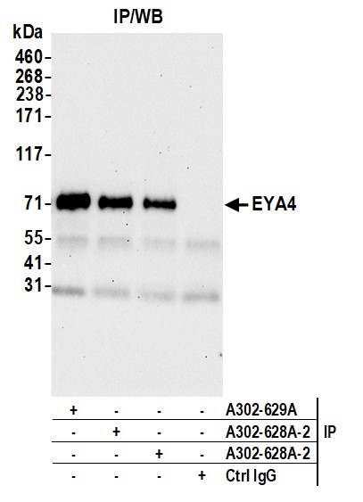
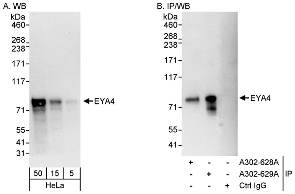
(Detection of human EYA4 by western blot and immunoprecipitation. Samples: Whole cell lysate (5, 15 and 50 ug for WB; 1 mg for IP, 20% of IP loaded) from HeLa cells. Antibodies: Affinity purified rabbit anti-EYA4 antibody AAA211949 used for WB at 0.04 ug/ml (A) and 1 ug/ml (B) and used for IP at 3 ug/mg lysate. EYA4 was also immunoprecipitated by rabbit anti-EYA4 antibody which recognizes an upstream epitope. Detection: Chemiluminescence with exposure times of 30 seconds (A) and 3 seconds (B).)
WB (Western Blot)
(Detection of human EYA4 by western blot and immunoprecipitation. Samples: Whole cell lysate (5, 15 and 50 ug for WB; 1 mg for IP, 20% of IP loaded) from HeLa cells. Antibodies: Affinity purified rabbit anti-EYA4 antibody AAA211949 used for WB at 0.04 ug/ml (A) and 1 ug/ml (B) and used for IP at 3 ug/mg lysate. EYA4 was also immunoprecipitated by rabbit anti-EYA4 antibody which recognizes an upstream epitope. Detection: Chemiluminescence with exposure times of 30 seconds (A) and 3 seconds (B).)
EYA4, Polyclonal Antibody (Cat# AAA211949)
 WB (Western Blot)
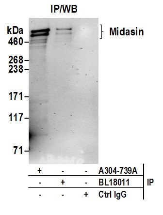
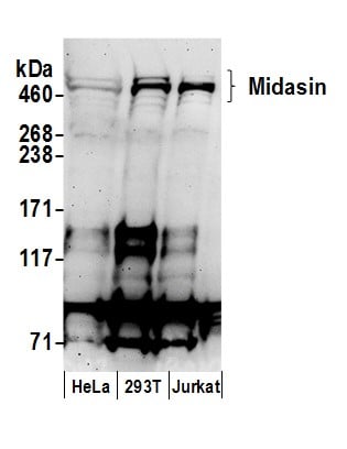
(Detection of human Midasin by western blot. Samples: Whole cell lysate (50 ug) from HeLa, HEK293T, and Jurkat cells prepared using NETN lysis buffer. Antibodies: Affinity purified rabbit anti-Midasin antibody AAA212900 (lot AAA212900-1) used for WB at 0.4 ug/ml. Detection: Chemiluminescence with an exposure time of 3 minutes.)
WB (Western Blot)
(Detection of human Midasin by western blot. Samples: Whole cell lysate (50 ug) from HeLa, HEK293T, and Jurkat cells prepared using NETN lysis buffer. Antibodies: Affinity purified rabbit anti-Midasin antibody AAA212900 (lot AAA212900-1) used for WB at 0.4 ug/ml. Detection: Chemiluminescence with an exposure time of 3 minutes.)
Midasin, Polyclonal Antibody (Cat# AAA212900)
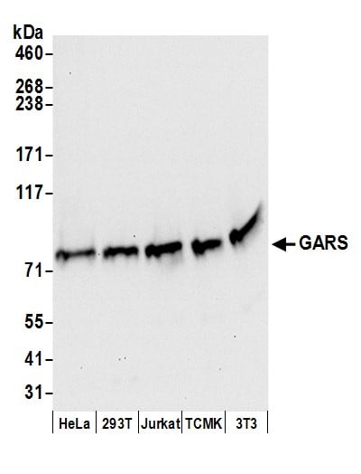 WB (Western Blot)
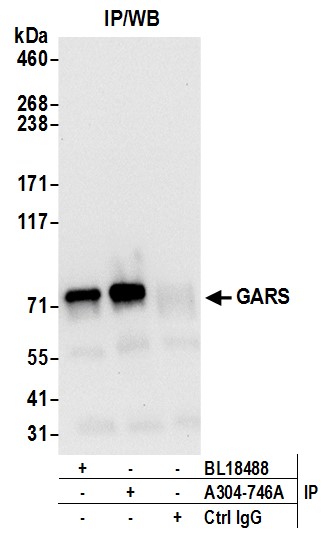
(Detection of human and mouse GARS by western blot. Samples: Whole cell lysate (50 ug) from HeLa, HEK293T, Jurkat, mouse TCMK-1, and mouse NIH 3T3 cells prepared using NETN lysis buffer. Antibodies: Affinity purified rabbit anti-GARS antibody AAA212905 (lot AAA212905-1) used for WB at 0.1 ug/ml. Detection: Chemiluminescence with an exposure time of 10 seconds.)
WB (Western Blot)
(Detection of human and mouse GARS by western blot. Samples: Whole cell lysate (50 ug) from HeLa, HEK293T, Jurkat, mouse TCMK-1, and mouse NIH 3T3 cells prepared using NETN lysis buffer. Antibodies: Affinity purified rabbit anti-GARS antibody AAA212905 (lot AAA212905-1) used for WB at 0.1 ug/ml. Detection: Chemiluminescence with an exposure time of 10 seconds.)
GARS, Polyclonal Antibody (Cat# AAA212905)
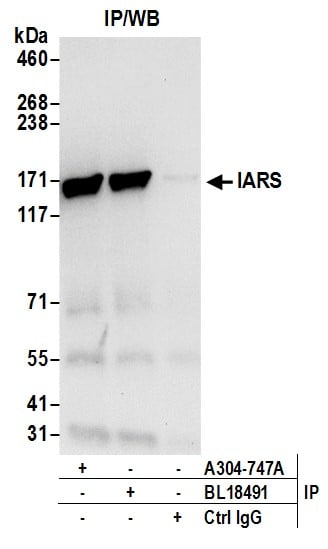
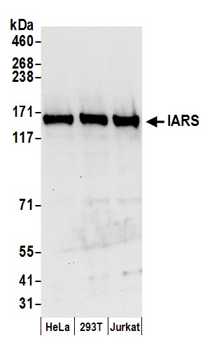 WB (Western Blot)
(Detection of human IARS by western blot. Samples: Whole cell lysate (50 ug) from HeLa, HEK293T, and Jurkat cells prepared using NETN lysis buffer. Antibodies: Affinity purified rabbit anti-IARS antibody AAA212906 (lot AAA212906-1) used for WB at 0.1 ug/ml. Detection: Chemiluminescence with an exposure time of 10 seconds.)
WB (Western Blot)
(Detection of human IARS by western blot. Samples: Whole cell lysate (50 ug) from HeLa, HEK293T, and Jurkat cells prepared using NETN lysis buffer. Antibodies: Affinity purified rabbit anti-IARS antibody AAA212906 (lot AAA212906-1) used for WB at 0.1 ug/ml. Detection: Chemiluminescence with an exposure time of 10 seconds.)
IARS, Polyclonal Antibody (Cat# AAA212906)
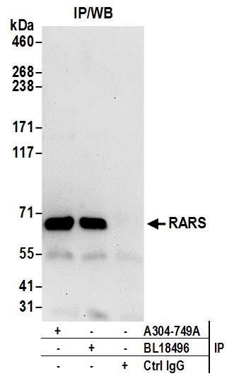
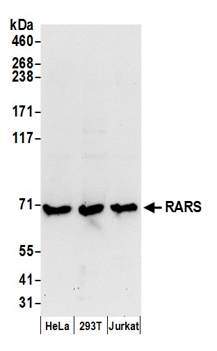 WB (Western Blot)
(Detection of human RARS by western blot. Samples: Whole cell lysate (50 ug) from HeLa, HEK293T, and Jurkat cells prepared using NETN lysis buffer. Antibodies: Affinity purified rabbit anti-RARS antibody AAA212908 (lot AAA212908-1) used for WB at 0.1 ug/ml. Detection: Chemiluminescence with an exposure time of 30 seconds.)
WB (Western Blot)
(Detection of human RARS by western blot. Samples: Whole cell lysate (50 ug) from HeLa, HEK293T, and Jurkat cells prepared using NETN lysis buffer. Antibodies: Affinity purified rabbit anti-RARS antibody AAA212908 (lot AAA212908-1) used for WB at 0.1 ug/ml. Detection: Chemiluminescence with an exposure time of 30 seconds.)
RARS, Polyclonal Antibody (Cat# AAA212908)
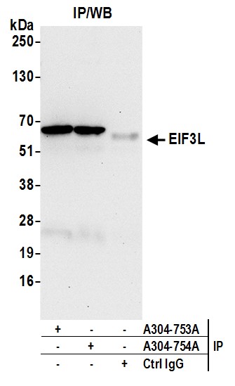
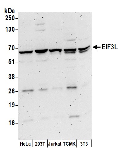 WB (Western Blot)
(Detection of human and mouse EIF3L by western blot. Samples: Whole cell lysate (50 ug) from HeLa, HEK293T, Jurkat, mouse TCMK-1, and mouse NIH 3T3 cells prepared using NETN lysis buffer. Antibodies: Affinity purified rabbit anti-EIF3L antibody AAA212913 (lot AAA212913-1) used for WB at 0.1 ug/ml. Detection: Chemiluminescence with an exposure time of 3 minutes.)
WB (Western Blot)
(Detection of human and mouse EIF3L by western blot. Samples: Whole cell lysate (50 ug) from HeLa, HEK293T, Jurkat, mouse TCMK-1, and mouse NIH 3T3 cells prepared using NETN lysis buffer. Antibodies: Affinity purified rabbit anti-EIF3L antibody AAA212913 (lot AAA212913-1) used for WB at 0.1 ug/ml. Detection: Chemiluminescence with an exposure time of 3 minutes.)
EIF3L, Polyclonal Antibody (Cat# AAA212913)
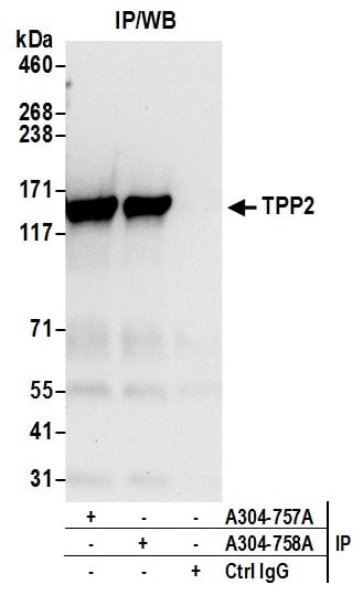
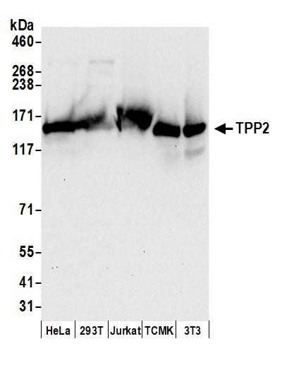 WB (Western Blot)
(Detection of human and mouse TPP2 by western blot. Samples: Whole cell lysate (50 ug) from HeLa, HEK293T, Jurkat, mouse TCMK-1, and mouse NIH 3T3 cells prepared using NETN lysis buffer. Antibodies: Affinity purified rabbit anti-TPP2 antibody AAA212916 (lot AAA212916-1) used for WB at 0.1 ug/ml. Detection: Chemiluminescence with an exposure time of 10 seconds.)
WB (Western Blot)
(Detection of human and mouse TPP2 by western blot. Samples: Whole cell lysate (50 ug) from HeLa, HEK293T, Jurkat, mouse TCMK-1, and mouse NIH 3T3 cells prepared using NETN lysis buffer. Antibodies: Affinity purified rabbit anti-TPP2 antibody AAA212916 (lot AAA212916-1) used for WB at 0.1 ug/ml. Detection: Chemiluminescence with an exposure time of 10 seconds.)
TPP2, Polyclonal Antibody (Cat# AAA212916)
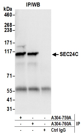
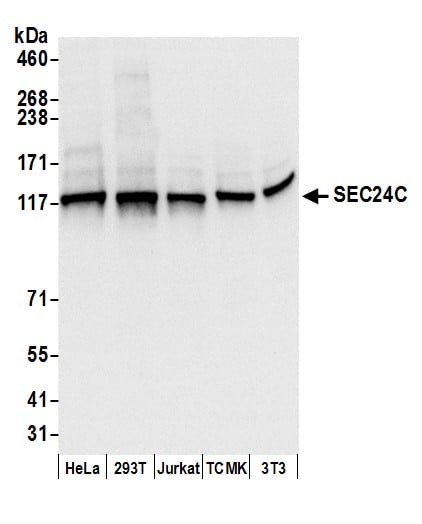 WB (Western Blot)
(Detection of human and mouse SEC24C by western blot. Samples: Whole cell lysate (50 ug) from HeLa, HEK293T, Jurkat, mouse TCMK-1, and mouse NIH 3T3 cells prepared using NETN lysis buffer. Antibodies: Affinity purified rabbit anti-SEC24C antibody AAA212917 (lot AAA212917-1) used for WB at 0.1 ug/ml. Detection: Chemiluminescence with an exposure time of 1 second.)
WB (Western Blot)
(Detection of human and mouse SEC24C by western blot. Samples: Whole cell lysate (50 ug) from HeLa, HEK293T, Jurkat, mouse TCMK-1, and mouse NIH 3T3 cells prepared using NETN lysis buffer. Antibodies: Affinity purified rabbit anti-SEC24C antibody AAA212917 (lot AAA212917-1) used for WB at 0.1 ug/ml. Detection: Chemiluminescence with an exposure time of 1 second.)
SEC24C, Polyclonal Antibody (Cat# AAA212917)
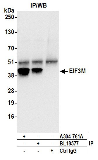
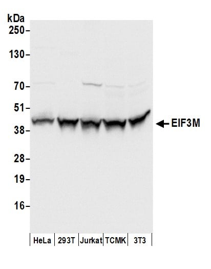 WB (Western Blot)
(Detection of human and mouse EIF3M by western blot. Samples: Whole cell lysate (50 ug) from HeLa, HEK293T, Jurkat, mouse TCMK-1, and mouse NIH 3T3 cells prepared using NETN lysis buffer. Antibodies: Affinity purified rabbit anti-EIF3M antibody AAA212918 (lot AAA212918-1) used for WB at 0.1 ug/ml. Detection: Chemiluminescence with an exposure time of 1 second.)
WB (Western Blot)
(Detection of human and mouse EIF3M by western blot. Samples: Whole cell lysate (50 ug) from HeLa, HEK293T, Jurkat, mouse TCMK-1, and mouse NIH 3T3 cells prepared using NETN lysis buffer. Antibodies: Affinity purified rabbit anti-EIF3M antibody AAA212918 (lot AAA212918-1) used for WB at 0.1 ug/ml. Detection: Chemiluminescence with an exposure time of 1 second.)
EIF3M, Polyclonal Antibody (Cat# AAA212918)
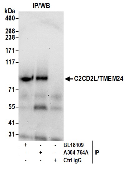
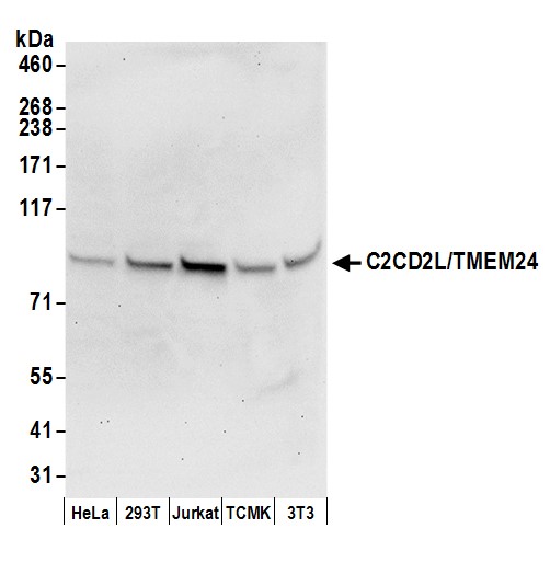 WB (Western Blot)
(Detection of human and mouse C2CD2L/TMEM24 by western blot. Samples: Whole cell lysate (50 ug) from HeLa, HEK293T, Jurkat, mouse TCMK-1, and mouse NIH 3T3 cells prepared using NETN lysis buffer. Antibody: Affinity purified rabbit anti-C2CD2L/TMEM24 antibody AAA212920 (lot AAA212920-1) used for WB at 0.1 ug/ml. Detection: Chemiluminescence with an exposure time of 30 seconds.)
WB (Western Blot)
(Detection of human and mouse C2CD2L/TMEM24 by western blot. Samples: Whole cell lysate (50 ug) from HeLa, HEK293T, Jurkat, mouse TCMK-1, and mouse NIH 3T3 cells prepared using NETN lysis buffer. Antibody: Affinity purified rabbit anti-C2CD2L/TMEM24 antibody AAA212920 (lot AAA212920-1) used for WB at 0.1 ug/ml. Detection: Chemiluminescence with an exposure time of 30 seconds.)
C2CD2L/TMEM24, Polyclonal Antibody (Cat# AAA212920)
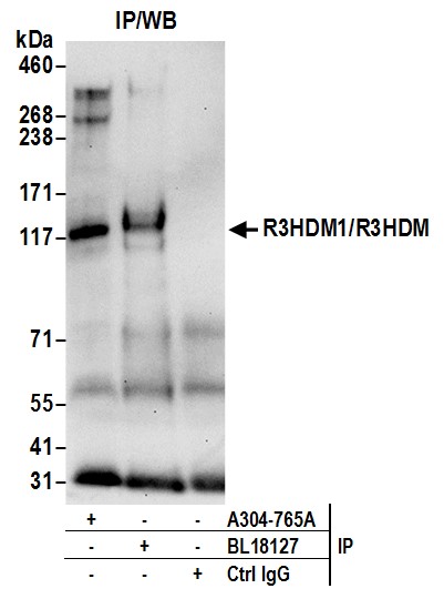
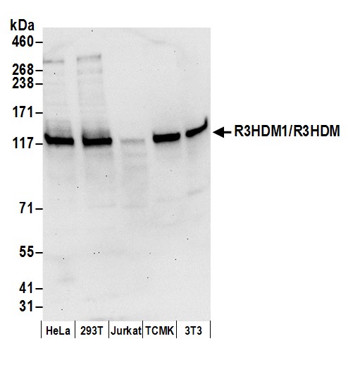 WB (Western Blot)
(Detection of human and mouse R3HDM1/R3HDM by western blot. Samples: Whole cell lysate (50 ug) from HeLa, HEK293T, Jurkat, mouse TCMK-1, and mouse NIH 3T3 cells prepared using NETN lysis buffer. Antibody: Affinity purified rabbit anti-R3HDM1/R3HDM antibody AAA212921 (lot AAA212921-1) used for WB at 0.1 ug/ml. Detection: Chemiluminescence with an exposure time of 30 seconds.)
WB (Western Blot)
(Detection of human and mouse R3HDM1/R3HDM by western blot. Samples: Whole cell lysate (50 ug) from HeLa, HEK293T, Jurkat, mouse TCMK-1, and mouse NIH 3T3 cells prepared using NETN lysis buffer. Antibody: Affinity purified rabbit anti-R3HDM1/R3HDM antibody AAA212921 (lot AAA212921-1) used for WB at 0.1 ug/ml. Detection: Chemiluminescence with an exposure time of 30 seconds.)
R3HDM1/R3HDM, Polyclonal Antibody (Cat# AAA212921)
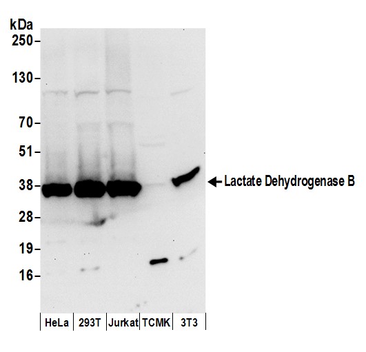 WB (Western Blot)
(Detection of human and mouse Lactate Dehydrogenase B by western blot. Samples: Whole cell lysate (50 ug) from HeLa, HEK293T, Jurkat, mouse TCMK-1, and mouse NIH 3T3 cells prepared using NETN lysis buffer. Antibody: Affinity purified rabbit anti-Lactate Dehydrogenase B antibody AAA212925 (lot AAA212925-1) used for WB at 0.1 ug/ml. Detection: Chemiluminescence with an exposure time of 10 seconds.)
WB (Western Blot)
(Detection of human and mouse Lactate Dehydrogenase B by western blot. Samples: Whole cell lysate (50 ug) from HeLa, HEK293T, Jurkat, mouse TCMK-1, and mouse NIH 3T3 cells prepared using NETN lysis buffer. Antibody: Affinity purified rabbit anti-Lactate Dehydrogenase B antibody AAA212925 (lot AAA212925-1) used for WB at 0.1 ug/ml. Detection: Chemiluminescence with an exposure time of 10 seconds.)
Lactate Dehydrogenase B/LDHB, Polyclonal Antibody (Cat# AAA212925)
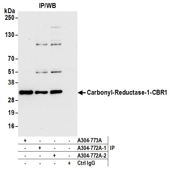
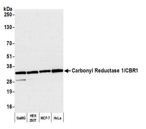 WB (Western Blot)
(Detection of human Carbonyl Reductase 1/CBR1 by western blot. Samples: Whole cell lysate (10 ug) from GaMG, HEK293T, MCF-7, and HeLa cells prepared using NETN lysis buffer. Antibody: Affinity purified rabbit anti-Carbonyl Reductase 1/CBR1 antibody AAA212927 Lot 2 used for WB at 0.04 ug/ml. Detection: Chemiluminescence with an exposure time of 10 seconds.)
WB (Western Blot)
(Detection of human Carbonyl Reductase 1/CBR1 by western blot. Samples: Whole cell lysate (10 ug) from GaMG, HEK293T, MCF-7, and HeLa cells prepared using NETN lysis buffer. Antibody: Affinity purified rabbit anti-Carbonyl Reductase 1/CBR1 antibody AAA212927 Lot 2 used for WB at 0.04 ug/ml. Detection: Chemiluminescence with an exposure time of 10 seconds.)
Carbonyl Reductase 1/CBR1, Polyclonal Antibody (Cat# AAA212927)
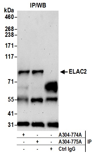
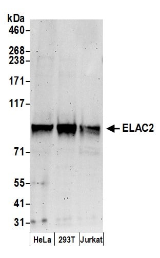 WB (Western Blot)
(Detection of human ELAC2 by western blot. Samples: Whole cell lysate (50 ug) from HeLa, HEK293T, and Jurkat cells prepared using NETN lysis buffer. Antibody: Affinity purified rabbit anti-ELAC2 antibody AAA212929 (lot AAA212929-1) used for WB at 0.4 ug/ml. Detection: Chemiluminescence with an exposure time of 3 minutes.)
WB (Western Blot)
(Detection of human ELAC2 by western blot. Samples: Whole cell lysate (50 ug) from HeLa, HEK293T, and Jurkat cells prepared using NETN lysis buffer. Antibody: Affinity purified rabbit anti-ELAC2 antibody AAA212929 (lot AAA212929-1) used for WB at 0.4 ug/ml. Detection: Chemiluminescence with an exposure time of 3 minutes.)
ELAC2, Polyclonal Antibody (Cat# AAA212929)
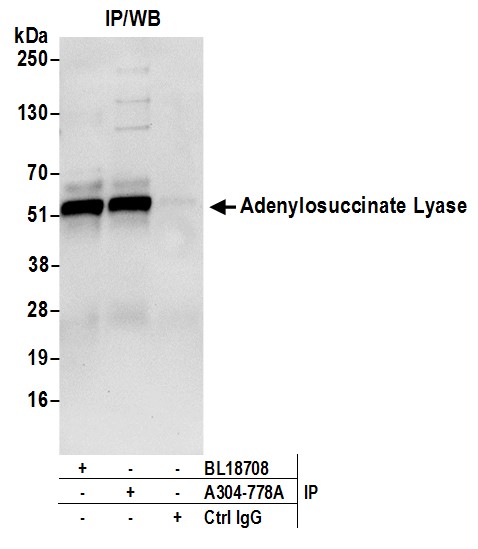
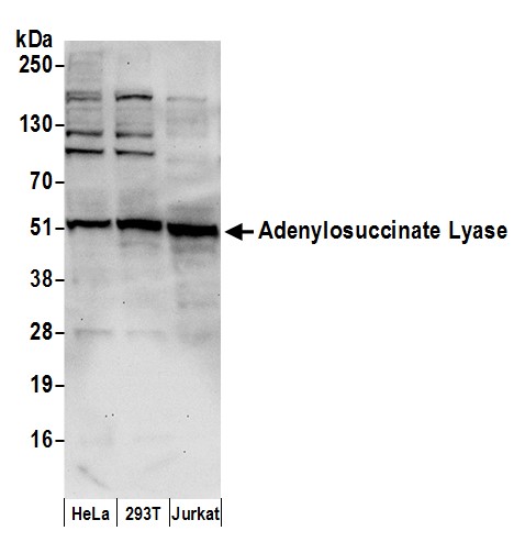 WB (Western Blot)
(Detection of human Adenylosuccinate Lyase/ADSL by western blot. Samples: Whole cell lysate (50 ug) from HeLa, HEK293T, and Jurkat cells prepared using NETN lysis buffer. Antibody: Affinity purified rabbit anti-Adenylosuccinate Lyase/ADSL antibody AAA212933 (lot AAA212933-1) used for WB at 0.1 ug/ml. Detection: Chemiluminescence with an exposure time of 30 seconds.)
WB (Western Blot)
(Detection of human Adenylosuccinate Lyase/ADSL by western blot. Samples: Whole cell lysate (50 ug) from HeLa, HEK293T, and Jurkat cells prepared using NETN lysis buffer. Antibody: Affinity purified rabbit anti-Adenylosuccinate Lyase/ADSL antibody AAA212933 (lot AAA212933-1) used for WB at 0.1 ug/ml. Detection: Chemiluminescence with an exposure time of 30 seconds.)
Adenylosuccinate Lyase/ADSL, Polyclonal Antibody (Cat# AAA212933)
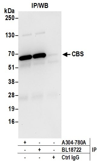
 WB (Western Blot)
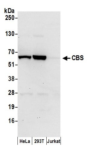
(Detection of human CBS by western blot. Samples: Whole cell lysate (50 ug) from HeLa, HEK293T, and Jurkat cells prepared using NETN lysis buffer. Antibody: Affinity purified rabbit anti-CBS antibody AAA212935 (lot AAA212935-1) used for WB at 0.1 ug/ml. Detection: Chemiluminescence with an exposure time of 30 seconds.)
WB (Western Blot)
(Detection of human CBS by western blot. Samples: Whole cell lysate (50 ug) from HeLa, HEK293T, and Jurkat cells prepared using NETN lysis buffer. Antibody: Affinity purified rabbit anti-CBS antibody AAA212935 (lot AAA212935-1) used for WB at 0.1 ug/ml. Detection: Chemiluminescence with an exposure time of 30 seconds.)
CBS, Polyclonal Antibody (Cat# AAA212935)
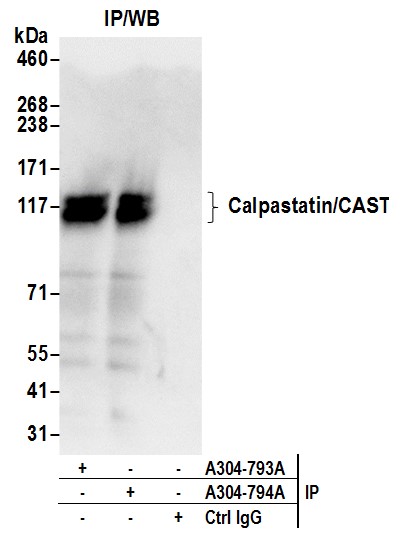
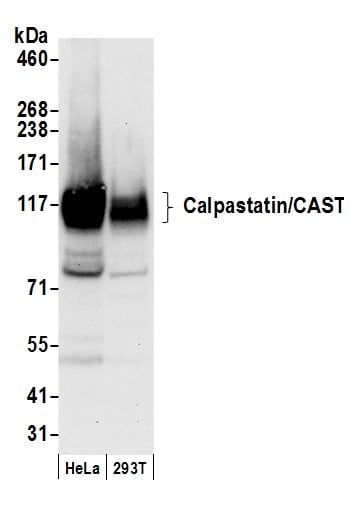 WB (Western Blot)
(Detection of human Calpastatin/CAST by western blot. Samples: Whole cell lysate (50 ug) from HeLa and 293T cells prepared using NETN lysis buffer. Antibody: Affinity purified rabbit anti-Calpastatin/CAST antibody AAA212942 (lot AAA212942-1) used for WB at 0.1 ug/ml. Detection: Chemiluminescence with an exposure time of 10 seconds.)
WB (Western Blot)
(Detection of human Calpastatin/CAST by western blot. Samples: Whole cell lysate (50 ug) from HeLa and 293T cells prepared using NETN lysis buffer. Antibody: Affinity purified rabbit anti-Calpastatin/CAST antibody AAA212942 (lot AAA212942-1) used for WB at 0.1 ug/ml. Detection: Chemiluminescence with an exposure time of 10 seconds.)
Calpastatin/CAST, Polyclonal Antibody (Cat# AAA212942)
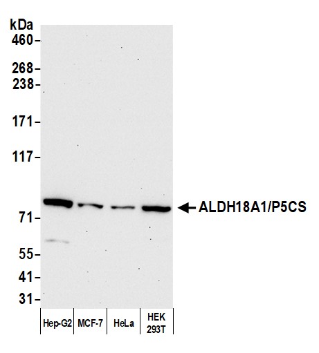
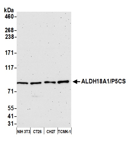 WB (Western Blot)
(Detection of mouse ALDH18A1/P5CS by western blot. Samples: Whole cell lysate (10 ug) from NIH 3T3, CT26, CH27, and TCMK-1 cells prepared using NETN lysis buffer. Antibody: Affinity purified rabbit anti-ALDH18A1/P5CS antibody AAA212945 Lot 1 used for WB at 0.04 ug/ml. Detection: Chemiluminescence with an exposure time of 30 seconds.)
WB (Western Blot)
(Detection of mouse ALDH18A1/P5CS by western blot. Samples: Whole cell lysate (10 ug) from NIH 3T3, CT26, CH27, and TCMK-1 cells prepared using NETN lysis buffer. Antibody: Affinity purified rabbit anti-ALDH18A1/P5CS antibody AAA212945 Lot 1 used for WB at 0.04 ug/ml. Detection: Chemiluminescence with an exposure time of 30 seconds.)
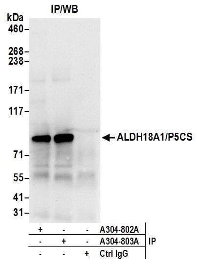
ALDH18A1/P5CS, Polyclonal Antibody (Cat# AAA212945)
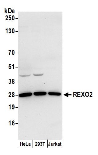 WB (Western Blot)
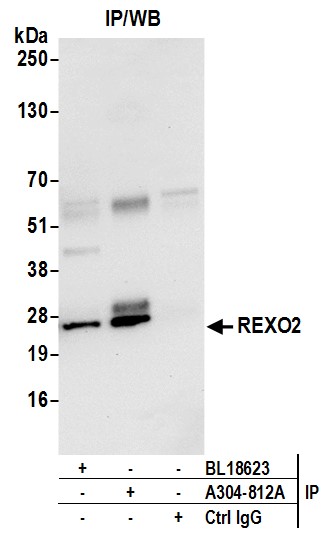
(Detection of human REXO2 by western blot. Samples: Whole cell lysate (50 ug) from HeLa, HEK293T, and Jurkat cells prepared using NETN lysis buffer. Antibody: Affinity purified rabbit anti-REXO2 antibody AAA212953 (lot AAA212953-1) used for WB at 0.4 ug/ml. Detection: Chemiluminescence with an exposure time of 10 seconds.)
WB (Western Blot)
(Detection of human REXO2 by western blot. Samples: Whole cell lysate (50 ug) from HeLa, HEK293T, and Jurkat cells prepared using NETN lysis buffer. Antibody: Affinity purified rabbit anti-REXO2 antibody AAA212953 (lot AAA212953-1) used for WB at 0.4 ug/ml. Detection: Chemiluminescence with an exposure time of 10 seconds.)
REXO2, Polyclonal Antibody (Cat# AAA212953)
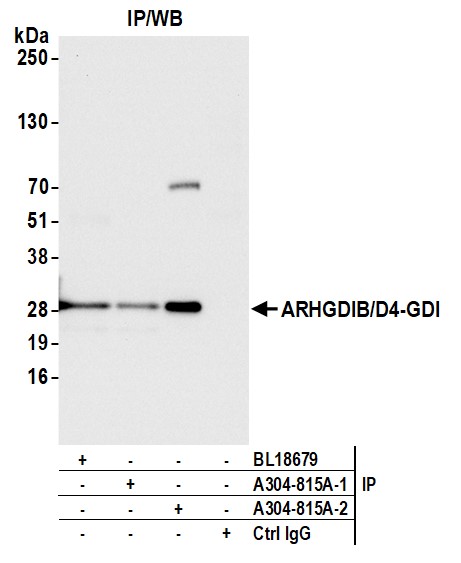
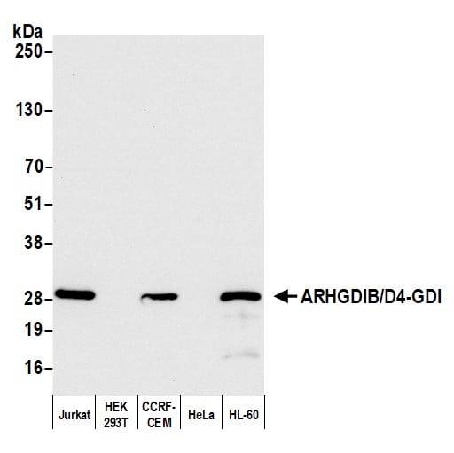 WB (Western Blot)
(Detection of human ARHGDIB/D4-GDI/RhoGDI2 by western blot. Samples: Whole cell lysate (10 ug) from Jurkat, HEK293T, CCRF-CEM, HeLa, and HL-60 cells prepared using NETN lysis buffer. Antibody: Affinity purified rabbit anti-ARHGDIB/D4-GDI/RhoGDI2 antibody (AAA212955 lot 2) used for WB at 0.1 ug/ml. Detection: Chemiluminescence with an exposure time of 3 seconds.)
WB (Western Blot)
(Detection of human ARHGDIB/D4-GDI/RhoGDI2 by western blot. Samples: Whole cell lysate (10 ug) from Jurkat, HEK293T, CCRF-CEM, HeLa, and HL-60 cells prepared using NETN lysis buffer. Antibody: Affinity purified rabbit anti-ARHGDIB/D4-GDI/RhoGDI2 antibody (AAA212955 lot 2) used for WB at 0.1 ug/ml. Detection: Chemiluminescence with an exposure time of 3 seconds.)
ARHGDIB/D4-GDI/RhoGDI2, Polyclonal Antibody (Cat# AAA212955)
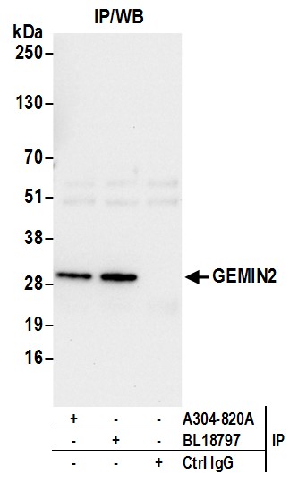
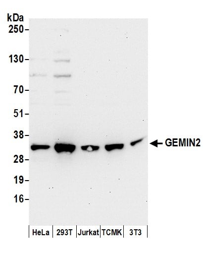 WB (Western Blot)
(Detection of human and mouse GEMIN2 by western blot. Samples: Whole cell lysate (50 ug) from HeLa, HEK293T, Jurkat, mouse TCMK-1, and mouse NIH 3T3 cells prepared using NETN lysis buffer. Antibody: Affinity purified rabbit anti-GEMIN2 antibody AAA212958 (lot AAA212958-1) used for WB at 0.1 ug/ml. Detection: Chemiluminescence with an exposure time of 30 seconds.)
WB (Western Blot)
(Detection of human and mouse GEMIN2 by western blot. Samples: Whole cell lysate (50 ug) from HeLa, HEK293T, Jurkat, mouse TCMK-1, and mouse NIH 3T3 cells prepared using NETN lysis buffer. Antibody: Affinity purified rabbit anti-GEMIN2 antibody AAA212958 (lot AAA212958-1) used for WB at 0.1 ug/ml. Detection: Chemiluminescence with an exposure time of 30 seconds.)
GEMIN2, Polyclonal Antibody (Cat# AAA212958)
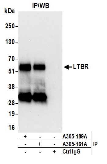
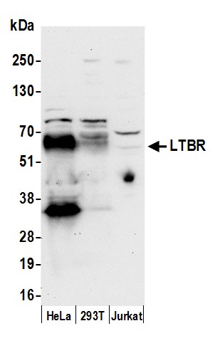 WB (Western Blot)
(Detection of human LTBR by western blot. Samples: Whole cell lysate (50 ug) from HeLa, HEK293T, and Jurkat cells prepared using NETN lysis buffer. Antibody: Affinity purified rabbit anti-LTBR antibody AAA213142 (lot AAA213142-1) used for WB at 0.1 ug/ml. Detection: Chemiluminescence with an exposure time of 30 seconds.)
WB (Western Blot)
(Detection of human LTBR by western blot. Samples: Whole cell lysate (50 ug) from HeLa, HEK293T, and Jurkat cells prepared using NETN lysis buffer. Antibody: Affinity purified rabbit anti-LTBR antibody AAA213142 (lot AAA213142-1) used for WB at 0.1 ug/ml. Detection: Chemiluminescence with an exposure time of 30 seconds.)
LTBR/Lymphotoxin-beta Receptor, Polyclonal Antibody (Cat# AAA213142)

 WB (Western Blot)
(Detection of human and mouse FMR1 by western blot. Samples: Whole cell lysate (50 ug) from HeLa, HEK293T, Jurkat, mouse TCMK-1, and mouse NIH 3T3 cells prepared using NETN lysis buffer. Antibody: Affinity purified rabbit anti-FMR1 antibody AAA213145 (lot AAA213145-1) used for WB at 0.1 ug/ml. Detection: Chemiluminescence with an exposure time of 10 seconds.)
WB (Western Blot)
(Detection of human and mouse FMR1 by western blot. Samples: Whole cell lysate (50 ug) from HeLa, HEK293T, Jurkat, mouse TCMK-1, and mouse NIH 3T3 cells prepared using NETN lysis buffer. Antibody: Affinity purified rabbit anti-FMR1 antibody AAA213145 (lot AAA213145-1) used for WB at 0.1 ug/ml. Detection: Chemiluminescence with an exposure time of 10 seconds.)
FMR1/FMRP, Polyclonal Antibody (Cat# AAA213145)

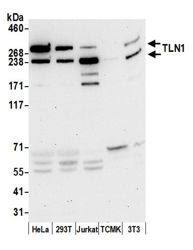 WB (Western Blot)
(Detection of human and mouse TLN1 by western blot. Samples: Whole cell lysate (50 ug) from HeLa, HEK293T, Jurkat, mouse TCMK-1, and mouse NIH 3T3 cells prepared using NETN lysis buffer. Antibody: Affinity purified rabbit anti-TLN1 antibody AAA213149 (lot AAA213149-1) used for WB at 0.1 ug/ml. Detection: Chemiluminescence with an exposure time of 30 seconds.)
WB (Western Blot)
(Detection of human and mouse TLN1 by western blot. Samples: Whole cell lysate (50 ug) from HeLa, HEK293T, Jurkat, mouse TCMK-1, and mouse NIH 3T3 cells prepared using NETN lysis buffer. Antibody: Affinity purified rabbit anti-TLN1 antibody AAA213149 (lot AAA213149-1) used for WB at 0.1 ug/ml. Detection: Chemiluminescence with an exposure time of 30 seconds.)
TLN1/Talin-1, Polyclonal Antibody (Cat# AAA213149)
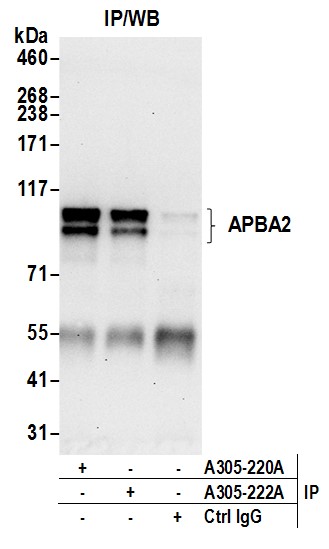
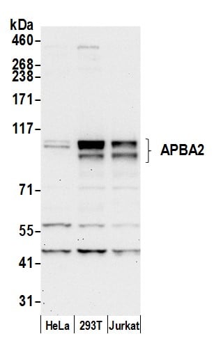 WB (Western Blot)
(Detection of human APBA2 by western blot. Samples: Whole cell lysate (50 ug) from HeLa, HEK293T, and Jurkat cells prepared using NETN lysis buffer. Antibody: Affinity purified rabbit anti-APBA2 antibody AAA213158 (lot AAA213158-1) used for WB at 0.1 ug/ml. Detection: Chemiluminescence with an exposure time of 30 seconds.)
WB (Western Blot)
(Detection of human APBA2 by western blot. Samples: Whole cell lysate (50 ug) from HeLa, HEK293T, and Jurkat cells prepared using NETN lysis buffer. Antibody: Affinity purified rabbit anti-APBA2 antibody AAA213158 (lot AAA213158-1) used for WB at 0.1 ug/ml. Detection: Chemiluminescence with an exposure time of 30 seconds.)
APBA2, Polyclonal Antibody (Cat# AAA213158)
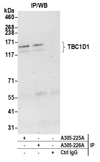
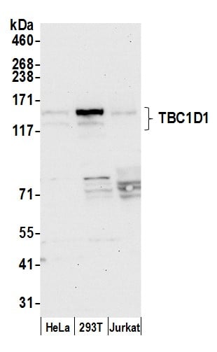 WB (Western Blot)
(Detection of human TBC1D1 by western blot. Samples: Whole cell lysate (50 ug) from HeLa, HEK293T, and Jurkat cells prepared using NETN lysis buffer. Antibody: Affinity purified rabbit anti-TBC1D1 antibody AAA213161 (lot AAA213161-1) used for WB at 1 ug/ml. Detection: Chemiluminescence with an exposure time of 30 seconds.)
WB (Western Blot)
(Detection of human TBC1D1 by western blot. Samples: Whole cell lysate (50 ug) from HeLa, HEK293T, and Jurkat cells prepared using NETN lysis buffer. Antibody: Affinity purified rabbit anti-TBC1D1 antibody AAA213161 (lot AAA213161-1) used for WB at 1 ug/ml. Detection: Chemiluminescence with an exposure time of 30 seconds.)
TBC1D1, Polyclonal Antibody (Cat# AAA213161)
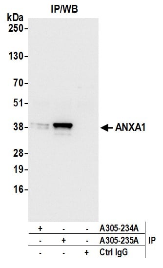
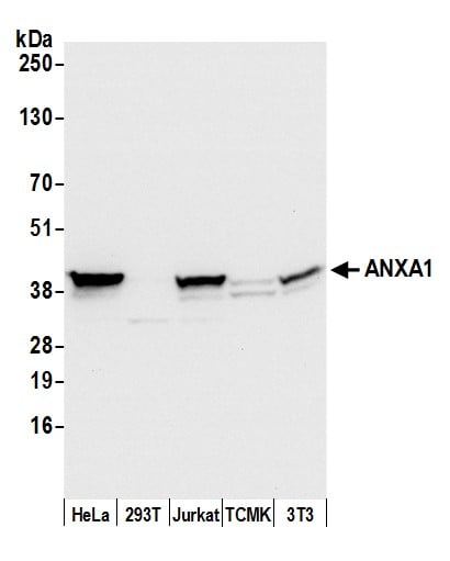 WB (Western Blot)
(Detection of human and mouse ANXA1 by western blot. Samples: Whole cell lysate (50 ug) from HeLa, HEK293T, Jurkat, mouse TCMK-1, and mouse NIH 3T3 cells prepared using NETN lysis buffer. Antibody: Affinity purified rabbit anti-ANXA1 antibody AAA213165 (lot AAA213165-1) used for WB at 0.1 ug/ml. Detection: Chemiluminescence with an exposure time of 3 seconds.)
WB (Western Blot)
(Detection of human and mouse ANXA1 by western blot. Samples: Whole cell lysate (50 ug) from HeLa, HEK293T, Jurkat, mouse TCMK-1, and mouse NIH 3T3 cells prepared using NETN lysis buffer. Antibody: Affinity purified rabbit anti-ANXA1 antibody AAA213165 (lot AAA213165-1) used for WB at 0.1 ug/ml. Detection: Chemiluminescence with an exposure time of 3 seconds.)
ANXA1/Annexin A1, Polyclonal Antibody (Cat# AAA213165)
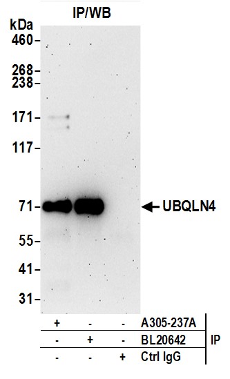
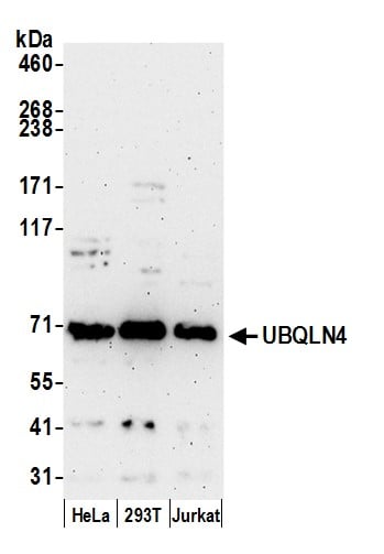 WB (Western Blot)
(Detection of human UBQLN4 by western blot. Samples: Whole cell lysate (50 ug) from HeLa, HEK293T, and Jurkat cells prepared using NETN lysis buffer. Antibody: Affinity purified rabbit anti-UBQLN4 antibody AAA213167 (lot AAA213167-1) used for WB at 0.1 ug/ml. Detection: Chemiluminescence with an exposure time of 3 minutes.)
WB (Western Blot)
(Detection of human UBQLN4 by western blot. Samples: Whole cell lysate (50 ug) from HeLa, HEK293T, and Jurkat cells prepared using NETN lysis buffer. Antibody: Affinity purified rabbit anti-UBQLN4 antibody AAA213167 (lot AAA213167-1) used for WB at 0.1 ug/ml. Detection: Chemiluminescence with an exposure time of 3 minutes.)
UBQLN4/CIP75/Ubiquilin 4, Polyclonal Antibody (Cat# AAA213167)
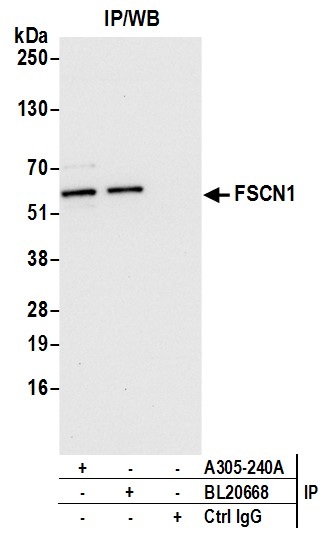
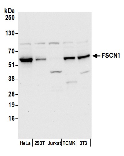 WB (Western Blot)
(Detection of human and mouse FSCN1 by western blot. Samples: Whole cell lysate (50 ug) from HeLa, 293T, Jurkat, mouse TCMK-1, and mouse NIH3T3 cells prepared using NETN lysis buffer. Antibody: Affinity purified rabbit anti-FSCN1 antibody AAA213170 (lot AAA213170-1) used for WB at 0.4 ug/ml. Detection: Chemiluminescence with an exposure time of 10 seconds.)
WB (Western Blot)
(Detection of human and mouse FSCN1 by western blot. Samples: Whole cell lysate (50 ug) from HeLa, 293T, Jurkat, mouse TCMK-1, and mouse NIH3T3 cells prepared using NETN lysis buffer. Antibody: Affinity purified rabbit anti-FSCN1 antibody AAA213170 (lot AAA213170-1) used for WB at 0.4 ug/ml. Detection: Chemiluminescence with an exposure time of 10 seconds.)
FSCN1, Polyclonal Antibody (Cat# AAA213170)
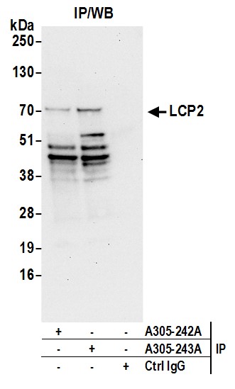
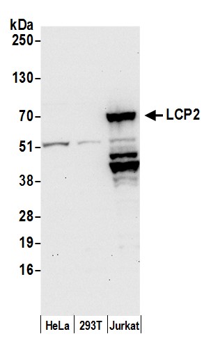 WB (Western Blot)
(Detection of human LCP2 by western blot. Samples: Whole cell lysate (50 ug) from HeLa, HEK293T, and Jurkat cells prepared using NETN lysis buffer. Antibody: Affinity purified rabbit anti-LCP2 antibody AAA213172 (lot AAA213172-1) used for WB at 0.1 ug/ml. Detection: Chemiluminescence with an exposure time of 30 seconds.)
WB (Western Blot)
(Detection of human LCP2 by western blot. Samples: Whole cell lysate (50 ug) from HeLa, HEK293T, and Jurkat cells prepared using NETN lysis buffer. Antibody: Affinity purified rabbit anti-LCP2 antibody AAA213172 (lot AAA213172-1) used for WB at 0.1 ug/ml. Detection: Chemiluminescence with an exposure time of 30 seconds.)
LCP2/SLP76, Polyclonal Antibody (Cat# AAA213172)
 WB (Western Blot)
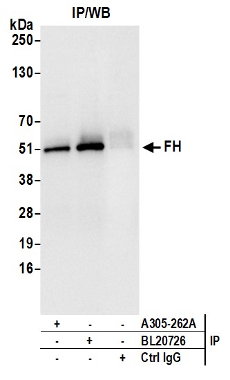
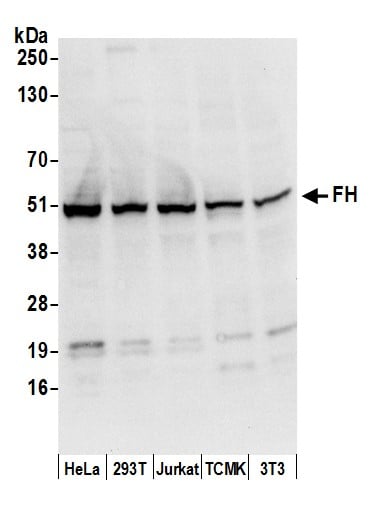
(Detection of human and mouse FH by western blot. Samples: Whole cell lysate (50 ug) from HeLa, HEK293T, Jurkat, mouse TCMK-1, and mouse NIH 3T3 cells prepared using NETN lysis buffer. Antibody: Affinity purified rabbit anti-FH antibody AAA213181 (lot AAA213181-1) used for WB at 0.1 ug/ml. Detection: Chemiluminescence with an exposure time of 3 seconds.)
WB (Western Blot)
(Detection of human and mouse FH by western blot. Samples: Whole cell lysate (50 ug) from HeLa, HEK293T, Jurkat, mouse TCMK-1, and mouse NIH 3T3 cells prepared using NETN lysis buffer. Antibody: Affinity purified rabbit anti-FH antibody AAA213181 (lot AAA213181-1) used for WB at 0.1 ug/ml. Detection: Chemiluminescence with an exposure time of 3 seconds.)
FH/Fumarate Hydratase, Polyclonal Antibody (Cat# AAA213181)
 WB (Western Blot)
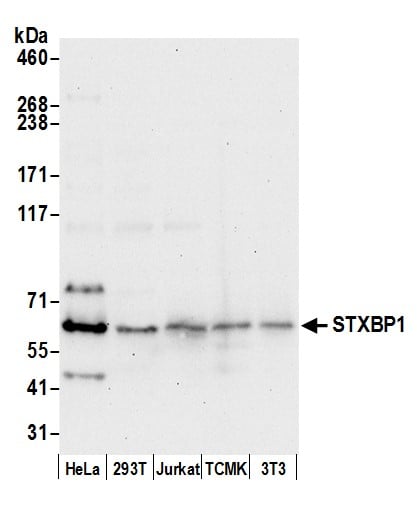
(Detection of human and mouse STXBP1 by western blot. Samples: Whole cell lysate (50 ug) from HeLa, HEK293T, Jurkat, mouse TCMK-1, and mouse NIH 3T3 cells prepared using NETN lysis buffer. Antibody: Affinity purified rabbit anti-STXBP1 antibody AAA213182 (lot AAA213182-1) used for WB at 0.1 ug/ml. Detection: Chemiluminescence with an exposure time of 30 seconds.)
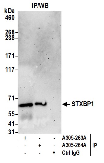
WB (Western Blot)
(Detection of human and mouse STXBP1 by western blot. Samples: Whole cell lysate (50 ug) from HeLa, HEK293T, Jurkat, mouse TCMK-1, and mouse NIH 3T3 cells prepared using NETN lysis buffer. Antibody: Affinity purified rabbit anti-STXBP1 antibody AAA213182 (lot AAA213182-1) used for WB at 0.1 ug/ml. Detection: Chemiluminescence with an exposure time of 30 seconds.)
STXBP1/MUNC18-1, Polyclonal Antibody (Cat# AAA213182)
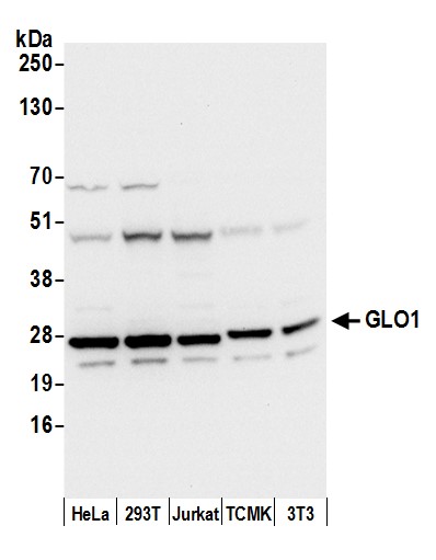 WB (Western Blot)
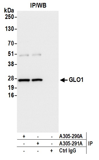
(Detection of human and mouse GLO1/Glyoxalase I by western blot. Samples: Whole cell lysate (50 ug) from HeLa, HEK293T, Jurkat, mouse TCMK-1, and mouse NIH 3T3 cells prepared using NETN lysis buffer. Antibody: Affinity purified rabbit anti-GLO1/Glyoxalase I antibody AAA213197 (lot AAA213197-1) used for WB at 0.1 ug/ml. Detection: Chemiluminescence with an exposure time of 3 seconds.)
WB (Western Blot)
(Detection of human and mouse GLO1/Glyoxalase I by western blot. Samples: Whole cell lysate (50 ug) from HeLa, HEK293T, Jurkat, mouse TCMK-1, and mouse NIH 3T3 cells prepared using NETN lysis buffer. Antibody: Affinity purified rabbit anti-GLO1/Glyoxalase I antibody AAA213197 (lot AAA213197-1) used for WB at 0.1 ug/ml. Detection: Chemiluminescence with an exposure time of 3 seconds.)
GLO1/Glyoxalase I, Polyclonal Antibody (Cat# AAA213197)
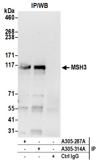
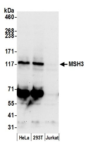 WB (Western Blot)
(Detection of human MSH3 by western blot. Samples: Whole cell lysate (50 ug) from HeLa, HEK293T, and Jurkat cells prepared using NETN lysis buffer. Antibody: Affinity purified rabbit anti-MSH3 antibody AAA213208 (lot AAA213208-1) used for WB at 0.1 ug/ml. Detection: Chemiluminescence with an exposure time of 30 seconds.)
WB (Western Blot)
(Detection of human MSH3 by western blot. Samples: Whole cell lysate (50 ug) from HeLa, HEK293T, and Jurkat cells prepared using NETN lysis buffer. Antibody: Affinity purified rabbit anti-MSH3 antibody AAA213208 (lot AAA213208-1) used for WB at 0.1 ug/ml. Detection: Chemiluminescence with an exposure time of 30 seconds.)
MSH3, Polyclonal Antibody (Cat# AAA213208)
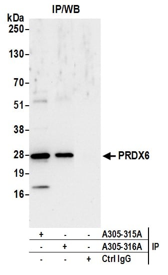
 WB (Western Blot)
(Detection of human and mouse PRDX6 by western blot. Samples: Whole cell lysate (15 ug) from HeLa, HEK293T, Jurkat, mouse TCMK-1, and mouse NIH 3T3 cells prepared using NETN lysis buffer. Antibody: Affinity purified rabbit anti-PRDX6 antibody AAA213209 (lot AAA213209-1) used for WB at 0.1 ug/ml. Detection: Chemiluminescence with an exposure time of 10 seconds.)
WB (Western Blot)
(Detection of human and mouse PRDX6 by western blot. Samples: Whole cell lysate (15 ug) from HeLa, HEK293T, Jurkat, mouse TCMK-1, and mouse NIH 3T3 cells prepared using NETN lysis buffer. Antibody: Affinity purified rabbit anti-PRDX6 antibody AAA213209 (lot AAA213209-1) used for WB at 0.1 ug/ml. Detection: Chemiluminescence with an exposure time of 10 seconds.)
PRDX6, Polyclonal Antibody (Cat# AAA213209)

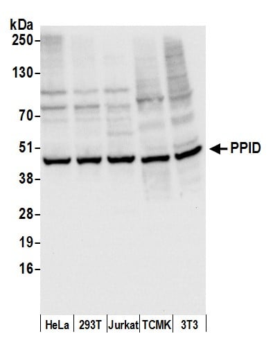 WB (Western Blot)
(Detection of human and mouse PPID by western blot. Samples: Whole cell lysate (50 ug) from HeLa, HEK293T, Jurkat, mouse TCMK-1, and mouse NIH 3T3 cells prepared using NETN lysis buffer. Antibody: Affinity purified rabbit anti-PPID antibody AAA213217 (lot AAA213217-1) used for WB at 0.1 ug/ml. Detection: Chemiluminescence with an exposure time of 3 seconds.)
WB (Western Blot)
(Detection of human and mouse PPID by western blot. Samples: Whole cell lysate (50 ug) from HeLa, HEK293T, Jurkat, mouse TCMK-1, and mouse NIH 3T3 cells prepared using NETN lysis buffer. Antibody: Affinity purified rabbit anti-PPID antibody AAA213217 (lot AAA213217-1) used for WB at 0.1 ug/ml. Detection: Chemiluminescence with an exposure time of 3 seconds.)
PPID, Polyclonal Antibody (Cat# AAA213217)
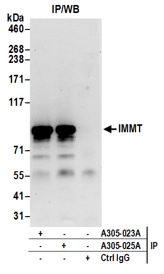
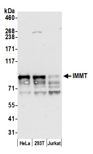 WB (Western Blot)
(Detection of human IMMT by western blot. Samples: Whole cell lysate (50 ug) from HeLa, HEK293T, and Jurkat cells prepared using NETN lysis buffer. Antibody: Affinity purified rabbit anti-IMMT antibody AAA213065 (lot AAA213065-1) used for WB at 0.04 ug/ml. Detection: Chemiluminescence with an exposure time of 30 seconds.)
WB (Western Blot)
(Detection of human IMMT by western blot. Samples: Whole cell lysate (50 ug) from HeLa, HEK293T, and Jurkat cells prepared using NETN lysis buffer. Antibody: Affinity purified rabbit anti-IMMT antibody AAA213065 (lot AAA213065-1) used for WB at 0.04 ug/ml. Detection: Chemiluminescence with an exposure time of 30 seconds.)
IMMT/Mitofilin, Polyclonal Antibody (Cat# AAA213065)
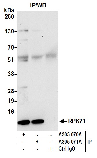
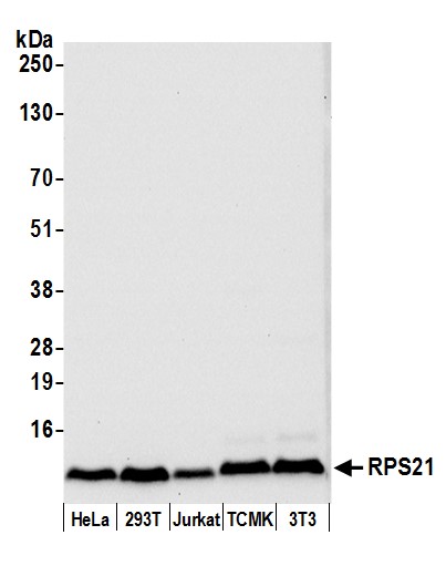 WB (Western Blot)
(Detection of human and mouse RPS21 by western blot. Samples: Whole cell lysate (50 ug) from HeLa, HEK293T, Jurkat, mouse TCMK-1, and mouse NIH 3T3 cells prepared using NETN lysis buffer. Antibody: Affinity purified rabbit anti-RPS21 antibody AAA213085 (lot AAA213085-1) used for WB at 0.04 ug/ml. Detection: Chemiluminescence with an exposure time of 10 seconds.)
WB (Western Blot)
(Detection of human and mouse RPS21 by western blot. Samples: Whole cell lysate (50 ug) from HeLa, HEK293T, Jurkat, mouse TCMK-1, and mouse NIH 3T3 cells prepared using NETN lysis buffer. Antibody: Affinity purified rabbit anti-RPS21 antibody AAA213085 (lot AAA213085-1) used for WB at 0.04 ug/ml. Detection: Chemiluminescence with an exposure time of 10 seconds.)
RPS21/Ribosomal Protein S21, Polyclonal Antibody (Cat# AAA213085)
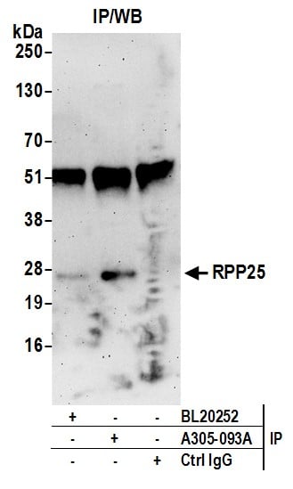
 WB (Western Blot)
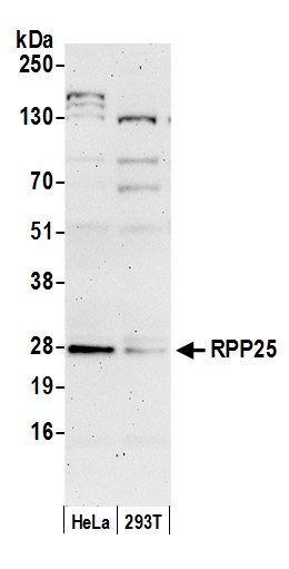
(Detection of human RPP25 by western blot. Samples: Whole cell lysate (50 ug) from HeLa and 293T cells prepared using NETN lysis buffer. Antibody: Affinity purified rabbit anti-RPP25 antibody AAA213096 (lot AAA213096-1) used for WB at 0.1 ug/ml. Detection: Chemiluminescence with an exposure time of 3 minutes.)
WB (Western Blot)
(Detection of human RPP25 by western blot. Samples: Whole cell lysate (50 ug) from HeLa and 293T cells prepared using NETN lysis buffer. Antibody: Affinity purified rabbit anti-RPP25 antibody AAA213096 (lot AAA213096-1) used for WB at 0.1 ug/ml. Detection: Chemiluminescence with an exposure time of 3 minutes.)
RPP25, Polyclonal Antibody (Cat# AAA213096)
What are Polyclonal Antibodies?
Polyclonal antibodies are antibodies that come from multiple B cell clones of a host animal. The typical hosts used for the majority of polyclonal antibody production are rabbits, goats, sheep, and donkeys. These polyclonal antibodies, once having identified their target, will bind to different epitopes located at different regions or sequences on the same protein/antigen. As a result, they are ideal at locating and binding to the target, even if the target is in very low concentrations (due to many different antibodies being able to bind to the same target molecule, which allows for significant amplification of a downstream signal).
Polyclonal antibodies are typically produced by injecting an antigen into a host animal, which causes the animal’s immune system to attack the foreign antigen by mass generating antibodies against it. After a period of time, serum is collected from the animal and purified using physicochemical fractionation, class-specific affinity purification, and/or antigen-affinity purification.
Key Uses of Polyclonal Antibodies
- Western Blotting: This method is used to find specific proteins in biological samples after separating them by size.
- Immunohistochemistry: IHC helps visualize the location of proteins in tissue sections using various staining techniques.
- ELISA: (Enzyme-Linked Immunosorbent Assay) is typically used to identify specific protein quantities in a sample. ELISAs can be either “Quantitative” or “Qualitative”.
- Flow Cytometry: technique that identifies and measures the specific protein on the surface or inside the cells in a fluid suspension.
- Immunoprecipitation: IP isolates and studies a specific protein from a complex mixture using antibodies.
Why Buy Polyclonal Antibodies from AAA Biotech?
1. Ideal for Various Applications
Our antibodies are generally going to be validated for use in multiple types of assays, including ELISA, Western Blotting, Immunohistochemistry, Immunoprecipitation, amongst others. They are ideal for a wide range of research applications.
2. Rigorous Quality Control
All of the antibodies in our catalog undergo strict quality testing to ensure specificity, sensitivity, and consistent performance. We are confident in the ability of our antibodies to provide you with accurate results.
3. Wide Assortment of Antibodies
Antibodies in are catalog can be found for both common and exotic species, and these antibodies are also available in both conjugated and recombinant forms to suit many diverse experimental needs.
4. Highly Purified
Our antibodies are available in purified forms with over 85% purity, as confirmed by SDS-PAGE. They are also available with tags such as His, Flag, GST, or MBP. We cater to customers worldwide.
FAQ
1. How are polyclonal antibodies produced?
Traditionally, polyclonal antibodies are produced by injecting an antigen into a host animal (such as a rabbit or goat), which then triggers an immune response from the host animal. The animal’s B cells produce antibodies that will recognize different parts of the injected antigen. These antibodies are then collected from the animal’s blood and purified for use.
2. How do polyclonal antibodies differ from monoclonal antibodies?
Polyclonal antibodies are a mix of antibodies that bind to different locations (epitopes) of the same antigen, while monoclonal antibodies are identical and bind to just one specific epitope. This makes polyclonal antibodies more versatile and better at detecting proteins that may be present in low quantities or in altered/modified forms.
3. How should I store polyclonal antibodies?
Polyclonal antibodies should be stored at 4°C for short-term use (up to a few weeks) and at -20°C or -80°C for long-term storage. Avoid repeated freeze-thaw cycles by dividing them into small aliquots. Always check the datasheet for specific storage instructions.