Filters
▼Clonality
▼Type
▼Reactivity
▼Gene Name
▼Isotype
▼Host
▼Application
▼Clone
▼Polyclonal Antibodies
At AAA Biotech also known as AAA Bio or AAABio, we provide a broad range of purified polyclonal antibodies (pAbs) that are able to all be browsed online through our website. Due to their high specificity and strong binding affinity, these antibodies are ideal for wide swathes of research and experimental applications.
Our polyclonal antibodies can easily support your work, whether you use them for Western Blotting, Immunocytochemistry (with or without Immunofluorescence used in conjunction), Immunohistochemistry, Immunoprecipitation, and ELISA tests. We highly encourage you to browse our range of pAbs and choose the one that best suits your experimental model.
Viewing 4600-4650 of 96812 product results
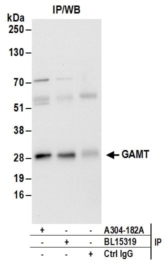
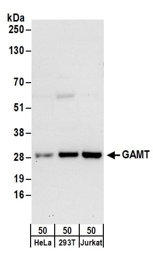 WB (Western Blot)
(Detection of human GAMT by western blot. Samples: Whole cell lysate (50 ug) from HeLa, HEK293T, and Jurkat cells. Antibodies: Affinity purified rabbit anti-GAMT antibody AAA212535 (lot AAA212535-1) used for WB at 0.4 ug/ml. Detection: Chemiluminescence with an exposure time of 30 seconds.)
WB (Western Blot)
(Detection of human GAMT by western blot. Samples: Whole cell lysate (50 ug) from HeLa, HEK293T, and Jurkat cells. Antibodies: Affinity purified rabbit anti-GAMT antibody AAA212535 (lot AAA212535-1) used for WB at 0.4 ug/ml. Detection: Chemiluminescence with an exposure time of 30 seconds.)
GAMT, Polyclonal Antibody (Cat# AAA212535)
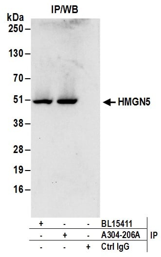
 WB (Western Blot)
(Detection of human HMGN5 by western blot. Samples: Whole cell lysate (50 ug) from HeLa, HEK293T, and Jurkat cells. Antibodies: Affinity purified rabbit anti-HMGN5 antibody AAA212543 (lot AAA212543-1) used for WB at 0.1 ug/ml. Detection: Chemiluminescence with an exposure time of 30 seconds.)
WB (Western Blot)
(Detection of human HMGN5 by western blot. Samples: Whole cell lysate (50 ug) from HeLa, HEK293T, and Jurkat cells. Antibodies: Affinity purified rabbit anti-HMGN5 antibody AAA212543 (lot AAA212543-1) used for WB at 0.1 ug/ml. Detection: Chemiluminescence with an exposure time of 30 seconds.)
HMGN5, Polyclonal Antibody (Cat# AAA212543)
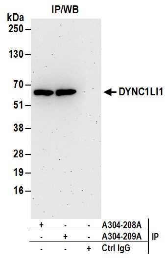
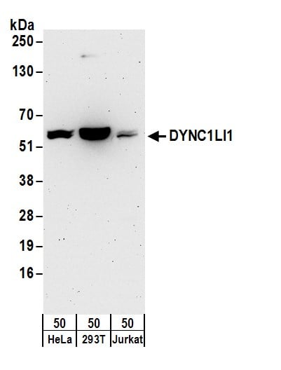 WB (Western Blot)
(Detection of human DYNC1LI1 by western blot. Samples: Whole cell lysate (50 ug) from HeLa, HEK293T, and Jurkat cells. Antibodies: Affinity purified rabbit anti-DYNC1LI1 antibody AAA212545 (lot AAA212545-1) used for WB at 0.1 ug/ml. Detection: Chemiluminescence with an exposure time of 3 minutes.)
WB (Western Blot)
(Detection of human DYNC1LI1 by western blot. Samples: Whole cell lysate (50 ug) from HeLa, HEK293T, and Jurkat cells. Antibodies: Affinity purified rabbit anti-DYNC1LI1 antibody AAA212545 (lot AAA212545-1) used for WB at 0.1 ug/ml. Detection: Chemiluminescence with an exposure time of 3 minutes.)
DYNC1LI1, Polyclonal Antibody (Cat# AAA212545)
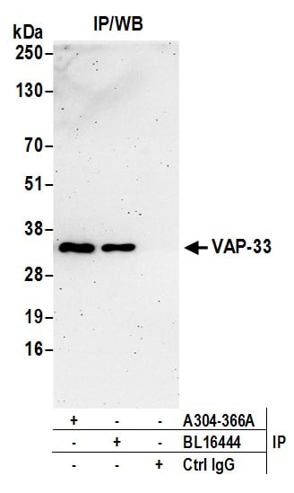
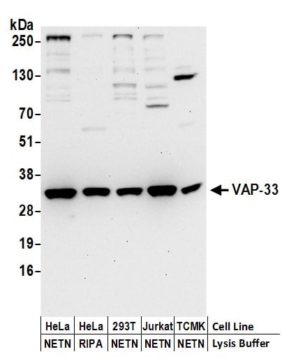 WB (Western Blot)
(Detection of human and mouse VAP-33 by western blot. Samples: Whole cell lysate (50 ug) prepared using NETN or RIPA buffer from HeLa, HEK293T, Jurkat, and mouse TCMK-1 cells. Antibodies: Affinity purified rabbit anti-VAP-33 antibody AAA212648 (lot AAA212648-1) used for WB at 0.4 ug/ml. Detection: Chemiluminescence with an exposure time of 30 seconds.)
WB (Western Blot)
(Detection of human and mouse VAP-33 by western blot. Samples: Whole cell lysate (50 ug) prepared using NETN or RIPA buffer from HeLa, HEK293T, Jurkat, and mouse TCMK-1 cells. Antibodies: Affinity purified rabbit anti-VAP-33 antibody AAA212648 (lot AAA212648-1) used for WB at 0.4 ug/ml. Detection: Chemiluminescence with an exposure time of 30 seconds.)
VAP-33, Polyclonal Antibody (Cat# AAA212648)
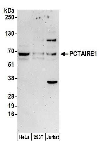 WB (Western Blot)
(Detection of human PCTAIRE1 by western blot. Samples: Whole cell lysate (50 ug) prepared using NETN buffer from HeLa, HEK293T, and Jurkat cells. Antibodies: Affinity purified rabbit anti-PCTAIRE1 antibody AAA212651 (lot AAA212651-1) used for WB at 0.1 ug/ml. Detection: Chemiluminescence with an exposure time of 3 minutes.)
WB (Western Blot)
(Detection of human PCTAIRE1 by western blot. Samples: Whole cell lysate (50 ug) prepared using NETN buffer from HeLa, HEK293T, and Jurkat cells. Antibodies: Affinity purified rabbit anti-PCTAIRE1 antibody AAA212651 (lot AAA212651-1) used for WB at 0.1 ug/ml. Detection: Chemiluminescence with an exposure time of 3 minutes.)
PCTAIRE1, Polyclonal Antibody (Cat# AAA212651)
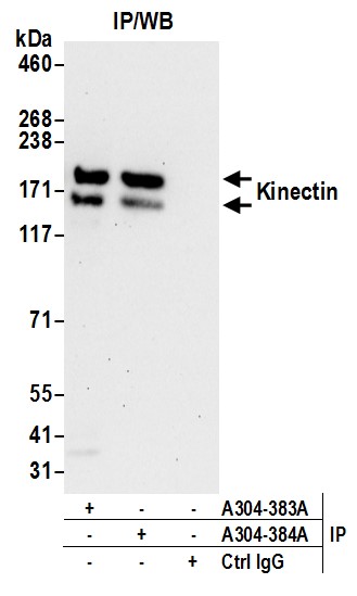
 WB (Western Blot)
(Detection of human Kinectin by western blot. Samples: Whole cell lysate (50 ug) prepared using NETN buffer from HeLa, HEK293T, and Jurkat cells. Antibodies: Affinity purified rabbit anti-Kinectin antibody AAA212659 (lot AAA212659-1) used for WB at 0.1 ug/ml. Detection: Chemiluminescence with an exposure time of 30 seconds.)
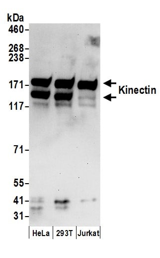
WB (Western Blot)
(Detection of human Kinectin by western blot. Samples: Whole cell lysate (50 ug) prepared using NETN buffer from HeLa, HEK293T, and Jurkat cells. Antibodies: Affinity purified rabbit anti-Kinectin antibody AAA212659 (lot AAA212659-1) used for WB at 0.1 ug/ml. Detection: Chemiluminescence with an exposure time of 30 seconds.)
Kinectin, Polyclonal Antibody (Cat# AAA212659)
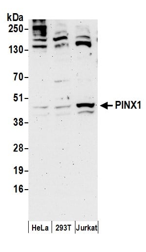 WB (Western Blot)
(Detection of human PINX1 by western blot. Samples: Whole cell lysate (50 ug) prepared using NETN buffer from HeLa, HEK293T, and Jurkat cells. Antibodies: Affinity purified rabbit anti-PINX1 antibody AAA212664 (lot AAA212664-1) used for WB at 0.4 ug/ml. Detection: Chemiluminescence with an exposure time of 3 minutes.)
WB (Western Blot)
(Detection of human PINX1 by western blot. Samples: Whole cell lysate (50 ug) prepared using NETN buffer from HeLa, HEK293T, and Jurkat cells. Antibodies: Affinity purified rabbit anti-PINX1 antibody AAA212664 (lot AAA212664-1) used for WB at 0.4 ug/ml. Detection: Chemiluminescence with an exposure time of 3 minutes.)
PINX1, Polyclonal Antibody (Cat# AAA212664)
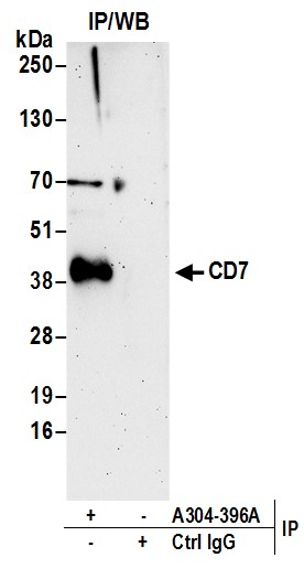
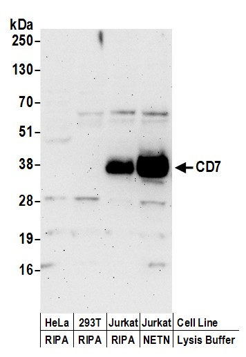 WB (Western Blot)
(Detection of human CD7 by western blot. Samples: Whole cell lysate (50 ug) prepared using NETN or RIPA buffer from HeLa, HEK293T, and Jurkat cells. Antibodies: Affinity purified rabbit anti-CD7 antibody AAA212667 (lot AAA212667-1) used for WB at 0.4 ug/ml. Detection: Chemiluminescence with an exposure time of 3 minutes.)
WB (Western Blot)
(Detection of human CD7 by western blot. Samples: Whole cell lysate (50 ug) prepared using NETN or RIPA buffer from HeLa, HEK293T, and Jurkat cells. Antibodies: Affinity purified rabbit anti-CD7 antibody AAA212667 (lot AAA212667-1) used for WB at 0.4 ug/ml. Detection: Chemiluminescence with an exposure time of 3 minutes.)
CD7, Polyclonal Antibody (Cat# AAA212667)
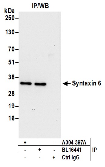
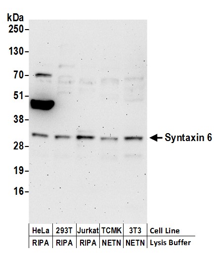 WB (Western Blot)
(Detection of human and mouse Syntaxin 6 by western blot. Samples: Whole cell lysate (50 ug) prepared using NETN or RIPA buffer from HeLa, HEK293T, Jurkat, mouse TCMK-1, and mouse NIH 3T3 cells. Antibodies: Affinity purified rabbit anti-Syntaxin 6 antibody AAA212668 (lot AAA212668-1) used for WB at 0.1 ug/ml. Detection: Chemiluminescence with an exposure time of 3 minutes.)
WB (Western Blot)
(Detection of human and mouse Syntaxin 6 by western blot. Samples: Whole cell lysate (50 ug) prepared using NETN or RIPA buffer from HeLa, HEK293T, Jurkat, mouse TCMK-1, and mouse NIH 3T3 cells. Antibodies: Affinity purified rabbit anti-Syntaxin 6 antibody AAA212668 (lot AAA212668-1) used for WB at 0.1 ug/ml. Detection: Chemiluminescence with an exposure time of 3 minutes.)
Syntaxin 6, Polyclonal Antibody (Cat# AAA212668)
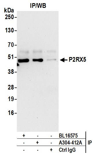
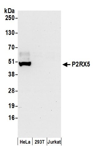 WB (Western Blot)
(Detection of human P2RX5 by western blot. Samples: Whole cell lysate (50 ug) prepared using NETN buffer from HeLa, HEK293T, and Jurkat cells. Antibodies: Affinity purified rabbit anti-P2RX5 antibody AAA212677 (lot AAA212677-1) used for WB at 0.1 ug/ml. Detection: Chemiluminescence with an exposure time of 30 seconds.)
WB (Western Blot)
(Detection of human P2RX5 by western blot. Samples: Whole cell lysate (50 ug) prepared using NETN buffer from HeLa, HEK293T, and Jurkat cells. Antibodies: Affinity purified rabbit anti-P2RX5 antibody AAA212677 (lot AAA212677-1) used for WB at 0.1 ug/ml. Detection: Chemiluminescence with an exposure time of 30 seconds.)
P2RX5, Polyclonal Antibody (Cat# AAA212677)
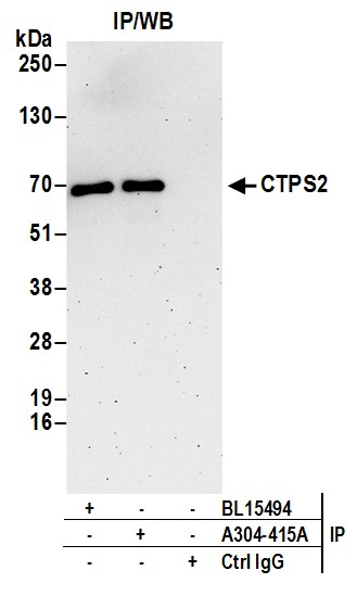
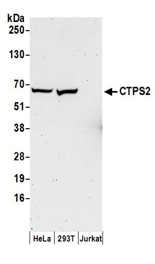 WB (Western Blot)
(Detection of human CTPS2 by western blot. Samples: Whole cell lysate (50 ug) prepared using NETN buffer from HeLa, HEK293T, and Jurkat cells. Antibodies: Affinity purified rabbit anti-CTPS2 antibody AAA212678 (lot AAA212678-1) used for WB at 0.4 ug/ml. Detection: Chemiluminescence with an exposure time of 3 minutes.)
WB (Western Blot)
(Detection of human CTPS2 by western blot. Samples: Whole cell lysate (50 ug) prepared using NETN buffer from HeLa, HEK293T, and Jurkat cells. Antibodies: Affinity purified rabbit anti-CTPS2 antibody AAA212678 (lot AAA212678-1) used for WB at 0.4 ug/ml. Detection: Chemiluminescence with an exposure time of 3 minutes.)
CTPS2, Polyclonal Antibody (Cat# AAA212678)
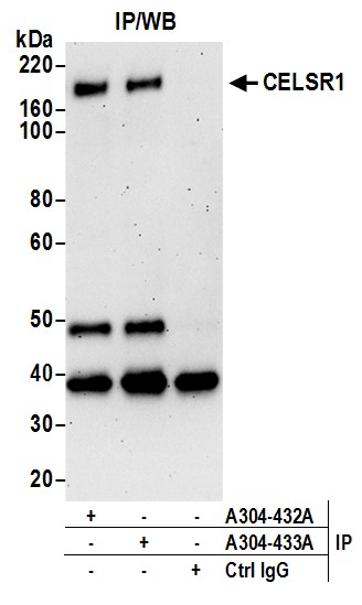
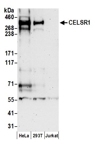 WB (Western Blot)
(Detection of human CELSR1 by western blot. Samples: Whole cell lysate (50 ug) prepared using NETN buffer from HeLa, HEK293T, and Jurkat cells. Antibodies: Affinity purified rabbit anti-CELSR1 antibody AAA212687 (lot AAA212687-1) used for WB at 0.4 ug/ml. Detection: Chemiluminescence with an exposure time of 3 minutes.)
WB (Western Blot)
(Detection of human CELSR1 by western blot. Samples: Whole cell lysate (50 ug) prepared using NETN buffer from HeLa, HEK293T, and Jurkat cells. Antibodies: Affinity purified rabbit anti-CELSR1 antibody AAA212687 (lot AAA212687-1) used for WB at 0.4 ug/ml. Detection: Chemiluminescence with an exposure time of 3 minutes.)
CELSR1, Polyclonal Antibody (Cat# AAA212687)
 WB (Western Blot)
(Detection of human MCT4 by western blot. Samples: Whole cell lysate (50 ug) from HeLa, GaMG, Hep-G2, LNCaP, and Jurkat cells prepared using NETN lysis buffer. Antibody: Affinity purified rabbit anti-MCT4 antibody AAA212693 (lot AAA212693-1) used for WB at 0.1 ug/ml. Detection: Chemiluminescence with an exposure time of 75 seconds.)
WB (Western Blot)
(Detection of human MCT4 by western blot. Samples: Whole cell lysate (50 ug) from HeLa, GaMG, Hep-G2, LNCaP, and Jurkat cells prepared using NETN lysis buffer. Antibody: Affinity purified rabbit anti-MCT4 antibody AAA212693 (lot AAA212693-1) used for WB at 0.1 ug/ml. Detection: Chemiluminescence with an exposure time of 75 seconds.)
MCT4, Polyclonal Antibody (Cat# AAA212693)
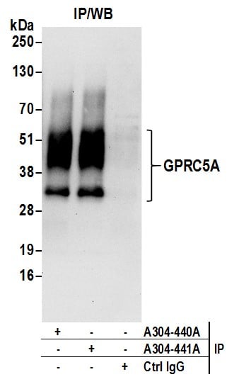
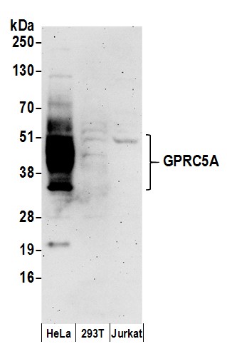 WB (Western Blot)
(Detection of human GPRC5A by western blot. Samples: Whole cell lysate (50 ug) prepared using NETN buffer from HeLa, HEK293T, and Jurkat cells. Antibodies: Affinity purified rabbit anti-GPRC5A antibody AAA212694 (lot AAA212694-1) used for WB at 0.1 ug/ml. Detection: Chemiluminescence with an exposure time of 3 minutes.)
WB (Western Blot)
(Detection of human GPRC5A by western blot. Samples: Whole cell lysate (50 ug) prepared using NETN buffer from HeLa, HEK293T, and Jurkat cells. Antibodies: Affinity purified rabbit anti-GPRC5A antibody AAA212694 (lot AAA212694-1) used for WB at 0.1 ug/ml. Detection: Chemiluminescence with an exposure time of 3 minutes.)
GPRC5A, Polyclonal Antibody (Cat# AAA212694)
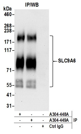
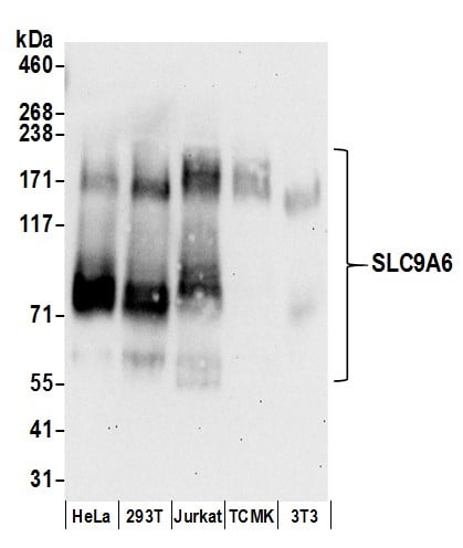 WB (Western Blot)
(Detection of human and mouse SLC9A6 by western blot. Samples: Whole cell lysate (50 ug) prepared using NETN buffer from HeLa, HEK293T, Jurkat, mouse TCMK-1, and mouse NIH 3T3 cells. Antibodies: Affinity purified rabbit anti-SLC9A6 antibody AAA212699 (lot AAA212699-1) used for WB at 0.1 ug/ml. Detection: Chemiluminescence with an exposure time of 30 seconds.)
WB (Western Blot)
(Detection of human and mouse SLC9A6 by western blot. Samples: Whole cell lysate (50 ug) prepared using NETN buffer from HeLa, HEK293T, Jurkat, mouse TCMK-1, and mouse NIH 3T3 cells. Antibodies: Affinity purified rabbit anti-SLC9A6 antibody AAA212699 (lot AAA212699-1) used for WB at 0.1 ug/ml. Detection: Chemiluminescence with an exposure time of 30 seconds.)
SLC9A6, Polyclonal Antibody (Cat# AAA212699)
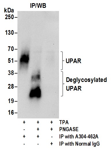
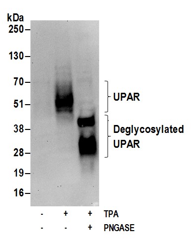 WB (Western Blot)
(Detection of human glycosylated and deglycosylated UPAR by western blot. Samples: Whole cell lysate (50 ug) from U-937 cells incubated with (+) or without (-) TPA (200nM, 72 hrs) and lysed using NETN lysis buffer. The lysate was then treated (+) or mock treated (-) with PnGase F. Antibodies: Affinity purified rabbit anti-UPAR antibody AAA212709 (lot AAA212709-1) used for WB at 1.0 ug/ml. Detection: Chemiluminescence with an exposure time of 30 seconds.)
WB (Western Blot)
(Detection of human glycosylated and deglycosylated UPAR by western blot. Samples: Whole cell lysate (50 ug) from U-937 cells incubated with (+) or without (-) TPA (200nM, 72 hrs) and lysed using NETN lysis buffer. The lysate was then treated (+) or mock treated (-) with PnGase F. Antibodies: Affinity purified rabbit anti-UPAR antibody AAA212709 (lot AAA212709-1) used for WB at 1.0 ug/ml. Detection: Chemiluminescence with an exposure time of 30 seconds.)
UPAR, Polyclonal Antibody (Cat# AAA212709)
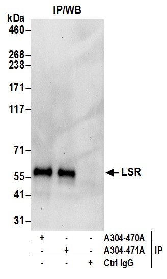
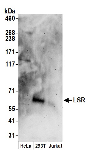 WB (Western Blot)
(Detection of human LSR by western blot. Samples: Whole cell lysate (50 ug) from HeLa, HEK293T, and Jurkat cells prepared using RIPA lysis buffer. Antibodies: Affinity purified rabbit anti-LSR antibody AAA212715 (lot AAA212715-1) used for WB at 0.1 ug/ml. Detection: Chemiluminescence with an exposure time of 3 minutes.)
WB (Western Blot)
(Detection of human LSR by western blot. Samples: Whole cell lysate (50 ug) from HeLa, HEK293T, and Jurkat cells prepared using RIPA lysis buffer. Antibodies: Affinity purified rabbit anti-LSR antibody AAA212715 (lot AAA212715-1) used for WB at 0.1 ug/ml. Detection: Chemiluminescence with an exposure time of 3 minutes.)
LSR, Polyclonal Antibody (Cat# AAA212715)
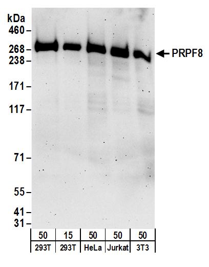 WB (Western Blot)
(Detection of human and mouse PRPF8 by western blot. Samples: Whole cell lysate from HEK293T (15 and 50 ug), HeLa (50ug), Jurkat (50ug), and mouse NIH 3T3 (50ug) cells. Antibodies: Affinity purified rabbit anti-PRPF8 antibody AAA212377 (lot AAA212377-1) used for WB at 0.1 ug/ml. Detection: Chemiluminescence with an exposure time of 3 minutes.)
WB (Western Blot)
(Detection of human and mouse PRPF8 by western blot. Samples: Whole cell lysate from HEK293T (15 and 50 ug), HeLa (50ug), Jurkat (50ug), and mouse NIH 3T3 (50ug) cells. Antibodies: Affinity purified rabbit anti-PRPF8 antibody AAA212377 (lot AAA212377-1) used for WB at 0.1 ug/ml. Detection: Chemiluminescence with an exposure time of 3 minutes.)
PRPF8, Polyclonal Antibody (Cat# AAA212377)
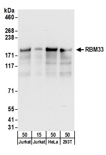
 WB (Western Blot)
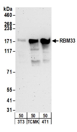
(Detection of mouse RBM33 by western blot. Samples: Whole cell lysate from mouse NIH 3T3 (50ug), TCMK-1 (50ug), and 4T1 (50ug) cells. Antibodies: Affinity purified rabbit anti-RBM33 antibody AAA212381 (lot AAA212381-1) used for WB at 0.4 ug/ml. Detection: Chemiluminescence with an exposure time of 3 minutes.)
WB (Western Blot)
(Detection of mouse RBM33 by western blot. Samples: Whole cell lysate from mouse NIH 3T3 (50ug), TCMK-1 (50ug), and 4T1 (50ug) cells. Antibodies: Affinity purified rabbit anti-RBM33 antibody AAA212381 (lot AAA212381-1) used for WB at 0.4 ug/ml. Detection: Chemiluminescence with an exposure time of 3 minutes.)
RBM33, Polyclonal Antibody (Cat# AAA212381)
 WB (Western Blot)
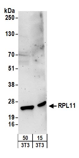
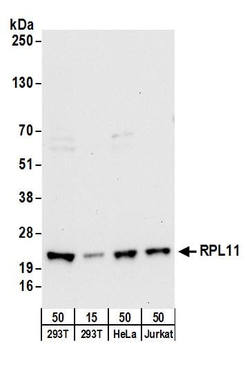
(Detection of human RPL11 by western blot. Samples: Whole cell lysate from HEK293T (15 and 50 ug), HeLa (50ug), and Jurkat (50ug) cells. Antibodies: Affinity purified rabbit anti-RPL11 antibody AAA212384 (lot AAA212384-1) used for WB at 0.4 ug/ml. Detection: Chemiluminescence with an exposure time of 30 seconds.)
WB (Western Blot)
(Detection of human RPL11 by western blot. Samples: Whole cell lysate from HEK293T (15 and 50 ug), HeLa (50ug), and Jurkat (50ug) cells. Antibodies: Affinity purified rabbit anti-RPL11 antibody AAA212384 (lot AAA212384-1) used for WB at 0.4 ug/ml. Detection: Chemiluminescence with an exposure time of 30 seconds.)
RPL11, Polyclonal Antibody (Cat# AAA212384)

 WB (Western Blot)
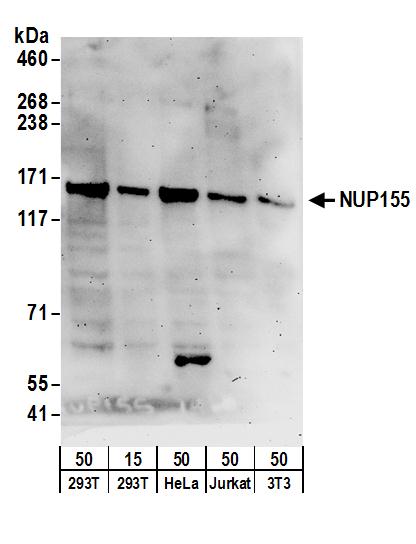
(Detection of human and mouse NUP155 by western blot. Samples: Whole cell lysate from HEK293T (15 and 50 ug), HeLa (50ug), Jurkat (50ug), and mouse NIH 3T3 (50ug) cells. Antibodies: Affinity purified rabbit anti-NUP155 antibody AAA212387 (lot AAA212387-1) used for WB at 0.1 ug/ml. Detection: Chemiluminescence with an exposure time of 3 minutes.)
WB (Western Blot)
(Detection of human and mouse NUP155 by western blot. Samples: Whole cell lysate from HEK293T (15 and 50 ug), HeLa (50ug), Jurkat (50ug), and mouse NIH 3T3 (50ug) cells. Antibodies: Affinity purified rabbit anti-NUP155 antibody AAA212387 (lot AAA212387-1) used for WB at 0.1 ug/ml. Detection: Chemiluminescence with an exposure time of 3 minutes.)
NUP155, Polyclonal Antibody (Cat# AAA212387)

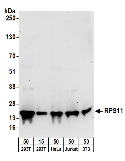
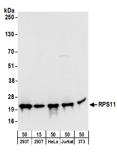 WB (Western Blot)
(Detection of human and mouse RPS11 by western blot. Samples: Whole cell lysate from HEK293T (15 and 50 ug), HeLa (50ug), Jurkat (50ug), and mouse NIH 3T3 (50ug) cells. Antibodies: Affinity purified rabbit anti-RPS11 antibody AAA212389 (lot AAA212389-1) used for WB at 0.1 ug/ml. Detection: Chemiluminescence with an exposure time of 10 seconds.)
WB (Western Blot)
(Detection of human and mouse RPS11 by western blot. Samples: Whole cell lysate from HEK293T (15 and 50 ug), HeLa (50ug), Jurkat (50ug), and mouse NIH 3T3 (50ug) cells. Antibodies: Affinity purified rabbit anti-RPS11 antibody AAA212389 (lot AAA212389-1) used for WB at 0.1 ug/ml. Detection: Chemiluminescence with an exposure time of 10 seconds.)
RPS11, Polyclonal Antibody (Cat# AAA212389)
 WB (Western Blot)
(Detection of human and mouse RPS11 by western blot. Samples: Whole cell lysate from HEK293T (15 and 50 ug), HeLa (50ug), Jurkat (50ug), and mouse NIH 3T3 (50ug) cells. Antibodies: Affinity purified rabbit anti-RPS11 antibody AAA212390 (lot AAA212390-1) used for WB at 0.1 ug/ml. Detection: Chemiluminescence with an exposure time of 30 seconds.)
WB (Western Blot)
(Detection of human and mouse RPS11 by western blot. Samples: Whole cell lysate from HEK293T (15 and 50 ug), HeLa (50ug), Jurkat (50ug), and mouse NIH 3T3 (50ug) cells. Antibodies: Affinity purified rabbit anti-RPS11 antibody AAA212390 (lot AAA212390-1) used for WB at 0.1 ug/ml. Detection: Chemiluminescence with an exposure time of 30 seconds.)
RPS11, Polyclonal Antibody (Cat# AAA212390)
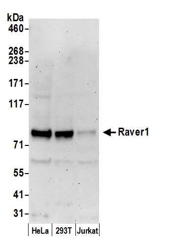 WB (Western Blot)
(Detection of human Raver1 by western blot. Samples: Whole cell lysate (50 ug) from HeLa, HEK293T, and Jurkat cells prepared using NETN lysis buffer. Antibody: Affinity purified rabbit anti-Raver1 antibody AAA212392 (lot AAA212392-1) used for WB at 1 ug/ml. Detection: Chemiluminescence with an exposure time of 3 minutes.)
WB (Western Blot)
(Detection of human Raver1 by western blot. Samples: Whole cell lysate (50 ug) from HeLa, HEK293T, and Jurkat cells prepared using NETN lysis buffer. Antibody: Affinity purified rabbit anti-Raver1 antibody AAA212392 (lot AAA212392-1) used for WB at 1 ug/ml. Detection: Chemiluminescence with an exposure time of 3 minutes.)
Raver1, Polyclonal Antibody (Cat# AAA212392)
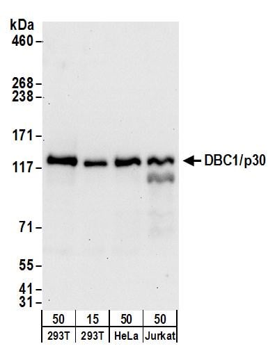
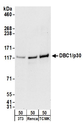 WB (Western Blot)
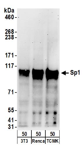
(Detection of mouse DBC1/p30 by western blot. Samples: Whole cell lysate (50 ug) from NIH 3T3, Renca, and TCMK-1 cells. Antibodies: Affinity purified goat anti-DBC1/p30 antibody AAA212394 (lot AAA212394-1) used for WB at 0.1 ug/ml. Detection: Chemiluminescence with an exposure time of 30 seconds.)
WB (Western Blot)
(Detection of mouse DBC1/p30 by western blot. Samples: Whole cell lysate (50 ug) from NIH 3T3, Renca, and TCMK-1 cells. Antibodies: Affinity purified goat anti-DBC1/p30 antibody AAA212394 (lot AAA212394-1) used for WB at 0.1 ug/ml. Detection: Chemiluminescence with an exposure time of 30 seconds.)
DBC1/p30 DBC, Polyclonal Antibody (Cat# AAA212394)
 WB (Western Blot)
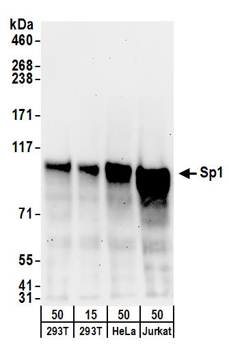
(Detection of human Sp1 by western blot. Samples: Whole cell lysate from HEK293T (15 and 50 ug), HeLa (50ug), and Jurkat (50ug) cells. Antibodies: Affinity purified goat anti-Sp1 antibody AAA212396 (lot AAA212396-1) used for WB at 0.1 ug/ml. Detection: Chemiluminescence with an exposure time of 3 minutes.)
WB (Western Blot)
(Detection of human Sp1 by western blot. Samples: Whole cell lysate from HEK293T (15 and 50 ug), HeLa (50ug), and Jurkat (50ug) cells. Antibodies: Affinity purified goat anti-Sp1 antibody AAA212396 (lot AAA212396-1) used for WB at 0.1 ug/ml. Detection: Chemiluminescence with an exposure time of 3 minutes.)
Sp1, Polyclonal Antibody (Cat# AAA212396)
 WB (Western Blot)
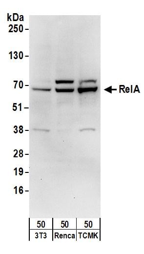
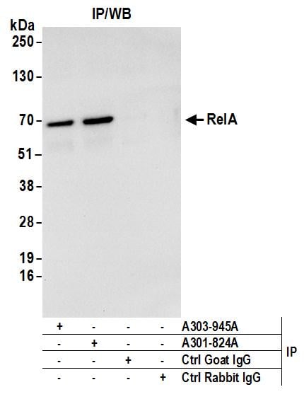
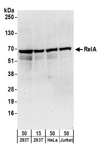
(Detection of human RelA by western blot. Samples: Whole cell lysate from HEK293T (15 and 50 ug), HeLa (50ug), and Jurkat (50ug) cells. Antibodies: Affinity purified goat anti-RelA antibody AAA212397 (lot AAA212397-1) used for WB at 0.1 ug/ml. Detection: Chemiluminescence with an exposure time of 3 minutes.)
WB (Western Blot)
(Detection of human RelA by western blot. Samples: Whole cell lysate from HEK293T (15 and 50 ug), HeLa (50ug), and Jurkat (50ug) cells. Antibodies: Affinity purified goat anti-RelA antibody AAA212397 (lot AAA212397-1) used for WB at 0.1 ug/ml. Detection: Chemiluminescence with an exposure time of 3 minutes.)
RelA, Polyclonal Antibody (Cat# AAA212397)
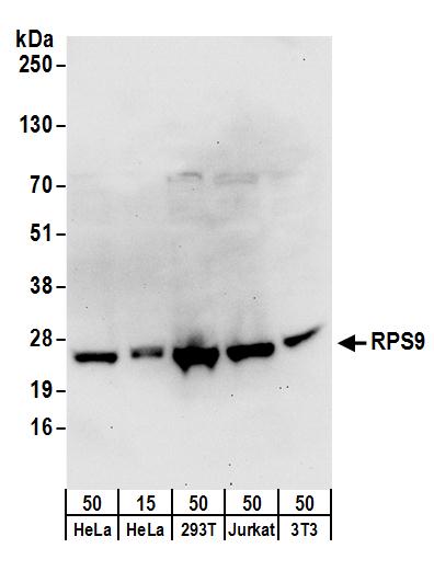 WB (Western Blot)
(Detection of human and mouse RPS9 by western blot. Samples: Whole cell lysate from HeLa (15 and 50 ug), HEK293T (50ug), Jurkat (50ug), and mouse NIH 3T3 (50ug) cells. Antibodies: Affinity purified rabbit anti-RPS9 antibody AAA212398 (lot AAA212398-1) used for WB at 0.4 ug/ml. Detection: Chemiluminescence with an exposure time of 30 seconds.)
WB (Western Blot)
(Detection of human and mouse RPS9 by western blot. Samples: Whole cell lysate from HeLa (15 and 50 ug), HEK293T (50ug), Jurkat (50ug), and mouse NIH 3T3 (50ug) cells. Antibodies: Affinity purified rabbit anti-RPS9 antibody AAA212398 (lot AAA212398-1) used for WB at 0.4 ug/ml. Detection: Chemiluminescence with an exposure time of 30 seconds.)
RPS9, Polyclonal Antibody (Cat# AAA212398)
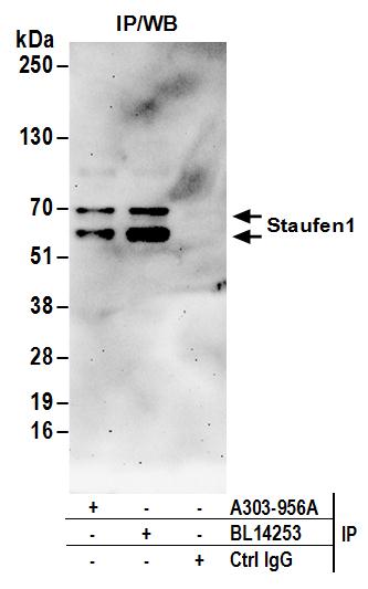
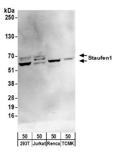 WB (Western Blot)
(Detection of human and mouse Staufen1 by western blot. Samples: Whole cell lysate (50 ug) from HEK293T, Jurkat, mouse Renca, and mouse TCMK-1 cells. Antibodies: Affinity purified rabbit anti-Staufen1 antibody AAA212405 (lot AAA212405-1) used for WB at 0.4 ug/ml. Detection: Chemiluminescence with an exposure time of 30 seconds.)
WB (Western Blot)
(Detection of human and mouse Staufen1 by western blot. Samples: Whole cell lysate (50 ug) from HEK293T, Jurkat, mouse Renca, and mouse TCMK-1 cells. Antibodies: Affinity purified rabbit anti-Staufen1 antibody AAA212405 (lot AAA212405-1) used for WB at 0.4 ug/ml. Detection: Chemiluminescence with an exposure time of 30 seconds.)
Staufen1, Polyclonal Antibody (Cat# AAA212405)
 WB (Western Blot)
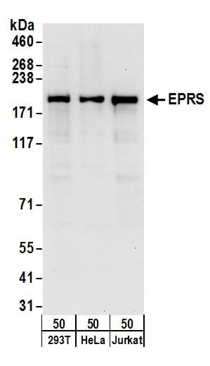
(Detection of human EPRS by western blot. Samples: Whole cell lysate (50 ug) from HEK293T, HeLa, and Jurkat cells. Antibodies: Affinity purified rabbit anti-EPRS antibody AAA212406 (lot AAA212406-1) used for WB at 0.1 ug/ml. Detection: Chemiluminescence with an exposure time of 10 seconds.)
WB (Western Blot)
(Detection of human EPRS by western blot. Samples: Whole cell lysate (50 ug) from HEK293T, HeLa, and Jurkat cells. Antibodies: Affinity purified rabbit anti-EPRS antibody AAA212406 (lot AAA212406-1) used for WB at 0.1 ug/ml. Detection: Chemiluminescence with an exposure time of 10 seconds.)
EPRS, Polyclonal Antibody (Cat# AAA212406)
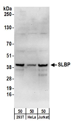 WB (Western Blot)
(Detection of human SLBP by western blot. Samples: Whole cell lysate (50 ug) from HEK293T, HeLa, and Jurkat cells. Antibodies: Affinity purified rabbit anti-SLBP antibody AAA212415 (lot AAA212415-1) used for WB at 0.4 ug/ml. Detection: Chemiluminescence with an exposure time of 30 seconds.)
WB (Western Blot)
(Detection of human SLBP by western blot. Samples: Whole cell lysate (50 ug) from HEK293T, HeLa, and Jurkat cells. Antibodies: Affinity purified rabbit anti-SLBP antibody AAA212415 (lot AAA212415-1) used for WB at 0.4 ug/ml. Detection: Chemiluminescence with an exposure time of 30 seconds.)
SLBP, Polyclonal Antibody (Cat# AAA212415)
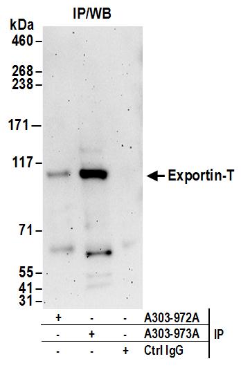
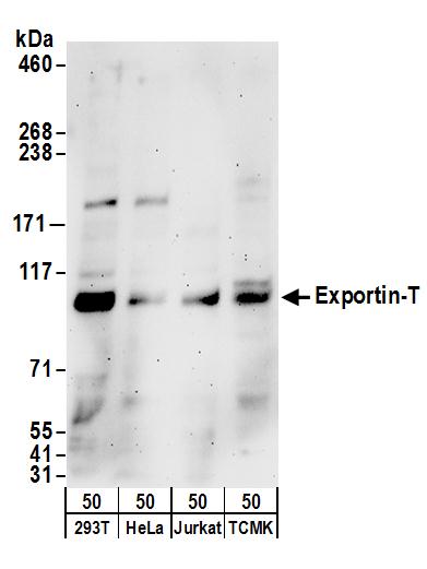 WB (Western Blot)
(Detection of human and mouse Exportin-T by western blot. Samples: Whole cell lysate (50 ug) from HEK293T, HeLa, Jurkat, and mouse TCMK-1 cells. Antibodies: Affinity purified rabbit anti-Exportin-T antibody AAA212418 (lot AAA212418-1) used for WB at 0.4 ug/ml. Detection: Chemiluminescence with an exposure time of 3 minutes.)
WB (Western Blot)
(Detection of human and mouse Exportin-T by western blot. Samples: Whole cell lysate (50 ug) from HEK293T, HeLa, Jurkat, and mouse TCMK-1 cells. Antibodies: Affinity purified rabbit anti-Exportin-T antibody AAA212418 (lot AAA212418-1) used for WB at 0.4 ug/ml. Detection: Chemiluminescence with an exposure time of 3 minutes.)
Exportin-T, Polyclonal Antibody (Cat# AAA212418)
 WB (Western Blot)
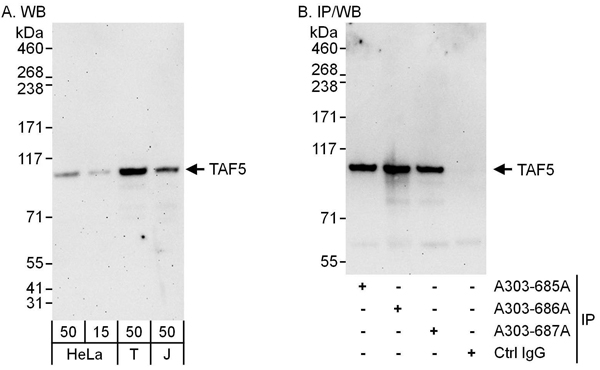
(Detection of human TAF5 by western blot and immunoprecipitation. Samples: Whole cell lysate from HeLa (15 and 50 ug for WB; 1 mg for IP, 20% of IP loaded), HEK293T (T; 50 ug) and Jurkat (J; 50 ug) cells. Antibodies: Affinity purified rabbit anti-TAF5 antibody AAA212260 used for WB at 0.1 ug/ml (A) and 1 ug/ml (B) and used for IP at 6 ug/mg lysate. TAF5 was also immunoprecipitated by rabbit anti-TAF5 antibodies and which recognize other epitopes. Detection: Chemiluminescence with exposure times of 3 minutes (A) and 30 seconds (B).)
WB (Western Blot)
(Detection of human TAF5 by western blot and immunoprecipitation. Samples: Whole cell lysate from HeLa (15 and 50 ug for WB; 1 mg for IP, 20% of IP loaded), HEK293T (T; 50 ug) and Jurkat (J; 50 ug) cells. Antibodies: Affinity purified rabbit anti-TAF5 antibody AAA212260 used for WB at 0.1 ug/ml (A) and 1 ug/ml (B) and used for IP at 6 ug/mg lysate. TAF5 was also immunoprecipitated by rabbit anti-TAF5 antibodies and which recognize other epitopes. Detection: Chemiluminescence with exposure times of 3 minutes (A) and 30 seconds (B).)
TAF5, Polyclonal Antibody (Cat# AAA212260)

 WB (Western Blot)
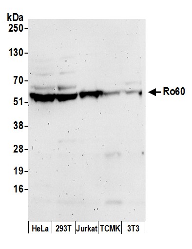
(Detection of human and mouse Ro60 by western blot. Samples: Whole cell lysate (50 ug) from HeLa, HEK293T, Jurkat, mouse TCMK-1, and mouse NIH 3T3 cells prepared using NETN lysis buffer. Antibody: Affinity purified rabbit anti-Ro60 antibody AAA212264 (lot AAA212264-2) used for WB at 0.1 ug/ml. Detection: Chemiluminescence with an exposure time of 3 minutes.)
WB (Western Blot)
(Detection of human and mouse Ro60 by western blot. Samples: Whole cell lysate (50 ug) from HeLa, HEK293T, Jurkat, mouse TCMK-1, and mouse NIH 3T3 cells prepared using NETN lysis buffer. Antibody: Affinity purified rabbit anti-Ro60 antibody AAA212264 (lot AAA212264-2) used for WB at 0.1 ug/ml. Detection: Chemiluminescence with an exposure time of 3 minutes.)
Ro60, Polyclonal Antibody (Cat# AAA212264)
 WB (Western Blot)
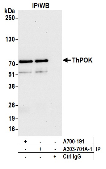
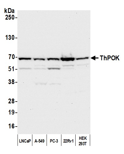
(Detection of human ThPOK by western blot. Samples: Whole cell lysate (50 ug) from LNCaP, A-549, PC-3, 22Rv1, and HEK293T cells prepared using NETN lysis buffer. Antibody: Affinity purified rabbit anti-ThPOK antibody (AAA212268 lot 1) used for WB at 0.1 ug/ml. Detection: Chemiluminescence with an exposure time of 10 seconds.)
WB (Western Blot)
(Detection of human ThPOK by western blot. Samples: Whole cell lysate (50 ug) from LNCaP, A-549, PC-3, 22Rv1, and HEK293T cells prepared using NETN lysis buffer. Antibody: Affinity purified rabbit anti-ThPOK antibody (AAA212268 lot 1) used for WB at 0.1 ug/ml. Detection: Chemiluminescence with an exposure time of 10 seconds.)
ThPOK, Polyclonal Antibody (Cat# AAA212268)

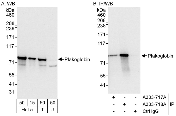 WB (Western Blot)
(Detection of human Plakoglobin by western blot and immunoprecipitation. Samples: Whole cell lysate from HeLa (15 and 50 ug for WB; 1 mg for IP, 20% of IP loaded), HEK293T (T; 50 ug) and Jurkat (J; 50 ug) cells. Antibodies: Affinity purified rabbit anti-Plakoglobin antibody AAA212271 used for WB at 0.1 ug/ml (A) and 1 ug/ml (B) and used for IP at 6 ug/mg lysate. Plakoglobin was also immunoprecipitated by rabbit anti-Plakoglobin antibody which recognizes an upstream epitope. Detection: Chemiluminescence with exposure times of 10 seconds (A) and 3 seconds (B).)
WB (Western Blot)
(Detection of human Plakoglobin by western blot and immunoprecipitation. Samples: Whole cell lysate from HeLa (15 and 50 ug for WB; 1 mg for IP, 20% of IP loaded), HEK293T (T; 50 ug) and Jurkat (J; 50 ug) cells. Antibodies: Affinity purified rabbit anti-Plakoglobin antibody AAA212271 used for WB at 0.1 ug/ml (A) and 1 ug/ml (B) and used for IP at 6 ug/mg lysate. Plakoglobin was also immunoprecipitated by rabbit anti-Plakoglobin antibody which recognizes an upstream epitope. Detection: Chemiluminescence with exposure times of 10 seconds (A) and 3 seconds (B).)
Plakoglobin, Polyclonal Antibody (Cat# AAA212271)
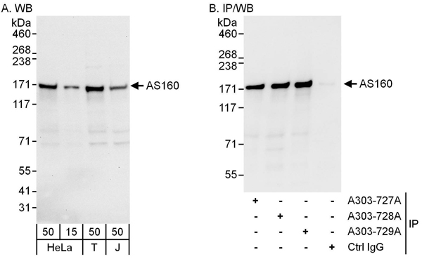 WB (Western Blot)
(Detection of human AS160 by western blot and immunoprecipitation. Samples: Whole cell lysate from HeLa (15 and 50 ug for WB; 1 mg for IP, 20% of IP loaded), HEK293T (T; 50 ug) and Jurkat (J; 50 ug) cells. Antibodies: Affinity purified rabbit anti-AS160 antibody AAA212273 used for WB at 0.1 ug/ml (A) and 1 ug/ml (B) and used for IP at 6 ug/mg lysate. AS160 was also immunoprecipitated by rabbit anti-AS160 antibodies and which recognize other epitopes. Detection: Chemiluminescence with exposure times of 10 seconds (A and B).)
WB (Western Blot)
(Detection of human AS160 by western blot and immunoprecipitation. Samples: Whole cell lysate from HeLa (15 and 50 ug for WB; 1 mg for IP, 20% of IP loaded), HEK293T (T; 50 ug) and Jurkat (J; 50 ug) cells. Antibodies: Affinity purified rabbit anti-AS160 antibody AAA212273 used for WB at 0.1 ug/ml (A) and 1 ug/ml (B) and used for IP at 6 ug/mg lysate. AS160 was also immunoprecipitated by rabbit anti-AS160 antibodies and which recognize other epitopes. Detection: Chemiluminescence with exposure times of 10 seconds (A and B).)
AS160, Polyclonal Antibody (Cat# AAA212273)
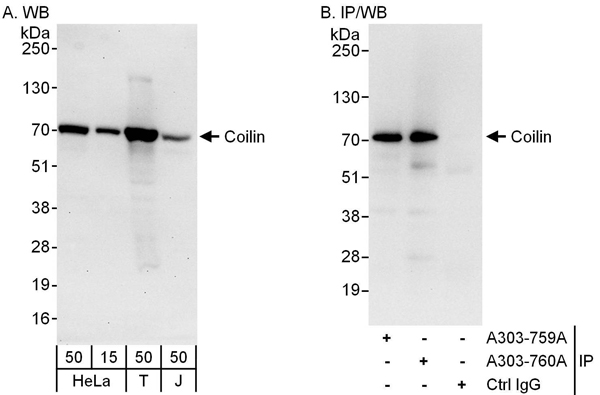 WB (Western Blot)
(Detection of human Coilin by western blot and immunoprecipitation. Samples: Whole cell lysate from HeLa (15 and 50 ug for WB; 1 mg for IP, 20% of IP loaded), HEK293T (T; 50 ug) and Jurkat (J; 50 ug) cells. Antibodies: Affinity purified rabbit anti-Coilin antibody AAA212278 used for WB at 0.1 ug/ml (A) and 1 ug/ml (B) and used for IP at 6 ug/mg lysate. Coilin was also immunoprecipitated by rabbit anti-Coilin antibody which recognizes an upstream epitope. Detection: Chemiluminescence with exposure times of 30 seconds (A) and 10 seconds (B).)
WB (Western Blot)
(Detection of human Coilin by western blot and immunoprecipitation. Samples: Whole cell lysate from HeLa (15 and 50 ug for WB; 1 mg for IP, 20% of IP loaded), HEK293T (T; 50 ug) and Jurkat (J; 50 ug) cells. Antibodies: Affinity purified rabbit anti-Coilin antibody AAA212278 used for WB at 0.1 ug/ml (A) and 1 ug/ml (B) and used for IP at 6 ug/mg lysate. Coilin was also immunoprecipitated by rabbit anti-Coilin antibody which recognizes an upstream epitope. Detection: Chemiluminescence with exposure times of 30 seconds (A) and 10 seconds (B).)
Coilin, Polyclonal Antibody (Cat# AAA212278)
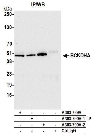
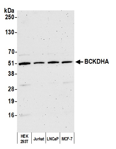 WB (Western Blot)
(Detection of human BCKDHA by western blot. Samples: Whole cell lysate (10 ug) from HEK293T, Jurkat, LNCaP, and MCF-7 cells prepared using NETN lysis buffer. Antibody: Affinity purified rabbit anti-BCKDHA antibody AAA212287 (lot AAA212287-2) used for WB at 0.04 ug/ml. Detection: Chemiluminescence with an exposure time of 3 minutes.)
WB (Western Blot)
(Detection of human BCKDHA by western blot. Samples: Whole cell lysate (10 ug) from HEK293T, Jurkat, LNCaP, and MCF-7 cells prepared using NETN lysis buffer. Antibody: Affinity purified rabbit anti-BCKDHA antibody AAA212287 (lot AAA212287-2) used for WB at 0.04 ug/ml. Detection: Chemiluminescence with an exposure time of 3 minutes.)
BCKDHA, Polyclonal Antibody (Cat# AAA212287)
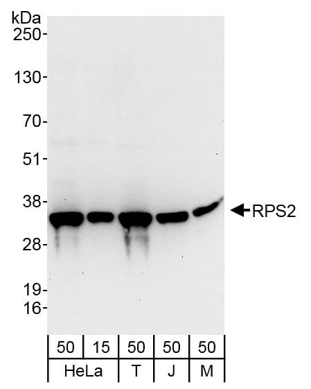 WB (Western Blot)
(Detection of human and mouse RPS2 by western blot. Samples: Whole cell lysate from HeLa (15 and 50 ug), HEK293T (T; 50 ug), Jurkat (J; 50 ug) and mouse NIH 3T3 (M; 50 ug) cells. Antibody: Affinity purified rabbit anti-RPS2 antibody AAA212288 (lot AAA212288-1) used at 0.1 ug/ml. Detection: Chemiluminescence with an exposure time of 10 seconds.)
WB (Western Blot)
(Detection of human and mouse RPS2 by western blot. Samples: Whole cell lysate from HeLa (15 and 50 ug), HEK293T (T; 50 ug), Jurkat (J; 50 ug) and mouse NIH 3T3 (M; 50 ug) cells. Antibody: Affinity purified rabbit anti-RPS2 antibody AAA212288 (lot AAA212288-1) used at 0.1 ug/ml. Detection: Chemiluminescence with an exposure time of 10 seconds.)
RPS2, Polyclonal Antibody (Cat# AAA212288)
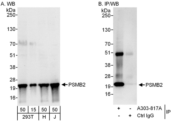 WB (Western Blot)
(Detection of human PSMB2 by western blot and immunoprecipitation. Samples: Whole cell lysate from HEK293T (15 and 50 ug for WB; 1 mg for IP, 20% of IP loaded), HeLa (H; 50 ug) and Jurkat (J; 50 ug) cells. Antibodies: Affinity purified rabbit anti-PSMB2 antibody AAA212295 used for WB at 0.1 ug/ml (A) and 1 ug/ml (B) and used for IP at 6 ug/mg lysate. Detection: Chemiluminescence with exposure times of 30 seconds (A and B).)
WB (Western Blot)
(Detection of human PSMB2 by western blot and immunoprecipitation. Samples: Whole cell lysate from HEK293T (15 and 50 ug for WB; 1 mg for IP, 20% of IP loaded), HeLa (H; 50 ug) and Jurkat (J; 50 ug) cells. Antibodies: Affinity purified rabbit anti-PSMB2 antibody AAA212295 used for WB at 0.1 ug/ml (A) and 1 ug/ml (B) and used for IP at 6 ug/mg lysate. Detection: Chemiluminescence with exposure times of 30 seconds (A and B).)
PSMB2, Polyclonal Antibody (Cat# AAA212295)
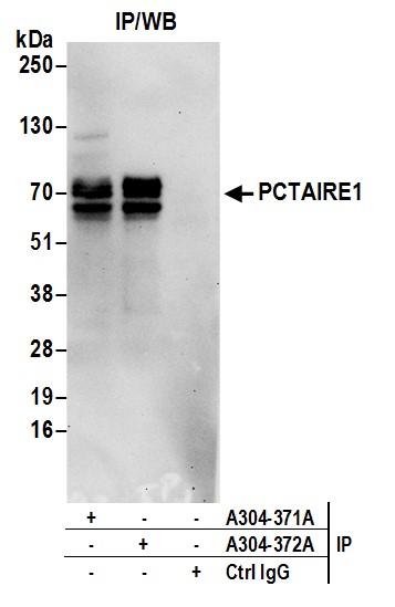
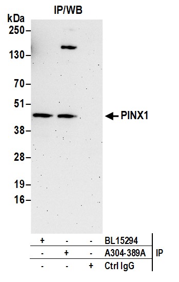
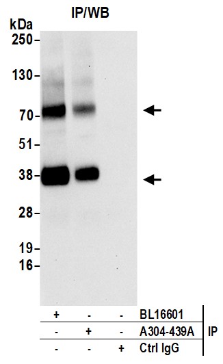
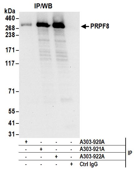
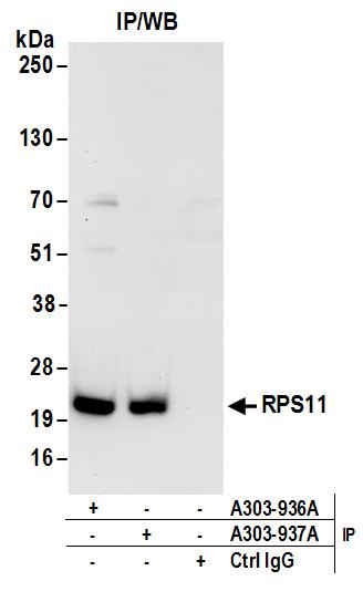
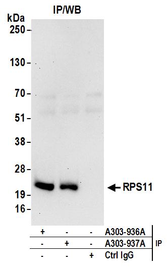
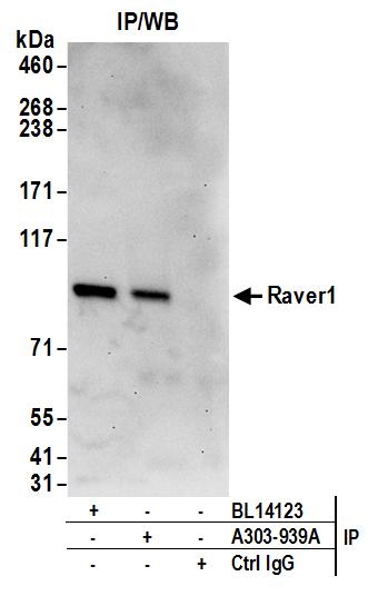
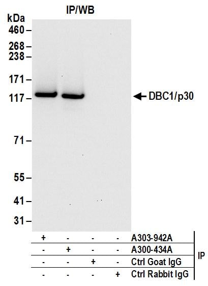
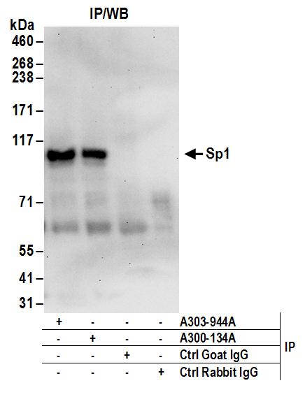
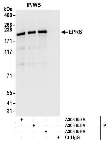
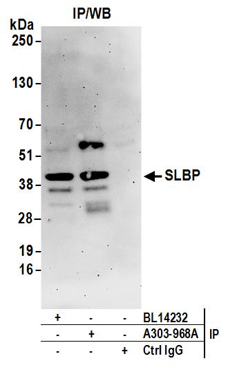
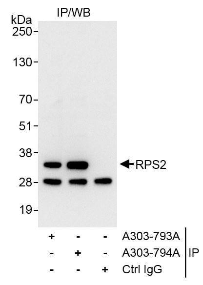
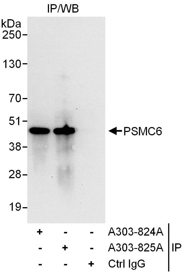 IP (Immunoprecipitation)
(Detection of human PSMC6 by western blot of immunoprecipitates. Samples: Whole cell lysate (1 mg for IP, 20% of IP loaded) from HEK293T cells. Antibodies: Affinity purified rabbit anti-PSMC6 antibody AAA212299 used for IP at 6 ug/mg lysate. PSMC6 was also immunoprecipitated by rabbit anti-PSMC6 antibody which recognizes a downstream epitope. For blotting immunoprecipitated PSMC6, was used at 1 ug/ml. Detection: Chemiluminescence with an exposure time of 10 seconds.)
IP (Immunoprecipitation)
(Detection of human PSMC6 by western blot of immunoprecipitates. Samples: Whole cell lysate (1 mg for IP, 20% of IP loaded) from HEK293T cells. Antibodies: Affinity purified rabbit anti-PSMC6 antibody AAA212299 used for IP at 6 ug/mg lysate. PSMC6 was also immunoprecipitated by rabbit anti-PSMC6 antibody which recognizes a downstream epitope. For blotting immunoprecipitated PSMC6, was used at 1 ug/ml. Detection: Chemiluminescence with an exposure time of 10 seconds.)
PSMC6, Polyclonal Antibody (Cat# AAA212299)
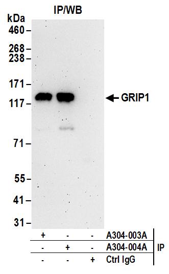
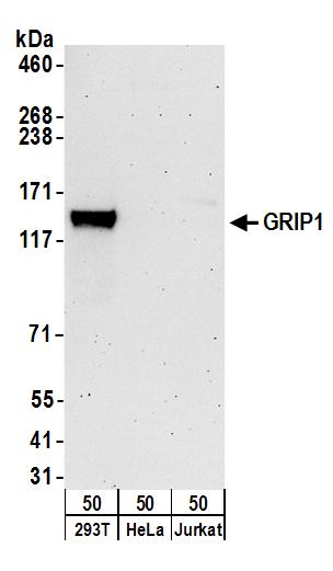 WB (Western Blot)
(Detection of human GRIP1 by western blot. Samples: Whole cell lysate (50 ug) from HEK293T, HeLa, and Jurkat cells. Antibodies: Affinity purified rabbit anti-GRIP1 antibody AAA212441 (lot AAA212441-1) used for WB at 0.4 ug/ml. Detection: Chemiluminescence with an exposure time of 3 minutes.)
WB (Western Blot)
(Detection of human GRIP1 by western blot. Samples: Whole cell lysate (50 ug) from HEK293T, HeLa, and Jurkat cells. Antibodies: Affinity purified rabbit anti-GRIP1 antibody AAA212441 (lot AAA212441-1) used for WB at 0.4 ug/ml. Detection: Chemiluminescence with an exposure time of 3 minutes.)
GRIP1, Polyclonal Antibody (Cat# AAA212441)
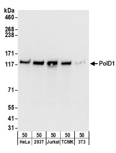 WB (Western Blot)
(Detection of human and mouse PolD1 by western blot. Samples: Whole cell lysate (50 ug) from HeLa, HEK293T, Jurkat, mouse TCMK-1, and mouse NIH 3T3 cells. Antibodies: Affinity purified rabbit anti-PolD1 antibody AAA212442 (lot AAA212442-1) used for WB at 0.1 ug/ml. Detection: Chemiluminescence with an exposure time of 10 seconds.)
WB (Western Blot)
(Detection of human and mouse PolD1 by western blot. Samples: Whole cell lysate (50 ug) from HeLa, HEK293T, Jurkat, mouse TCMK-1, and mouse NIH 3T3 cells. Antibodies: Affinity purified rabbit anti-PolD1 antibody AAA212442 (lot AAA212442-1) used for WB at 0.1 ug/ml. Detection: Chemiluminescence with an exposure time of 10 seconds.)
PolD1, Polyclonal Antibody (Cat# AAA212442)
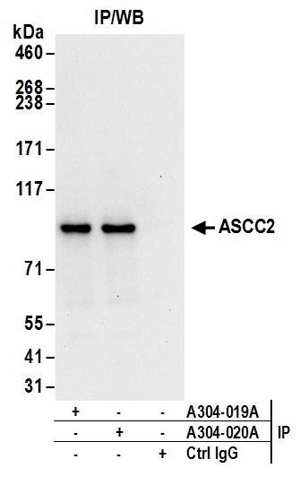
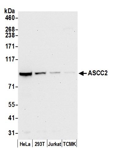 WB (Western Blot)
(Detection of human and mouse ASCC2 by western blot. Samples: Whole cell lysate (50 ug) from HeLa, HEK293T, Jurkat, and mouse TCMK-1 cells prepared using NETN lysis buffer. Antibody: Affinity purified rabbit anti-ASCC2 antibody AAA212455 (lot AAA212455-1) used for WB at 0.1 ug/ml. Detection: Chemiluminescence with an exposure time of 30 seconds.)
WB (Western Blot)
(Detection of human and mouse ASCC2 by western blot. Samples: Whole cell lysate (50 ug) from HeLa, HEK293T, Jurkat, and mouse TCMK-1 cells prepared using NETN lysis buffer. Antibody: Affinity purified rabbit anti-ASCC2 antibody AAA212455 (lot AAA212455-1) used for WB at 0.1 ug/ml. Detection: Chemiluminescence with an exposure time of 30 seconds.)
ASCC2, Polyclonal Antibody (Cat# AAA212455)
 WB (Western Blot)
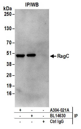
(Detection of human and mouse RRAGC/RagC by western blot. Samples: Whole cell lysate (50 ug) from HEK293T, HeLa, Jurkat, mouse TCMK-1, and mouse NIH 3T3 cells. Antibodies: Affinity purified rabbit anti-RRAGC/RagC antibody AAA212456 (lot AAA212456-1) used for WB at 0.1 ug/ml. Detection: Chemiluminescence with an exposure time of 3 minutes.)
WB (Western Blot)
(Detection of human and mouse RRAGC/RagC by western blot. Samples: Whole cell lysate (50 ug) from HEK293T, HeLa, Jurkat, mouse TCMK-1, and mouse NIH 3T3 cells. Antibodies: Affinity purified rabbit anti-RRAGC/RagC antibody AAA212456 (lot AAA212456-1) used for WB at 0.1 ug/ml. Detection: Chemiluminescence with an exposure time of 3 minutes.)
RRAGC/RagC, Polyclonal Antibody (Cat# AAA212456)
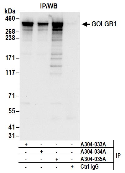
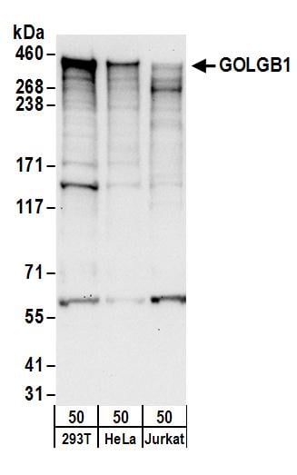 WB (Western Blot)
(Detection of human GOLGB1 by western blot. Samples: Whole cell lysate (50 ug) from HEK293T, HeLa, and Jurkat cells. Antibodies: Affinity purified rabbit anti-GOLGB1 antibody AAA212463 (lot AAA212463-1) used for WB at 0.1 ug/ml. Detection: Chemiluminescence with an exposure time of 30 seconds.)
WB (Western Blot)
(Detection of human GOLGB1 by western blot. Samples: Whole cell lysate (50 ug) from HEK293T, HeLa, and Jurkat cells. Antibodies: Affinity purified rabbit anti-GOLGB1 antibody AAA212463 (lot AAA212463-1) used for WB at 0.1 ug/ml. Detection: Chemiluminescence with an exposure time of 30 seconds.)
GOLGB1, Polyclonal Antibody (Cat# AAA212463)
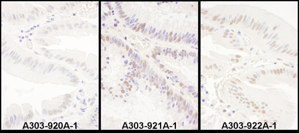
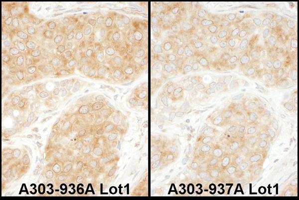
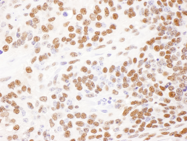
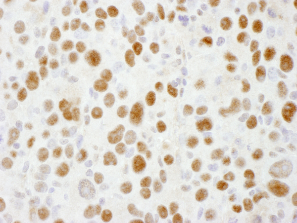
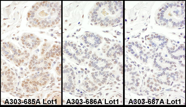
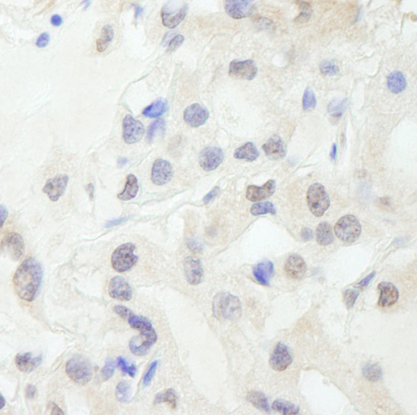
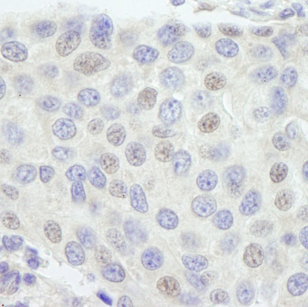
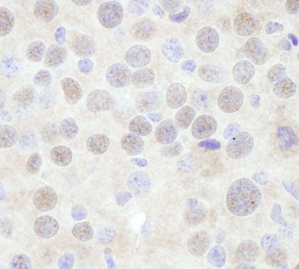 IHC (Immunohistochemisry)
(Detection of mouse CHD3 by immunohistochemistry. Sample: FFPE section of mouse renal cell carcinoma. Antibody: Affinity purified rabbit anti-CHD3 (Cat. No. AAA213894) used at a dilution of 1:250. Detection: DAB)
IHC (Immunohistochemisry)
(Detection of mouse CHD3 by immunohistochemistry. Sample: FFPE section of mouse renal cell carcinoma. Antibody: Affinity purified rabbit anti-CHD3 (Cat. No. AAA213894) used at a dilution of 1:250. Detection: DAB)
CHD3, Polyclonal Antibody (Cat# AAA213894)
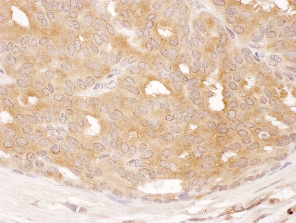
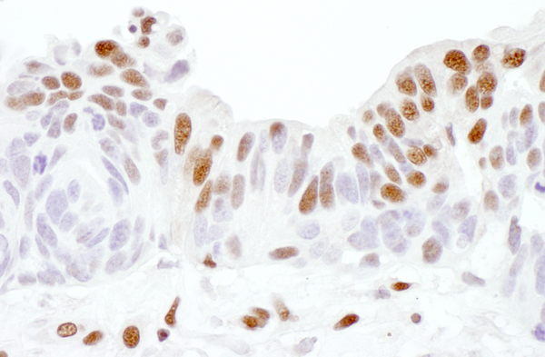
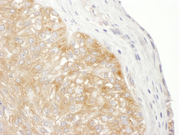
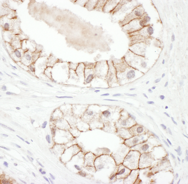
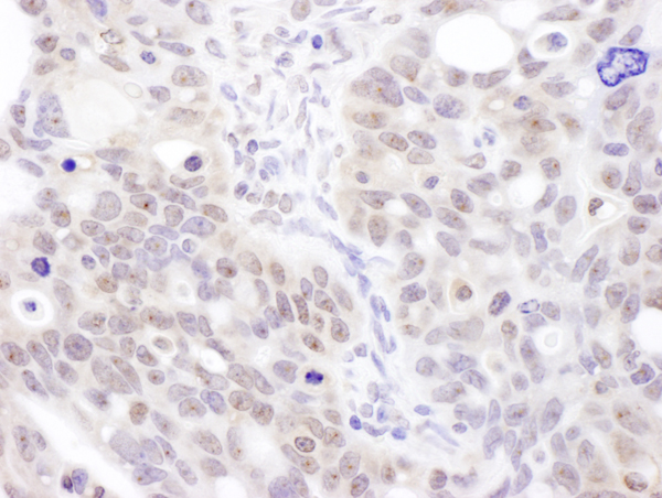
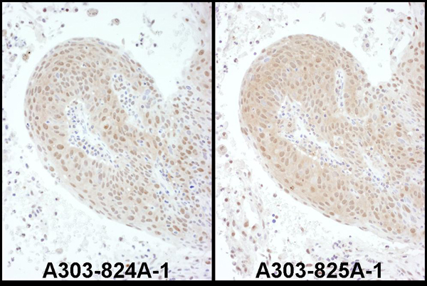
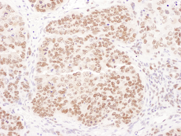
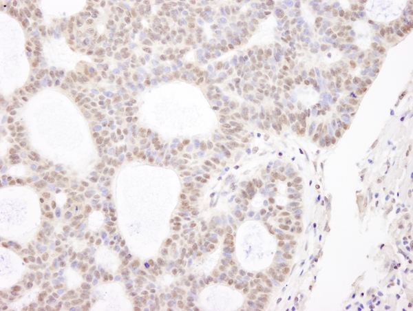
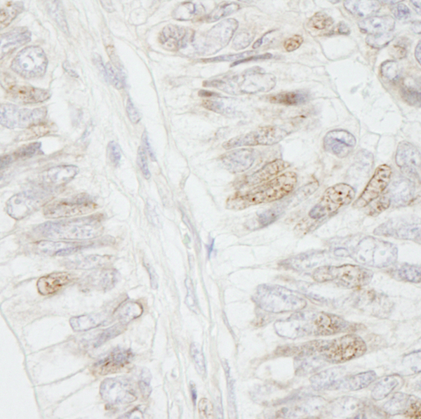 IHC (Immunohiostchemistry)
(Detection of human APC by immunohistochemistry. Sample: FFPE section of human colon carcinoma. Antibody: Affinity purified rabbit anti-APC (Cat. No. AAA213897) used at a dilution of 1:250. Detection: DAB)
IHC (Immunohiostchemistry)
(Detection of human APC by immunohistochemistry. Sample: FFPE section of human colon carcinoma. Antibody: Affinity purified rabbit anti-APC (Cat. No. AAA213897) used at a dilution of 1:250. Detection: DAB)
APC, Polyclonal Antibody (Cat# AAA213897)
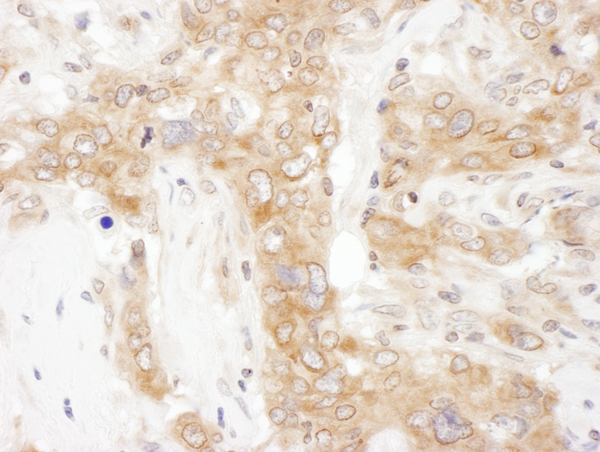
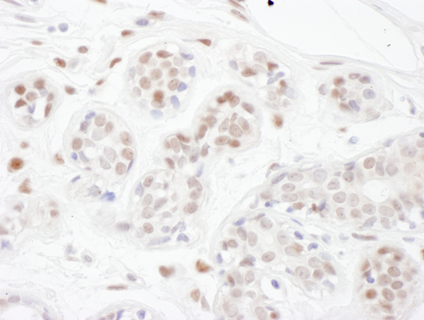
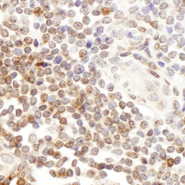
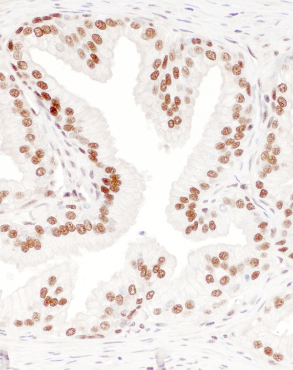
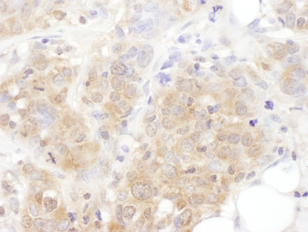
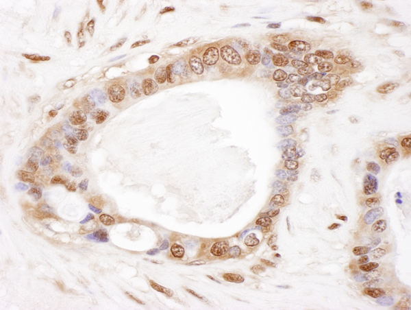
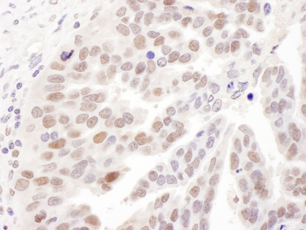
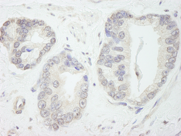 IHC (Immunohistochemisry)
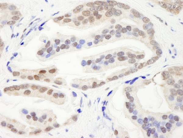
(Detection of human GTF3C1/TFIIIC220 by immunohistochemistry. Sample: FFPE section of human prostate carcinoma. Antibody: Affinity purified rabbit anti-GTF3C1/TFIIIC220 (Cat. No. AAA213898) used at a dilution of 1:250. Detection: DAB)
IHC (Immunohistochemisry)
(Detection of human GTF3C1/TFIIIC220 by immunohistochemistry. Sample: FFPE section of human prostate carcinoma. Antibody: Affinity purified rabbit anti-GTF3C1/TFIIIC220 (Cat. No. AAA213898) used at a dilution of 1:250. Detection: DAB)
GTF3C1/TFIII220, Polyclonal Antibody (Cat# AAA213898)
What are Polyclonal Antibodies?
Polyclonal antibodies are antibodies that come from multiple B cell clones of a host animal. The typical hosts used for the majority of polyclonal antibody production are rabbits, goats, sheep, and donkeys. These polyclonal antibodies, once having identified their target, will bind to different epitopes located at different regions or sequences on the same protein/antigen. As a result, they are ideal at locating and binding to the target, even if the target is in very low concentrations (due to many different antibodies being able to bind to the same target molecule, which allows for significant amplification of a downstream signal).
Polyclonal antibodies are typically produced by injecting an antigen into a host animal, which causes the animal’s immune system to attack the foreign antigen by mass generating antibodies against it. After a period of time, serum is collected from the animal and purified using physicochemical fractionation, class-specific affinity purification, and/or antigen-affinity purification.
Key Uses of Polyclonal Antibodies
- Western Blotting: This method is used to find specific proteins in biological samples after separating them by size.
- Immunohistochemistry: IHC helps visualize the location of proteins in tissue sections using various staining techniques.
- ELISA: (Enzyme-Linked Immunosorbent Assay) is typically used to identify specific protein quantities in a sample. ELISAs can be either “Quantitative” or “Qualitative”.
- Flow Cytometry: technique that identifies and measures the specific protein on the surface or inside the cells in a fluid suspension.
- Immunoprecipitation: IP isolates and studies a specific protein from a complex mixture using antibodies.
Why Buy Polyclonal Antibodies from AAA Biotech?
1. Ideal for Various Applications
Our antibodies are generally going to be validated for use in multiple types of assays, including ELISA, Western Blotting, Immunohistochemistry, Immunoprecipitation, amongst others. They are ideal for a wide range of research applications.
2. Rigorous Quality Control
All of the antibodies in our catalog undergo strict quality testing to ensure specificity, sensitivity, and consistent performance. We are confident in the ability of our antibodies to provide you with accurate results.
3. Wide Assortment of Antibodies
Antibodies in are catalog can be found for both common and exotic species, and these antibodies are also available in both conjugated and recombinant forms to suit many diverse experimental needs.
4. Highly Purified
Our antibodies are available in purified forms with over 85% purity, as confirmed by SDS-PAGE. They are also available with tags such as His, Flag, GST, or MBP. We cater to customers worldwide.
FAQ
1. How are polyclonal antibodies produced?
Traditionally, polyclonal antibodies are produced by injecting an antigen into a host animal (such as a rabbit or goat), which then triggers an immune response from the host animal. The animal’s B cells produce antibodies that will recognize different parts of the injected antigen. These antibodies are then collected from the animal’s blood and purified for use.
2. How do polyclonal antibodies differ from monoclonal antibodies?
Polyclonal antibodies are a mix of antibodies that bind to different locations (epitopes) of the same antigen, while monoclonal antibodies are identical and bind to just one specific epitope. This makes polyclonal antibodies more versatile and better at detecting proteins that may be present in low quantities or in altered/modified forms.
3. How should I store polyclonal antibodies?
Polyclonal antibodies should be stored at 4°C for short-term use (up to a few weeks) and at -20°C or -80°C for long-term storage. Avoid repeated freeze-thaw cycles by dividing them into small aliquots. Always check the datasheet for specific storage instructions.