Filters
▼Clonality
▼Type
▼Reactivity
▼Gene Name
▼Isotype
▼Host
▼Application
▼Clone
▼Polyclonal Antibodies
At AAA Biotech also known as AAA Bio or AAABio, we provide a broad range of purified polyclonal antibodies (pAbs) that are able to all be browsed online through our website. Due to their high specificity and strong binding affinity, these antibodies are ideal for wide swathes of research and experimental applications.
Our polyclonal antibodies can easily support your work, whether you use them for Western Blotting, Immunocytochemistry (with or without Immunofluorescence used in conjunction), Immunohistochemistry, Immunoprecipitation, and ELISA tests. We highly encourage you to browse our range of pAbs and choose the one that best suits your experimental model.
Viewing 4400-4450 of 96812 product results
 WB (Western Blot)
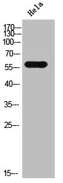
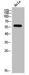
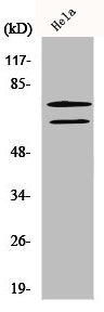
(Western Blot analysis of HELA cells using ALS2CR13 Polyclonal Antibody)
WB (Western Blot)
(Western Blot analysis of HELA cells using ALS2CR13 Polyclonal Antibody)
FAM117B, Polyclonal Antibody (Cat# AAA236592)
 WB (Western Blot)
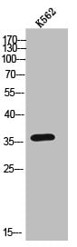
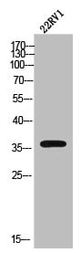
(Western Blot analysis of 22RV1 cells using AR-beta3 Polyclonal Antibody)
WB (Western Blot)
(Western Blot analysis of 22RV1 cells using AR-beta3 Polyclonal Antibody)
ADRB3, Polyclonal Antibody (Cat# AAA236593)
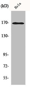
 WB (Western Blot)
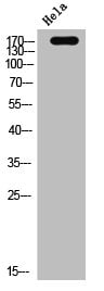
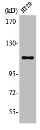
(Western Blot analysis of HELA cells using EGFR Polyclonal Antibody)
WB (Western Blot)
(Western Blot analysis of HELA cells using EGFR Polyclonal Antibody)
EGFR, Polyclonal Antibody (Cat# AAA236597)
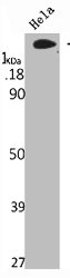
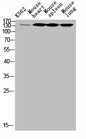 WB (Western Blot)
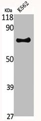
(Western Blot analysis of k562 mouse-heart mouse-spleen mouse-lung cells using ErbB-3 Polyclonal Antibody)
WB (Western Blot)
(Western Blot analysis of k562 mouse-heart mouse-spleen mouse-lung cells using ErbB-3 Polyclonal Antibody)
ERBB3, Polyclonal Antibody (Cat# AAA236601)
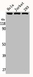
 WB (Western Blot)
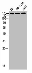
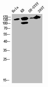
(Western blot analysis of Hela KB SH-SY5Y 293T lysis using Flt-1 antibody.)
WB (Western Blot)
(Western blot analysis of Hela KB SH-SY5Y 293T lysis using Flt-1 antibody.)
FLT1, Polyclonal Antibody (Cat# AAA236605)
 WB (Western Blot)
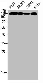
(Western Blot analysis of 293T AD293 22RV1 HELA cells using Phospho-MYPT1 (T853) Polyclonal Antibody)
WB (Western Blot)
(Western Blot analysis of 293T AD293 22RV1 HELA cells using Phospho-MYPT1 (T853) Polyclonal Antibody)
PPP1R12A, Polyclonal Antibody (Cat# AAA236612)
 WB (Western Blot)
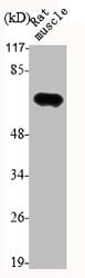
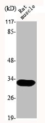
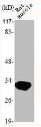
(Western Blot analysis of RAT-MUCLE cells using Lamin A/C Polyclonal Antibody)
WB (Western Blot)
(Western Blot analysis of RAT-MUCLE cells using Lamin A/C Polyclonal Antibody)
LMNA, Polyclonal Antibody (Cat# AAA236275)
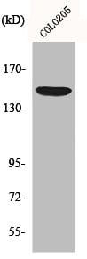
 WB (Western Blot)
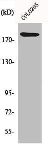
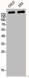
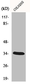
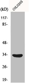
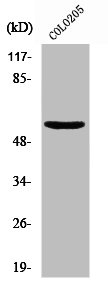
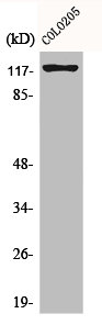
(Western Blot analysis of COLO 293 cells using Laminin alpha-4 Polyclonal Antibody)
WB (Western Blot)
(Western Blot analysis of COLO 293 cells using Laminin alpha-4 Polyclonal Antibody)
LAMA4, Polyclonal Antibody (Cat# AAA236276)
 WB (Western Blot)
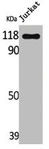
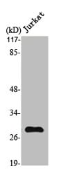
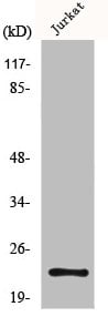
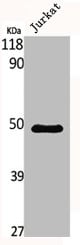
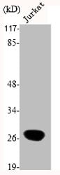
(Western Blot analysis of Jurkat cells using Lfc Polyclonal Antibody)
WB (Western Blot)
(Western Blot analysis of Jurkat cells using Lfc Polyclonal Antibody)
ARHGEF2, Polyclonal Antibody (Cat# AAA236278)
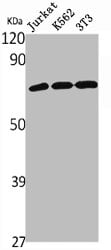
 WB (Western Blot)
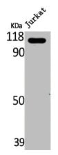
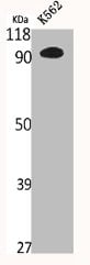
(Western Blot analysis of Jurkat cells using LRG1 Polyclonal Antibody)
WB (Western Blot)
(Western Blot analysis of Jurkat cells using LRG1 Polyclonal Antibody)
LRG1, Polyclonal Antibody (Cat# AAA236282)
 WB (Western Blot)
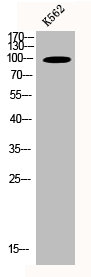
(Western Blot analysis of Jurkat cells using LZK Polyclonal Antibody)
WB (Western Blot)
(Western Blot analysis of Jurkat cells using LZK Polyclonal Antibody)
MAP3K13, Polyclonal Antibody (Cat# AAA236286)
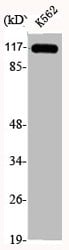
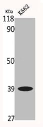
 WB (Western Blot)
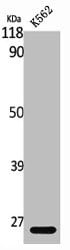
(Western Blot analysis of K562 cells using MAD2 Polyclonal Antibody)
WB (Western Blot)
(Western Blot analysis of K562 cells using MAD2 Polyclonal Antibody)
MAD2L1, Polyclonal Antibody (Cat# AAA236287)
 WB (Western Blot)
(Western Blot analysis of Jurkat cells using MMP-7 Polyclonal Antibody)
WB (Western Blot)
(Western Blot analysis of Jurkat cells using MMP-7 Polyclonal Antibody)
MMP7, Polyclonal Antibody (Cat# AAA236291)
 WB (Western Blot)
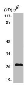
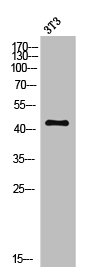
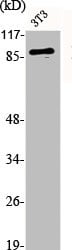
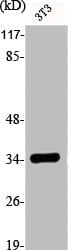
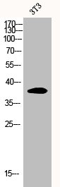
(Western Blot analysis of NIH-3T3 cells using MC5-R Polyclonal Antibody)
WB (Western Blot)
(Western Blot analysis of NIH-3T3 cells using MC5-R Polyclonal Antibody)
MC5R, Polyclonal Antibody (Cat# AAA236292)
 WB (Western Blot)
(Western Blot analysis of various cells using MEK-3 Polyclonal Antibody)
WB (Western Blot)
(Western Blot analysis of various cells using MEK-3 Polyclonal Antibody)
MAP2K3, Polyclonal Antibody (Cat# AAA236297)
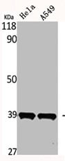 WB (Western Blot)
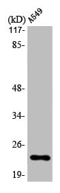
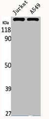
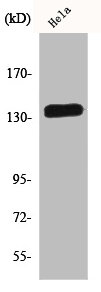
(Western Blot analysis of HELA A549 cells using MEL-1B-R Polyclonal Antibody)
WB (Western Blot)
(Western Blot analysis of HELA A549 cells using MEL-1B-R Polyclonal Antibody)
MTNR1B, Polyclonal Antibody (Cat# AAA236298)
 WB (Western Blot)
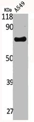
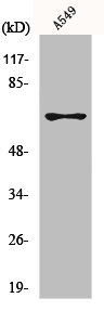
(Western Blot analysis of A549 cells using Menin Polyclonal Antibody)
WB (Western Blot)
(Western Blot analysis of A549 cells using Menin Polyclonal Antibody)
MEN1, Polyclonal Antibody (Cat# AAA236300)
 WB (Western Blot)
(Western Blot analysis of various cells using MerTK Polyclonal Antibody)
WB (Western Blot)
(Western Blot analysis of various cells using MerTK Polyclonal Antibody)
MERTK, Polyclonal Antibody (Cat# AAA236301)
 WB (Western Blot)
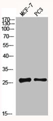
(Western Blot analysis of MCF7 PC3 cells using MGMT Polyclonal Antibody)
WB (Western Blot)
(Western Blot analysis of MCF7 PC3 cells using MGMT Polyclonal Antibody)
MGMT, Polyclonal Antibody (Cat# AAA236306)
 WB (Western Blot)
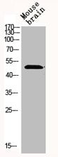
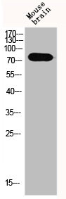
(Western Blot analysis of MOUSE-BRAIN cells using Mnk1 Polyclonal Antibody)
WB (Western Blot)
(Western Blot analysis of MOUSE-BRAIN cells using Mnk1 Polyclonal Antibody)
MKNK1, Polyclonal Antibody (Cat# AAA236312)
 WB (Western Blot)
(Western Blot analysis of Jurkat cells using MNT Polyclonal Antibody)
WB (Western Blot)
(Western Blot analysis of Jurkat cells using MNT Polyclonal Antibody)
MNT, Polyclonal Antibody (Cat# AAA236313)
 WB (Western Blot)
(Western Blot analysis of RAT-MUSCLE cells using MOX-2 Polyclonal Antibody)
WB (Western Blot)
(Western Blot analysis of RAT-MUSCLE cells using MOX-2 Polyclonal Antibody)
MEOX2, Polyclonal Antibody (Cat# AAA236314)
 WB (Western Blot)
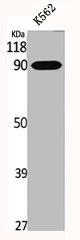
(Western Blot analysis of K562 cells using MRE11 Polyclonal Antibody)
WB (Western Blot)
(Western Blot analysis of K562 cells using MRE11 Polyclonal Antibody)
MRE11A, Polyclonal Antibody (Cat# AAA236315)
 WB (Western Blot)
(Western Blot analysis of HELA K562 293 cells using MRP3 Polyclonal Antibody)
WB (Western Blot)
(Western Blot analysis of HELA K562 293 cells using MRP3 Polyclonal Antibody)
ABCC3, Polyclonal Antibody (Cat# AAA236316)
 WB (Western Blot)
(Western Blot analysis of Jurkat A549 cells using MRP7 Polyclonal Antibody)
WB (Western Blot)
(Western Blot analysis of Jurkat A549 cells using MRP7 Polyclonal Antibody)
ABCC10, Polyclonal Antibody (Cat# AAA236317)
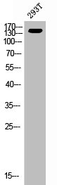
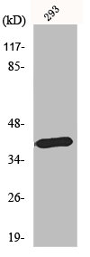
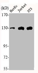
 WB (Western Blot)
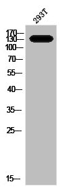
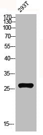
(Western blot analysis of 293T lysis using MRP-L10 antibody.)
WB (Western Blot)
(Western blot analysis of 293T lysis using MRP-L10 antibody.)
MRPL10, Polyclonal Antibody (Cat# AAA236318)
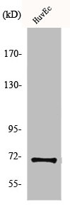
 WB (Western Blot)
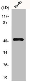
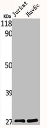
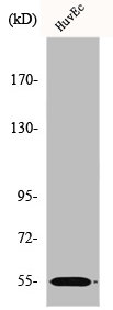
(Western Blot analysis of Jurkat HuvEc cells using MRP-S34 Polyclonal Antibody)
WB (Western Blot)
(Western Blot analysis of Jurkat HuvEc cells using MRP-S34 Polyclonal Antibody)
MRPS34, Polyclonal Antibody (Cat# AAA236322)
 WB (Western Blot)
(Western Blot analysis of RAT-MUSCLE cells using MyD88 Polyclonal Antibody)
WB (Western Blot)
(Western Blot analysis of RAT-MUSCLE cells using MyD88 Polyclonal Antibody)
MYD88, Polyclonal Antibody (Cat# AAA236327)
 WB (Western Blot)
(Western Blot analysis of Jurkat cells using Myf-5 Polyclonal Antibody)
WB (Western Blot)
(Western Blot analysis of Jurkat cells using Myf-5 Polyclonal Antibody)
MYF5, Polyclonal Antibody (Cat# AAA236328)
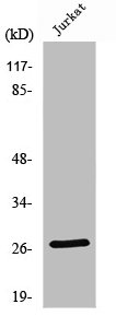
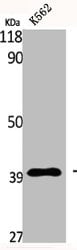
 WB (Western Blot)
(Western Blot analysis of K562 cells using N4BP1 Polyclonal Antibody)
WB (Western Blot)
(Western Blot analysis of K562 cells using N4BP1 Polyclonal Antibody)
N4BP1, Polyclonal Antibody (Cat# AAA236334)
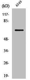
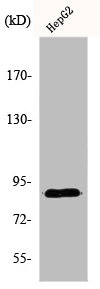
 WB (Western Blot)
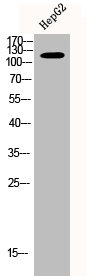
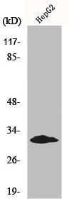
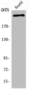
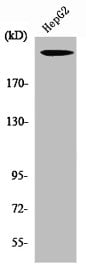
(Western Blot analysis of HepG2 cells using Neurofibromin Polyclonal Antibody)
WB (Western Blot)
(Western Blot analysis of HepG2 cells using Neurofibromin Polyclonal Antibody)
NF1, Polyclonal Antibody (Cat# AAA236341)
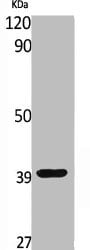
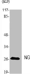
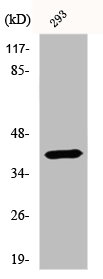
 WB (Western Blot)
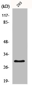
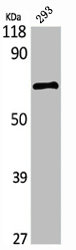
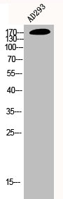
(Western Blot analysis of 293 cells using Neuroglycan C Polyclonal Antibody)
WB (Western Blot)
(Western Blot analysis of 293 cells using Neuroglycan C Polyclonal Antibody)
CSPG5, Polyclonal Antibody (Cat# AAA236342)
 WB (Western Blot)
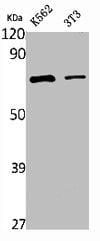
(Western Blot analysis of K562 NIH-3T3 cells using NFkappaB-p65 Polyclonal Antibody)
WB (Western Blot)
(Western Blot analysis of K562 NIH-3T3 cells using NFkappaB-p65 Polyclonal Antibody)
RELA, Polyclonal Antibody (Cat# AAA236347)
 WB (Western Blot)
(Western Blot analysis of various cells using NGF Polyclonal Antibody)
WB (Western Blot)
(Western Blot analysis of various cells using NGF Polyclonal Antibody)
NGF, Polyclonal Antibody (Cat# AAA236353)
 WB (Western Blot)
(Western Blot analysis of Jurkat K562 NIH-3T3 cells using NHE-9 Polyclonal Antibody)
WB (Western Blot)
(Western Blot analysis of Jurkat K562 NIH-3T3 cells using NHE-9 Polyclonal Antibody)
SLC9A9, Polyclonal Antibody (Cat# AAA236355)

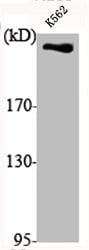 WB (Western Blot)
(Western Blot analysis of K562 cells using DNA-PKCS Polyclonal Antibody)
WB (Western Blot)
(Western Blot analysis of K562 cells using DNA-PKCS Polyclonal Antibody)
PRKDC, Polyclonal Antibody (Cat# AAA236100)
 WB (Western Blot)
(Western Blot analysis of K562 cells using DRS-1 Polyclonal Antibody)
WB (Western Blot)
(Western Blot analysis of K562 cells using DRS-1 Polyclonal Antibody)
ECI2, Polyclonal Antibody (Cat# AAA236106)

 WB (Western Blot)
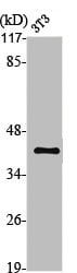
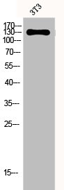
(Western Blot analysis of NIH-3T3 cells using Dynamin I Polyclonal Antibody)
WB (Western Blot)
(Western Blot analysis of NIH-3T3 cells using Dynamin I Polyclonal Antibody)
DNM1, Polyclonal Antibody (Cat# AAA236108)
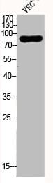 WB (Western Blot)
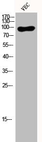
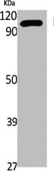
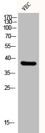
(Western Blot analysis of VEC cells using Dyrk1A Polyclonal Antibody)
WB (Western Blot)
(Western Blot analysis of VEC cells using Dyrk1A Polyclonal Antibody)
DYRK1A, Polyclonal Antibody (Cat# AAA236109)
 WB (Western Blot)
(Western Blot analysis of NIH-3T3 cells using E2F-6 Polyclonal Antibody)
WB (Western Blot)
(Western Blot analysis of NIH-3T3 cells using E2F-6 Polyclonal Antibody)
E2F6, Polyclonal Antibody (Cat# AAA236110)
 WB (Western Blot)
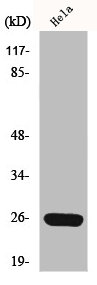
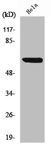
(Western Blot analysis of 293 cells using E-cadherin Polyclonal Antibody)
WB (Western Blot)
(Western Blot analysis of 293 cells using E-cadherin Polyclonal Antibody)
CDH1/CDH2/CDH3/CDH4, Polyclonal Antibody (Cat# AAA236112)
 WB (Western Blot)
(Western Blot analysis of various cells using EF-2 Polyclonal Antibody)
WB (Western Blot)
(Western Blot analysis of various cells using EF-2 Polyclonal Antibody)
EEF2, Polyclonal Antibody (Cat# AAA236117)
 WB (Western Blot)
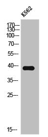
(Western Blot analysis of K562 cells using Eg5 Polyclonal Antibody)
WB (Western Blot)
(Western Blot analysis of K562 cells using Eg5 Polyclonal Antibody)
KIF11, Polyclonal Antibody (Cat# AAA236118)
 WB (Western Blot)
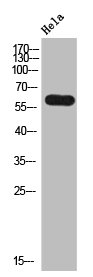
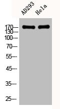
(Western Blot analysis of AD293 HELA cells using EGFR Polyclonal Antibody)
WB (Western Blot)
(Western Blot analysis of AD293 HELA cells using EGFR Polyclonal Antibody)
EGFR, Polyclonal Antibody (Cat# AAA236119)
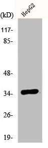
 WB (Western Blot)
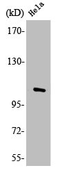
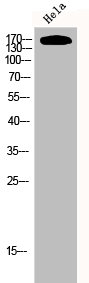
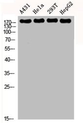
(Western Blot analysis of A431 HELA 293T HepG2 cells using EGFR Polyclonal Antibody)
WB (Western Blot)
(Western Blot analysis of A431 HELA 293T HepG2 cells using EGFR Polyclonal Antibody)
EGFR, Polyclonal Antibody (Cat# AAA236120)
 WB (Western Blot)
(Western Blot analysis of K562 cells using eIF2alpha Polyclonal Antibody)
WB (Western Blot)
(Western Blot analysis of K562 cells using eIF2alpha Polyclonal Antibody)
EIF2S1, Polyclonal Antibody (Cat# AAA236122)
 WB (Western Blot)
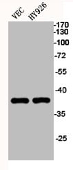
(Western Blot analysis of VEC HY926 cells using EKLF/CKLF/UKLF Polyclonal Antibody)
WB (Western Blot)
(Western Blot analysis of VEC HY926 cells using EKLF/CKLF/UKLF Polyclonal Antibody)
KLF1/KLF5/KLF7, Polyclonal Antibody (Cat# AAA236123)
 WB (Western Blot)
(Western Blot analysis of K562 cells using EphA2/3/4 Polyclonal Antibody)
WB (Western Blot)
(Western Blot analysis of K562 cells using EphA2/3/4 Polyclonal Antibody)
EPHA2/EPHA3/EPHA4, Polyclonal Antibody (Cat# AAA236131)
 WB (Western Blot)
(Western Blot analysis of K562 cells using Epsin 1 Polyclonal Antibody)
WB (Western Blot)
(Western Blot analysis of K562 cells using Epsin 1 Polyclonal Antibody)
EPN1, Polyclonal Antibody (Cat# AAA236137)
 WB (Western Blot)
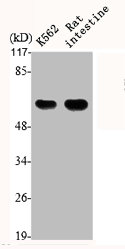
(Western Blot analysis of K562 RAT-intestine cells using ER81 Polyclonal Antibody)
WB (Western Blot)
(Western Blot analysis of K562 RAT-intestine cells using ER81 Polyclonal Antibody)
ETV1, Polyclonal Antibody (Cat# AAA236138)
What are Polyclonal Antibodies?
Polyclonal antibodies are antibodies that come from multiple B cell clones of a host animal. The typical hosts used for the majority of polyclonal antibody production are rabbits, goats, sheep, and donkeys. These polyclonal antibodies, once having identified their target, will bind to different epitopes located at different regions or sequences on the same protein/antigen. As a result, they are ideal at locating and binding to the target, even if the target is in very low concentrations (due to many different antibodies being able to bind to the same target molecule, which allows for significant amplification of a downstream signal).
Polyclonal antibodies are typically produced by injecting an antigen into a host animal, which causes the animal’s immune system to attack the foreign antigen by mass generating antibodies against it. After a period of time, serum is collected from the animal and purified using physicochemical fractionation, class-specific affinity purification, and/or antigen-affinity purification.
Key Uses of Polyclonal Antibodies
- Western Blotting: This method is used to find specific proteins in biological samples after separating them by size.
- Immunohistochemistry: IHC helps visualize the location of proteins in tissue sections using various staining techniques.
- ELISA: (Enzyme-Linked Immunosorbent Assay) is typically used to identify specific protein quantities in a sample. ELISAs can be either “Quantitative” or “Qualitative”.
- Flow Cytometry: technique that identifies and measures the specific protein on the surface or inside the cells in a fluid suspension.
- Immunoprecipitation: IP isolates and studies a specific protein from a complex mixture using antibodies.
Why Buy Polyclonal Antibodies from AAA Biotech?
1. Ideal for Various Applications
Our antibodies are generally going to be validated for use in multiple types of assays, including ELISA, Western Blotting, Immunohistochemistry, Immunoprecipitation, amongst others. They are ideal for a wide range of research applications.
2. Rigorous Quality Control
All of the antibodies in our catalog undergo strict quality testing to ensure specificity, sensitivity, and consistent performance. We are confident in the ability of our antibodies to provide you with accurate results.
3. Wide Assortment of Antibodies
Antibodies in are catalog can be found for both common and exotic species, and these antibodies are also available in both conjugated and recombinant forms to suit many diverse experimental needs.
4. Highly Purified
Our antibodies are available in purified forms with over 85% purity, as confirmed by SDS-PAGE. They are also available with tags such as His, Flag, GST, or MBP. We cater to customers worldwide.
FAQ
1. How are polyclonal antibodies produced?
Traditionally, polyclonal antibodies are produced by injecting an antigen into a host animal (such as a rabbit or goat), which then triggers an immune response from the host animal. The animal’s B cells produce antibodies that will recognize different parts of the injected antigen. These antibodies are then collected from the animal’s blood and purified for use.
2. How do polyclonal antibodies differ from monoclonal antibodies?
Polyclonal antibodies are a mix of antibodies that bind to different locations (epitopes) of the same antigen, while monoclonal antibodies are identical and bind to just one specific epitope. This makes polyclonal antibodies more versatile and better at detecting proteins that may be present in low quantities or in altered/modified forms.
3. How should I store polyclonal antibodies?
Polyclonal antibodies should be stored at 4°C for short-term use (up to a few weeks) and at -20°C or -80°C for long-term storage. Avoid repeated freeze-thaw cycles by dividing them into small aliquots. Always check the datasheet for specific storage instructions.