Filters
▼Clonality
▼Type
▼Reactivity
▼Gene Name
▼Isotype
▼Host
▼Application
▼Clone
▼Polyclonal Antibodies
At AAA Biotech also known as AAA Bio or AAABio, we provide a broad range of purified polyclonal antibodies (pAbs) that are able to all be browsed online through our website. Due to their high specificity and strong binding affinity, these antibodies are ideal for wide swathes of research and experimental applications.
Our polyclonal antibodies can easily support your work, whether you use them for Western Blotting, Immunocytochemistry (with or without Immunofluorescence used in conjunction), Immunohistochemistry, Immunoprecipitation, and ELISA tests. We highly encourage you to browse our range of pAbs and choose the one that best suits your experimental model.
Viewing 4300-4350 of 96812 product results
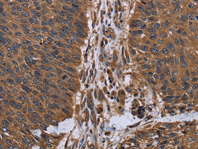
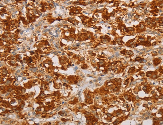
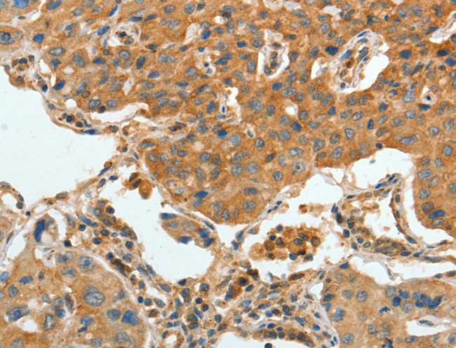
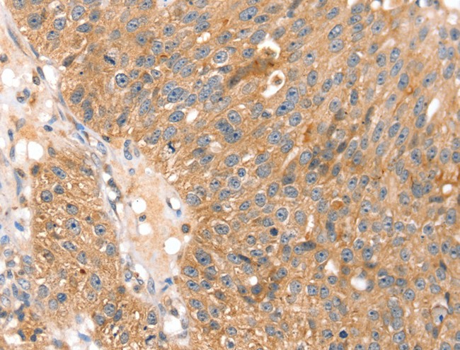
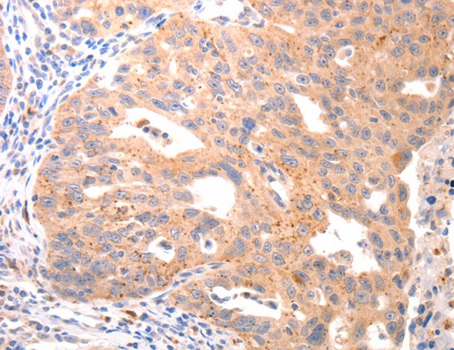
 IHC (Immunohiostchemistry)
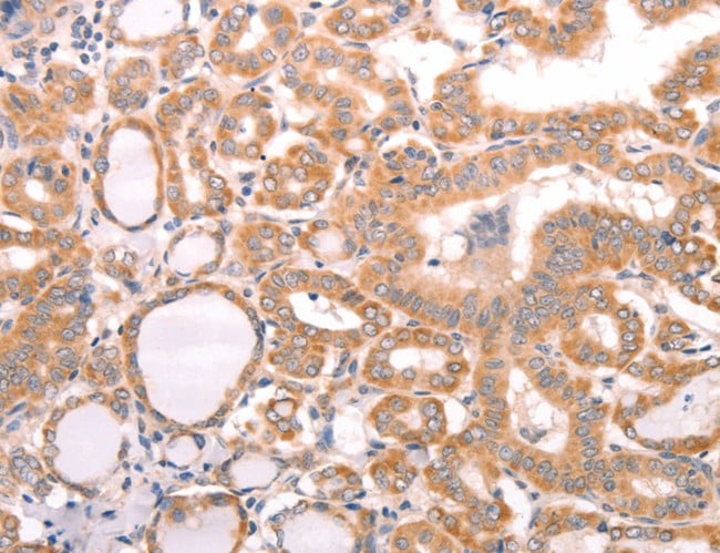
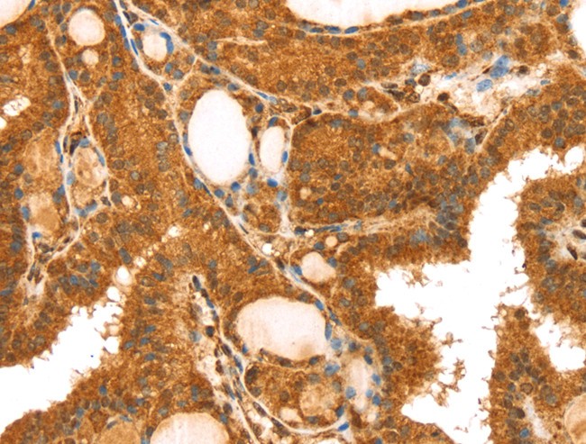
(Immunohistochemistry of paraffin-embedded Human thyroid cancer tissue using ABCF2 Polyclonal Antibody at dilution 1:15)
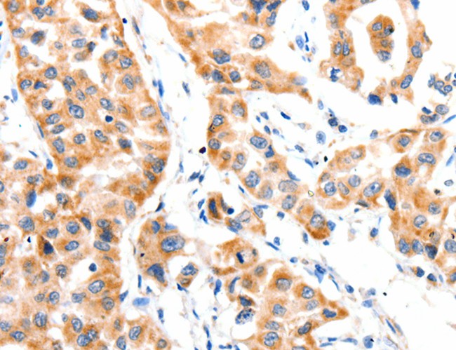
IHC (Immunohiostchemistry)
(Immunohistochemistry of paraffin-embedded Human thyroid cancer tissue using ABCF2 Polyclonal Antibody at dilution 1:15)
ABCF2, Polyclonal Antibody (Cat# AAA166839)
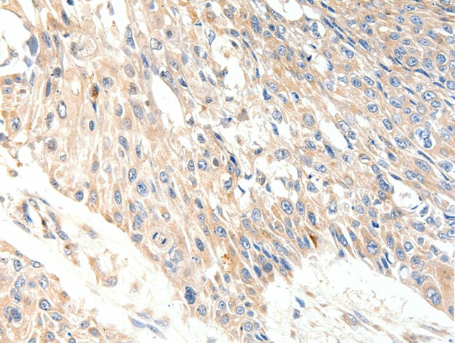
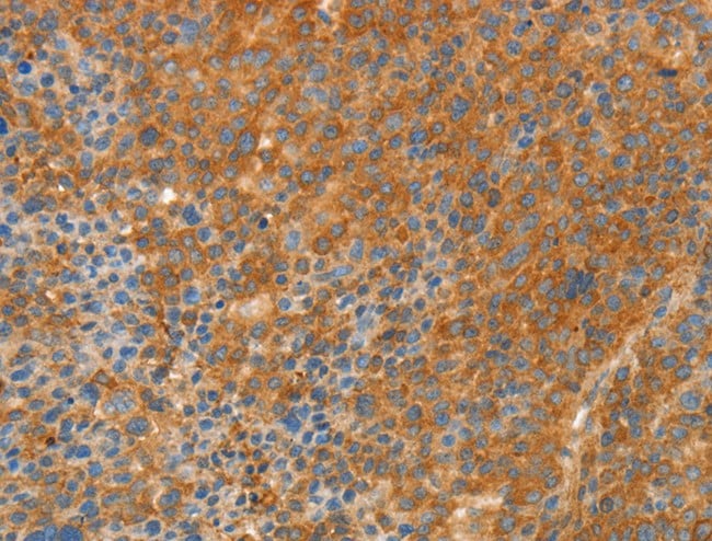
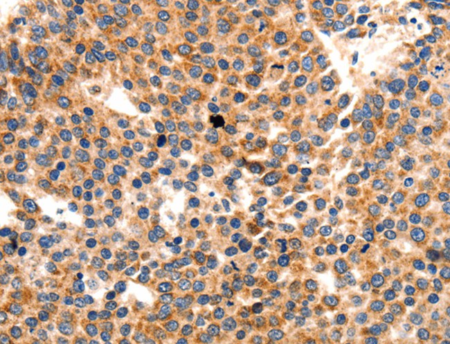
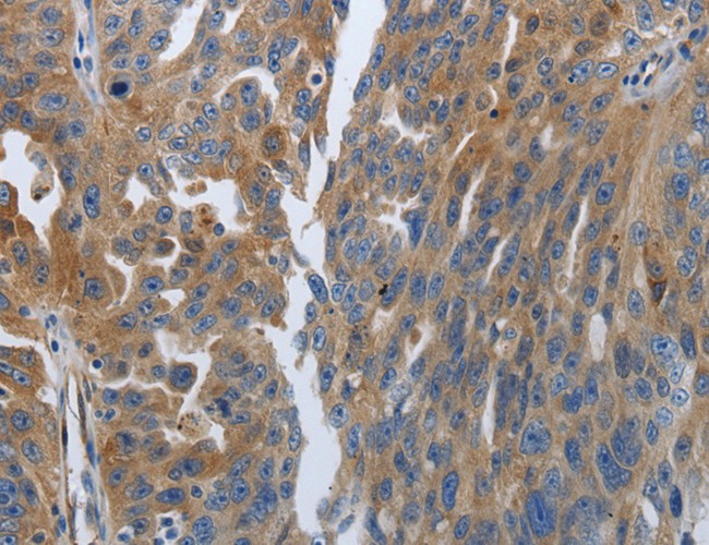
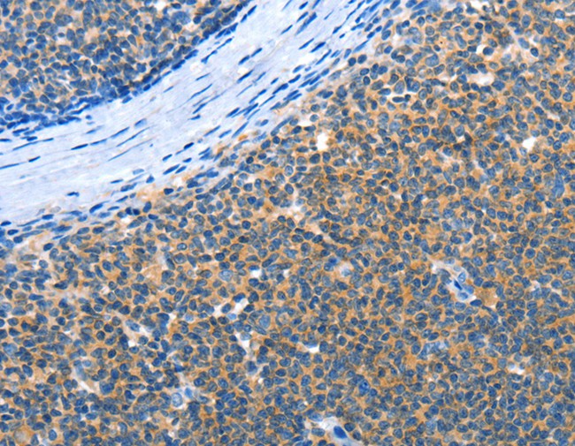
 IHC (Immunohistochemisry)
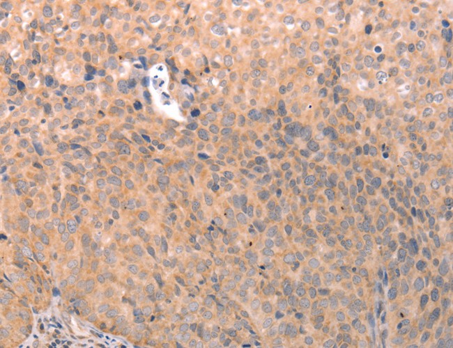
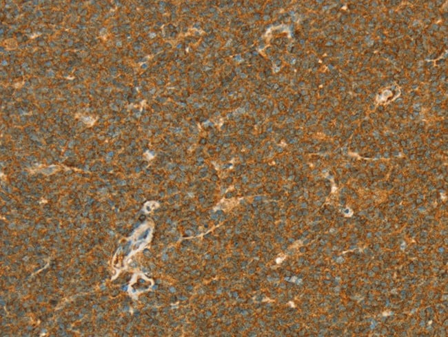
(Immunohistochemistry of paraffin-embedded Human lymphoma using APOL2 Polyclonal Antibody at dilution of 1:80)
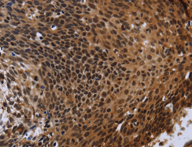
IHC (Immunohistochemisry)
(Immunohistochemistry of paraffin-embedded Human lymphoma using APOL2 Polyclonal Antibody at dilution of 1:80)
APOL2, Polyclonal Antibody (Cat# AAA166842)
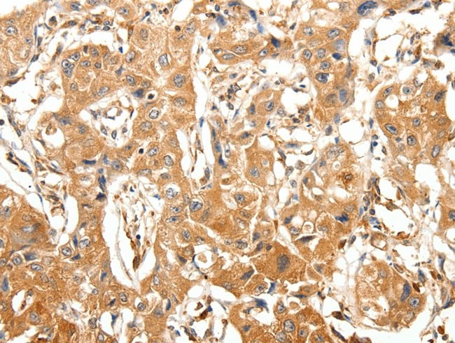
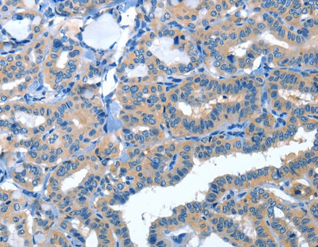
 IHC (Immunohiostchemistry)
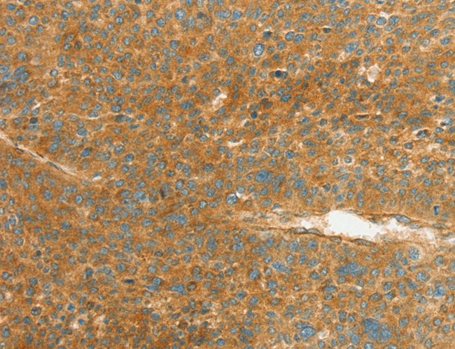
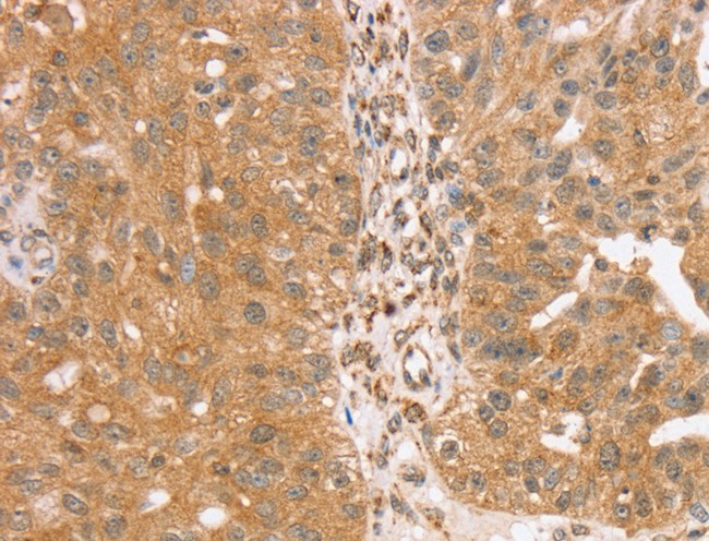
(Immunohistochemistry of paraffin-embedded Human breast cancer tissue using TRIM25 Polyclonal Antibody at dilution 1:40)
IHC (Immunohiostchemistry)
(Immunohistochemistry of paraffin-embedded Human breast cancer tissue using TRIM25 Polyclonal Antibody at dilution 1:40)
TRIM25, Polyclonal Antibody (Cat# AAA166844)
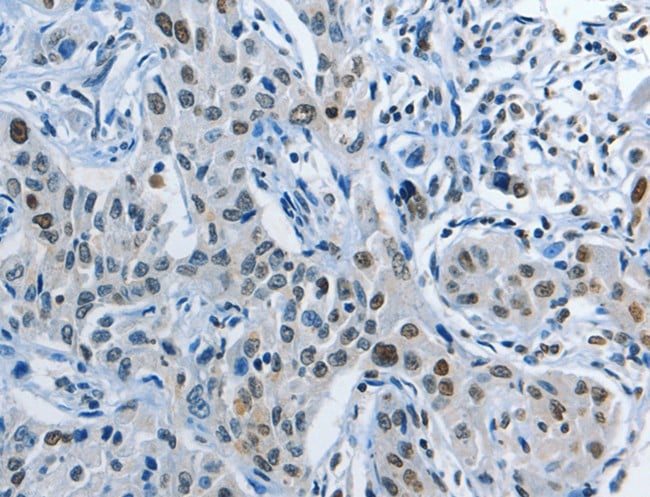
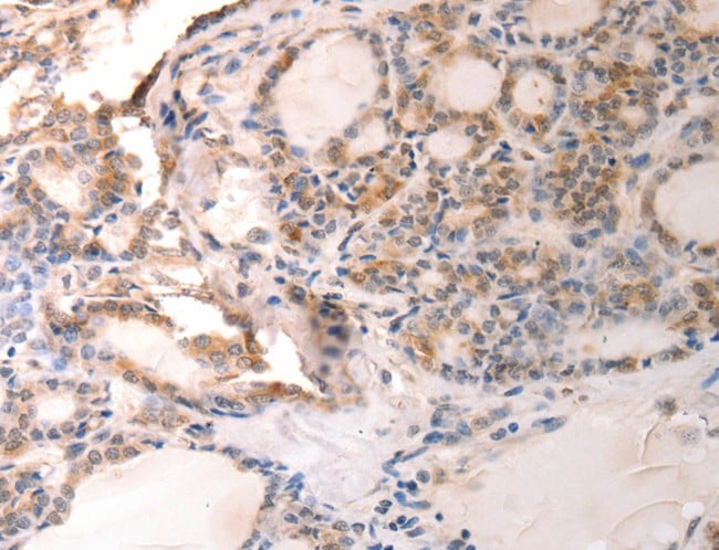
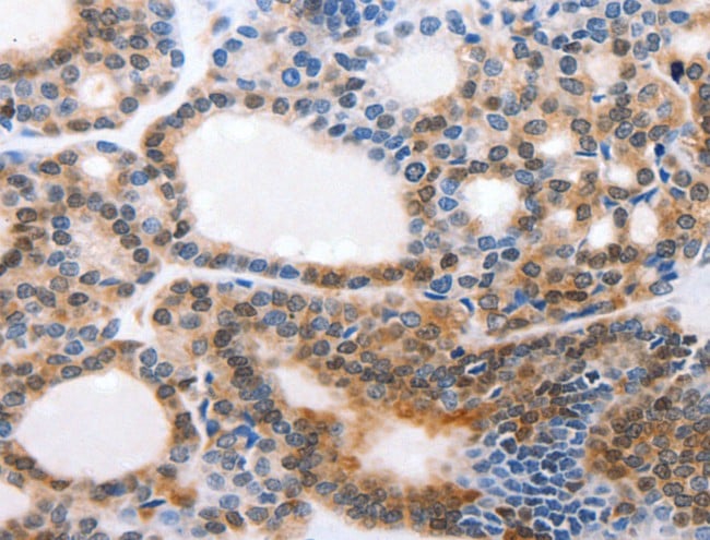 IHC (Immunohistochemisry)
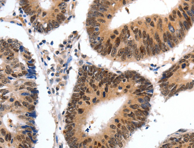
(Immunohistochemistry of paraffin-embedded Human thyroid cancer using CMTM3 Polyclonal Antibody at dilution of 1:20)
IHC (Immunohistochemisry)
(Immunohistochemistry of paraffin-embedded Human thyroid cancer using CMTM3 Polyclonal Antibody at dilution of 1:20)
CMTM3, Polyclonal Antibody (Cat# AAA166846)
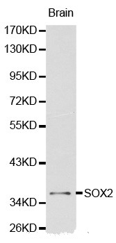
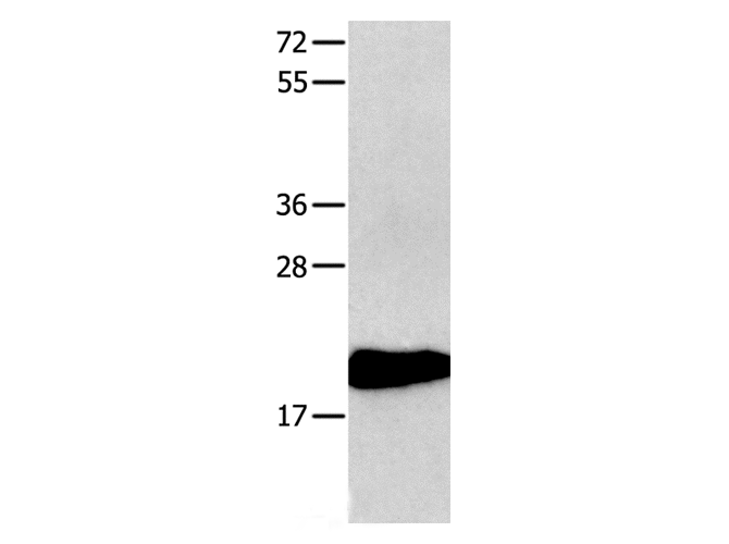
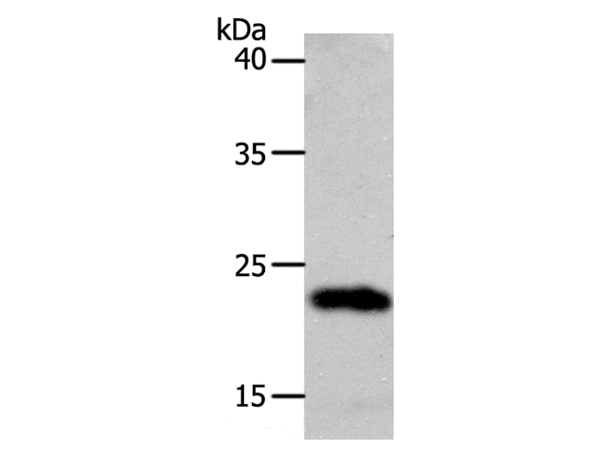
 Application Data
SOX2, Polyclonal Antibody (Cat# AAA166851)
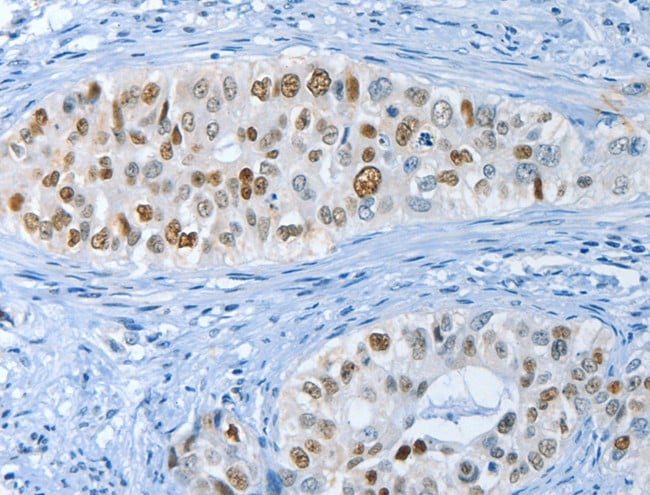
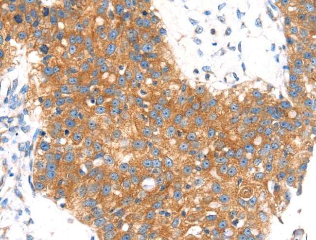
 IHC (Immunohiostchemistry)
(Immunohistochemistry of paraffin-embedded Human lung cancer tissue using NEK4 Polyclonal Antibody at dilution 1:20)
IHC (Immunohiostchemistry)
(Immunohistochemistry of paraffin-embedded Human lung cancer tissue using NEK4 Polyclonal Antibody at dilution 1:20)
NEK4, Polyclonal Antibody (Cat# AAA166860)
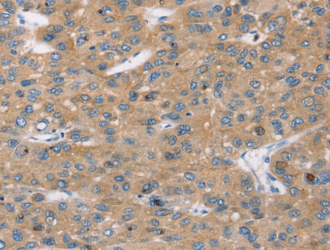 IHC (Immunohiostchemistry)
(Immunohistochemistry of paraffin-embedded Human ovarian cancer tissue using AP1G1 Polyclonal Antibody at dilution 1:16)
IHC (Immunohiostchemistry)
(Immunohistochemistry of paraffin-embedded Human ovarian cancer tissue using AP1G1 Polyclonal Antibody at dilution 1:16)
AP1G1, Polyclonal Antibody (Cat# AAA166863)
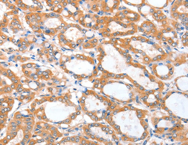
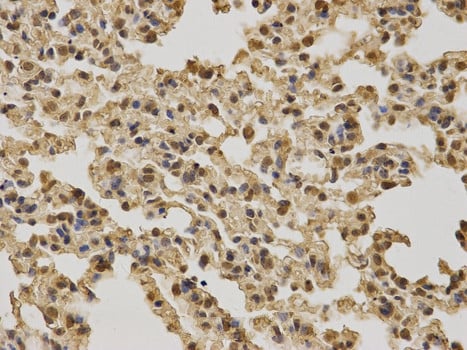
 IHC (Immunohiostchemistry)
(Immunohistochemistry of paraffin-embedded Human lung cancer tissue using ACSBG1 Polyclonal Antibody at dilution 1:25)
IHC (Immunohiostchemistry)
(Immunohistochemistry of paraffin-embedded Human lung cancer tissue using ACSBG1 Polyclonal Antibody at dilution 1:25)
ACSBG1, Polyclonal Antibody (Cat# AAA166866)

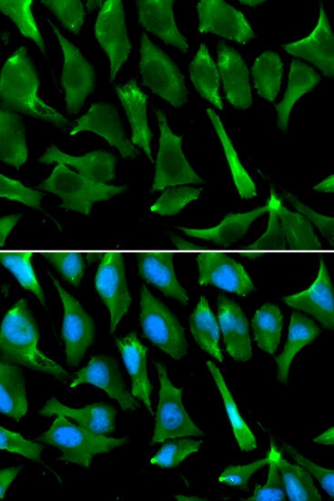 IF (Immunofluorescence)
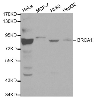
(Immunofluorescence analysis of A549 cell using BRCA1 antibody. Blue: DAPI for nuclear staining.)
IF (Immunofluorescence)
(Immunofluorescence analysis of A549 cell using BRCA1 antibody. Blue: DAPI for nuclear staining.)
BRCA1, Polyclonal Antibody (Cat# AAA166867)

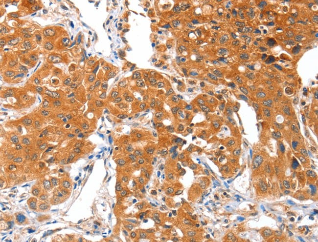
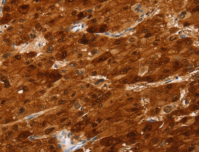
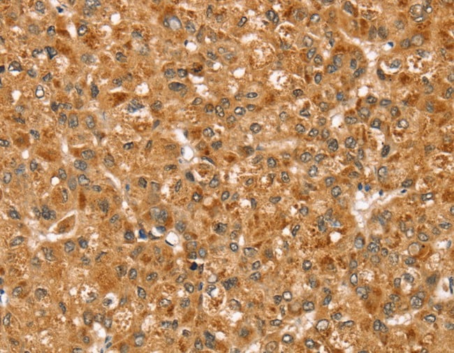
 IHC (Immunohiostchemistry)
(Immunohistochemistry of paraffin-embedded Human liver cancer tissue using ID2 Polyclonal Antibody at dilution 1:30)
IHC (Immunohiostchemistry)
(Immunohistochemistry of paraffin-embedded Human liver cancer tissue using ID2 Polyclonal Antibody at dilution 1:30)
ID2, Polyclonal Antibody (Cat# AAA166890)
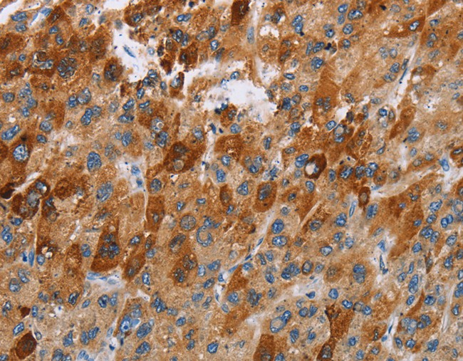
 IHC (Immunohiostchemistry)
(Immunohistochemistry of paraffin-embedded Human liver cancer using SULT2A1 Polyclonal Antibody at dilution of 1:25)
IHC (Immunohiostchemistry)
(Immunohistochemistry of paraffin-embedded Human liver cancer using SULT2A1 Polyclonal Antibody at dilution of 1:25)
SULT2A1, Polyclonal Antibody (Cat# AAA166892)
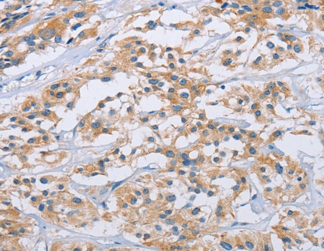
 IHC (Immunohistochemisry)
(Immunohistochemistry of paraffin-embedded Human lung cancer using ITGA2B Polyclonal Antibody at dilution of 1:40)
IHC (Immunohistochemisry)
(Immunohistochemistry of paraffin-embedded Human lung cancer using ITGA2B Polyclonal Antibody at dilution of 1:40)
ITGA2B, Polyclonal Antibody (Cat# AAA166894)
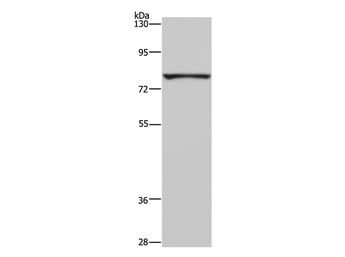
 IHC (Immunohiostchemistry)
(Immunohistochemistry of paraffin-embedded Human lung cancer tissue using SLC27A1 Polyclonal Antibody at dilution 1:40)
IHC (Immunohiostchemistry)
(Immunohistochemistry of paraffin-embedded Human lung cancer tissue using SLC27A1 Polyclonal Antibody at dilution 1:40)
SLC27A1, Polyclonal Antibody (Cat# AAA166895)
 IHC (Immunohistochemisry)
(Immunohistochemistry of paraffin-embedded Human liver cancer using BNIP3 Polyclonal Antibody at dilution of 1:20)
IHC (Immunohistochemisry)
(Immunohistochemistry of paraffin-embedded Human liver cancer using BNIP3 Polyclonal Antibody at dilution of 1:20)
BNIP3, Polyclonal Antibody (Cat# AAA166896)

 IHC (Immunohistochemisry)
(Immunohistochemistry of paraffin-embedded Human lung cancer using APPL1 Polyclonal Antibody at dilution of 1:30)
IHC (Immunohistochemisry)
(Immunohistochemistry of paraffin-embedded Human lung cancer using APPL1 Polyclonal Antibody at dilution of 1:30)
APPL1, Polyclonal Antibody (Cat# AAA166897)
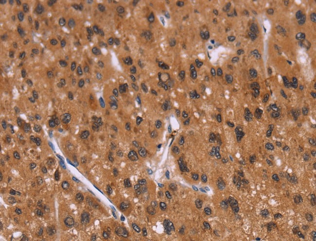
 IHC (Immunohiostchemistry)
(Immunohistochemistry of paraffin-embedded Human liver cancer tissue using PAFAH1B1 Polyclonal Antibody at dilution 1:25)
IHC (Immunohiostchemistry)
(Immunohistochemistry of paraffin-embedded Human liver cancer tissue using PAFAH1B1 Polyclonal Antibody at dilution 1:25)
PAFAH1B1, Polyclonal Antibody (Cat# AAA166991)
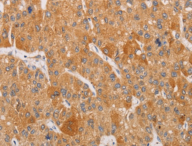
 IHC (Immunohistochemisry)
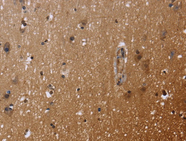
(Immunohistochemistry of paraffin-embedded Human brain using FHL3 Polyclonal Antibody at dilution of 1:40)
IHC (Immunohistochemisry)
(Immunohistochemistry of paraffin-embedded Human brain using FHL3 Polyclonal Antibody at dilution of 1:40)
FHL3, Polyclonal Antibody (Cat# AAA166996)
 IHC (Immunohiostchemistry)
(Immunohistochemistry of paraffin-embedded Human thyroid cancer using CWC27 Polyclonal Antibody at dilution of 1:40)
IHC (Immunohiostchemistry)
(Immunohistochemistry of paraffin-embedded Human thyroid cancer using CWC27 Polyclonal Antibody at dilution of 1:40)
CWC27, Polyclonal Antibody (Cat# AAA167001)
 IHC (Immunohistochemisry)
(Immunohistochemistry of paraffin-embedded Human breast cancer using SOX11 Polyclonal Antibody at dilution of 1:20)
IHC (Immunohistochemisry)
(Immunohistochemistry of paraffin-embedded Human breast cancer using SOX11 Polyclonal Antibody at dilution of 1:20)
SOX11, Polyclonal Antibody (Cat# AAA167015)
 IHC (Immunohistochemisry)
(Immunohistochemistry of paraffin-embedded Human gastric cancer using SOCS1 Polyclonal Antibody at dilution of 1:70)
IHC (Immunohistochemisry)
(Immunohistochemistry of paraffin-embedded Human gastric cancer using SOCS1 Polyclonal Antibody at dilution of 1:70)
SOCS1, Polyclonal Antibody (Cat# AAA167032)
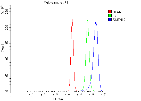 FCM/FACS (Flow Cytometry)
(Figure 3. Flow Cytometry analysis of MCF-7 cells using anti-SMTNL2 antibody (AAA127919).Overlay histogram showing MCF-7 cells stained with AAA127919 (Blue line). To facilitate intracellular staining, cells were fixed with 4% paraformaldehyde and permeabilized with permeabilization buffer. The cells were blocked with 10% normal goat serum. And then incubated with rabbit anti-SMTNL2 Antibody (AAA127919, 1ug/1x106 cells) for 30 min at 20 degree C. DyLight488 conjugated goat anti-rabbit IgG was used as secondary antibody for 30 minutes at 20 degree C. Isotype control antibody (Green line) was rabbit IgG (1ug/1x106) used under the same conditions. Unlabelled sample (Red line) was also used as a control.)
FCM/FACS (Flow Cytometry)
(Figure 3. Flow Cytometry analysis of MCF-7 cells using anti-SMTNL2 antibody (AAA127919).Overlay histogram showing MCF-7 cells stained with AAA127919 (Blue line). To facilitate intracellular staining, cells were fixed with 4% paraformaldehyde and permeabilized with permeabilization buffer. The cells were blocked with 10% normal goat serum. And then incubated with rabbit anti-SMTNL2 Antibody (AAA127919, 1ug/1x106 cells) for 30 min at 20 degree C. DyLight488 conjugated goat anti-rabbit IgG was used as secondary antibody for 30 minutes at 20 degree C. Isotype control antibody (Green line) was rabbit IgG (1ug/1x106) used under the same conditions. Unlabelled sample (Red line) was also used as a control.)
SMTNL2, Polyclonal Antibody (Cat# AAA127919)
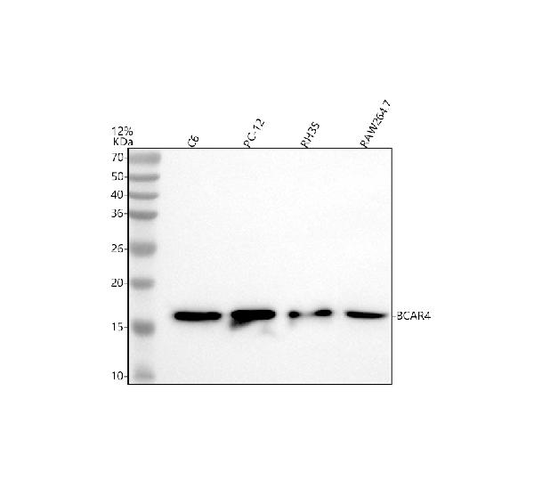
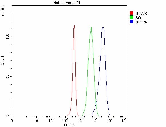 FCM/FACS (Flow Cytometry)
(Figure 2. Flow Cytometry analysis of 293T cells using anti-BCAR4 antibody (AAA127947).Overlay histogram showing 293T cells stained with AAA127947 (Blue line). The cells were fixed with 4% paraformaldehyde and blocked with 10% normal goat serum. And then incubated with rabbit anti-BCAR4 Antibody (AAA127947, 1ug/1x106 cells) for 30 min at 20 degree C. DyLight488 conjugated goat anti-rabbit IgG was used as secondary antibody for 30 minutes at 20 degree C. Isotype control antibody (Green line) was rabbit IgG (1ug/1x106) used under the same conditions. Unlabelled sample without incubation with primary antibody and secondary antibody (Red line) was used as a blank control.)
FCM/FACS (Flow Cytometry)
(Figure 2. Flow Cytometry analysis of 293T cells using anti-BCAR4 antibody (AAA127947).Overlay histogram showing 293T cells stained with AAA127947 (Blue line). The cells were fixed with 4% paraformaldehyde and blocked with 10% normal goat serum. And then incubated with rabbit anti-BCAR4 Antibody (AAA127947, 1ug/1x106 cells) for 30 min at 20 degree C. DyLight488 conjugated goat anti-rabbit IgG was used as secondary antibody for 30 minutes at 20 degree C. Isotype control antibody (Green line) was rabbit IgG (1ug/1x106) used under the same conditions. Unlabelled sample without incubation with primary antibody and secondary antibody (Red line) was used as a blank control.)
BCAR4, Polyclonal Antibody (Cat# AAA127947)
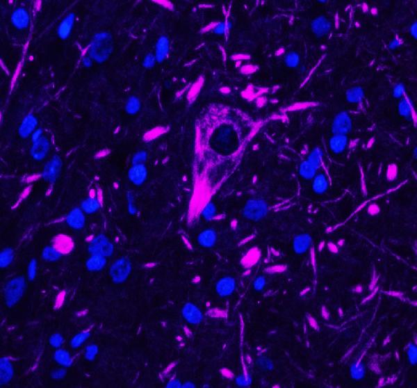
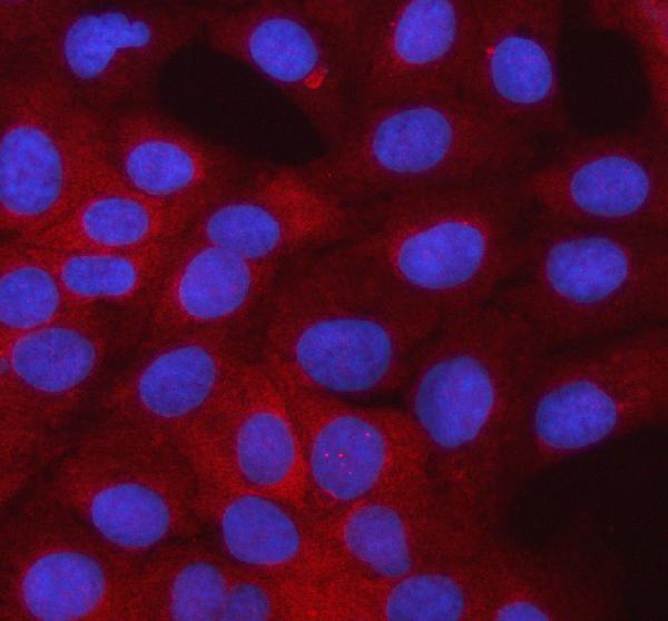
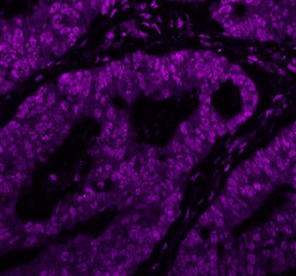 IF (Immunofluorescence)
(Figure 3. IF analysis of NEFH using anti-NEFH antibody (MA1071).NEFH was detected in a paraffin-embedded section of human colon cancer tissue. Heat mediated antigen retrieval was performed in EDTA buffer (pH 8.0, epitope retrieval solution). The tissue section was blocked with 10% goat serum. The tissue section was then incubated with 5ug/mL mouse anti-NEFH Antibody (MA1071) overnight at 4 degree C. DyLight 647 Conjugated Goat Anti-Mouse IgG (AAA127949) was used as secondary antibody at 1:500 dilution and incubated for 30 minutes at 37 degree C. The section was counterstained with DAPI. Visualize using a fluorescence microscope and filter sets appropriate for the label used.)
IF (Immunofluorescence)
(Figure 3. IF analysis of NEFH using anti-NEFH antibody (MA1071).NEFH was detected in a paraffin-embedded section of human colon cancer tissue. Heat mediated antigen retrieval was performed in EDTA buffer (pH 8.0, epitope retrieval solution). The tissue section was blocked with 10% goat serum. The tissue section was then incubated with 5ug/mL mouse anti-NEFH Antibody (MA1071) overnight at 4 degree C. DyLight 647 Conjugated Goat Anti-Mouse IgG (AAA127949) was used as secondary antibody at 1:500 dilution and incubated for 30 minutes at 37 degree C. The section was counterstained with DAPI. Visualize using a fluorescence microscope and filter sets appropriate for the label used.)
IgG, Polyclonal Secondary Antibody (Cat# AAA127949)
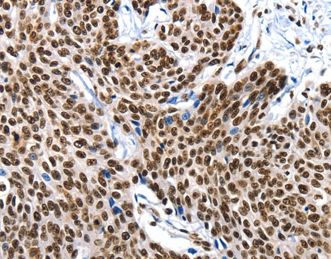
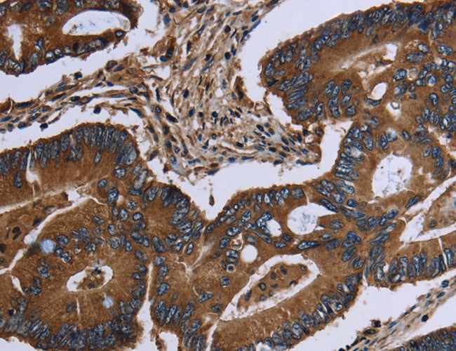
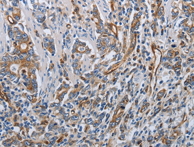
 IHC (Immunohiostchemistry)
(Immunohistochemistry of paraffin-embedded Human colon cancer tissue using XRCC6 Polyclonal Antibody at dilution of 1:10)
IHC (Immunohiostchemistry)
(Immunohistochemistry of paraffin-embedded Human colon cancer tissue using XRCC6 Polyclonal Antibody at dilution of 1:10)
XRCC6, Polyclonal Antibody (Cat# AAA166108)
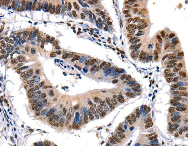
 IHC (Immunohistochemisry)
(Immunohistochemistry of paraffin-embedded Human esophagus cancer using APOH Polyclonal Antibody at dilution of 1:30)
IHC (Immunohistochemisry)
(Immunohistochemistry of paraffin-embedded Human esophagus cancer using APOH Polyclonal Antibody at dilution of 1:30)
APOH, Polyclonal Antibody (Cat# AAA166111)
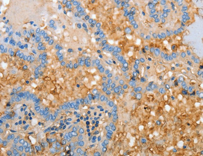
 IHC (Immunohiostchemistry)
(Immunohistochemistry of paraffin-embedded Human thyroid cancer tissue using PARP3 Polyclonal Antibody at dilution 1:25)
IHC (Immunohiostchemistry)
(Immunohistochemistry of paraffin-embedded Human thyroid cancer tissue using PARP3 Polyclonal Antibody at dilution 1:25)
PARP3, Polyclonal Antibody (Cat# AAA166112)
 IHC (Immunohiostchemistry)
(Immunohistochemistry of paraffin-embedded Human lung cancer tissue using ANKRA2 Polyclonal Antibody at dilution 1:25)
IHC (Immunohiostchemistry)
(Immunohistochemistry of paraffin-embedded Human lung cancer tissue using ANKRA2 Polyclonal Antibody at dilution 1:25)
ANKRA2, Polyclonal Antibody (Cat# AAA166117)

 IHC (Immunohiostchemistry)
(Immunohistochemistry of paraffin-embedded Human sarcoma using LAMB3 Polyclonal Antibody at dilution of 1:40)
IHC (Immunohiostchemistry)
(Immunohistochemistry of paraffin-embedded Human sarcoma using LAMB3 Polyclonal Antibody at dilution of 1:40)
LAMB3, Polyclonal Antibody (Cat# AAA166127)
 IHC (Immunohiostchemistry)
(Immunohistochemistry of paraffin-embedded Human liver cancer using STX3 Polyclonal Antibody at dilution of 1:35)
IHC (Immunohiostchemistry)
(Immunohistochemistry of paraffin-embedded Human liver cancer using STX3 Polyclonal Antibody at dilution of 1:35)
STX3, Polyclonal Antibody (Cat# AAA166144)
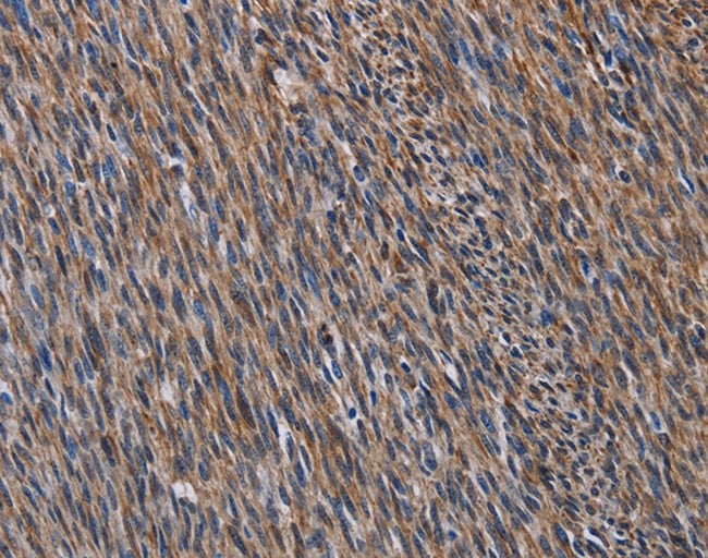
 IHC (Immunohiostchemistry)
(Immunohistochemistry of paraffin-embedded Human thyroid cancer tissue using CCR9 Polyclonal Antibody at dilution 1:40)
IHC (Immunohiostchemistry)
(Immunohistochemistry of paraffin-embedded Human thyroid cancer tissue using CCR9 Polyclonal Antibody at dilution 1:40)
CCR9, Polyclonal Antibody (Cat# AAA166162)
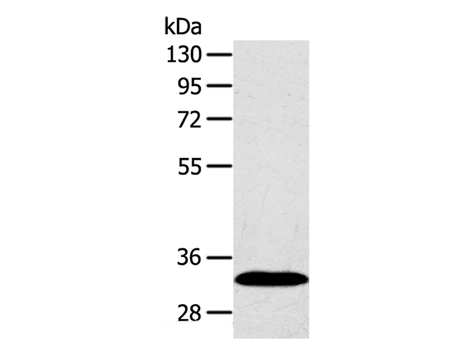
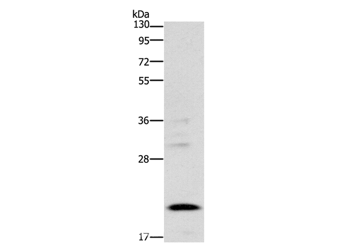
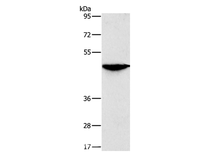
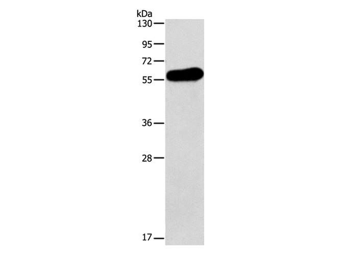
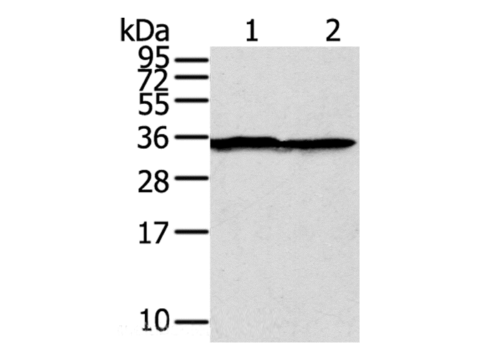
 WB (Western Blot)
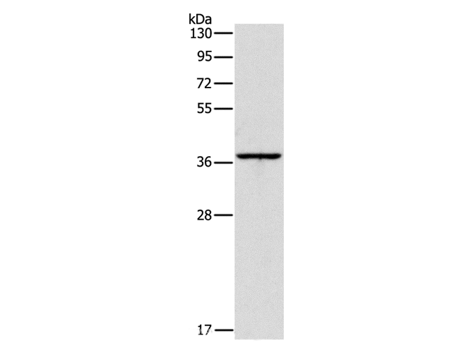
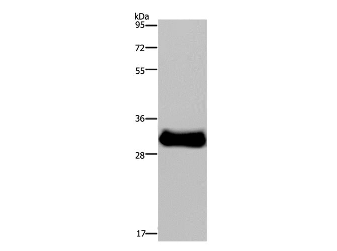
(Western Blot analysis of Raji cell using FASTKD3 Polyclonal Antibody at dilution of 1:200)
WB (Western Blot)
(Western Blot analysis of Raji cell using FASTKD3 Polyclonal Antibody at dilution of 1:200)
FASTKD3, Polyclonal Antibody (Cat# AAA166169)
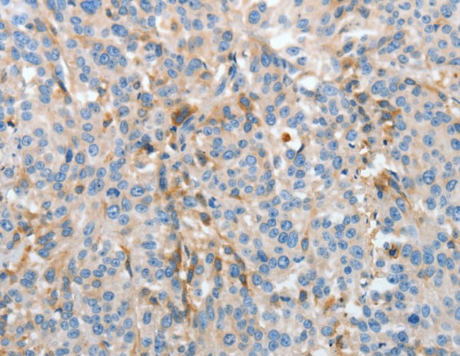
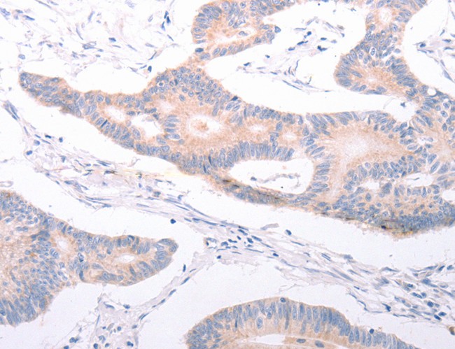
 IHC (Immunohiostchemistry)
(Immunohistochemistry of paraffin-embedded Human thyroid cancer tissue using VTCN1 Polyclonal Antibody at dilution 1:20)
IHC (Immunohiostchemistry)
(Immunohistochemistry of paraffin-embedded Human thyroid cancer tissue using VTCN1 Polyclonal Antibody at dilution 1:20)
VTCN1, Polyclonal Antibody (Cat# AAA166171)
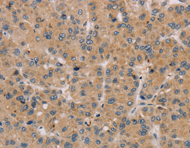
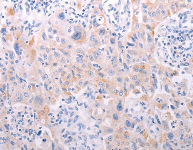
 IHC (Immunohiostchemistry)
(Immunohistochemistry of paraffin-embedded Human thyroid cancer tissue using AMDHD2 Polyclonal Antibody at dilution 1:30)
IHC (Immunohiostchemistry)
(Immunohistochemistry of paraffin-embedded Human thyroid cancer tissue using AMDHD2 Polyclonal Antibody at dilution 1:30)
AMDHD2, Polyclonal Antibody (Cat# AAA166174)
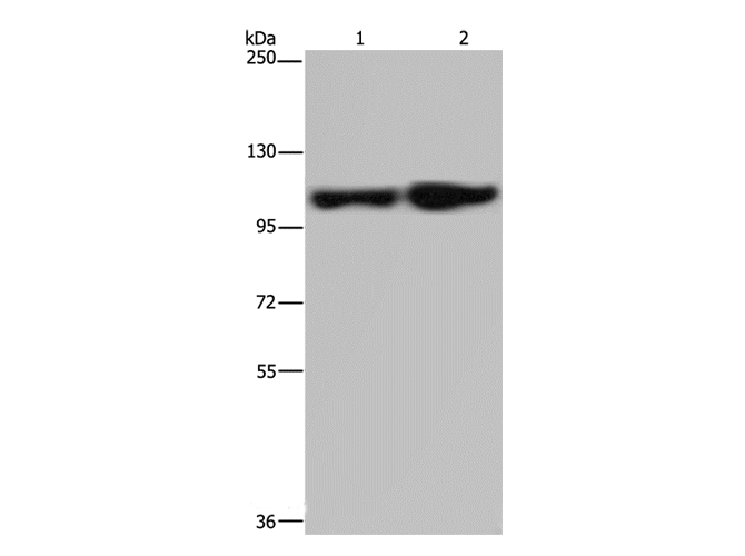
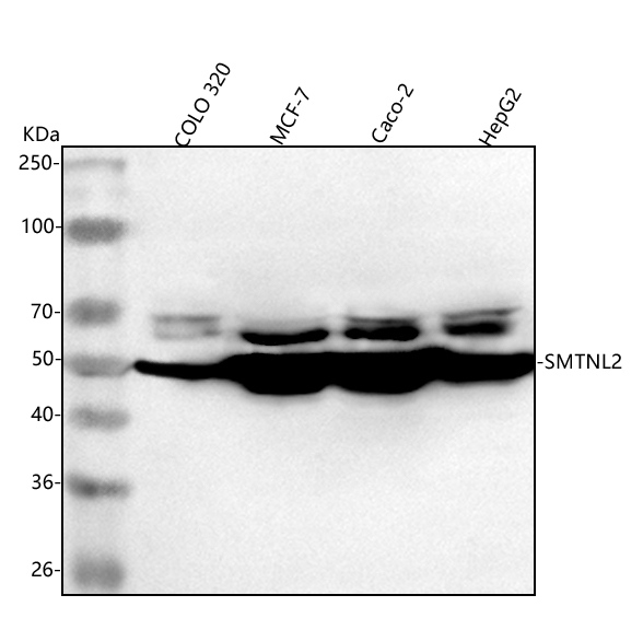
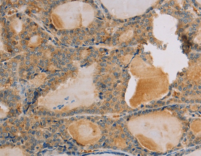
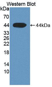
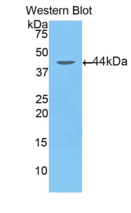
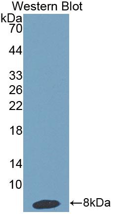
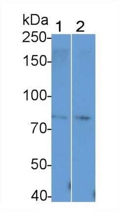 WB (Western Blot)
(Western Blot; Sample: Lane1: Mouse Testis lysate; Lane2: Rat Testis lysate Primary Ab: 1 ug/ml Rabbit Anti-Mouse TAGAP Antibody Second Ab: 0.2ug/mL HRP-Linked Caprine Anti-Rabbit IgG Polyclonal Antibody)
WB (Western Blot)
(Western Blot; Sample: Lane1: Mouse Testis lysate; Lane2: Rat Testis lysate Primary Ab: 1 ug/ml Rabbit Anti-Mouse TAGAP Antibody Second Ab: 0.2ug/mL HRP-Linked Caprine Anti-Rabbit IgG Polyclonal Antibody)
T-Cell Activation Rho GTPase Activating Protein (TAGAP), Polyclonal Antibody (Cat# AAA152615)


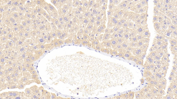
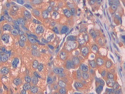
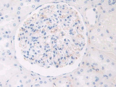
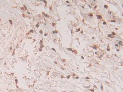
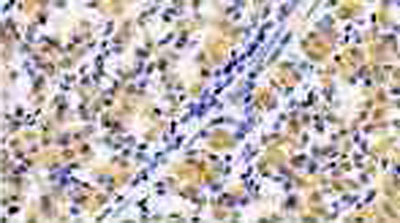 IHC (Immunohistochemisry)
(DAB staining on IHC-P;Samples: Human Stomach Tissue;Primary Ab: 20ug/ml Rabbit Anti-Human IL1RA AntibodySecond Ab: 2ug/mL HRP-Linked Caprine Anti-Rabbit IgG Polyclonal Antibody)
IHC (Immunohistochemisry)
(DAB staining on IHC-P;Samples: Human Stomach Tissue;Primary Ab: 20ug/ml Rabbit Anti-Human IL1RA AntibodySecond Ab: 2ug/mL HRP-Linked Caprine Anti-Rabbit IgG Polyclonal Antibody)
Interleukin 1 Receptor Antagonist (IL1RA), Polyclonal Antibody (Cat# AAA131549)
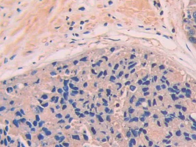
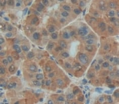
 IHC (Immunohistochemistry)
(DAB staining on IHC-P; Samples: Human Breast cancer Tissue))
IHC (Immunohistochemistry)
(DAB staining on IHC-P; Samples: Human Breast cancer Tissue))
Complement Factor B (CFB), Polyclonal Antibody (Cat# AAA131593)
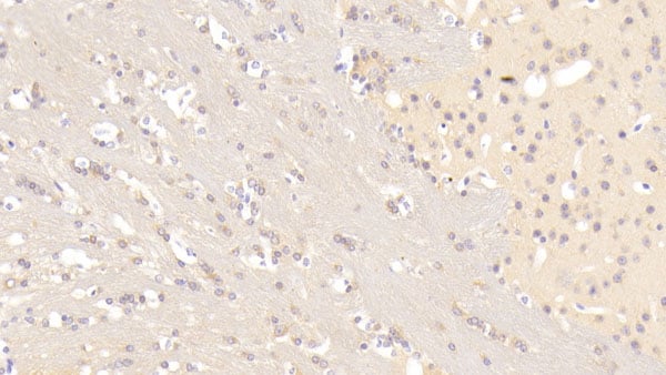
 IHC (Immunohiostchemistry)
(DABstainingonIHC-P.Samples:HumanTissue))
IHC (Immunohiostchemistry)
(DABstainingonIHC-P.Samples:HumanTissue))
Aspartate Aminotransferase 2 (AST2), Polyclonal Antibody (Cat# AAA131610)
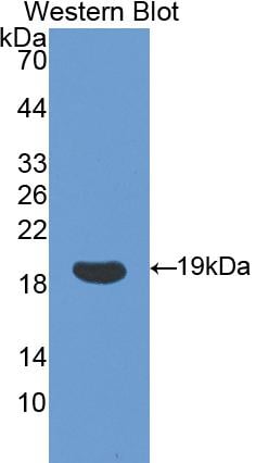
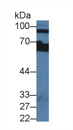
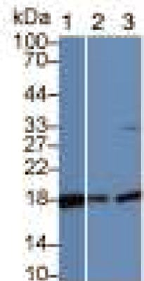 WB (Western Blot)
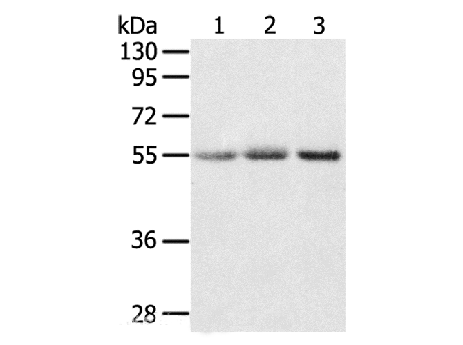
(Sample:Lane 1: Porcine Cerebrum lysate;Lane 2: Mouse Cerebrum lysate;Lane 3: Hela cell lysatePrimary Ab: 1ug/ml Rabbit Anti-Porcine CYPA AntibodySecond Ab: 0.2ug/mL HRP-Linked Caprine Anti-Rabbit IgG Polyclonal Antibody)
WB (Western Blot)
(Sample:Lane 1: Porcine Cerebrum lysate;Lane 2: Mouse Cerebrum lysate;Lane 3: Hela cell lysatePrimary Ab: 1ug/ml Rabbit Anti-Porcine CYPA AntibodySecond Ab: 0.2ug/mL HRP-Linked Caprine Anti-Rabbit IgG Polyclonal Antibody)
Cyclophilin A (CYPA), Polyclonal Antibody (Cat# AAA131840)
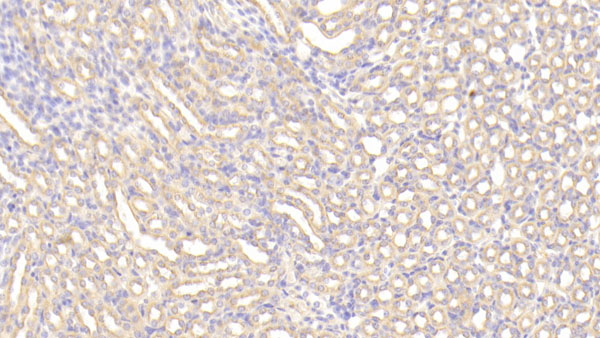
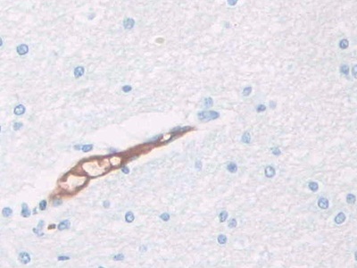 IHC (Immunohistochemisry)
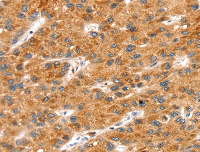
(DAB staining on IHC-P; Samples: Human Cerebrum Tissue))
IHC (Immunohistochemisry)
(DAB staining on IHC-P; Samples: Human Cerebrum Tissue))
Haptoglobin Related Protein (HPR), Polyclonal Antibody (Cat# AAA131841)

 IHC (Immunohistochemisry)
(DAB staining on IHC-P; Samples: Human Breast Cancer Tissue.)
IHC (Immunohistochemisry)
(DAB staining on IHC-P; Samples: Human Breast Cancer Tissue.)
S100 Calcium Binding Protein A11 (S100A11), Polyclonal Antibody (Cat# AAA131856)
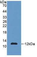
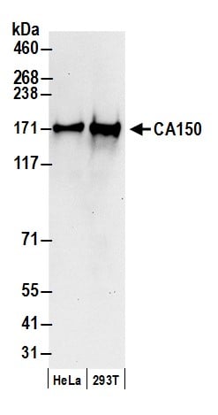 WB (Western Blot)
(Detection of human CA150 by western blot. Samples: Whole cell lysate (50 ug) from HeLa and HEK293T cells prepared using NETN lysis buffer. Antibody: Affinity purified rabbit anti-CA150 antibody AAA210905 (lot AAA210905-2) used for WB at 0.1 ug/ml. Detection: Chemiluminescence with an exposure time of 10 seconds.)
WB (Western Blot)
(Detection of human CA150 by western blot. Samples: Whole cell lysate (50 ug) from HeLa and HEK293T cells prepared using NETN lysis buffer. Antibody: Affinity purified rabbit anti-CA150 antibody AAA210905 (lot AAA210905-2) used for WB at 0.1 ug/ml. Detection: Chemiluminescence with an exposure time of 10 seconds.)
CA150, Polyclonal Antibody (Cat# AAA210905)
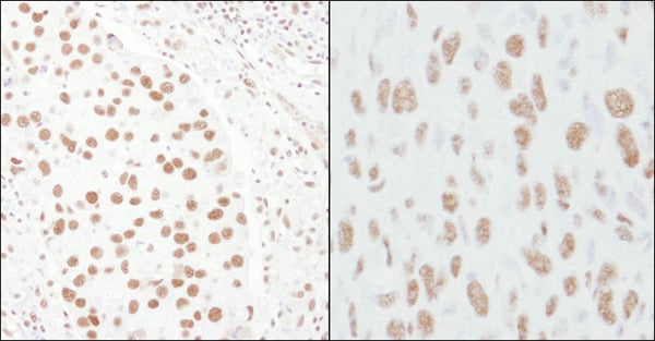
 WB (Western Blot)
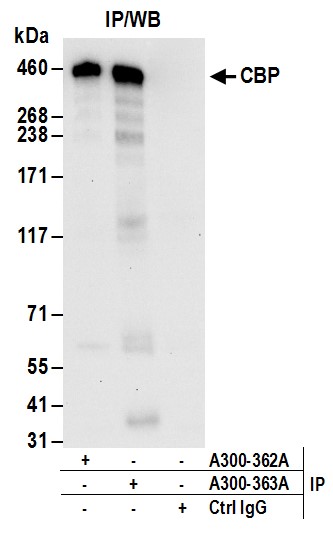
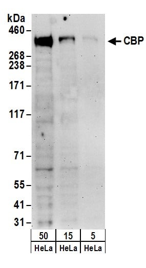
(Detection of human CBP by western blot. Samples: Whole cell lysate (5, 15 and 50 ug) from HeLa cells. Antibodies: Affinity purified rabbit anti-CBP antibody AAA210908 (lot AAA210908-1) used for WB at 0.4 ug/ml. Detection: Chemiluminescence with an exposure time of 3 minutes.)
WB (Western Blot)
(Detection of human CBP by western blot. Samples: Whole cell lysate (5, 15 and 50 ug) from HeLa cells. Antibodies: Affinity purified rabbit anti-CBP antibody AAA210908 (lot AAA210908-1) used for WB at 0.4 ug/ml. Detection: Chemiluminescence with an exposure time of 3 minutes.)
CBP, Polyclonal Antibody (Cat# AAA210908)
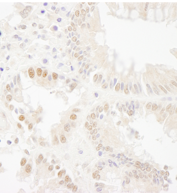
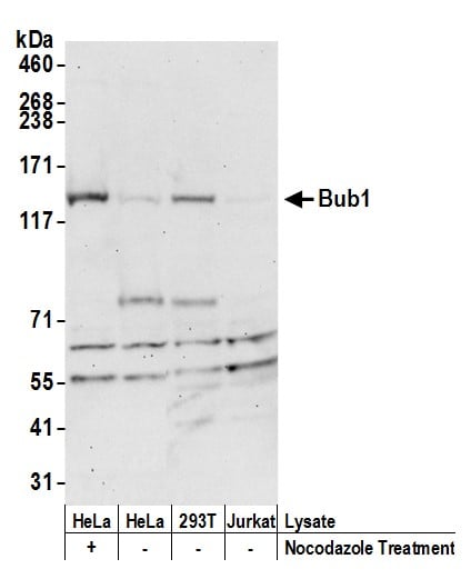 WB (Western Blot)
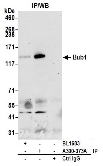
(Detection of human Bub1 by western blot. Samples: Whole cell lysate (50 ug) from HeLa treated with nacodazole (+) or mock treated (-), HEK293T. and Jurkat cells prepared using NETN lysis buffer. Antibody: Affinity purified rabbit anti-Bub1 antibody AAA210912 (lot AAA210912-2) used for WB at 0.4 ug/ml. Detection: Chemiluminescence with an exposure time of 30 seconds)
WB (Western Blot)
(Detection of human Bub1 by western blot. Samples: Whole cell lysate (50 ug) from HeLa treated with nacodazole (+) or mock treated (-), HEK293T. and Jurkat cells prepared using NETN lysis buffer. Antibody: Affinity purified rabbit anti-Bub1 antibody AAA210912 (lot AAA210912-2) used for WB at 0.4 ug/ml. Detection: Chemiluminescence with an exposure time of 30 seconds)
Bub1, Polyclonal Antibody (Cat# AAA210912)
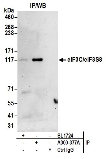
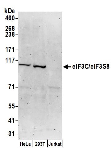 WB (Western Blot)
(Detection of human eIF3C/eIF3S8 by western blot. Samples: Whole cell lysate (50 ug) from HeLa, HEK293T, and Jurkat cells prepared using NETN lysis buffer. Antibodies: Affinity purified rabbit anti-eIF3C/eIF3S8 antibody AAA210914 (lot AAA210914-2) used for WB at 0.1 ug/ml. Detection: Chemiluminescence with an exposure time of 3 minutes.)
WB (Western Blot)
(Detection of human eIF3C/eIF3S8 by western blot. Samples: Whole cell lysate (50 ug) from HeLa, HEK293T, and Jurkat cells prepared using NETN lysis buffer. Antibodies: Affinity purified rabbit anti-eIF3C/eIF3S8 antibody AAA210914 (lot AAA210914-2) used for WB at 0.1 ug/ml. Detection: Chemiluminescence with an exposure time of 3 minutes.)
eIF3C/eIF3S8, Polyclonal Antibody (Cat# AAA210914)
 WB (Western Blot)
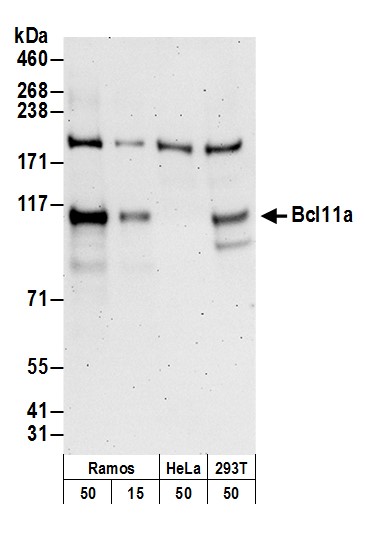
(Detection of human Bcl11a by western blot. Samples: Whole cell lysate from Ramos (50 and 15 ug), HeLa (50 ug), and HEK293T (50 ug) cells prepared using NETN lysis buffer. Antibody: Affinity purified rabbit anti-Bcl11a antibody AAA210916 (lot AAA210916-2) used for WB at 0.1 ug/ml. Detection: Chemiluminescence with an exposure time of 3 minutes.)
WB (Western Blot)
(Detection of human Bcl11a by western blot. Samples: Whole cell lysate from Ramos (50 and 15 ug), HeLa (50 ug), and HEK293T (50 ug) cells prepared using NETN lysis buffer. Antibody: Affinity purified rabbit anti-Bcl11a antibody AAA210916 (lot AAA210916-2) used for WB at 0.1 ug/ml. Detection: Chemiluminescence with an exposure time of 3 minutes.)
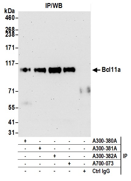
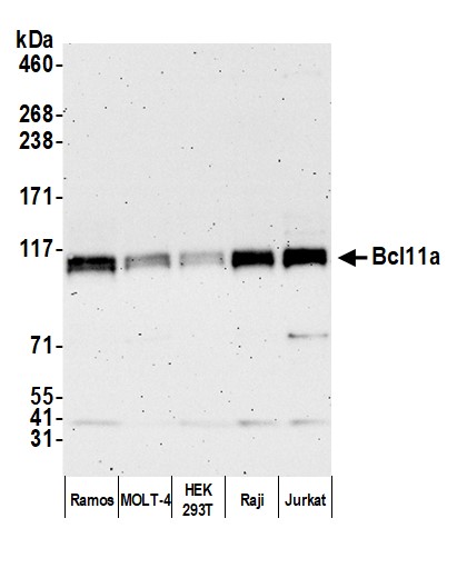
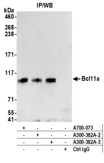
Bcl11a, Polyclonal Antibody (Cat# AAA210916)
 WB (Western Blot)
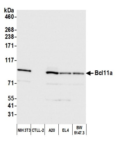
(Detection of mouse Bcl11a by western blot. Samples: Whole cell lysate (10 ug) from NIH 3T3, CTLL-2, A20, EL4, and BW5147.3 cells prepared using NETN lysis buffer. Antibody: Affinity purified rabbit anti-Bcl11a antibody (AAA210917 lot 3) used for WB at 0.04 ug/ml. Detection: Chemiluminescence with an exposure time of 3 seconds.)
WB (Western Blot)
(Detection of mouse Bcl11a by western blot. Samples: Whole cell lysate (10 ug) from NIH 3T3, CTLL-2, A20, EL4, and BW5147.3 cells prepared using NETN lysis buffer. Antibody: Affinity purified rabbit anti-Bcl11a antibody (AAA210917 lot 3) used for WB at 0.04 ug/ml. Detection: Chemiluminescence with an exposure time of 3 seconds.)
Bcl11a, Polyclonal Antibody (Cat# AAA210917)
 WB (Western Blot)
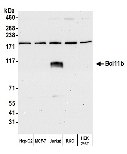
(Detection of human Bcl11b by western blot. Samples: Whole cell lysate (5 ug) from Hep-G2, MCF-7, Jurkat, RKO, and HEK293T cells prepared using NETN lysis buffer. Antibody: Affinity purified rabbit anti-Bcl11b antibody (AAA210918 lot 3) used for WB at 0.04 ug/ml. Detection: Chemiluminescence with an exposure time of 3 seconds.)
WB (Western Blot)
(Detection of human Bcl11b by western blot. Samples: Whole cell lysate (5 ug) from Hep-G2, MCF-7, Jurkat, RKO, and HEK293T cells prepared using NETN lysis buffer. Antibody: Affinity purified rabbit anti-Bcl11b antibody (AAA210918 lot 3) used for WB at 0.04 ug/ml. Detection: Chemiluminescence with an exposure time of 3 seconds.)
Bcl11b, Polyclonal Antibody (Cat# AAA210918)
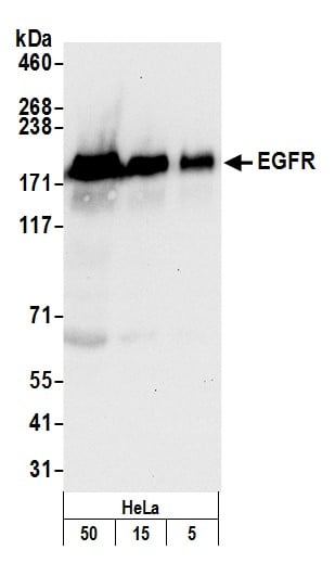 WB (Western Blot)
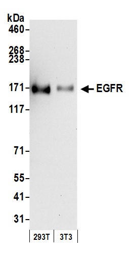
(Detection of human EGFR by western blot. Samples: Whole cell lysate (5, 15 and 50 ug) from HeLa cells prepared using NETN lysis buffer. Antibody: Affinity purified rabbit anti-EGFR antibody AAA210923 (lot AAA210923-2) used for WB at 0.04 ug/ml. Detection: Chemiluminescence with an exposure time of 10 seconds.)
WB (Western Blot)
(Detection of human EGFR by western blot. Samples: Whole cell lysate (5, 15 and 50 ug) from HeLa cells prepared using NETN lysis buffer. Antibody: Affinity purified rabbit anti-EGFR antibody AAA210923 (lot AAA210923-2) used for WB at 0.04 ug/ml. Detection: Chemiluminescence with an exposure time of 10 seconds.)
EGFR, Polyclonal Antibody (Cat# AAA210923)
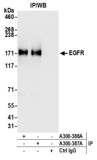
 WB (Western Blot)
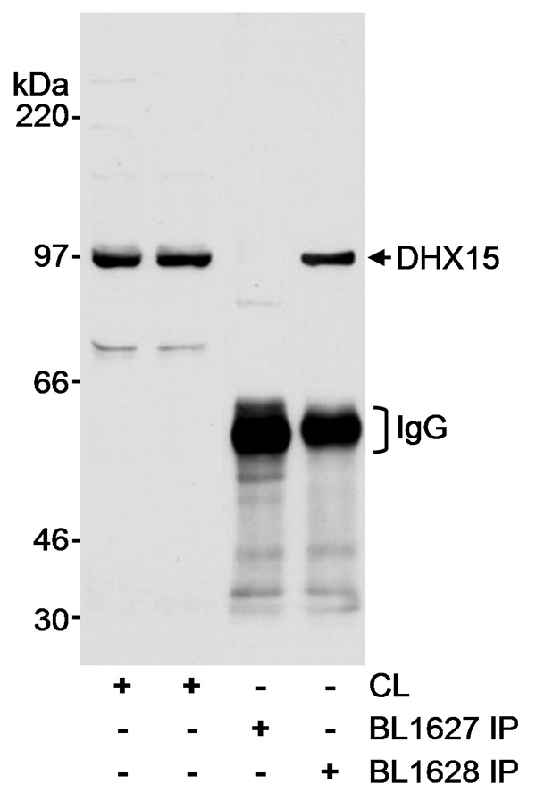
(Detection of human DHX15 by western blot and immunoprecipitation. Samples: Whole cell lysate (CL; 60 ug for WB, 1 mg for IP) from HEK293T cells. Antibodies: Affinity purified rabbit anti-DHX15 antibody BL1627 (Cat. No. AAA210924) used at 0.1 ug/ml for WB and at 1 ug/mg lysate for IP, which did not precipitate DHX15. Successful IP of DHX15 was accomplished using affinity purified rabbit anti-DHX15 antibody BL1628 (Cat. No. at 1 ug/mg lysate. Detection: Chemiluminescence with an exposure time of 15 seconds.)
WB (Western Blot)
(Detection of human DHX15 by western blot and immunoprecipitation. Samples: Whole cell lysate (CL; 60 ug for WB, 1 mg for IP) from HEK293T cells. Antibodies: Affinity purified rabbit anti-DHX15 antibody BL1627 (Cat. No. AAA210924) used at 0.1 ug/ml for WB and at 1 ug/mg lysate for IP, which did not precipitate DHX15. Successful IP of DHX15 was accomplished using affinity purified rabbit anti-DHX15 antibody BL1628 (Cat. No. at 1 ug/mg lysate. Detection: Chemiluminescence with an exposure time of 15 seconds.)
DHX15, Polyclonal Antibody (Cat# AAA210924)
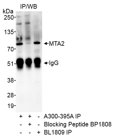
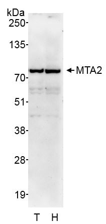 WB (Western Blot)
(Detection of human MTA2 by western blot. Samples: Whole cell lysate (50 ug) from HEK293T (T) and HeLa (H) cells prepared using NETN lysis buffer. Antibody: Affinity purified rabbit anti-MTA2 antibody AAA210926 (lot AAA210926-1) used for WB at 0.1 ug/ml. Detection: Chemiluminescence with exposure time of 2 minutes.)
WB (Western Blot)
(Detection of human MTA2 by western blot. Samples: Whole cell lysate (50 ug) from HEK293T (T) and HeLa (H) cells prepared using NETN lysis buffer. Antibody: Affinity purified rabbit anti-MTA2 antibody AAA210926 (lot AAA210926-1) used for WB at 0.1 ug/ml. Detection: Chemiluminescence with exposure time of 2 minutes.)
MTA2, Polyclonal Antibody (Cat# AAA210926)
What are Polyclonal Antibodies?
Polyclonal antibodies are antibodies that come from multiple B cell clones of a host animal. The typical hosts used for the majority of polyclonal antibody production are rabbits, goats, sheep, and donkeys. These polyclonal antibodies, once having identified their target, will bind to different epitopes located at different regions or sequences on the same protein/antigen. As a result, they are ideal at locating and binding to the target, even if the target is in very low concentrations (due to many different antibodies being able to bind to the same target molecule, which allows for significant amplification of a downstream signal).
Polyclonal antibodies are typically produced by injecting an antigen into a host animal, which causes the animal’s immune system to attack the foreign antigen by mass generating antibodies against it. After a period of time, serum is collected from the animal and purified using physicochemical fractionation, class-specific affinity purification, and/or antigen-affinity purification.
Key Uses of Polyclonal Antibodies
- Western Blotting: This method is used to find specific proteins in biological samples after separating them by size.
- Immunohistochemistry: IHC helps visualize the location of proteins in tissue sections using various staining techniques.
- ELISA: (Enzyme-Linked Immunosorbent Assay) is typically used to identify specific protein quantities in a sample. ELISAs can be either “Quantitative” or “Qualitative”.
- Flow Cytometry: technique that identifies and measures the specific protein on the surface or inside the cells in a fluid suspension.
- Immunoprecipitation: IP isolates and studies a specific protein from a complex mixture using antibodies.
Why Buy Polyclonal Antibodies from AAA Biotech?
1. Ideal for Various Applications
Our antibodies are generally going to be validated for use in multiple types of assays, including ELISA, Western Blotting, Immunohistochemistry, Immunoprecipitation, amongst others. They are ideal for a wide range of research applications.
2. Rigorous Quality Control
All of the antibodies in our catalog undergo strict quality testing to ensure specificity, sensitivity, and consistent performance. We are confident in the ability of our antibodies to provide you with accurate results.
3. Wide Assortment of Antibodies
Antibodies in are catalog can be found for both common and exotic species, and these antibodies are also available in both conjugated and recombinant forms to suit many diverse experimental needs.
4. Highly Purified
Our antibodies are available in purified forms with over 85% purity, as confirmed by SDS-PAGE. They are also available with tags such as His, Flag, GST, or MBP. We cater to customers worldwide.
FAQ
1. How are polyclonal antibodies produced?
Traditionally, polyclonal antibodies are produced by injecting an antigen into a host animal (such as a rabbit or goat), which then triggers an immune response from the host animal. The animal’s B cells produce antibodies that will recognize different parts of the injected antigen. These antibodies are then collected from the animal’s blood and purified for use.
2. How do polyclonal antibodies differ from monoclonal antibodies?
Polyclonal antibodies are a mix of antibodies that bind to different locations (epitopes) of the same antigen, while monoclonal antibodies are identical and bind to just one specific epitope. This makes polyclonal antibodies more versatile and better at detecting proteins that may be present in low quantities or in altered/modified forms.
3. How should I store polyclonal antibodies?
Polyclonal antibodies should be stored at 4°C for short-term use (up to a few weeks) and at -20°C or -80°C for long-term storage. Avoid repeated freeze-thaw cycles by dividing them into small aliquots. Always check the datasheet for specific storage instructions.