Filters
▼Clonality
▼Type
▼Reactivity
▼Gene Name
▼Isotype
▼Host
▼Application
▼Clone
▼Polyclonal Antibodies
At AAA Biotech also known as AAA Bio or AAABio, we provide a broad range of purified polyclonal antibodies (pAbs) that are able to all be browsed online through our website. Due to their high specificity and strong binding affinity, these antibodies are ideal for wide swathes of research and experimental applications.
Our polyclonal antibodies can easily support your work, whether you use them for Western Blotting, Immunocytochemistry (with or without Immunofluorescence used in conjunction), Immunohistochemistry, Immunoprecipitation, and ELISA tests. We highly encourage you to browse our range of pAbs and choose the one that best suits your experimental model.
Viewing 4350-4400 of 96812 product results
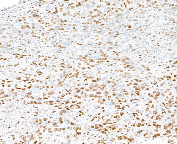
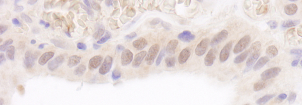
 WB (Western Blot)
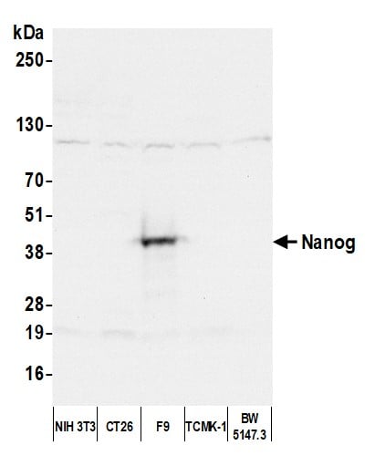
(Detection of mouse Nanog by western blot. Samples: Whole cell lysate (50 ug) from NIH 3T3, CT26, F9, TCMK-1, and BW5147.3 cells prepared using NETN lysis buffer. Antibody: Affinity purified rabbit anti-Nanog antibody AAA210927 (lot AAA210927-4) used for WB at 0.1 ug/ml. Detection: Chemiluminescence with an exposure time of 3 seconds.)
WB (Western Blot)
(Detection of mouse Nanog by western blot. Samples: Whole cell lysate (50 ug) from NIH 3T3, CT26, F9, TCMK-1, and BW5147.3 cells prepared using NETN lysis buffer. Antibody: Affinity purified rabbit anti-Nanog antibody AAA210927 (lot AAA210927-4) used for WB at 0.1 ug/ml. Detection: Chemiluminescence with an exposure time of 3 seconds.)
Nanog, Polyclonal Antibody (Cat# AAA210927)
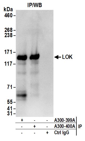
 WB (Western Blot)
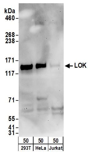
(Detection of human LOK by western blot. Samples: Whole cell lysate (50 ug) from HEK293T, HeLa, and Jurkat cells. Antibodies: Affinity purified rabbit anti-LOK antibody AAA210928 (lot AAA210928-2) used for WB at 0.1 ug/ml. Detection: Chemiluminescence with an exposure time of 3 minutes.)
WB (Western Blot)
(Detection of human LOK by western blot. Samples: Whole cell lysate (50 ug) from HEK293T, HeLa, and Jurkat cells. Antibodies: Affinity purified rabbit anti-LOK antibody AAA210928 (lot AAA210928-2) used for WB at 0.1 ug/ml. Detection: Chemiluminescence with an exposure time of 3 minutes.)
LOK, Polyclonal Antibody (Cat# AAA210928)
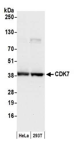
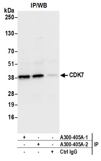
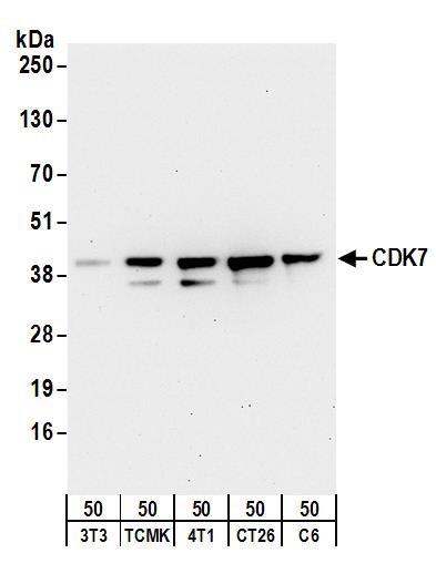 WB (Western Blot)
(Detection of mouse and rat CDK7 by western blot. Samples: Whole cell lysate (50 ug) from NIH 3T3, TCMK-1, 4T1, CT26.WT, and rat C6 cells. Antibodies: Affinity purified rabbit anti-CDK7 antibody AAA210932 (lot AAA210932-1) used for WB at 1 ug/ml. Detection: Chemiluminescence with an exposure time of 30 seconds.)
WB (Western Blot)
(Detection of mouse and rat CDK7 by western blot. Samples: Whole cell lysate (50 ug) from NIH 3T3, TCMK-1, 4T1, CT26.WT, and rat C6 cells. Antibodies: Affinity purified rabbit anti-CDK7 antibody AAA210932 (lot AAA210932-1) used for WB at 1 ug/ml. Detection: Chemiluminescence with an exposure time of 30 seconds.)
CDK7, Polyclonal Antibody (Cat# AAA210932)
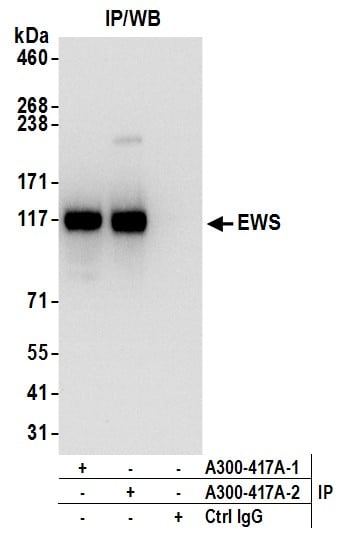
 WB (Western Blot)
(Detection of human and mouse EWS by western blot. Samples: Whole cell lysate (5 ug) from HeLa, HEK293T, Jurkat, mouse TCMK-1, and mouse NIH 3T3 cells prepared using NETN lysis buffer. Antibody: Affinity purified rabbit anti-EWS antibody AAA210940 (lot AAA210940-2) used for WB at 0.04 ug/ml. Detection: Chemiluminescence with an exposure time of 1 second.)
WB (Western Blot)
(Detection of human and mouse EWS by western blot. Samples: Whole cell lysate (5 ug) from HeLa, HEK293T, Jurkat, mouse TCMK-1, and mouse NIH 3T3 cells prepared using NETN lysis buffer. Antibody: Affinity purified rabbit anti-EWS antibody AAA210940 (lot AAA210940-2) used for WB at 0.04 ug/ml. Detection: Chemiluminescence with an exposure time of 1 second.)
EWS, Polyclonal Antibody (Cat# AAA210940)

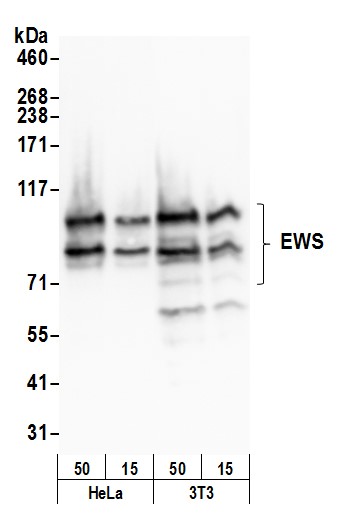 WB (Western Blot)
(Detection of human and mouse EWS by western blot. Samples: Whole cell lysate (15 and 50 ug) from HeLa and mouse NIH 3T3 cells prepared using NETN lysis buffer. Antibody: Affinity purified rabbit anti-EWS antibody AAA210941 (lot AAA210941-2) used for WB at 0.04 ug/ml. Detection: Chemiluminescence with an exposure time of 3 seconds.)
WB (Western Blot)
(Detection of human and mouse EWS by western blot. Samples: Whole cell lysate (15 and 50 ug) from HeLa and mouse NIH 3T3 cells prepared using NETN lysis buffer. Antibody: Affinity purified rabbit anti-EWS antibody AAA210941 (lot AAA210941-2) used for WB at 0.04 ug/ml. Detection: Chemiluminescence with an exposure time of 3 seconds.)
EWS, Polyclonal Antibody (Cat# AAA210941)
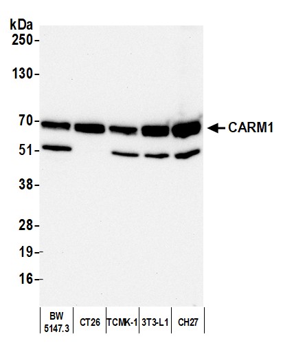
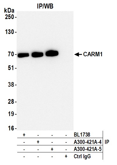
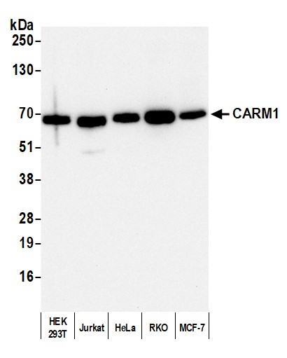 WB (Western Blot)
(Detection of human CARM1 by western blot. Samples: Whole cell lysate (10 ug) from HEK293T, Jurkat, HeLa, RKO, and MCF-7 cells prepared using NETN lysis buffer. Antibody: Affinity purified rabbit anti-CARM1 antibody (AAA210942 lot 5) used for WB at 0.04 ug/ml. Detection: Chemiluminescence with an exposure time of 30 seconds.)
WB (Western Blot)
(Detection of human CARM1 by western blot. Samples: Whole cell lysate (10 ug) from HEK293T, Jurkat, HeLa, RKO, and MCF-7 cells prepared using NETN lysis buffer. Antibody: Affinity purified rabbit anti-CARM1 antibody (AAA210942 lot 5) used for WB at 0.04 ug/ml. Detection: Chemiluminescence with an exposure time of 30 seconds.)
CARM1, Polyclonal Antibody (Cat# AAA210942)
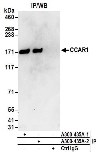
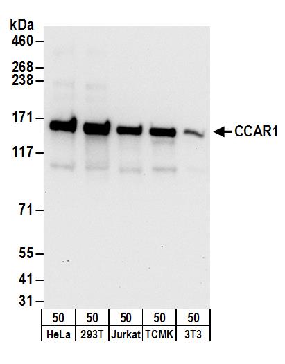 WB (Western Blot)
(Detection of human and mouse CCAR1 by western blot. Samples: Whole cell lysate (50 ug) from HeLa, HEK293T, Jurkat, mouse TCMK-1, and mouse NIH 3T3 cells. Antibodies: Affinity purified rabbit anti-CCAR1 antibody AAA210949 (lot AAA210949-2) used for WB at 0.1 ug/ml. Detection: Chemiluminescence with an exposure time of 10 seconds.)
WB (Western Blot)
(Detection of human and mouse CCAR1 by western blot. Samples: Whole cell lysate (50 ug) from HeLa, HEK293T, Jurkat, mouse TCMK-1, and mouse NIH 3T3 cells. Antibodies: Affinity purified rabbit anti-CCAR1 antibody AAA210949 (lot AAA210949-2) used for WB at 0.1 ug/ml. Detection: Chemiluminescence with an exposure time of 10 seconds.)
CCAR1, Polyclonal Antibody (Cat# AAA210949)
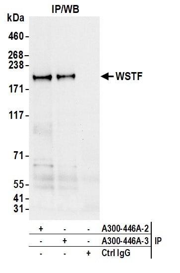
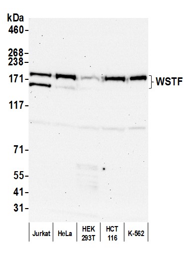 WB (Western Blot)
(Detection of human WSTF by western blot. Samples: Whole cell lysate (50 ug) from Jurkat, HeLa, HEK293T, HCT 116, and K-562 cells prepared using NETN lysis buffer. Antibody: Affinity purified rabbit anti-WSTF antibody (AAA210954 lot 3) used for WB at 0.1 ug/ml. Detection: Chemiluminescence with an exposure time of 30 seconds.)
WB (Western Blot)
(Detection of human WSTF by western blot. Samples: Whole cell lysate (50 ug) from Jurkat, HeLa, HEK293T, HCT 116, and K-562 cells prepared using NETN lysis buffer. Antibody: Affinity purified rabbit anti-WSTF antibody (AAA210954 lot 3) used for WB at 0.1 ug/ml. Detection: Chemiluminescence with an exposure time of 30 seconds.)
WSTF, Polyclonal Antibody (Cat# AAA210954)
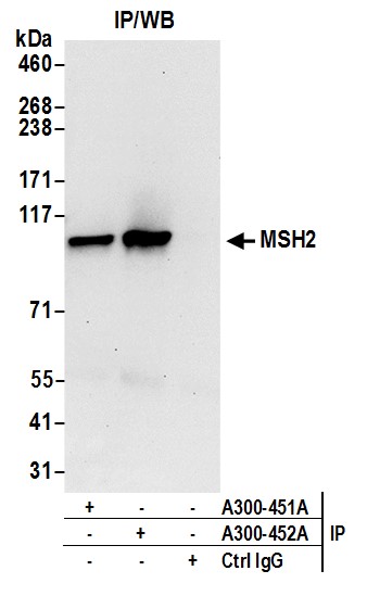
 WB (Western Blot)
(Detection of human and mouse MSH2 by western blot. Samples: Whole cell lysate (50 ug) from HeLa, HEK293T, and mouse NIH 3T3 cells prepared using NETN lysis buffer. Antibody: Affinity purified rabbit anti-MSH2 antibody AAA210958 (lot AAA210958-2) used for WB at 0.1 ug/ml. Detection: Chemiluminescence with an exposure time of 30 seconds.)
WB (Western Blot)
(Detection of human and mouse MSH2 by western blot. Samples: Whole cell lysate (50 ug) from HeLa, HEK293T, and mouse NIH 3T3 cells prepared using NETN lysis buffer. Antibody: Affinity purified rabbit anti-MSH2 antibody AAA210958 (lot AAA210958-2) used for WB at 0.1 ug/ml. Detection: Chemiluminescence with an exposure time of 30 seconds.)
MSH2, Polyclonal Antibody (Cat# AAA210958)
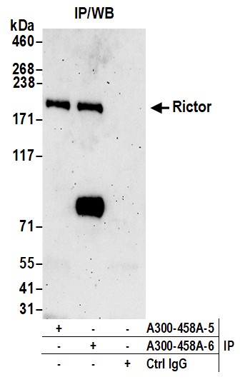
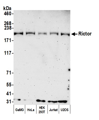 WB (Western Blot)
(Detection of human Rictor by western blot. Samples: Whole cell lysate (50 ug) from GaMG, HeLa, HEK293T, Jurkat, and U2OS cells prepared using NETN lysis buffer. Antibody: Affinity purified rabbit anti-Rictor antibody AAA210961 Lot 6 used for WB at 0.1 ug/ml. Detection: Chemiluminescence with an exposure time of 3 minutes.)
WB (Western Blot)
(Detection of human Rictor by western blot. Samples: Whole cell lysate (50 ug) from GaMG, HeLa, HEK293T, Jurkat, and U2OS cells prepared using NETN lysis buffer. Antibody: Affinity purified rabbit anti-Rictor antibody AAA210961 Lot 6 used for WB at 0.1 ug/ml. Detection: Chemiluminescence with an exposure time of 3 minutes.)
Rictor, Polyclonal Antibody (Cat# AAA210961)
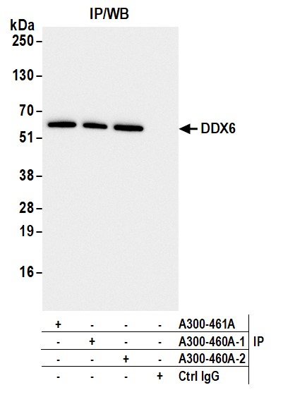
 WB (Western Blot)
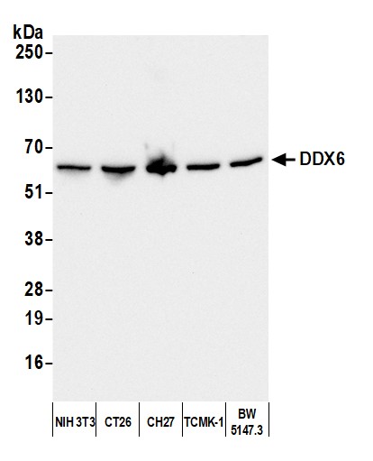
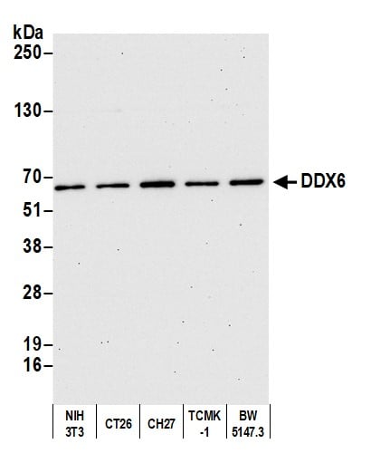
(Detection of mouse DDX6 by western blot. Samples: Whole cell lysate (10 ug) from NIH 3T3, CT26, CH27, TCMK-1, and BW5147.3 cells prepared using NETN lysis buffer. Antibody: Affinity purified rabbit anti-DDX6 antibody (AAA210963 lot 2) used for WB at 0.04 ug/ml. Detection: Chemiluminescence with an exposure time of 10 seconds.)
WB (Western Blot)
(Detection of mouse DDX6 by western blot. Samples: Whole cell lysate (10 ug) from NIH 3T3, CT26, CH27, TCMK-1, and BW5147.3 cells prepared using NETN lysis buffer. Antibody: Affinity purified rabbit anti-DDX6 antibody (AAA210963 lot 2) used for WB at 0.04 ug/ml. Detection: Chemiluminescence with an exposure time of 10 seconds.)
DDX6, Polyclonal Antibody (Cat# AAA210963)
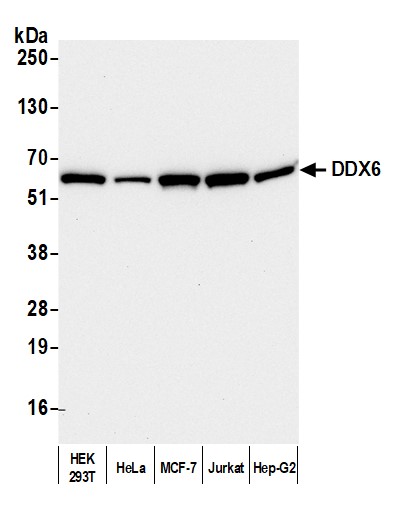
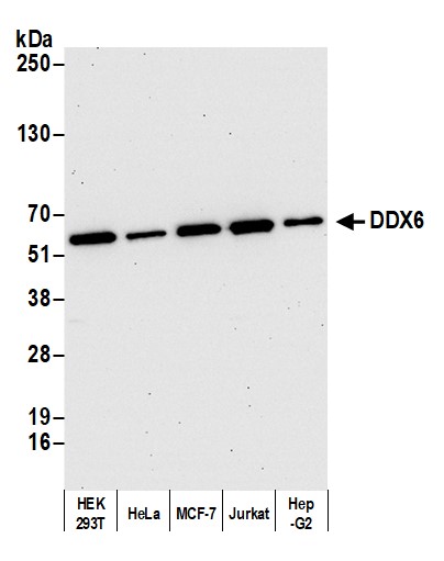 WB (Western Blot)
(Detection of human DDX6 by western blot. Samples: Whole cell lysate (2 ug) from HEK293T, HeLa, MCF-7, Jurkat, and Hep-G2 cells prepared using NETN lysis buffer. Antibody: Affinity purified rabbit anti-DDX6 antibody AAA210964 (lot AAA210964-4) used for WB at 0.04 ug/ml. Detection: Chemiluminescence with an exposure time of 30 seconds.)
WB (Western Blot)
(Detection of human DDX6 by western blot. Samples: Whole cell lysate (2 ug) from HEK293T, HeLa, MCF-7, Jurkat, and Hep-G2 cells prepared using NETN lysis buffer. Antibody: Affinity purified rabbit anti-DDX6 antibody AAA210964 (lot AAA210964-4) used for WB at 0.04 ug/ml. Detection: Chemiluminescence with an exposure time of 30 seconds.)
DDX6, Polyclonal Antibody (Cat# AAA210964)
 WB (Western Blot)
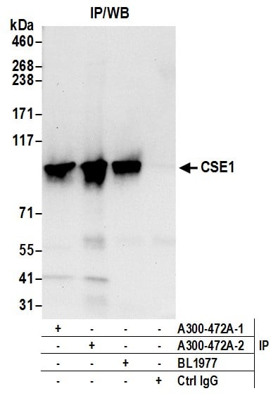
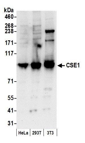
(Detection of human and mouse CSE1 by western blot. Samples: Whole cell lysate (50 ug) from HeLa, HEK293T, and mouse NIH 3T3 cells prepared using NETN lysis buffer. Antibody: Affinity purified rabbit anti-CSE1 antibody AAA210971 (lot AAA210971-2) used for WB at 0.4 ug/ml. Detection: Chemiluminescence with an exposure time of 10 seconds.)
WB (Western Blot)
(Detection of human and mouse CSE1 by western blot. Samples: Whole cell lysate (50 ug) from HeLa, HEK293T, and mouse NIH 3T3 cells prepared using NETN lysis buffer. Antibody: Affinity purified rabbit anti-CSE1 antibody AAA210971 (lot AAA210971-2) used for WB at 0.4 ug/ml. Detection: Chemiluminescence with an exposure time of 10 seconds.)
CSE1, Polyclonal Antibody (Cat# AAA210971)
 WB (Western Blot)
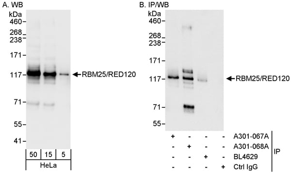
(Detection of human RBM25/RED120 by western blot and immunoprecipitation. Samples: Whole cell lysate (5, 15 and 50 ug for WB; 1 mg for IP, 20% of IP loaded) from HeLa cells. Antibodies: Affinity purified rabbit anti-RBM25/RED120 antibody AAA211303 used for WB at 0.04 ug/ml (A) and 1 ug/ml (B) and used for IP at 3 ug/mg lysate. RBM25/RED120 was also immunoprecipitated by rabbit anti-RBM25/RED120 antibodies and BL4629, which recognize other epitopes. Detection: Chemiluminescence with exposure times of 3 seconds (A and B).)
WB (Western Blot)
(Detection of human RBM25/RED120 by western blot and immunoprecipitation. Samples: Whole cell lysate (5, 15 and 50 ug for WB; 1 mg for IP, 20% of IP loaded) from HeLa cells. Antibodies: Affinity purified rabbit anti-RBM25/RED120 antibody AAA211303 used for WB at 0.04 ug/ml (A) and 1 ug/ml (B) and used for IP at 3 ug/mg lysate. RBM25/RED120 was also immunoprecipitated by rabbit anti-RBM25/RED120 antibodies and BL4629, which recognize other epitopes. Detection: Chemiluminescence with exposure times of 3 seconds (A and B).)
RBM25/RED120, Polyclonal Antibody (Cat# AAA211303)
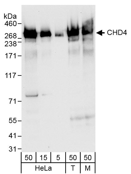 WB (Western Blot)
(Detection of human and mouse CHD4 by western blot. Samples: Whole cell lysate from HeLa (5, 15 and 50 ug, HEK293T (T; 50 ug) and mouse NIH 3T3 (M; 50 ug) cells. Antibodies: Affinity purified rabbit anti-CHD4 antibody AAA211307 (lot AAA211307-1) used at 0.04 ug/ml. Detection: Chemiluminescence with exposure times of 10 seconds.)
WB (Western Blot)
(Detection of human and mouse CHD4 by western blot. Samples: Whole cell lysate from HeLa (5, 15 and 50 ug, HEK293T (T; 50 ug) and mouse NIH 3T3 (M; 50 ug) cells. Antibodies: Affinity purified rabbit anti-CHD4 antibody AAA211307 (lot AAA211307-1) used at 0.04 ug/ml. Detection: Chemiluminescence with exposure times of 10 seconds.)
CHD4, Polyclonal Antibody (Cat# AAA211307)
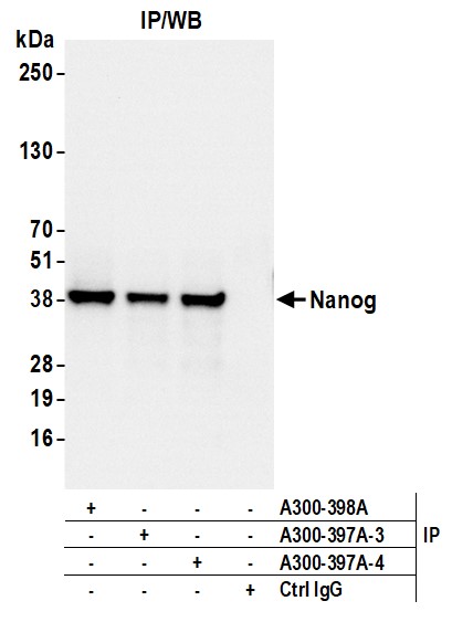
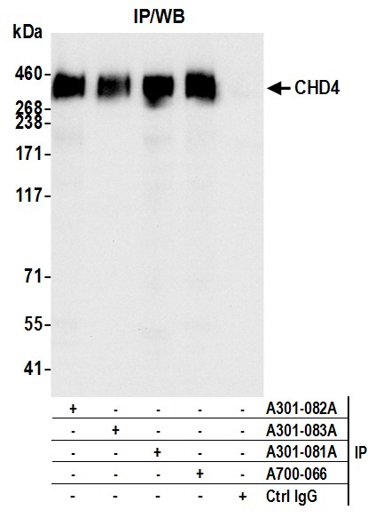
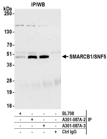
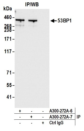
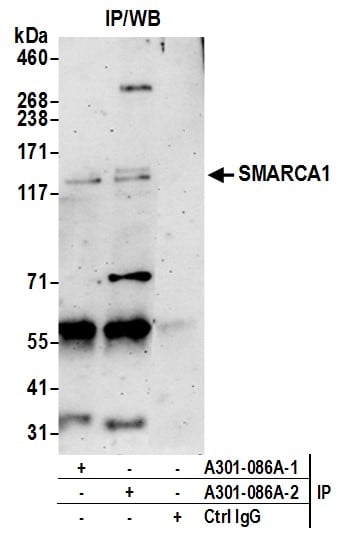 IP (Immunoprecipitation)
(Detection of human SMARCA1 by western blot of immunoprecipitates. Samples: Whole cell lysate (0.5 or 1.0 mg per IP reaction; 20% of IP loaded) from HeLa cells prepared using NETN lysis buffer. Antibodies: Affinity purified rabbit anti-SMARCA1 antibody AAA211310 (lot AAA211310-2) used for IP at 6 ug per reaction. SMARCA1 was also immunoprecipitated by a previous lot of this antibody (lot AAA211310-1). For blotting immunoprecipitated SMARCA1, AAA211310 was used at 1 ug/ml. Detection: Chemiluminescence with an exposure time of 3 minutes.)
IP (Immunoprecipitation)
(Detection of human SMARCA1 by western blot of immunoprecipitates. Samples: Whole cell lysate (0.5 or 1.0 mg per IP reaction; 20% of IP loaded) from HeLa cells prepared using NETN lysis buffer. Antibodies: Affinity purified rabbit anti-SMARCA1 antibody AAA211310 (lot AAA211310-2) used for IP at 6 ug per reaction. SMARCA1 was also immunoprecipitated by a previous lot of this antibody (lot AAA211310-1). For blotting immunoprecipitated SMARCA1, AAA211310 was used at 1 ug/ml. Detection: Chemiluminescence with an exposure time of 3 minutes.)
SMARCA1/SNF2L, Polyclonal Antibody (Cat# AAA211310)
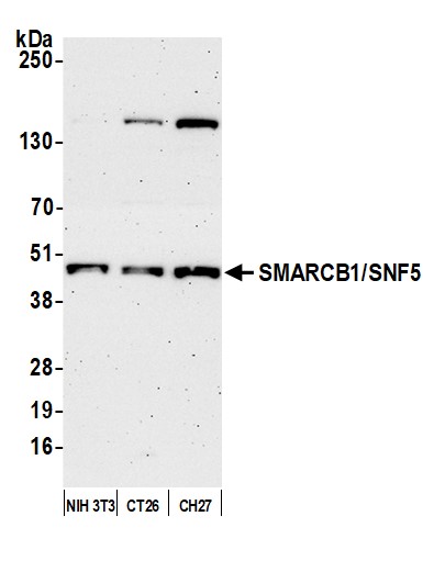
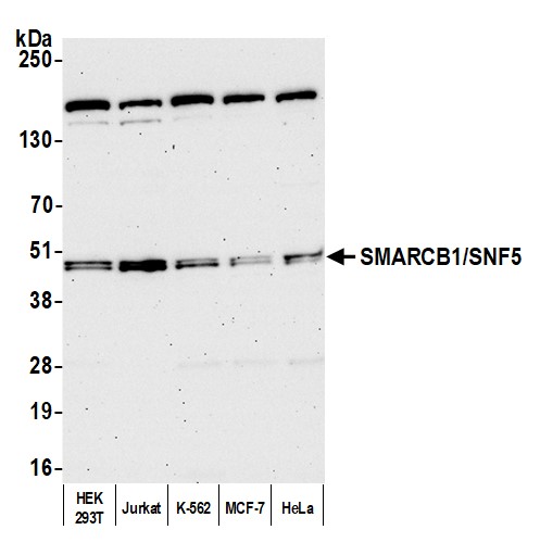 WB (Western Blot)
(Detection of human SMARCB1/SNF5 by western blot. Samples: Whole cell lysate (10 ug) from HEK293T, Jurkat, K-562, MCF-7, and HeLa cells prepared using NETN lysis buffer. Antibody: Affinity purified rabbit anti-SMARCB1/SNF5 antibody (AAA211311 lot 3) used for WB at 0.1 ug/ml. Detection: Chemiluminescence with an exposure time of 75 seconds.)
WB (Western Blot)
(Detection of human SMARCB1/SNF5 by western blot. Samples: Whole cell lysate (10 ug) from HEK293T, Jurkat, K-562, MCF-7, and HeLa cells prepared using NETN lysis buffer. Antibody: Affinity purified rabbit anti-SMARCB1/SNF5 antibody (AAA211311 lot 3) used for WB at 0.1 ug/ml. Detection: Chemiluminescence with an exposure time of 75 seconds.)
SMARCB1/SNF5, Polyclonal Antibody (Cat# AAA211311)
 WB (Western Blot)
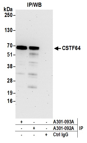
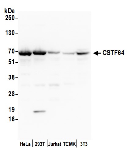
(Detection of human and mouse CSTF64 by western blot. Samples: Whole cell lysate (50 ug) from HeLa, HEK293T, Jurkat, mouse TCMK-1, and mouse NIH 3T3 cells prepared using NETN lysis buffer. Antibody: Affinity purified rabbit anti-CSTF64 antibody AAA211314 (lot AAA211314-2) used for WB at 0.1 ug/ml. Detection: Chemiluminescence with an exposure time of 30 seconds.)
WB (Western Blot)
(Detection of human and mouse CSTF64 by western blot. Samples: Whole cell lysate (50 ug) from HeLa, HEK293T, Jurkat, mouse TCMK-1, and mouse NIH 3T3 cells prepared using NETN lysis buffer. Antibody: Affinity purified rabbit anti-CSTF64 antibody AAA211314 (lot AAA211314-2) used for WB at 0.1 ug/ml. Detection: Chemiluminescence with an exposure time of 30 seconds.)
CSTF64, Polyclonal Antibody (Cat# AAA211314)
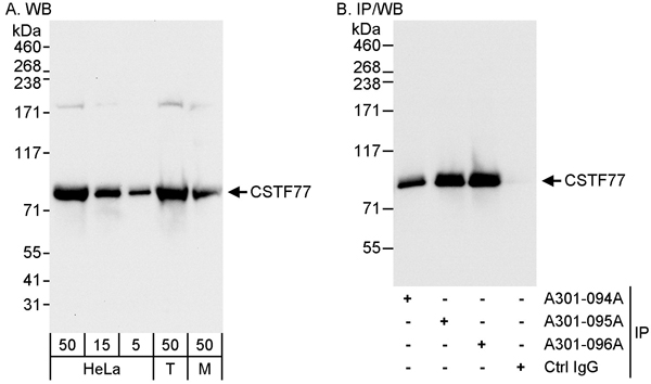 WB (Western Blot)
(Detection of human and mouse CSTF77 by western blot (h&m) and immunoprecipitation (h). Samples: Whole cell lysate from HeLa (5, 15 and 50 ug for WB; 1 mg for IP, 20% of IP loaded), HEK293T (T; 50 ug) and mouse NIH 3T3 (M; 50 ug) cells. Antibodies: Affinity purified rabbit anti-CSTF77 antibody AAA211317 used for WB at 0.04 ug/ml (A) and 1 ug/ml (B) and used for IP at 3 ug/mg lysate. CSTF77 was also immunoprecipitated by rabbit anti-CSTF77 antibodies and which recognize upstream epitopes. Detection: Chemiluminescence with exposure times of 10 seconds (A) and 1 second (B).)
WB (Western Blot)
(Detection of human and mouse CSTF77 by western blot (h&m) and immunoprecipitation (h). Samples: Whole cell lysate from HeLa (5, 15 and 50 ug for WB; 1 mg for IP, 20% of IP loaded), HEK293T (T; 50 ug) and mouse NIH 3T3 (M; 50 ug) cells. Antibodies: Affinity purified rabbit anti-CSTF77 antibody AAA211317 used for WB at 0.04 ug/ml (A) and 1 ug/ml (B) and used for IP at 3 ug/mg lysate. CSTF77 was also immunoprecipitated by rabbit anti-CSTF77 antibodies and which recognize upstream epitopes. Detection: Chemiluminescence with exposure times of 10 seconds (A) and 1 second (B).)
CSTF77, Polyclonal Antibody (Cat# AAA211317)
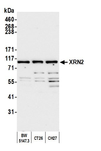
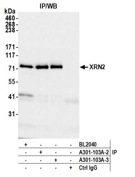
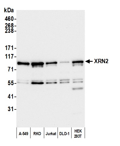 WB (Western Blot)
(Detection of human XRN2 by western blot. Samples: Whole cell lysate (10 ug) from A-549, RKO, Jurkat, DLD-1, and HEK293T cells prepared using NETN lysis buffer. Antibody: Affinity purified rabbit anti-XRN2 antibody (AAA211318 lot 3) used for WB at 0.04 ug/ml. Detection: Chemiluminescence with an exposure time of 30 seconds.)
WB (Western Blot)
(Detection of human XRN2 by western blot. Samples: Whole cell lysate (10 ug) from A-549, RKO, Jurkat, DLD-1, and HEK293T cells prepared using NETN lysis buffer. Antibody: Affinity purified rabbit anti-XRN2 antibody (AAA211318 lot 3) used for WB at 0.04 ug/ml. Detection: Chemiluminescence with an exposure time of 30 seconds.)
XRN2, Polyclonal Antibody (Cat# AAA211318)
 WB (Western Blot)
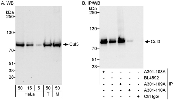
(Detection of human and mouse Cul3 by western blot (h&m) and immunoprecipitation (h). Samples: Whole cell lysate from HeLa (15 and 50 ug for WB; 1 mg for IP, 20% of IP loaded), HEK293T (T; 50 ug) and mouse NIH 3T3 (M; 50 ug) cells. Antibodies: Affinity purified rabbit anti-Cul3 antibody AAA211321 used for WB at 0.04 ug/ml (A) and 1 ug/ml (B) and used for IP at 3 ug/mg lysate. Cul3 was also immunoprecipitated by rabbit anti-Cul3 antibodies A301-108A and BL4592, which recognize upstream epitopes. Detection: Chemiluminescence with exposure times of 30 seconds (A) and 10 seconds (B).)
WB (Western Blot)
(Detection of human and mouse Cul3 by western blot (h&m) and immunoprecipitation (h). Samples: Whole cell lysate from HeLa (15 and 50 ug for WB; 1 mg for IP, 20% of IP loaded), HEK293T (T; 50 ug) and mouse NIH 3T3 (M; 50 ug) cells. Antibodies: Affinity purified rabbit anti-Cul3 antibody AAA211321 used for WB at 0.04 ug/ml (A) and 1 ug/ml (B) and used for IP at 3 ug/mg lysate. Cul3 was also immunoprecipitated by rabbit anti-Cul3 antibodies A301-108A and BL4592, which recognize upstream epitopes. Detection: Chemiluminescence with exposure times of 30 seconds (A) and 10 seconds (B).)
Cul3, Polyclonal Antibody (Cat# AAA211321)
 WB (Western Blot)
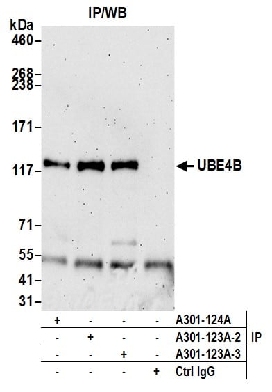
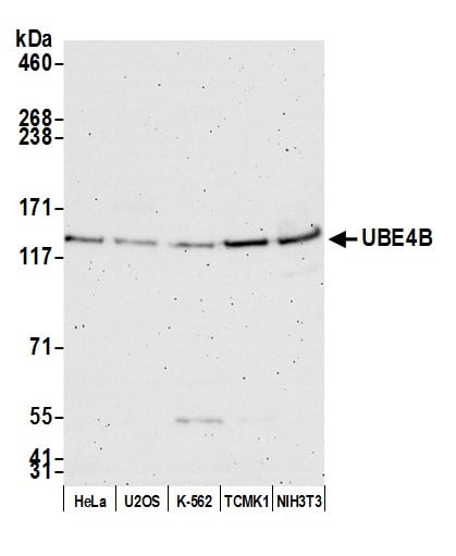
(Detection of human and mouse UBE4B by western blot. Samples: Whole cell lysate (50 ug) from HeLa, U2OS, K-562, TCMK-1, and NIH 3T3 cells prepared using NETN lysis buffer. Antibody: Affinity purified rabbit anti-UBE4B antibody AAA211325 (lot AAA211325-3) used for WB at 0.1 ug/ml. Detection: Chemiluminescence with an exposure time of 3 minutes.)
WB (Western Blot)
(Detection of human and mouse UBE4B by western blot. Samples: Whole cell lysate (50 ug) from HeLa, U2OS, K-562, TCMK-1, and NIH 3T3 cells prepared using NETN lysis buffer. Antibody: Affinity purified rabbit anti-UBE4B antibody AAA211325 (lot AAA211325-3) used for WB at 0.1 ug/ml. Detection: Chemiluminescence with an exposure time of 3 minutes.)
UBE4B, Polyclonal Antibody (Cat# AAA211325)
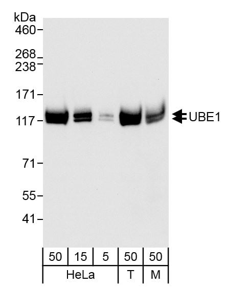 WB (Western Blot)
(Detection of human and mouse UBE1 by western blot. Samples: Whole cell lysate from HeLa (5, 15 and 50 ug), HEK293T (T; 50 ug) and mouse NIH 3T3 (M; 50 ug) cells. Antibodies: Affinity purified rabbit anti-UBE1 antibody AAA211327 (lot AAA211327-1) used for WB at 0.04 ug/ml. Detection: Chemiluminescence with exposure time of 10 seconds.)
WB (Western Blot)
(Detection of human and mouse UBE1 by western blot. Samples: Whole cell lysate from HeLa (5, 15 and 50 ug), HEK293T (T; 50 ug) and mouse NIH 3T3 (M; 50 ug) cells. Antibodies: Affinity purified rabbit anti-UBE1 antibody AAA211327 (lot AAA211327-1) used for WB at 0.04 ug/ml. Detection: Chemiluminescence with exposure time of 10 seconds.)
UBE1, Polyclonal Antibody (Cat# AAA211327)
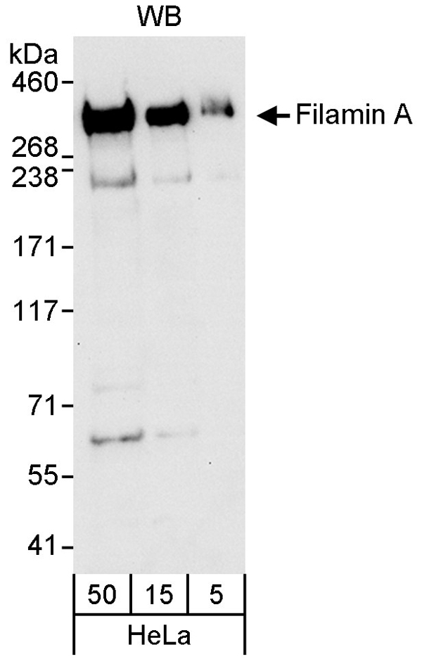 WB (Western Blot)
(Detection of human Filamin A by western blot. Samples: Whole cell lysate (5, 15 and 50 ug for WB) from HeLa cells. Antibody: Affinity purified rabbit anti-Filamin A antibody AAA211328 used for WB at 0.04 ug/ml. Detection: Chemiluminescence with an exposure time of 30 seconds.)
WB (Western Blot)
(Detection of human Filamin A by western blot. Samples: Whole cell lysate (5, 15 and 50 ug for WB) from HeLa cells. Antibody: Affinity purified rabbit anti-Filamin A antibody AAA211328 used for WB at 0.04 ug/ml. Detection: Chemiluminescence with an exposure time of 30 seconds.)
Filamin A, Polyclonal Antibody (Cat# AAA211328)

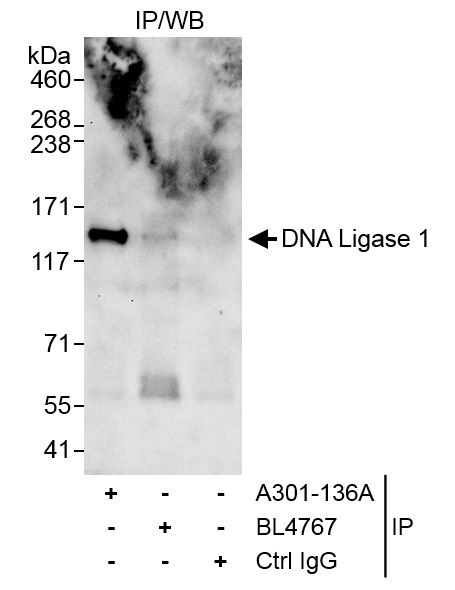 IP (Immunoprecipitation)
(Detection of human DNA Ligase 1 by western blot of immunoprecipitates. Samples: Whole cell lysate (1 mg for IP, 20% of IP loaded) from HeLa cells. Antibodies: Affinity purified rabbit anti-DNA Ligase 1 antibody AAA211331 used for IP at 3 ug/mg lysate. For blotting immunoprecipitated DNA Ligase 1, rabbit anti-DNA Ligase 1 antibody BL4767 was used at 1 ug/ml. Detection: Chemiluminescence with an exposure time of 30 seconds.)
IP (Immunoprecipitation)
(Detection of human DNA Ligase 1 by western blot of immunoprecipitates. Samples: Whole cell lysate (1 mg for IP, 20% of IP loaded) from HeLa cells. Antibodies: Affinity purified rabbit anti-DNA Ligase 1 antibody AAA211331 used for IP at 3 ug/mg lysate. For blotting immunoprecipitated DNA Ligase 1, rabbit anti-DNA Ligase 1 antibody BL4767 was used at 1 ug/ml. Detection: Chemiluminescence with an exposure time of 30 seconds.)
DNA Ligase 1, Polyclonal Antibody (Cat# AAA211331)
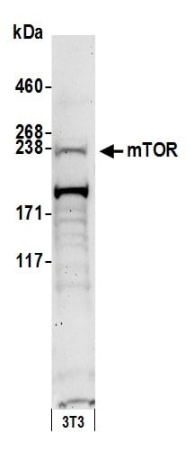
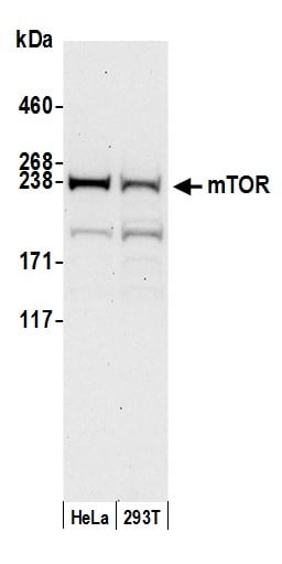 WB (Western Blot)
(Detection of human mTOR by western blot. Samples: Whole cell lysate (50 ug) from HeLa and HEK293T cells prepared using NETN lysis buffer. Antibody: Affinity purified rabbit anti-mTOR antibody AAA211332 (lot AAA211332-2) used for WB at 0.4 ug/ml. Detection: Chemiluminescence with an exposure time of 30 seconds.)
WB (Western Blot)
(Detection of human mTOR by western blot. Samples: Whole cell lysate (50 ug) from HeLa and HEK293T cells prepared using NETN lysis buffer. Antibody: Affinity purified rabbit anti-mTOR antibody AAA211332 (lot AAA211332-2) used for WB at 0.4 ug/ml. Detection: Chemiluminescence with an exposure time of 30 seconds.)
mTOR, Polyclonal Antibody (Cat# AAA211332)
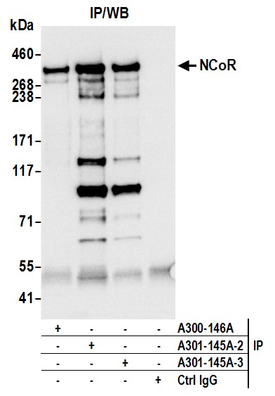
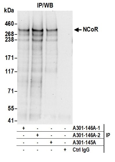
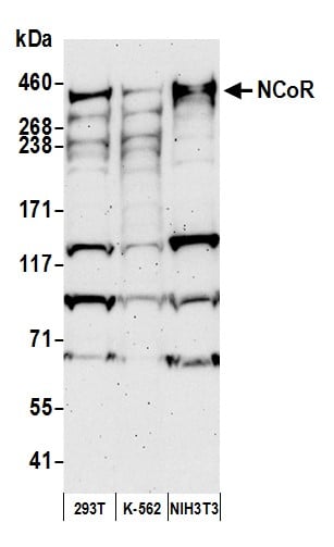 WB (Western Blot)
(Detection of human and mouse NCoR by western blot. Samples:Whole cell lysate (50 ug) from HEK293T, K-562, and NIH 3T3 cells prepared using NETN lysis buffer. Antibody: Affinity purified rabbit anti-NCoR antibody AAA211333 (lot AAA211333-3) used for WB at 0.1 ug/ml. Detection: Chemiluminescence with an exposure time of 3 minutes.)
WB (Western Blot)
(Detection of human and mouse NCoR by western blot. Samples:Whole cell lysate (50 ug) from HEK293T, K-562, and NIH 3T3 cells prepared using NETN lysis buffer. Antibody: Affinity purified rabbit anti-NCoR antibody AAA211333 (lot AAA211333-3) used for WB at 0.1 ug/ml. Detection: Chemiluminescence with an exposure time of 3 minutes.)
NCoR, Polyclonal Antibody (Cat# AAA211333)
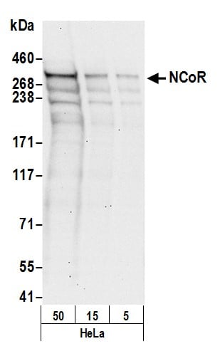 WB (Western Blot)
(Detection of human NCoR by western blot. Samples: Whole cell lysate (5, 15 and 50 ug) from HeLa cells prepared using NETN lysis buffer. Antibody: Affinity purified rabbit anti-NCoR antibody AAA211334 (lot AAA211334-2) used for WB at 0.1 ug/ml. Detection: Chemiluminescence with an exposure time of 30 seconds.)
WB (Western Blot)
(Detection of human NCoR by western blot. Samples: Whole cell lysate (5, 15 and 50 ug) from HeLa cells prepared using NETN lysis buffer. Antibody: Affinity purified rabbit anti-NCoR antibody AAA211334 (lot AAA211334-2) used for WB at 0.1 ug/ml. Detection: Chemiluminescence with an exposure time of 30 seconds.)
NCoR, Polyclonal Antibody (Cat# AAA211334)

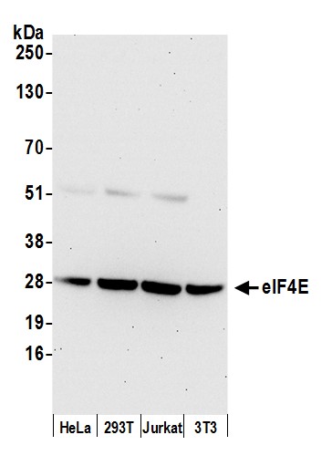 WB (Western Blot)
(Detection of human and mouse eIF4E by western blot. Samples: Whole cell lysate (50 ug) from HeLa, HEK293T, Jurkat, and mouse NIH 3T3 cells prepared using NETN lysis buffer. Antibody: Affinity purified rabbit anti-eIF4E antibody AAA211337 (lot AAA211337-3) used for WB at 0.1 ug/ml. Detection: Chemiluminescence with an exposure time of 30 seconds.)
WB (Western Blot)
(Detection of human and mouse eIF4E by western blot. Samples: Whole cell lysate (50 ug) from HeLa, HEK293T, Jurkat, and mouse NIH 3T3 cells prepared using NETN lysis buffer. Antibody: Affinity purified rabbit anti-eIF4E antibody AAA211337 (lot AAA211337-3) used for WB at 0.1 ug/ml. Detection: Chemiluminescence with an exposure time of 30 seconds.)
eIF4E, Polyclonal Antibody (Cat# AAA211337)
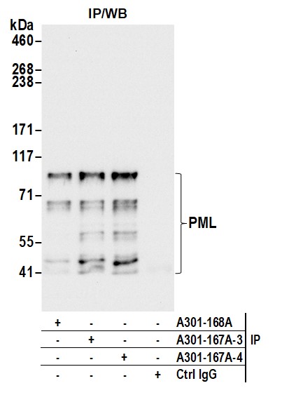
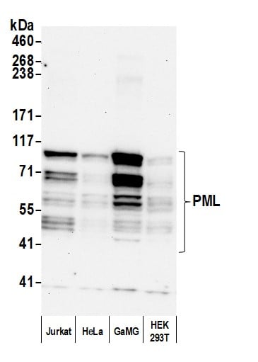 WB (Western Blot)
(Detection of human PML by western blot. Samples: Whole cell lysate (50 ug) from Jurkat, HeLa, GaMG, and HEK293T cells prepared using NETN lysis buffer. Antibody: Affinity purified rabbit anti-PML antibody AAA211341 (AAA211341 lot 4) used for WB at 0.04 ug/ml. Detection: Chemiluminescence with an exposure time of 3 minutes.)
WB (Western Blot)
(Detection of human PML by western blot. Samples: Whole cell lysate (50 ug) from Jurkat, HeLa, GaMG, and HEK293T cells prepared using NETN lysis buffer. Antibody: Affinity purified rabbit anti-PML antibody AAA211341 (AAA211341 lot 4) used for WB at 0.04 ug/ml. Detection: Chemiluminescence with an exposure time of 3 minutes.)
PML, Polyclonal Antibody (Cat# AAA211341)
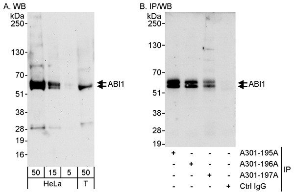 WB (Western Blot)
(Detection of human ABI1 by western blot and immunoprecipitation. Samples: Whole cell lysate from HeLa (5, 15 and 50 ug for WB; 1 mg for IP, 20% of IP loaded) and HEK293T (T; 50 ug) cells. Antibodies: Affinity purified rabbit anti-ABI1 antibody AAA211352 used for WB at 0.04 ug/ml (A) and 1 ug/ml (B) and used for IP at 3 ug/mg lysate. ABI1 was efficiently immunoprecipitated by rabbit anti-ABI1 antibodies and which recognize upstream epitopes. Detection: Chemiluminescence with exposure times of 3 minutes (A) and 10 seconds (B).)
WB (Western Blot)
(Detection of human ABI1 by western blot and immunoprecipitation. Samples: Whole cell lysate from HeLa (5, 15 and 50 ug for WB; 1 mg for IP, 20% of IP loaded) and HEK293T (T; 50 ug) cells. Antibodies: Affinity purified rabbit anti-ABI1 antibody AAA211352 used for WB at 0.04 ug/ml (A) and 1 ug/ml (B) and used for IP at 3 ug/mg lysate. ABI1 was efficiently immunoprecipitated by rabbit anti-ABI1 antibodies and which recognize upstream epitopes. Detection: Chemiluminescence with exposure times of 3 minutes (A) and 10 seconds (B).)
ABI1, Polyclonal Antibody (Cat# AAA211352)
 WB (Western Blot)
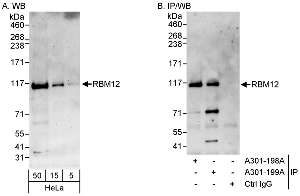
(Detection of human RBM12 by western blot and immunoprecipitation. Samples: Whole cell lysate (5, 15 and 50 ug for WB; 1 mg for IP, 20% of IP loaded) from HeLa cells. Antibodies: Affinity purified rabbit anti-RBM12 antibody AAA211354 used for WB at 0.04 ug/ml (A) and 1 ug/ml (B) and used for IP at 3 ug/mg lysate. RBM12 was also immunoprecipitated by rabbit anti-RBM12 antibody which recognizes an upstream epitope. Detection: Chemiluminescence with exposure times of 3 minutes (A) and 30 seconds (B).)
WB (Western Blot)
(Detection of human RBM12 by western blot and immunoprecipitation. Samples: Whole cell lysate (5, 15 and 50 ug for WB; 1 mg for IP, 20% of IP loaded) from HeLa cells. Antibodies: Affinity purified rabbit anti-RBM12 antibody AAA211354 used for WB at 0.04 ug/ml (A) and 1 ug/ml (B) and used for IP at 3 ug/mg lysate. RBM12 was also immunoprecipitated by rabbit anti-RBM12 antibody which recognizes an upstream epitope. Detection: Chemiluminescence with exposure times of 3 minutes (A) and 30 seconds (B).)
RBM12, Polyclonal Antibody (Cat# AAA211354)
 WB (Western Blot)
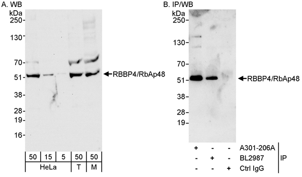
(Detection of human and mouse RBBP4/RbAp48 by western blot (h&m) and immunoprecipitation (h). Samples: Whole cell lysate from HeLa (5, 15, and 50 ug for WB; 1 mg for IP, 20% of IP loaded), HEK293T (T; 50 ug), and mouse NIH 3T3 (M; 50 ug) cells. Antibodies: Affinity purified rabbit anti-RBBP4/RbAp48 antibody AAA211355 used for WB at 0.04 ug/ml (A) and 0.1 ug/ml (B) and used for IP at 3 ug/mg lysate. RBBP4/RbAp48 was also immunoprecipitated by rabbit anti-RBBP4/RbAp48 antibody BL2987, which recognizes a downstream epitope. Detection: Chemiluminescence with exposure times of 30 seconds (A) and 10 seconds (B).)
WB (Western Blot)
(Detection of human and mouse RBBP4/RbAp48 by western blot (h&m) and immunoprecipitation (h). Samples: Whole cell lysate from HeLa (5, 15, and 50 ug for WB; 1 mg for IP, 20% of IP loaded), HEK293T (T; 50 ug), and mouse NIH 3T3 (M; 50 ug) cells. Antibodies: Affinity purified rabbit anti-RBBP4/RbAp48 antibody AAA211355 used for WB at 0.04 ug/ml (A) and 0.1 ug/ml (B) and used for IP at 3 ug/mg lysate. RBBP4/RbAp48 was also immunoprecipitated by rabbit anti-RBBP4/RbAp48 antibody BL2987, which recognizes a downstream epitope. Detection: Chemiluminescence with exposure times of 30 seconds (A) and 10 seconds (B).)
RbBP4, Polyclonal Antibody (Cat# AAA211355)
 WB (Western Blot)
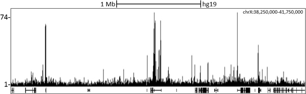
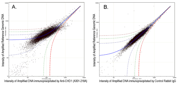
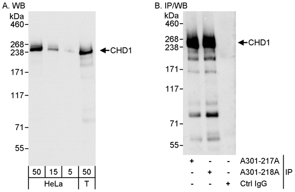
(Detection of human CHD1 by western blot and immunoprecipitation. Samples: Whole cell lysate from HeLa (5, 15 and 50 ug for WB; 1 mg for IP, 20% of IP loaded) and HEK293T (T; 50 ug) cells. Antibodies: Affinity purified rabbit anti-CHD1 antibody AAA211358 used for WB at 0.04 ug/ml (A) and 1 ug/ml (B) and used for IP at 3 ug/mg lysate. CHD1 was also immunoprecipitated by rabbit anti-CHD1 antibody which recognizes an upstream epitope. Detection: Chemiluminescence with exposure times of 3 seconds (A) and 10 seconds (B).)
WB (Western Blot)
(Detection of human CHD1 by western blot and immunoprecipitation. Samples: Whole cell lysate from HeLa (5, 15 and 50 ug for WB; 1 mg for IP, 20% of IP loaded) and HEK293T (T; 50 ug) cells. Antibodies: Affinity purified rabbit anti-CHD1 antibody AAA211358 used for WB at 0.04 ug/ml (A) and 1 ug/ml (B) and used for IP at 3 ug/mg lysate. CHD1 was also immunoprecipitated by rabbit anti-CHD1 antibody which recognizes an upstream epitope. Detection: Chemiluminescence with exposure times of 3 seconds (A) and 10 seconds (B).)
CHD1, Polyclonal Antibody (Cat# AAA211358)
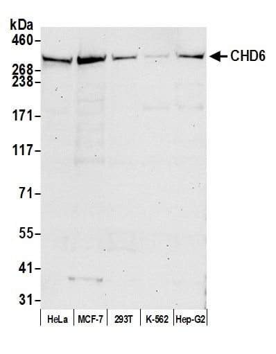 WB (Western Blot)
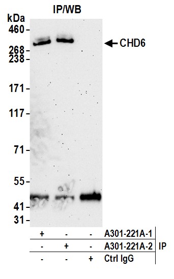
(Detection of human CHD6 by western blot. Samples: Whole cell lysate (50 ug) from HeLa, MCF-7, HEK293T, K-562, and Hep-G2 cells prepared using NETN lysis buffer. Antibody: Affinity purified rabbit anti-CHD6 antibody AAA211361 (lot AAA211361-2) used for WB at 1 ug/ml. Detection: Chemiluminescence with an exposure time of 30 seconds.)
WB (Western Blot)
(Detection of human CHD6 by western blot. Samples: Whole cell lysate (50 ug) from HeLa, MCF-7, HEK293T, K-562, and Hep-G2 cells prepared using NETN lysis buffer. Antibody: Affinity purified rabbit anti-CHD6 antibody AAA211361 (lot AAA211361-2) used for WB at 1 ug/ml. Detection: Chemiluminescence with an exposure time of 30 seconds.)
CHD6, Polyclonal Antibody (Cat# AAA211361)
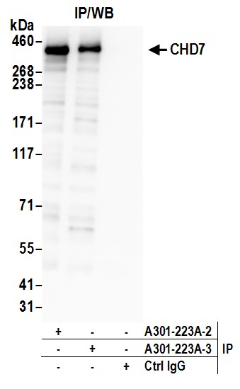
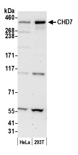 WB (Western Blot)
(Detection of human CHD7 by western blot. Samples: Whole cell lysate (50 ug) from HeLa and HEK293T cells prepared using NETN lysis buffer. Antibody: Affinity purified rabbit anti-CHD7 antibody AAA211362 (lot AAA211362-3) used for WB at 0.1 ug/ml. Detection: Chemiluminescence with an exposure time of 30 seconds.)
WB (Western Blot)
(Detection of human CHD7 by western blot. Samples: Whole cell lysate (50 ug) from HeLa and HEK293T cells prepared using NETN lysis buffer. Antibody: Affinity purified rabbit anti-CHD7 antibody AAA211362 (lot AAA211362-3) used for WB at 0.1 ug/ml. Detection: Chemiluminescence with an exposure time of 30 seconds.)
CHD7, Polyclonal Antibody (Cat# AAA211362)
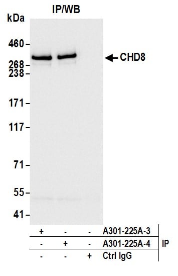
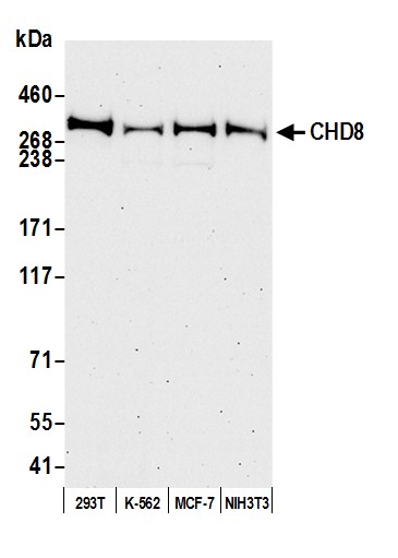 WB (Western Blot)
(Detection of human and mouse CHD8 by western blot. Samples: Whole cell lysate (50 ug) from HEK293T, K-562, MCF-7, and NIH 3T3 cells prepared using NETN lysis buffer. Antibody: Affinity purified Rabbit anti-CHD8 antibody AAA211364 (lot AAA211364-4) used for WB at 0.1 ug/ml. Detection: Chemiluminescence with an exposure time of 75 seconds.)
WB (Western Blot)
(Detection of human and mouse CHD8 by western blot. Samples: Whole cell lysate (50 ug) from HEK293T, K-562, MCF-7, and NIH 3T3 cells prepared using NETN lysis buffer. Antibody: Affinity purified Rabbit anti-CHD8 antibody AAA211364 (lot AAA211364-4) used for WB at 0.1 ug/ml. Detection: Chemiluminescence with an exposure time of 75 seconds.)
CHD8, Polyclonal Antibody (Cat# AAA211364)
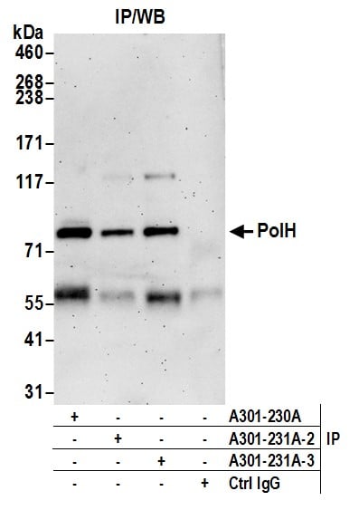
 WB (Western Blot)
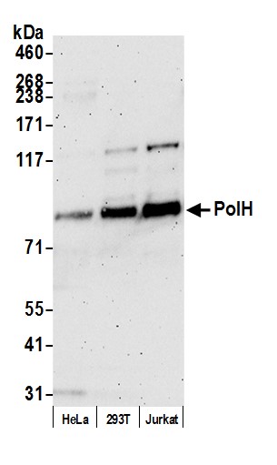
(Detection of human PolH by western blot. Samples: Whole cell lysate (50 ug) from HeLa, HEK293T, and Jurkat cells prepared using NETN lysis buffer. Antibody: Affinity purified Rabbit anti-PolH antibody AAA211367 (lot AAA211367-3) used for WB at 0.1 ug/ml. Detection: Chemiluminescence with an exposure time of 3 minutes.)
WB (Western Blot)
(Detection of human PolH by western blot. Samples: Whole cell lysate (50 ug) from HeLa, HEK293T, and Jurkat cells prepared using NETN lysis buffer. Antibody: Affinity purified Rabbit anti-PolH antibody AAA211367 (lot AAA211367-3) used for WB at 0.1 ug/ml. Detection: Chemiluminescence with an exposure time of 3 minutes.)
PolH, Polyclonal Antibody (Cat# AAA211367)
 WB (Western Blot)
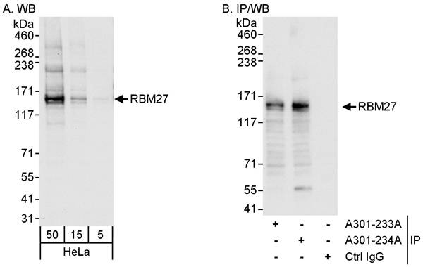
(Detection of human RBM27 by western blot and immunoprecipitation. Samples: Whole cell lysate (5, 15 and 50 ug for WB; 1 mg for IP, 20% of IP loaded) from HeLa cells. Antibodies: Affinity purified rabbit anti-RBM27 antibody AAA211369 used for WB at 0.04 ug/ml (A) and 1 ug/ml (B) and used for IP at 3 ug/mg lysate. RBM27 was also immunoprecipitated by rabbit anti-RBM27 antibody which recognizes an upstream epitope. Detection: Chemiluminescence with exposure times of 10 seconds (A) and 3 seconds (B).)
WB (Western Blot)
(Detection of human RBM27 by western blot and immunoprecipitation. Samples: Whole cell lysate (5, 15 and 50 ug for WB; 1 mg for IP, 20% of IP loaded) from HeLa cells. Antibodies: Affinity purified rabbit anti-RBM27 antibody AAA211369 used for WB at 0.04 ug/ml (A) and 1 ug/ml (B) and used for IP at 3 ug/mg lysate. RBM27 was also immunoprecipitated by rabbit anti-RBM27 antibody which recognizes an upstream epitope. Detection: Chemiluminescence with exposure times of 10 seconds (A) and 3 seconds (B).)
RBM27, Polyclonal Antibody (Cat# AAA211369)
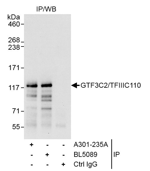 IP (Immunoprecipitation)
(Detection of human GTF3C2/TFIIIC110 by western blot of immunoprecipitates. Samples: Whole cell lysate (1 mg for IP, 20% of IP loaded) from HeLa cells. Antibodies: Affinity purified rabbit anti-GTF3C2/TFIIIC110 antibody AAA211370 used for IP at 3 ug/mg lysate. GTF3C2/TFIIIC110 was also immunoprecipitated by rabbit anti-GTF3C2/TFIIIC110 antibody BL5089, which recognizes a downstream epitope. For blotting immunoprecipitated GTF3C2/TFIIIC110, BL5089 was used at 1 ug/ml. Detection: Chemiluminescence with an exposure time of 30 seconds.)
IP (Immunoprecipitation)
(Detection of human GTF3C2/TFIIIC110 by western blot of immunoprecipitates. Samples: Whole cell lysate (1 mg for IP, 20% of IP loaded) from HeLa cells. Antibodies: Affinity purified rabbit anti-GTF3C2/TFIIIC110 antibody AAA211370 used for IP at 3 ug/mg lysate. GTF3C2/TFIIIC110 was also immunoprecipitated by rabbit anti-GTF3C2/TFIIIC110 antibody BL5089, which recognizes a downstream epitope. For blotting immunoprecipitated GTF3C2/TFIIIC110, BL5089 was used at 1 ug/ml. Detection: Chemiluminescence with an exposure time of 30 seconds.)
GTF3C2/TFIIIC110, Polyclonal Antibody (Cat# AAA211370)
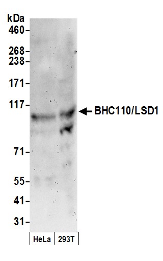 WB (Western Blot)
(Detection of human BHC110/LSD1 by western blot. Samples: Whole cell lysate (50 ug) from HeLa and HEK293T cells prepared using NETN lysis buffer. Antibody: Affinity purified rabbit anti-BHC110/LSD1 antibody AAA210822 (lot AAA210822-1) used for WB at 0.06 ug/ml. Detection: Chemiluminescence with an exposure time of 3 minutes.)
WB (Western Blot)
(Detection of human BHC110/LSD1 by western blot. Samples: Whole cell lysate (50 ug) from HeLa and HEK293T cells prepared using NETN lysis buffer. Antibody: Affinity purified rabbit anti-BHC110/LSD1 antibody AAA210822 (lot AAA210822-1) used for WB at 0.06 ug/ml. Detection: Chemiluminescence with an exposure time of 3 minutes.)
BHC110/LSD1, Polyclonal Antibody (Cat# AAA210822)
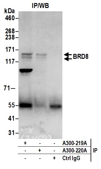
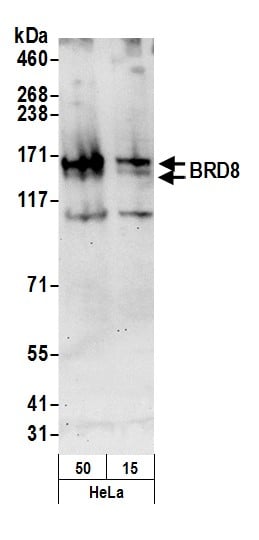 WB (Western Blot)
(Detection of human BRD8 by western blot. Samples: Nuclear extract (50 and 15 ug) from HeLa cells. Antibody: Affinity purified rabbit anti-BRD8 antibody AAA210823 (lot AAA210823-2) used for WB at 0.1 ug/ml. Detection: Chemiluminescence with an exposure time of 30 seconds.)
WB (Western Blot)
(Detection of human BRD8 by western blot. Samples: Nuclear extract (50 and 15 ug) from HeLa cells. Antibody: Affinity purified rabbit anti-BRD8 antibody AAA210823 (lot AAA210823-2) used for WB at 0.1 ug/ml. Detection: Chemiluminescence with an exposure time of 30 seconds.)
BRD8, Polyclonal Antibody (Cat# AAA210823)
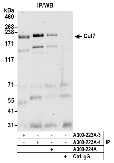
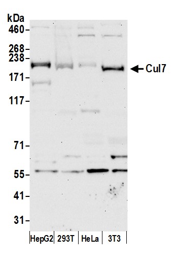 WB (Western Blot)
(Detection of human and mouse Cul7 by western blot. Samples: Whole cell lysate (50 ug) from Hep-G2, HEK293T, HeLa, and mouse NIH 3T3 cells prepared using NETN lysis buffer. Antibody: Affinity purified rabbit anti-Cul7 antibody AAA210826 (lot AAA210826-4) used for WB at 0.4 ug/ml. Detection: Chemiluminescence with an exposure time of 30 seconds.)
WB (Western Blot)
(Detection of human and mouse Cul7 by western blot. Samples: Whole cell lysate (50 ug) from Hep-G2, HEK293T, HeLa, and mouse NIH 3T3 cells prepared using NETN lysis buffer. Antibody: Affinity purified rabbit anti-Cul7 antibody AAA210826 (lot AAA210826-4) used for WB at 0.4 ug/ml. Detection: Chemiluminescence with an exposure time of 30 seconds.)
Cul7, Polyclonal Antibody (Cat# AAA210826)
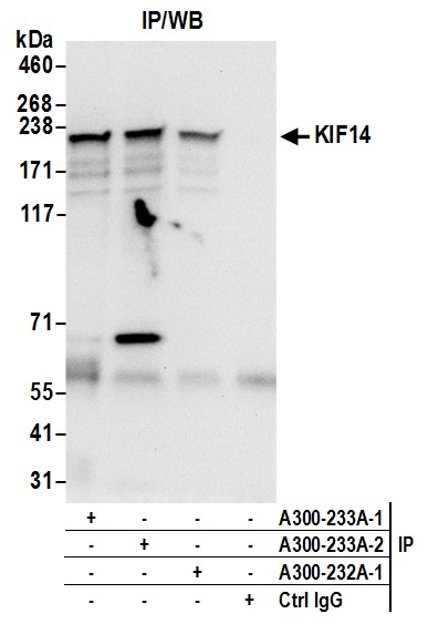
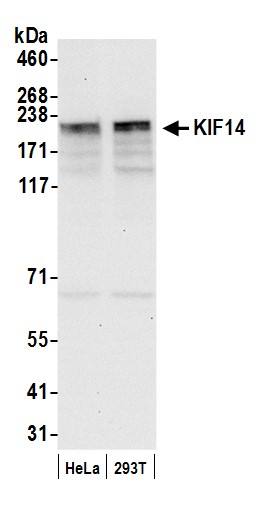 WB (Western Blot)
(Detection of human KIF14 by western blot. Samples: Whole cell lysate (50 ug) from HeLa and HEK293T cells prepared using NETN lysis buffer. Antibody: Affinity purified rabbit anti-KIF14 antibody AAA210829 (lot AAA210829-2) used for WB at 0.1 ug/ml. Detection: Chemiluminescence with an exposure time of 10 seconds.)
WB (Western Blot)
(Detection of human KIF14 by western blot. Samples: Whole cell lysate (50 ug) from HeLa and HEK293T cells prepared using NETN lysis buffer. Antibody: Affinity purified rabbit anti-KIF14 antibody AAA210829 (lot AAA210829-2) used for WB at 0.1 ug/ml. Detection: Chemiluminescence with an exposure time of 10 seconds.)
KIF14, Polyclonal Antibody (Cat# AAA210829)
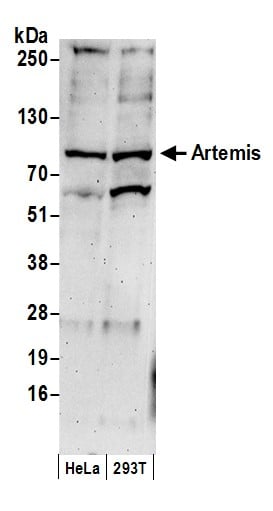 WB (Western Blot)
(Detection of human Artemis by western blot. Samples: Whole cell lysate (50 ug) from HeLa and HEK293T cells prepared using NETN lysis buffer. Antibody: Affinity purified rabbit anti-Artemis antibody AAA210830 (lot AAA210830-1) used for WB at 1 ug/ml. Detection: Chemiluminescence with an exposure time of 3 minutes.)
WB (Western Blot)
(Detection of human Artemis by western blot. Samples: Whole cell lysate (50 ug) from HeLa and HEK293T cells prepared using NETN lysis buffer. Antibody: Affinity purified rabbit anti-Artemis antibody AAA210830 (lot AAA210830-1) used for WB at 1 ug/ml. Detection: Chemiluminescence with an exposure time of 3 minutes.)
Artemis, Polyclonal Antibody (Cat# AAA210830)
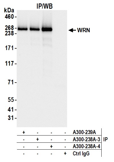
 WB (Western Blot)
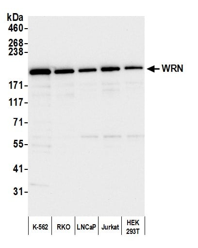
(Detection of human WRN by western blot. Samples: Whole cell lysate (10 ug) from K-562, RKO, LNCaP, Jurkat, and HEK293T cells prepared using NETN lysis buffer. Antibody: Affinity purified rabbit anti-WRN antibody (AAA210834 lot 4) used for WB at 0.1 ug/ml. Detection: Chemiluminescence with an exposure time of 30 seconds.)
WB (Western Blot)
(Detection of human WRN by western blot. Samples: Whole cell lysate (10 ug) from K-562, RKO, LNCaP, Jurkat, and HEK293T cells prepared using NETN lysis buffer. Antibody: Affinity purified rabbit anti-WRN antibody (AAA210834 lot 4) used for WB at 0.1 ug/ml. Detection: Chemiluminescence with an exposure time of 30 seconds.)
WRN, Polyclonal Antibody (Cat# AAA210834)
 WB (Western Blot)
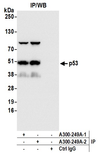
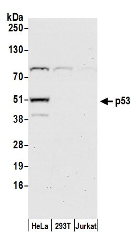
(Detection of human p53 by western blot. Samples: Whole cell lysate (50 ug) from HeLa, HEK293T, and Jurkat cells prepared using NETN lysis buffer. Antibody: Affinity purified rabbit anti-p53 antibody AAA210841 (lot AAA210841-2) used for WB at 0.1 ug/ml. Detection: Chemiluminescence with an exposure time of 30 seconds.)
WB (Western Blot)
(Detection of human p53 by western blot. Samples: Whole cell lysate (50 ug) from HeLa, HEK293T, and Jurkat cells prepared using NETN lysis buffer. Antibody: Affinity purified rabbit anti-p53 antibody AAA210841 (lot AAA210841-2) used for WB at 0.1 ug/ml. Detection: Chemiluminescence with an exposure time of 30 seconds.)
p53, Polyclonal Antibody (Cat# AAA210841)
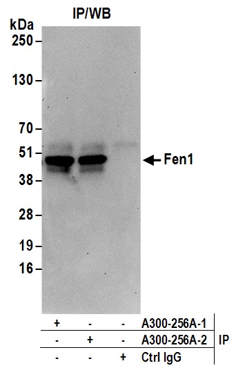
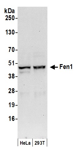 WB (Western Blot)
(Detection of human Fen1 by western blot. Samples: Whole cell lysate (50 ug) from HeLa and HEK293T cells prepared using NETN lysis buffer. Antibodies: Affinity purified rabbit anti-Fen1 antibody AAA210846 (lot AAA210846-2) used for WB at 0.1 ug/ml. Detection: Chemiluminescence with an exposure time of 30 seconds.)
WB (Western Blot)
(Detection of human Fen1 by western blot. Samples: Whole cell lysate (50 ug) from HeLa and HEK293T cells prepared using NETN lysis buffer. Antibodies: Affinity purified rabbit anti-Fen1 antibody AAA210846 (lot AAA210846-2) used for WB at 0.1 ug/ml. Detection: Chemiluminescence with an exposure time of 30 seconds.)
Fen1, Polyclonal Antibody (Cat# AAA210846)
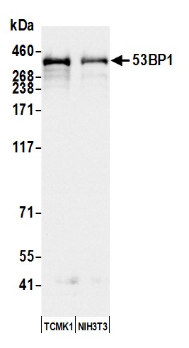
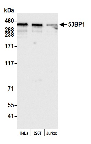 WB (Western Blot)
(Detection of human 53BP1 by western blot. Samples: Whole cell lysate (15 ug) from HeLa, HEK293T, and Jurkat cells prepared using NETN lysis buffer. Antibody: Affinity purified rabbit anti-53BP1 antibody AAA210853 (lot AAA210853-7) used for WB at 0.04 ug/ml. Detection: Chemiluminescence with an exposure time of 3 seconds.)
WB (Western Blot)
(Detection of human 53BP1 by western blot. Samples: Whole cell lysate (15 ug) from HeLa, HEK293T, and Jurkat cells prepared using NETN lysis buffer. Antibody: Affinity purified rabbit anti-53BP1 antibody AAA210853 (lot AAA210853-7) used for WB at 0.04 ug/ml. Detection: Chemiluminescence with an exposure time of 3 seconds.)
53BP1, Polyclonal Antibody (Cat# AAA210853)
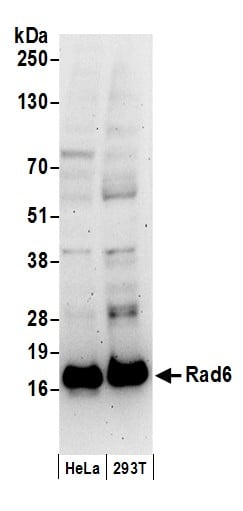 WB (Western Blot)
(Detection of human Rad6 by western blot. Samples: Whole cell lysate (50 ug) from HeLa and HEK293T cells prepared using NETN lysis buffer. Antibody: Affinity purified rabbit anti-Rad6 antibody AAA210860 (lot AAA210860-1) used for WB at 0.4 ug/ml. Detection: Chemiluminescence with an exposure time of 3 minutes.)
WB (Western Blot)
(Detection of human Rad6 by western blot. Samples: Whole cell lysate (50 ug) from HeLa and HEK293T cells prepared using NETN lysis buffer. Antibody: Affinity purified rabbit anti-Rad6 antibody AAA210860 (lot AAA210860-1) used for WB at 0.4 ug/ml. Detection: Chemiluminescence with an exposure time of 3 minutes.)
Rad6, Polyclonal Antibody (Cat# AAA210860)
What are Polyclonal Antibodies?
Polyclonal antibodies are antibodies that come from multiple B cell clones of a host animal. The typical hosts used for the majority of polyclonal antibody production are rabbits, goats, sheep, and donkeys. These polyclonal antibodies, once having identified their target, will bind to different epitopes located at different regions or sequences on the same protein/antigen. As a result, they are ideal at locating and binding to the target, even if the target is in very low concentrations (due to many different antibodies being able to bind to the same target molecule, which allows for significant amplification of a downstream signal).
Polyclonal antibodies are typically produced by injecting an antigen into a host animal, which causes the animal’s immune system to attack the foreign antigen by mass generating antibodies against it. After a period of time, serum is collected from the animal and purified using physicochemical fractionation, class-specific affinity purification, and/or antigen-affinity purification.
Key Uses of Polyclonal Antibodies
- Western Blotting: This method is used to find specific proteins in biological samples after separating them by size.
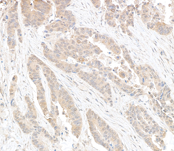
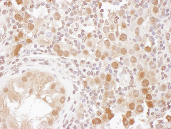
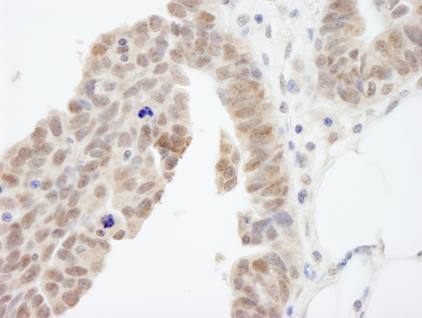
- Immunohistochemistry: IHC helps visualize the location of proteins in tissue sections using various staining techniques.
- ELISA: (Enzyme-Linked Immunosorbent Assay) is typically used to identify specific protein quantities in a sample. ELISAs can be either “Quantitative” or “Qualitative”.
- Flow Cytometry: technique that identifies and measures the specific protein on the surface or inside the cells in a fluid suspension.
- Immunoprecipitation: IP isolates and studies a specific protein from a complex mixture using antibodies.
Why Buy Polyclonal Antibodies from AAA Biotech?
1. Ideal for Various Applications
Our antibodies are generally going to be validated for use in multiple types of assays, including ELISA, Western Blotting, Immunohistochemistry, Immunoprecipitation, amongst others. They are ideal for a wide range of research applications.
2. Rigorous Quality Control
All of the antibodies in our catalog undergo strict quality testing to ensure specificity, sensitivity, and consistent performance. We are confident in the ability of our antibodies to provide you with accurate results.
3. Wide Assortment of Antibodies
Antibodies in are catalog can be found for both common and exotic species, and these antibodies are also available in both conjugated and recombinant forms to suit many diverse experimental needs.
4. Highly Purified
Our antibodies are available in purified forms with over 85% purity, as confirmed by SDS-PAGE. They are also available with tags such as His, Flag, GST, or MBP. We cater to customers worldwide.
FAQ
1. How are polyclonal antibodies produced?
Traditionally, polyclonal antibodies are produced by injecting an antigen into a host animal (such as a rabbit or goat), which then triggers an immune response from the host animal. The animal’s B cells produce antibodies that will recognize different parts of the injected antigen. These antibodies are then collected from the animal’s blood and purified for use.
2. How do polyclonal antibodies differ from monoclonal antibodies?
Polyclonal antibodies are a mix of antibodies that bind to different locations (epitopes) of the same antigen, while monoclonal antibodies are identical and bind to just one specific epitope. This makes polyclonal antibodies more versatile and better at detecting proteins that may be present in low quantities or in altered/modified forms.
3. How should I store polyclonal antibodies?
Polyclonal antibodies should be stored at 4°C for short-term use (up to a few weeks) and at -20°C or -80°C for long-term storage. Avoid repeated freeze-thaw cycles by dividing them into small aliquots. Always check the datasheet for specific storage instructions.