Filters
▼Clonality
▼Type
▼Reactivity
▼Gene Name
▼Isotype
▼Host
▼Application
▼Clone
▼Polyclonal Antibodies
At AAA Biotech also known as AAA Bio or AAABio, we provide a broad range of purified polyclonal antibodies (pAbs) that are able to all be browsed online through our website. Due to their high specificity and strong binding affinity, these antibodies are ideal for wide swathes of research and experimental applications.
Our polyclonal antibodies can easily support your work, whether you use them for Western Blotting, Immunocytochemistry (with or without Immunofluorescence used in conjunction), Immunohistochemistry, Immunoprecipitation, and ELISA tests. We highly encourage you to browse our range of pAbs and choose the one that best suits your experimental model.
Viewing 5250-5300 of 96812 product results
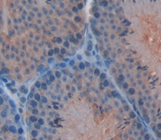
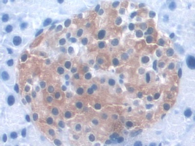
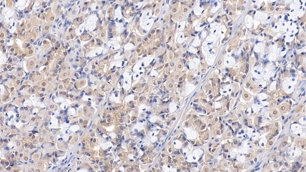
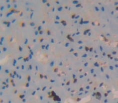
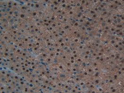
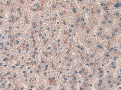
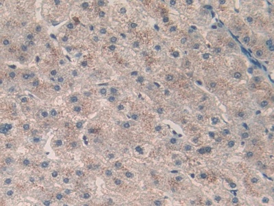
 IHC (Immunohistochemistry)
(DAB staining on IHC-P; Samples: Human Liver Tissue.)
IHC (Immunohistochemistry)
(DAB staining on IHC-P; Samples: Human Liver Tissue.)
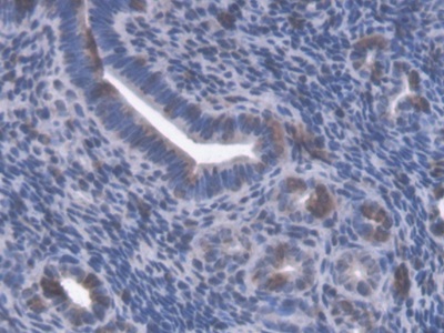
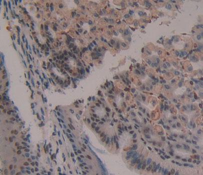
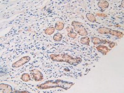
Retinoic Acid Receptor Alpha, Polyclonal Antibody (Cat# AAA145165)
 IHC (Immunohiostchemistry)
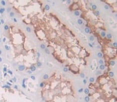
(DAB staining on fromalin fixed paraffin- embedded Kidney tissue))
IHC (Immunohiostchemistry)
(DAB staining on fromalin fixed paraffin- embedded Kidney tissue))
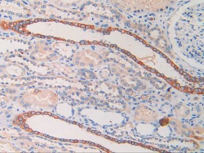
Glycophorin E, Polyclonal Antibody (Cat# AAA145180)
 IHC (Immunohiostchemistry)
(DAB staining on fromalin fixed paraffin- embedded Kidney tissue))
IHC (Immunohiostchemistry)
(DAB staining on fromalin fixed paraffin- embedded Kidney tissue))
Snurportin 1, Polyclonal Antibody (Cat# AAA145206)

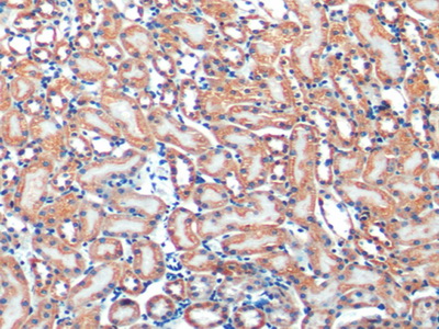
 IHC (Immunohistochemisry)
(DAB staining on IHC-P; Samples: Mouse Kidney Tissue))
IHC (Immunohistochemisry)
(DAB staining on IHC-P; Samples: Mouse Kidney Tissue))
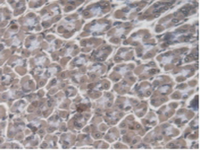
Proline Dehydrogenase, Polyclonal Antibody (Cat# AAA145229)
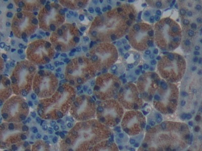
 IHC (Immunohiostchemistry)
(DAB staining on IHC-P; Samples: Mouse Pancreas Tissue))
IHC (Immunohiostchemistry)
(DAB staining on IHC-P; Samples: Mouse Pancreas Tissue))
Kidney And Brain Protein, Polyclonal Antibody (Cat# AAA145232)
 IHC (Immunohistochemisry)
(DAB staining on fromalin fixed paraffin- embedded Kidney tissue))
IHC (Immunohistochemisry)
(DAB staining on fromalin fixed paraffin- embedded Kidney tissue))
Protocadherin Beta 15, Polyclonal Antibody (Cat# AAA144745)
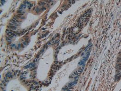
 IHC (Immunohistochemistry)
(DAB staining on IHC-P; Samples: Human Breast cancer Tissue))
IHC (Immunohistochemistry)
(DAB staining on IHC-P; Samples: Human Breast cancer Tissue))
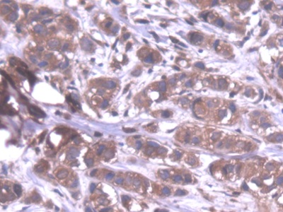
Protein Phosphatase 1, Polyclonal Antibody (Cat# AAA144746)
 IHC (Immunohistochemisry)
(DAB staining on IHC-P; Samples: Human Breast cancer Tissue))
IHC (Immunohistochemisry)
(DAB staining on IHC-P; Samples: Human Breast cancer Tissue))
Leucine Zipper, Down Regulated In Cancer 1, Polyclonal Antibody (Cat# AAA144748)
 IHC (Immunohistochemisry)
(DAB staining on fromalin fixed paraffin- embedded Kidney tissue))
IHC (Immunohistochemisry)
(DAB staining on fromalin fixed paraffin- embedded Kidney tissue))
Signal Regulatory Protein Alpha, Polyclonal Antibody (Cat# AAA144750)

 IHC (Immunohiostchemistry)
(DABstainingonIHC-P.Samples:MouseTissue))
IHC (Immunohiostchemistry)
(DABstainingonIHC-P.Samples:MouseTissue))
Calcium Channel, Voltage Dependent, T-Type, Alpha 1H Subunit, Polyclonal Antibody (Cat# AAA144770)
 IHC (Immunohiostchemistry)
(DAB staining on fromalin fixed paraffin- embedded kidney tissue))
IHC (Immunohiostchemistry)
(DAB staining on fromalin fixed paraffin- embedded kidney tissue))
Alpha-1,4-Galactosyltransferase, Polyclonal Antibody (Cat# AAA144795)
 IHC (Immunohiostchemistry)
(DAB staining on fromalin fixed paraffin- embedded Kidney tissue))
IHC (Immunohiostchemistry)
(DAB staining on fromalin fixed paraffin- embedded Kidney tissue))
Leupaxin, Polyclonal Antibody (Cat# AAA144800)
 IHC (Immunohiostchemistry)
(DAB staining on fromalin fixed paraffin- embedded kidney tissue))
IHC (Immunohiostchemistry)
(DAB staining on fromalin fixed paraffin- embedded kidney tissue))
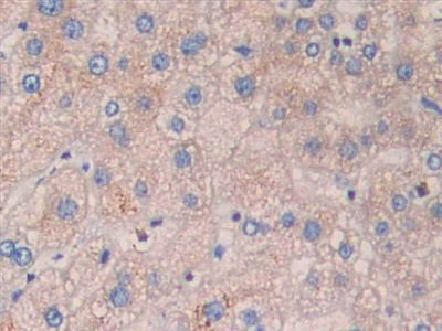
Wilms Tumor 1 Associated Protein, Polyclonal Antibody (Cat# AAA144820)
 IHC (Immunohistochemistry)
(DAB staining on IHC-P; Samples: Rat Liver Tissue))
IHC (Immunohistochemistry)
(DAB staining on IHC-P; Samples: Rat Liver Tissue))
Alanine Glyoxylate Aminotransferase, Polyclonal Antibody (Cat# AAA144822)
 IHC (Immunohistochemistry)
(DAB staining on IHC-P; Samples: Mouse Kidney Tissue)
IHC (Immunohistochemistry)
(DAB staining on IHC-P; Samples: Mouse Kidney Tissue)
Guanylate Binding Protein 4, Polyclonal Antibody (Cat# AAA144834)
 IHC (Immunohistochemisry)
(DAB staining on IHC-P; Samples: Rat Stomach Tissue))
IHC (Immunohistochemisry)
(DAB staining on IHC-P; Samples: Rat Stomach Tissue))
Receptor Interacting Serine Threonine Kinase 1, Polyclonal Antibody (Cat# AAA144852)

 IHC (Immunohistochemistry)
(DAB staining on IHC-P; Samples: Mouse Testis Tissue))
IHC (Immunohistochemistry)
(DAB staining on IHC-P; Samples: Mouse Testis Tissue))
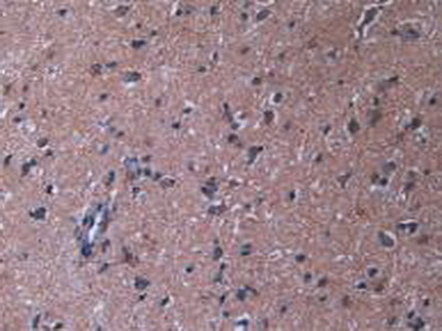
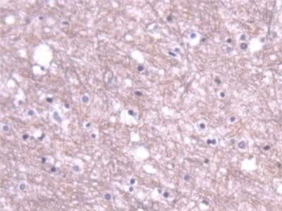
Sperm Protein 17, Polyclonal Antibody (Cat# AAA144865)
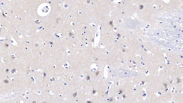
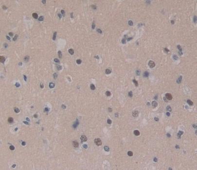 IHC (Immunohistochemisry)
(DAB staining on fromalin fixed paraffin- embedded brain tissue))
IHC (Immunohistochemisry)
(DAB staining on fromalin fixed paraffin- embedded brain tissue))
CCAAT/Enhancer Binding Protein Delta, Polyclonal Antibody (Cat# AAA144867)
 IHC (Immunohiostchemistry)
(DAB staining on fromalin fixed paraffin- embedded Kidney tissue))
IHC (Immunohiostchemistry)
(DAB staining on fromalin fixed paraffin- embedded Kidney tissue))
YY1 Associated Factor 2, Polyclonal Antibody (Cat# AAA144889)
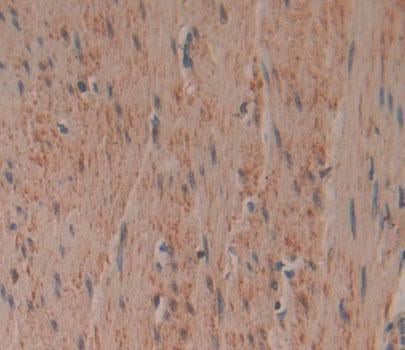
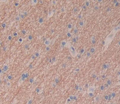 IHC (Immunohiostchemistry)
(DAB staining on fromalin fixed paraffin- embedded brain tissue))
IHC (Immunohiostchemistry)
(DAB staining on fromalin fixed paraffin- embedded brain tissue))
Tectorin Beta, Polyclonal Antibody (Cat# AAA144894)
 IHC (Immunohistochemisry)
(DAB staining on fromalin fixed paraffin- embedded Kidney tissue))
IHC (Immunohistochemisry)
(DAB staining on fromalin fixed paraffin- embedded Kidney tissue))
Schlafen Family Member 5, Polyclonal Antibody (Cat# AAA144914)
 IHC (Immunohistochemisry)
(DAB staining on fromalin fixed paraffin- embedded kidney tissue))
IHC (Immunohistochemisry)
(DAB staining on fromalin fixed paraffin- embedded kidney tissue))
Cholinergic Receptor, Polyclonal Antibody (Cat# AAA144926)
 IHC (Immunohiostchemistry)
(DAB staining on fromalin fixed paraffin- embedded Kidney tissue))
IHC (Immunohiostchemistry)
(DAB staining on fromalin fixed paraffin- embedded Kidney tissue))
Heart And Neural Crest Derivatives Expressed Protein 1, Polyclonal Antibody (Cat# AAA144933)
 IHC (Immunohiostchemistry)
(DAB staining on fromalin fixed paraffin- embedded Kidney tissue))
IHC (Immunohiostchemistry)
(DAB staining on fromalin fixed paraffin- embedded Kidney tissue))
Geminin, Polyclonal Antibody (Cat# AAA144938)
 IHC (Immunohistochemisry)
(DAB staining on fromalin fixed paraffin- embedded Kidney tissue))
IHC (Immunohistochemisry)
(DAB staining on fromalin fixed paraffin- embedded Kidney tissue))
Delta-Sleep Inducing Peptide, Polyclonal Antibody (Cat# AAA144943)

 IHC (Immunohiostchemistry)
(DAB staining on fromalin fixed paraffin- embedded kidney tissue))
IHC (Immunohiostchemistry)
(DAB staining on fromalin fixed paraffin- embedded kidney tissue))
Actinin Alpha 1, Polyclonal Antibody (Cat# AAA144980)
 IHC (Immunohiostchemistry)
(DABstainingonIHC-P.Samples:HumanTissue))
IHC (Immunohiostchemistry)
(DABstainingonIHC-P.Samples:HumanTissue))
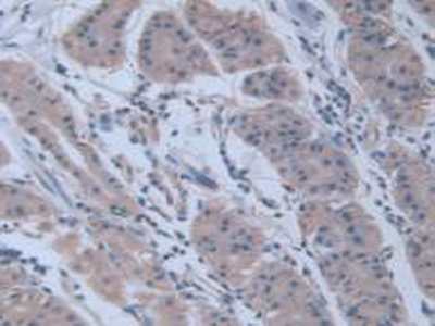
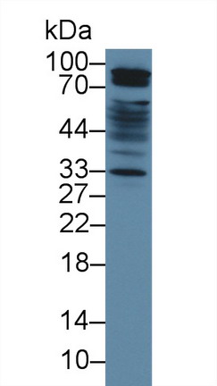
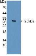
Retinoic Acid Receptor Responder 1, Polyclonal Antibody (Cat# AAA145069)
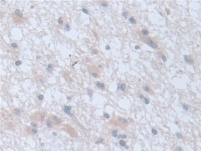
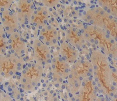
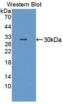
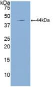
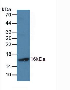
Hydroxyproline, Polyclonal Antibody (Cat# AAA145119)
 IHC (Immunohistochemistry)
(DAB staining on IHC-P; Samples: Human Kidney Tissue)
IHC (Immunohistochemistry)
(DAB staining on IHC-P; Samples: Human Kidney Tissue)
V-Erb A Erythroblastic Leukemia Viral Oncogene Homolog 4, Polyclonal Antibody (Cat# AAA144995)
 IHC (Immunohiostchemistry)
(DABstainingonIHC-P.Samples:RatTissue))
IHC (Immunohiostchemistry)
(DABstainingonIHC-P.Samples:RatTissue))
ATPase, Na+/K+ Transporting Beta 4 Polypeptide, Polyclonal Antibody (Cat# AAA145026)
 IHC (Immunohiostchemistry)
(DABstainingonIHC-P.Samples:RatTissue))
IHC (Immunohiostchemistry)
(DABstainingonIHC-P.Samples:RatTissue))
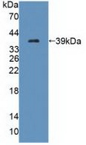
Lactase, Polyclonal Antibody (Cat# AAA145039)
 IHC (Immunohistochemisry)
(DAB staining on fromalin fixed paraffin- embedded Kidney tissue))
IHC (Immunohistochemisry)
(DAB staining on fromalin fixed paraffin- embedded Kidney tissue))
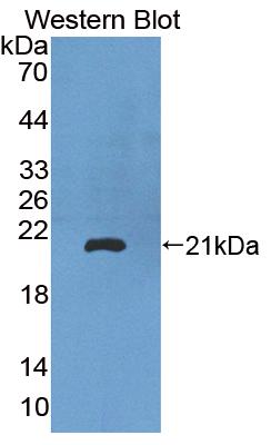
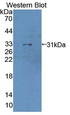
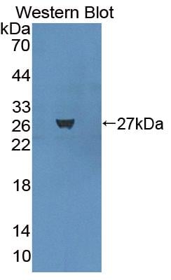
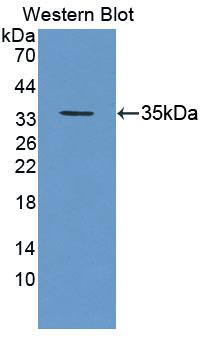
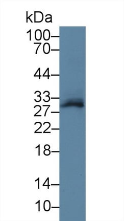
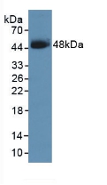
Bcl6 Corepressor, Polyclonal Antibody (Cat# AAA144707)
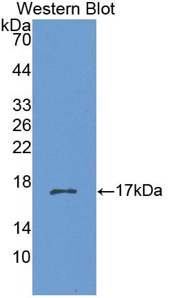
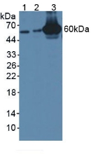
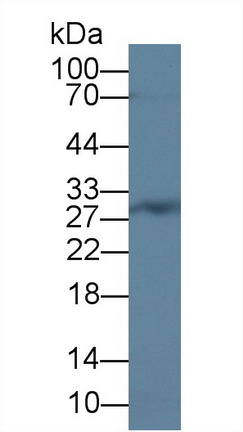
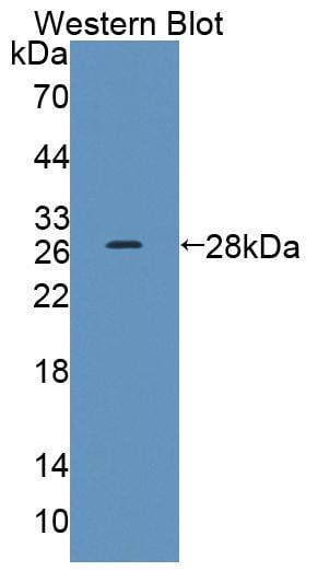
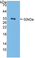
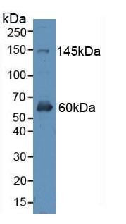
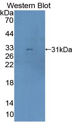
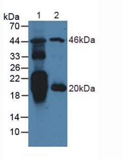
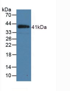
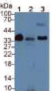
 WB (Western Blot)
(Western Blot;Sample: Lane 1: Porcine Cerebrum lysate;Lane 2: Mouse Heart lysatePrimary Ab: 2ug/ml Rabbit Anti-Human CHRNb3 AntibodySecond Ab: 0.2ug/mL HRP-Linked Caprine Anti-Rabbit IgG Polyclonal Antibody)
WB (Western Blot)
(Western Blot;Sample: Lane 1: Porcine Cerebrum lysate;Lane 2: Mouse Heart lysatePrimary Ab: 2ug/ml Rabbit Anti-Human CHRNb3 AntibodySecond Ab: 0.2ug/mL HRP-Linked Caprine Anti-Rabbit IgG Polyclonal Antibody)
Cholinergic Receptor, Polyclonal Antibody (Cat# AAA144711)
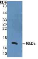
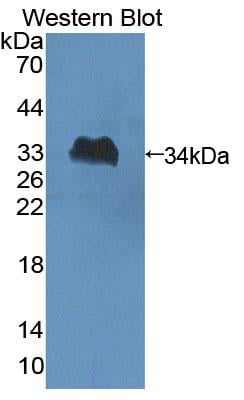
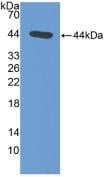
 WB (Western Blot)
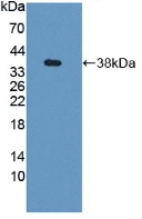
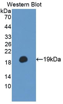
(Western Blot A: Sample: RecombinantNPPA,Mouse.)
WB (Western Blot)
(Western Blot A: Sample: RecombinantNPPA,Mouse.)
Natriuretic Peptide Precursor A (NPPA), Polyclonal Antibody (Cat# AAA147846)
 IHC (Immunohistochemistry)
(DAB staining on IHC-P; Samples: Rat Stomach Tissue.)
IHC (Immunohistochemistry)
(DAB staining on IHC-P; Samples: Rat Stomach Tissue.)
N-Terminal Pro-Atrial Natriuretic Peptide (NT-ProANP), Polyclonal Antibody (Cat# AAA147849)
 IHC (Immunohistochemistry)
(DAB staining on IHC-P; Samples: Mouse Lung Tissue.)
IHC (Immunohistochemistry)
(DAB staining on IHC-P; Samples: Mouse Lung Tissue.)
Natriuretic Peptide Precursor B (NPPB), Polyclonal Antibody (Cat# AAA147850)
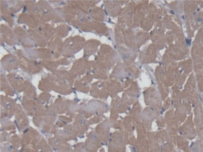
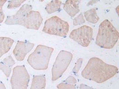
 IHC (Immunohiostchemistry)
(DAB staining on IHC-P; Samples: Porcine Cardiac Muscle Tissue))
IHC (Immunohiostchemistry)
(DAB staining on IHC-P; Samples: Porcine Cardiac Muscle Tissue))
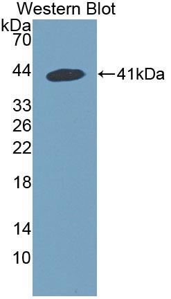
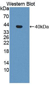
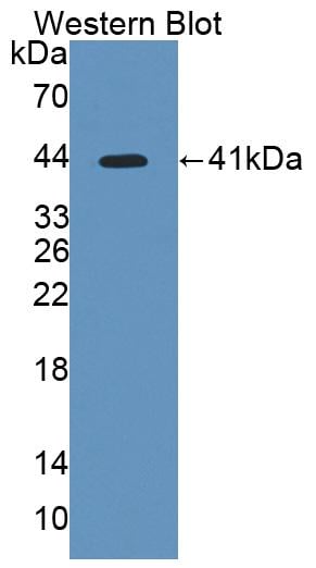
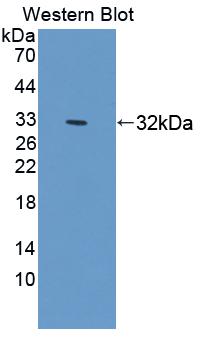
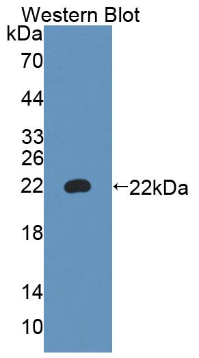
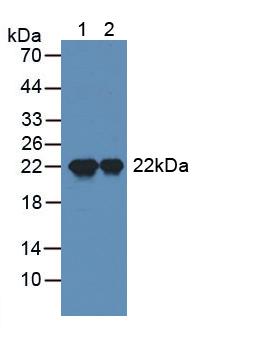
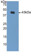
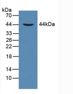
Natriuretic Peptide Precursor B (NPPB), Polyclonal Antibody (Cat# AAA147851)
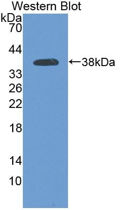
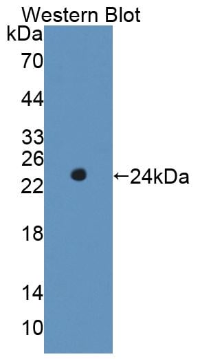
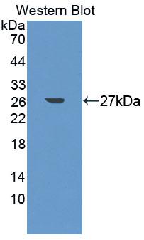
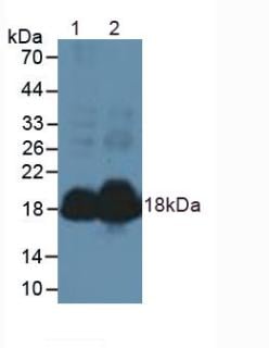
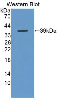
 WB (Western Blot)
(Western Blot: Sample: Recombinant GFP.)
WB (Western Blot)
(Western Blot: Sample: Recombinant GFP.)
Green Fluorescent Protein (GFP), Polyclonal Antibody (Cat# AAA147852)
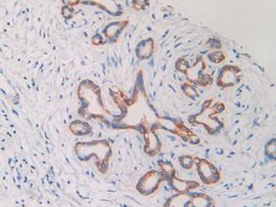
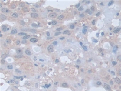
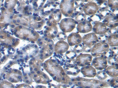
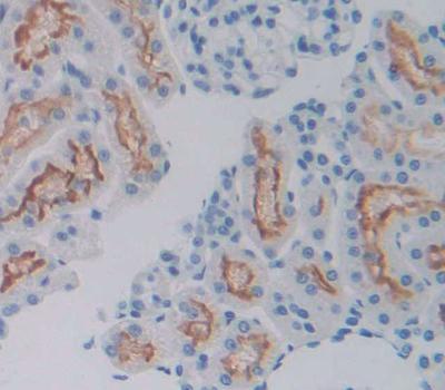
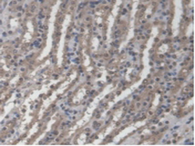
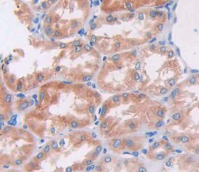
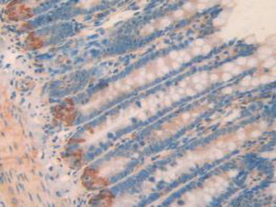
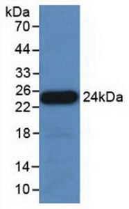
 IHC (Immunohistochemistry)
(DAB staining on IHC-P; Samples: Human Kidney Tissue.)
IHC (Immunohistochemistry)
(DAB staining on IHC-P; Samples: Human Kidney Tissue.)
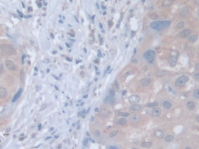
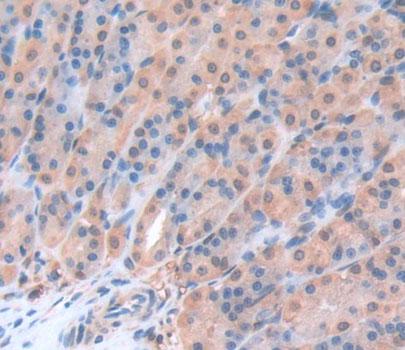
Cytokeratin Fragment Antigen 21-1 (CYFRA21-1), Polyclonal Antibody (Cat# AAA147876)
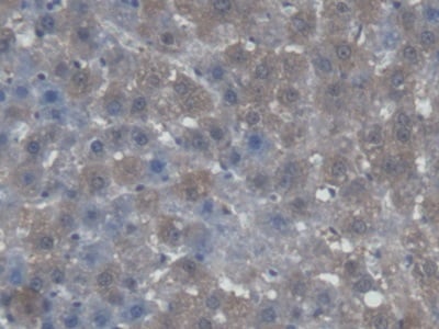
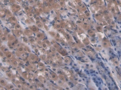
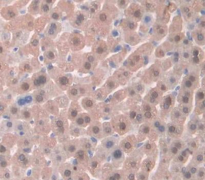
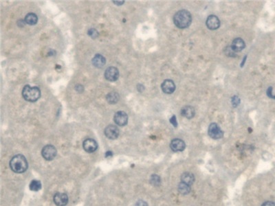
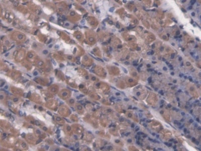
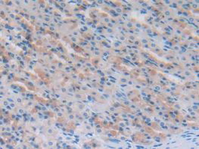
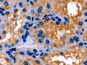
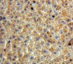
 IHC (Immunohistochemistry)
(DAB staining on IHC-P; Samples: Human Liver Tissue.)
IHC (Immunohistochemistry)
(DAB staining on IHC-P; Samples: Human Liver Tissue.)
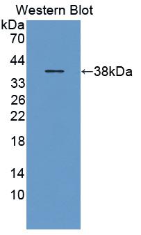
Natriuretic Peptide Precursor B (NPPB), Polyclonal Antibody (Cat# AAA147878)
 IHC (Immunohistochemisry)
(DAB staining on IHC-P;Samples: Porcine Kidney Tissue;Primary Ab: 20?g/ml Rabbit Anti-Porcine APOE AntibodySecond Ab: 2?g/mL HRP-Linked Caprine Anti-Rabbit IgG Polyclonal Antibody)
IHC (Immunohistochemisry)
(DAB staining on IHC-P;Samples: Porcine Kidney Tissue;Primary Ab: 20?g/ml Rabbit Anti-Porcine APOE AntibodySecond Ab: 2?g/mL HRP-Linked Caprine Anti-Rabbit IgG Polyclonal Antibody)
Apolipoprotein E (APOE), Polyclonal Antibody (Cat# AAA147879)
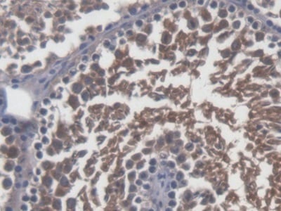
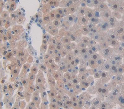
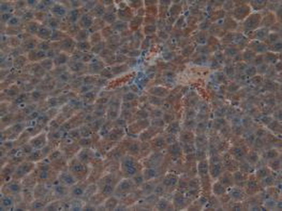
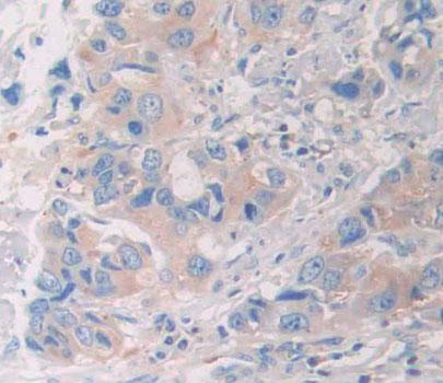
 IHC (Immunohiostchemistry)
(DAB staining on IHC-P; Samples: Human Liver Tissue)
IHC (Immunohiostchemistry)
(DAB staining on IHC-P; Samples: Human Liver Tissue)
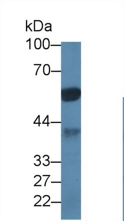
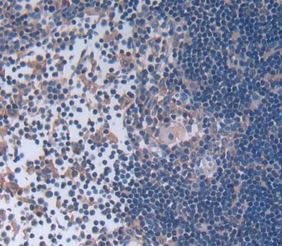
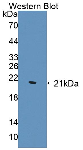
Calcitonin (CT), Polyclonal Antibody (Cat# AAA147889)

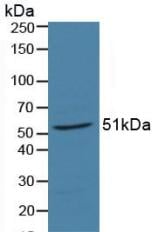
 WB (Western Blot)
(Western BlotSamples:Lane1: Human Plasma;Lane2: Human Plasma;Primary Ab: 0.05ug/ml Rabbit Anti-Human C4d AntibodySecond Ab: 0.2ug/ml HRP- Linked Caprine Anti-Rabbit IgG Polyclonal Antibody)
WB (Western Blot)
(Western BlotSamples:Lane1: Human Plasma;Lane2: Human Plasma;Primary Ab: 0.05ug/ml Rabbit Anti-Human C4d AntibodySecond Ab: 0.2ug/ml HRP- Linked Caprine Anti-Rabbit IgG Polyclonal Antibody)
Complement Component 4d (C4d), Polyclonal Antibody (Cat# AAA147890)
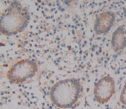
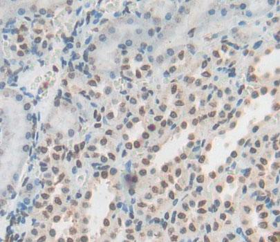
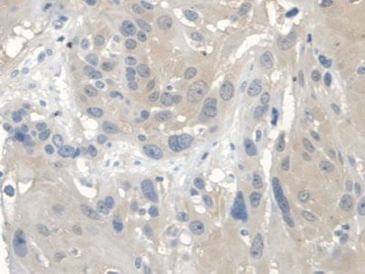
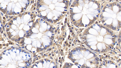 IHC (Immunohistochemistry)
(DAB staining on IHC-PSamples: Human Colon TissuePrimary Ab: 20ug/ml Rabbit Anti-Human AQP5 antibodySecond Ab: 2ug/mL HRP-Linked Caprine Anti-Rabbit IgG Polyclonal Antibody)
IHC (Immunohistochemistry)
(DAB staining on IHC-PSamples: Human Colon TissuePrimary Ab: 20ug/ml Rabbit Anti-Human AQP5 antibodySecond Ab: 2ug/mL HRP-Linked Caprine Anti-Rabbit IgG Polyclonal Antibody)
Aquaporin 5 (AQP5), Polyclonal Antibody (Cat# AAA147893)
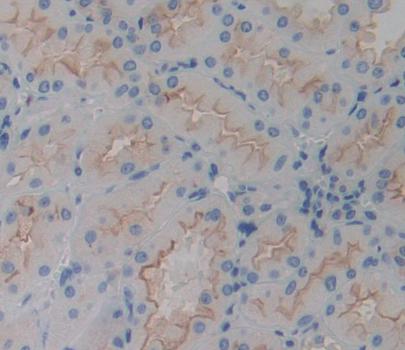
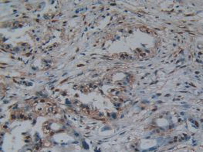
 IHC (Immunohiostchemistry)
(DAB staining on fromalin fixed paraffin-embedded Liver tissue))
IHC (Immunohiostchemistry)
(DAB staining on fromalin fixed paraffin-embedded Liver tissue))
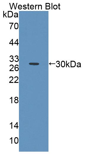
Squamous Cell Carcinoma Antigen 1 (SCCA1), Polyclonal Antibody (Cat# AAA147895)
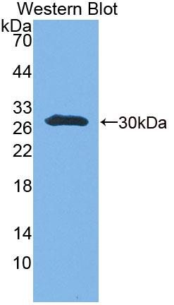
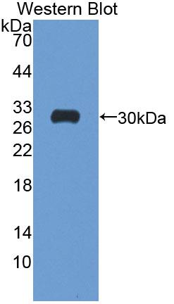
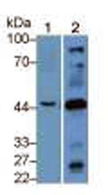
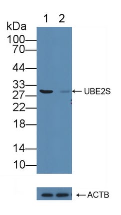 Knockout Validation
(Knockout Validation: Lane 1: Wild-type 293T cell lysate; Lane 2: UBE2S knockout 293T cell lysate; Predicted MW: 24kd Observed MW: 30kd Primary Ab: 1ug/ml Rabbit Anti-Human UBE2S Antibody Second Ab: 0.2ug/mL HRP-Linked Caprine Anti-Rabbit IgG Polyclonal Antibody)
Knockout Validation
(Knockout Validation: Lane 1: Wild-type 293T cell lysate; Lane 2: UBE2S knockout 293T cell lysate; Predicted MW: 24kd Observed MW: 30kd Primary Ab: 1ug/ml Rabbit Anti-Human UBE2S Antibody Second Ab: 0.2ug/mL HRP-Linked Caprine Anti-Rabbit IgG Polyclonal Antibody)
Ubiquitin Conjugating Enzyme E2S (UBE2S), Polyclonal Antibody (Cat# AAA147897)
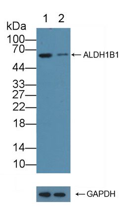
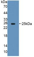
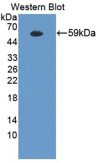
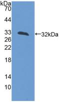
 Knockout Validation
(Knockout Validation: Lane 1: Wild-type K562 cell lysate; Lane 2: ALDH1B1 knockout K562 cell lysate; Predicted MW: 59kd Observed MW: 59kd Primary Ab: 1ug/ml Rabbit Anti-Human ALDH1B1 Antibody Second Ab: 0.2ug/mL HRP-Linked Caprine Anti-Rabbit IgG Polyclonal Antibody)
Knockout Validation
(Knockout Validation: Lane 1: Wild-type K562 cell lysate; Lane 2: ALDH1B1 knockout K562 cell lysate; Predicted MW: 59kd Observed MW: 59kd Primary Ab: 1ug/ml Rabbit Anti-Human ALDH1B1 Antibody Second Ab: 0.2ug/mL HRP-Linked Caprine Anti-Rabbit IgG Polyclonal Antibody)
Aldehyde Dehydrogenase 1 Family, Member B1 (ALDH1B1), Polyclonal Antibody (Cat# AAA147899)
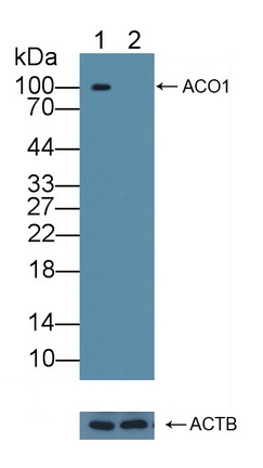
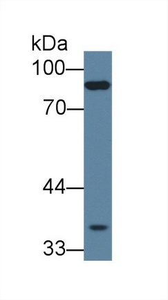
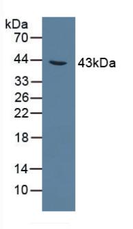
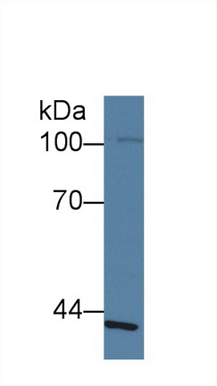
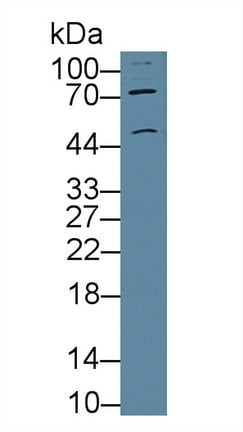
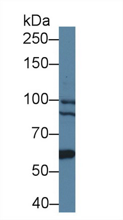
 Knockout Validation
(Knockout Validation: Lane 1: Wild-type HepG2 cell lysate; Lane 2: ACO1 knockout HepG2 cell lysate; Predicted MW: 98kd Observed MW: 100kd Primary Ab: 1ug/ml Rabbit Anti-Mouse ACO1 Antibody Second Ab: 0.2ug/mL HRP-Linked Caprine Anti-Rabbit IgG Polyclonal Antibody)
Knockout Validation
(Knockout Validation: Lane 1: Wild-type HepG2 cell lysate; Lane 2: ACO1 knockout HepG2 cell lysate; Predicted MW: 98kd Observed MW: 100kd Primary Ab: 1ug/ml Rabbit Anti-Mouse ACO1 Antibody Second Ab: 0.2ug/mL HRP-Linked Caprine Anti-Rabbit IgG Polyclonal Antibody)
Aconitase 1 (ACO1), Polyclonal Antibody (Cat# AAA147900)
Pyroglutamic Acid (PCA), Polyclonal Antibody (Cat# AAA147906)
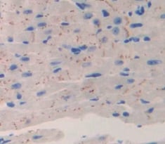
 IHC (Immunohistochemistry)
(DAB staining on IHC-P; Samples: Mouse Liver Tissue.)
IHC (Immunohistochemistry)
(DAB staining on IHC-P; Samples: Mouse Liver Tissue.)
Cytochrome C Oxidase Subunit VIc (COX6c), Polyclonal Antibody (Cat# AAA147907)
What are Polyclonal Antibodies?
Polyclonal antibodies are antibodies that come from multiple B cell clones of a host animal. The typical hosts used for the majority of polyclonal antibody production are rabbits, goats, sheep, and donkeys. These polyclonal antibodies, once having identified their target, will bind to different epitopes located at different regions or sequences on the same protein/antigen. As a result, they are ideal at locating and binding to the target, even if the target is in very low concentrations (due to many different antibodies being able to bind to the same target molecule, which allows for significant amplification of a downstream signal).
Polyclonal antibodies are typically produced by injecting an antigen into a host animal, which causes the animal’s immune system to attack the foreign antigen by mass generating antibodies against it. After a period of time, serum is collected from the animal and purified using physicochemical fractionation, class-specific affinity purification, and/or antigen-affinity purification.
Key Uses of Polyclonal Antibodies
- Western Blotting: This method is used to find specific proteins in biological samples after separating them by size.
- Immunohistochemistry: IHC helps visualize the location of proteins in tissue sections using various staining techniques.
- ELISA: (Enzyme-Linked Immunosorbent Assay) is typically used to identify specific protein quantities in a sample. ELISAs can be either “Quantitative” or “Qualitative”.
- Flow Cytometry: technique that identifies and measures the specific protein on the surface or inside the cells in a fluid suspension.
- Immunoprecipitation: IP isolates and studies a specific protein from a complex mixture using antibodies.
Why Buy Polyclonal Antibodies from AAA Biotech?
1. Ideal for Various Applications
Our antibodies are generally going to be validated for use in multiple types of assays, including ELISA, Western Blotting, Immunohistochemistry, Immunoprecipitation, amongst others. They are ideal for a wide range of research applications.
2. Rigorous Quality Control
All of the antibodies in our catalog undergo strict quality testing to ensure specificity, sensitivity, and consistent performance. We are confident in the ability of our antibodies to provide you with accurate results.
3. Wide Assortment of Antibodies
Antibodies in are catalog can be found for both common and exotic species, and these antibodies are also available in both conjugated and recombinant forms to suit many diverse experimental needs.
4. Highly Purified
Our antibodies are available in purified forms with over 85% purity, as confirmed by SDS-PAGE. They are also available with tags such as His, Flag, GST, or MBP. We cater to customers worldwide.
FAQ
1. How are polyclonal antibodies produced?
Traditionally, polyclonal antibodies are produced by injecting an antigen into a host animal (such as a rabbit or goat), which then triggers an immune response from the host animal. The animal’s B cells produce antibodies that will recognize different parts of the injected antigen. These antibodies are then collected from the animal’s blood and purified for use.
2. How do polyclonal antibodies differ from monoclonal antibodies?
Polyclonal antibodies are a mix of antibodies that bind to different locations (epitopes) of the same antigen, while monoclonal antibodies are identical and bind to just one specific epitope. This makes polyclonal antibodies more versatile and better at detecting proteins that may be present in low quantities or in altered/modified forms.
3. How should I store polyclonal antibodies?
Polyclonal antibodies should be stored at 4°C for short-term use (up to a few weeks) and at -20°C or -80°C for long-term storage. Avoid repeated freeze-thaw cycles by dividing them into small aliquots. Always check the datasheet for specific storage instructions.