Filters
▼Clonality
▼Type
▼Reactivity
▼Gene Name
▼Isotype
▼Host
▼Application
▼Clone
▼Phospho Antibodies
Phospho-specific antibodies’ typical purpose is to enable researchers to detect changes in proteins. They will exclusively bind to the amino acid sequence on a protein that has been phosphorylated (which is both a physical & chemical change) and do not bind to the same amino acid sequence on said protein if it lacks said phosphorylation. This aids in being able to clearly see and understand the data produced from this particular protein modification.
Viewing 4750-4800 of 5298 product results
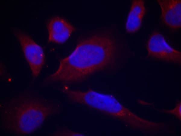
 IF (Immunofluorescence)
(Immunofluorescent analysis of TPL2 (phospho-Thr290) staining in HepG2 cells. Formalin-fixed cells were permeabilized with 0.1% Triton X-100 in TBS for 5-10 minutes and blocked with 3% BSA-PBS for 30 minutes at room temperature. Cells were probed with the primary antibody in 3% BSA-PBS and incubated overnight at 4 C in a humidified chamber. Cells were washed with PBST and incubated with a DyLight 594-conjugated secondary antibody (red) in PBS at room temperature in the dark. DAPI was used to stain the cell nuclei (blue).)
IF (Immunofluorescence)
(Immunofluorescent analysis of TPL2 (phospho-Thr290) staining in HepG2 cells. Formalin-fixed cells were permeabilized with 0.1% Triton X-100 in TBS for 5-10 minutes and blocked with 3% BSA-PBS for 30 minutes at room temperature. Cells were probed with the primary antibody in 3% BSA-PBS and incubated overnight at 4 C in a humidified chamber. Cells were washed with PBST and incubated with a DyLight 594-conjugated secondary antibody (red) in PBS at room temperature in the dark. DAPI was used to stain the cell nuclei (blue).)
TPL2 (phospho-Thr290), Polyclonal Antibody (Cat# AAA310507)
 IHC (Immunohiostchemistry)
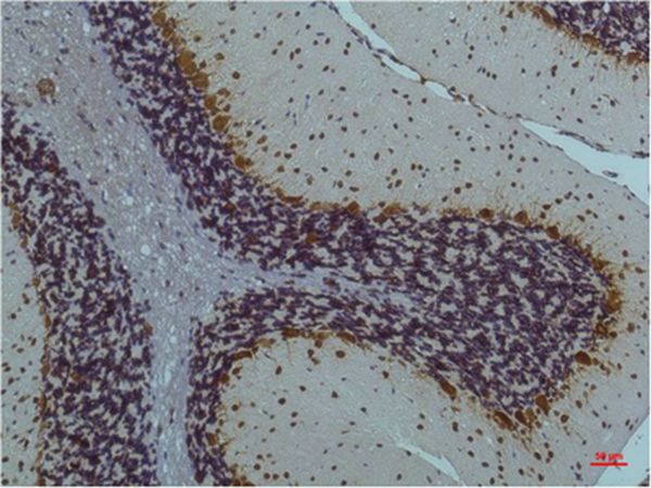
(Immunohistochemical analysis of paraffin-embedded mouse brain tissue using anti- phospho-Synapsin �� (S9) antibody. Counter stained with hematoxylin.)
IHC (Immunohiostchemistry)
(Immunohistochemical analysis of paraffin-embedded mouse brain tissue using anti- phospho-Synapsin �� (S9) antibody. Counter stained with hematoxylin.)
Synapsin, Monoclonal Antibody (Cat# AAA311020)
 IHC (Immunohiostchemistry)
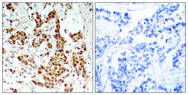
(Immunohistochemical analysis of paraffin-embedded Human Lung Carcinoma Tissue using Phospho-Akt Ser473 Mouse mAb diluted at 1:200.)
IHC (Immunohiostchemistry)
(Immunohistochemical analysis of paraffin-embedded Human Lung Carcinoma Tissue using Phospho-Akt Ser473 Mouse mAb diluted at 1:200.)
Akt, Monoclonal Antibody (Cat# AAA309602)
 IHC (Immunohiostchemistry)
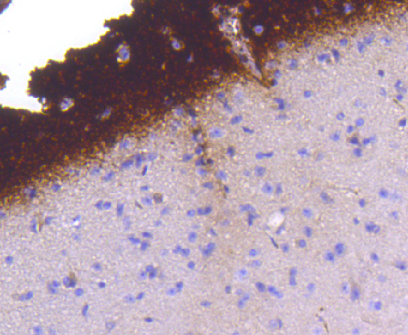
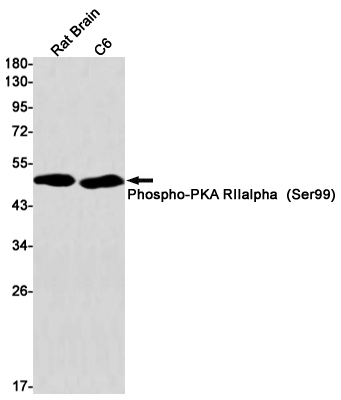
(Immunohistochemistry of PKA R2 (Phospho-Ser99) in paraffin-embedded Human tonsil using PKA R2 (Phospho-Ser99) Rabbit mAb at dilution 1/100)
IHC (Immunohiostchemistry)
(Immunohistochemistry of PKA R2 (Phospho-Ser99) in paraffin-embedded Human tonsil using PKA R2 (Phospho-Ser99) Rabbit mAb at dilution 1/100)
PKA RII alpha, Monoclonal Antibody (Cat# AAA314435)
 WB (Western Blot)
(Western blot analysis of extracts from Mouse brain tissue using DRP-2 (Phospho-Thr514) Antibody.The lane on the left is treated with the antigen-specific peptide.)
WB (Western Blot)
(Western blot analysis of extracts from Mouse brain tissue using DRP-2 (Phospho-Thr514) Antibody.The lane on the left is treated with the antigen-specific peptide.)
DPYSL2, Polyclonal Antibody (Cat# AAA243036)
 ICC (Immunocytochemistry)
(ICC staining Phospho-EGFR (Y1092) in BT-20 cells (green). The nuclear counter stain is DAPI (blue). Cells were fixed in paraformaldehyde, permeabilised with 0.25% Triton X100/PBS.)
ICC (Immunocytochemistry)
(ICC staining Phospho-EGFR (Y1092) in BT-20 cells (green). The nuclear counter stain is DAPI (blue). Cells were fixed in paraformaldehyde, permeabilised with 0.25% Triton X100/PBS.)
EGFR, Monoclonal Antibody (Cat# AAA310999)
 IHC (Immunohiostchemistry)
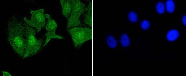
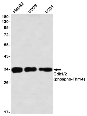
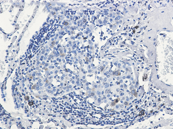
(Immunohistochemistry of Cdk1/2 (phospho-Thr14) in paraffin-embedded Human lung cancer tissue using Cdk1/2 (phospho-Thr14) Rabbit mAb at dilution 1/50)
IHC (Immunohiostchemistry)
(Immunohistochemistry of Cdk1/2 (phospho-Thr14) in paraffin-embedded Human lung cancer tissue using Cdk1/2 (phospho-Thr14) Rabbit mAb at dilution 1/50)
CDK1/2, Monoclonal Antibody (Cat# AAA314498)
 IF (Immunofluorescence)
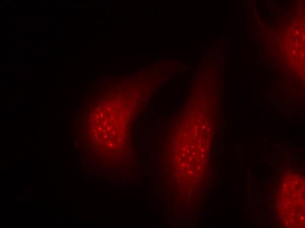
(Immunofluorescence staining of methanol-fixed Hela cells using p90RSK(Phospho-Thr348) Antibody.)
IF (Immunofluorescence)
(Immunofluorescence staining of methanol-fixed Hela cells using p90RSK(Phospho-Thr348) Antibody.)
p90RSK, Polyclonal Antibody (Cat# AAA303738)
 WB (Western Blot)
(Western blot analysis of lysates from Jurkat cells treated with Ca+ 40nM 30', using MYOD (Phospho-Ser200) Antibody.)
WB (Western Blot)
(Western blot analysis of lysates from Jurkat cells treated with Ca+ 40nM 30', using MYOD (Phospho-Ser200) Antibody.)
MYOD, Polyclonal Antibody (Cat# AAA318361)
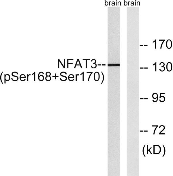 WB (Western Blot)
(Western blot analysis of NFAT3 (Phospho-Ser168+Ser170) Antibody. The lane on the right is blocked with the NFAT3 (Phospho-Ser168+Ser170) peptide.)
WB (Western Blot)
(Western blot analysis of NFAT3 (Phospho-Ser168+Ser170) Antibody. The lane on the right is blocked with the NFAT3 (Phospho-Ser168+Ser170) peptide.)
NFATc4, ELISA Kit (Cat# AAA319263)
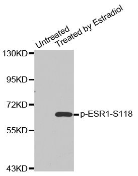
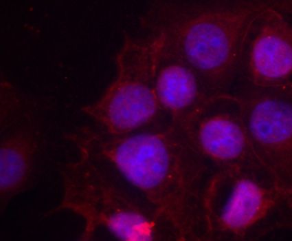 IF (Immunofluorescence)
(Immunofluorescence staining of methanol-fixed MCF-7 cells using Phospho-ESR1-S118 antibody.)
IF (Immunofluorescence)
(Immunofluorescence staining of methanol-fixed MCF-7 cells using Phospho-ESR1-S118 antibody.)
ESR1-S118, Antibody (Cat# AAA37388)
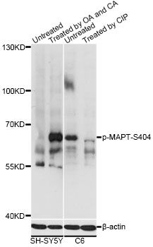
 WB (Western Blot)
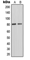
(Western blot analysis of TAU (pT548) expression in mouse brain (A) whole cell lysates.)
WB (Western Blot)
(Western blot analysis of TAU (pT548) expression in mouse brain (A) whole cell lysates.)
TAU (pT231), Polyclonal Antibody (Cat# AAA105103)
 IF (Immunofluorescence)
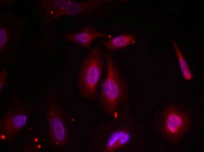
(Immunofluorescence staining of methanol-fixed Hela cells using ATF2(Phospho-Thr69 or 51) Antibody.)
IF (Immunofluorescence)
(Immunofluorescence staining of methanol-fixed Hela cells using ATF2(Phospho-Thr69 or 51) Antibody.)
ATF2, Polyclonal Antibody (Cat# AAA243261)
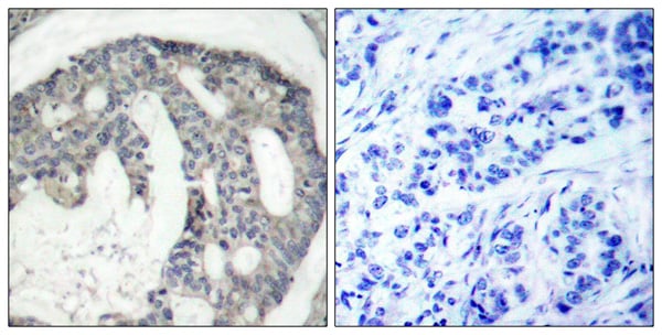
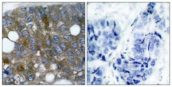
 IHC (Immunohiostchemistry)
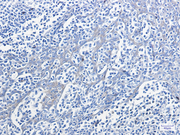
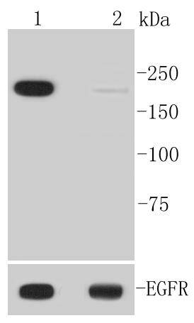
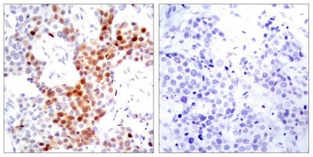
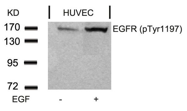
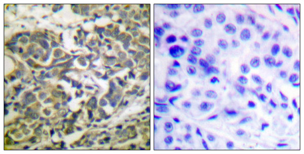
(Immunohistochemical analysis of paraffin-embedded human breast carcinoma tissue using EGFR(Phospho-Tyr1197) Antibody (left) or the same antibody preincubated with blocking peptide(right).)
IHC (Immunohiostchemistry)
(Immunohistochemical analysis of paraffin-embedded human breast carcinoma tissue using EGFR(Phospho-Tyr1197) Antibody (left) or the same antibody preincubated with blocking peptide(right).)
EGFR, Polyclonal Antibody (Cat# AAA305635)
 WB (Western Blot)
(Western blot analysis of lysates from COS7 cells treated with TNF 20ng/ml 5', using PEA-15 (Phospho-Ser104) Antibody.)
WB (Western Blot)
(Western blot analysis of lysates from COS7 cells treated with TNF 20ng/ml 5', using PEA-15 (Phospho-Ser104) Antibody.)
PEA-15, Polyclonal Antibody (Cat# AAA318372)
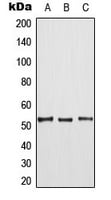
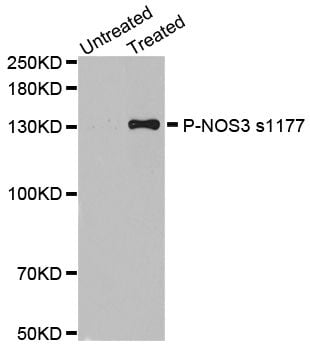
 WB (Western Blot)
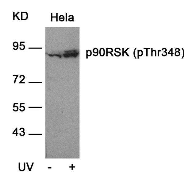
(Western blot analysis of extracts from Hela cells, untreated (-) or treated, 1:5000. Secondary antibody was diluted at 1:20000 cells nucleus extracted by Minute TM Cytoplasmic and Nuclear Fractionation kit (SC-003,Inventbiotech,MN,USA).)
WB (Western Blot)
(Western blot analysis of extracts from Hela cells, untreated (-) or treated, 1:5000. Secondary antibody was diluted at 1:20000 cells nucleus extracted by Minute TM Cytoplasmic and Nuclear Fractionation kit (SC-003,Inventbiotech,MN,USA).)
Histone H4, ELISA Kit (Cat# AAA319061)
 WB (Western Blot)
(Western Blot analysis of various cells using Phospho-EphA2 (Y588) Polyclonal Antibody)
WB (Western Blot)
(Western Blot analysis of various cells using Phospho-EphA2 (Y588) Polyclonal Antibody)
EphA2, ELISA Kit (Cat# AAA319103)
 WB (Western Blot)
(Western blot analysis of lysates from COS7 cells treated with UV 15', using Catenin-beta (Phospho-Tyr489) Antibody.)
WB (Western Blot)
(Western blot analysis of lysates from COS7 cells treated with UV 15', using Catenin-beta (Phospho-Tyr489) Antibody.)
Catenin-beta, Polyclonal Antibody (Cat# AAA318295)
 IF (Immunofluorescence)
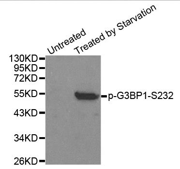
(Immunofluorescence staining of methanol-fixed HeLa cells using Phospho-G3BP1-S232 antibody.)
IF (Immunofluorescence)
(Immunofluorescence staining of methanol-fixed HeLa cells using Phospho-G3BP1-S232 antibody.)
G3BP1-S232, Antibody (Cat# AAA37371)
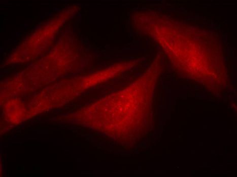
 IF (Immunofluorescence)
(Immunofluorescence staining of methanol-fixed HeLa cells using Phospho-NOS3-S1177 antibody.)
IF (Immunofluorescence)
(Immunofluorescence staining of methanol-fixed HeLa cells using Phospho-NOS3-S1177 antibody.)
NOS3-S1177, Antibody (Cat# AAA37413)
 IHC (Immunohiostchemistry)
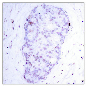
(Immunohistochemical analysis of paraffin-embedded human breast carcinoma tissue using NF-κB p105(phospho-Ser907) antibody.)
IHC (Immunohiostchemistry)
(Immunohistochemical analysis of paraffin-embedded human breast carcinoma tissue using NF-κB p105(phospho-Ser907) antibody.)
NFKB1, Polyclonal Antibody (Cat# AAA243174)
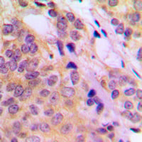
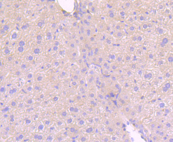
 IHC (Immunohiostchemistry)
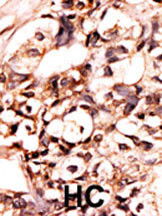
(Formalin-fixed and paraffin-embedded human cancer tissue reacted with the primary antibody, which was peroxidase-conjugated to the secondary antibody, followed by AEC staining. This data demonstrates the use of this antibody for immunohistochemistry; clinical relevance has not been evaluated. BC = breast carcinoma; HC = hepatocarcinoma.)
IHC (Immunohiostchemistry)
(Formalin-fixed and paraffin-embedded human cancer tissue reacted with the primary antibody, which was peroxidase-conjugated to the secondary antibody, followed by AEC staining. This data demonstrates the use of this antibody for immunohistochemistry; clinical relevance has not been evaluated. BC = breast carcinoma; HC = hepatocarcinoma.)
Phospho-DAXX (S213), Polyclonal Antibody (Cat# AAA288614)
 WB (Western Blot)
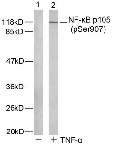
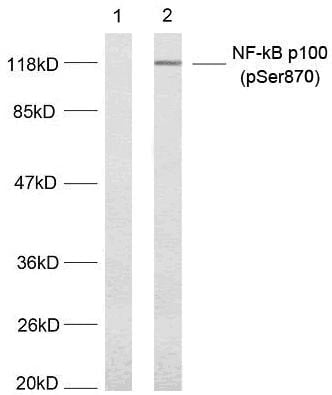
(Western blot analysis of extract from MDA-MB-435 cells untreated or treated with TNF-alpha (20ng/ml, 5min) using NF-kappaB p100(phospho-Ser870) antibody ().)
WB (Western Blot)
(Western blot analysis of extract from MDA-MB-435 cells untreated or treated with TNF-alpha (20ng/ml, 5min) using NF-kappaB p100(phospho-Ser870) antibody ().)
NFkappaB-p100/p52, Polyclonal Antibody (Cat# AAA303198)
 IHC (Immunohiostchemistry)
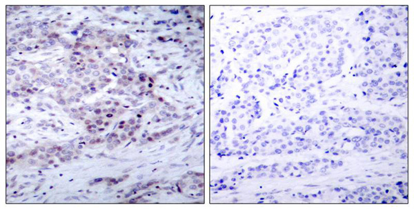
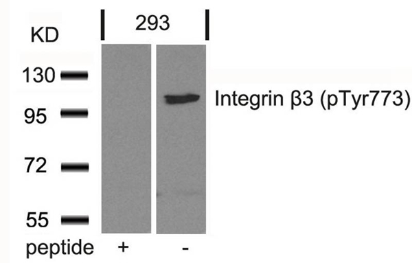
(Immunohistochemical analysis of paraffin-embedded human breast carcinoma tissue using Integrin b3(Phospho-Tyr773) Antibody (left) or the same antibody preincubated with blocking peptide(right).)
IHC (Immunohiostchemistry)
(Immunohistochemical analysis of paraffin-embedded human breast carcinoma tissue using Integrin b3(Phospho-Tyr773) Antibody (left) or the same antibody preincubated with blocking peptide(right).)
Integrin beta3, Polyclonal Antibody (Cat# AAA308011)
 IHC (Immunohiostchemistry)
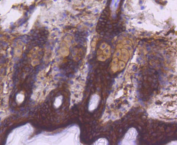
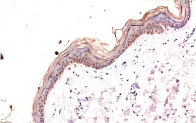
(Immunohistochemistry of paraffin-embedded Human skin tissue with Phospho-MLKL (Ser358) Monoclonal Antibody at dilution of 1:200)
IHC (Immunohiostchemistry)
(Immunohistochemistry of paraffin-embedded Human skin tissue with Phospho-MLKL (Ser358) Monoclonal Antibody at dilution of 1:200)
MLKL, Monoclonal Antibody (Cat# AAA173639)

 ICC (Immunocytochemistry)
(ICC staining Phospho-AMPK alpha 2 (S345) in Hela cells (green). The nuclear counter stain is DAPI (blue). Cells were fixed in paraformaldehyde, permeabilised with 0.25% Triton X100/PBS.)
ICC (Immunocytochemistry)
(ICC staining Phospho-AMPK alpha 2 (S345) in Hela cells (green). The nuclear counter stain is DAPI (blue). Cells were fixed in paraformaldehyde, permeabilised with 0.25% Triton X100/PBS.)
AMPK alpha 2, Monoclonal Antibody (Cat# AAA311042)
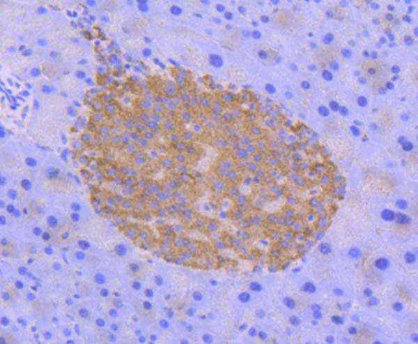
 IHC (Immunohistochemisry)
(Immunohistochemical analysis of paraffin-embedded mouse colon tissue using anti-MLKL (phospho S345) antibody. Counter stained with hematoxylin.)
IHC (Immunohistochemisry)
(Immunohistochemical analysis of paraffin-embedded mouse colon tissue using anti-MLKL (phospho S345) antibody. Counter stained with hematoxylin.)
MLKL, Monoclonal Antibody (Cat# AAA311047)
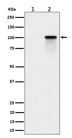 WB (Western Blot)
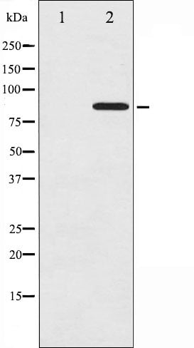
(Western blot analysis of Phospho-Nucleolin (T84) expression in (1) K562 cell lysate; (2) K562 cell treated with Calyculin A lysate.)
WB (Western Blot)
(Western blot analysis of Phospho-Nucleolin (T84) expression in (1) K562 cell lysate; (2) K562 cell treated with Calyculin A lysate.)
Nucleolin, Monoclonal Recombinant Antibody (Cat# AAA314882)
 WB (Western Blot)
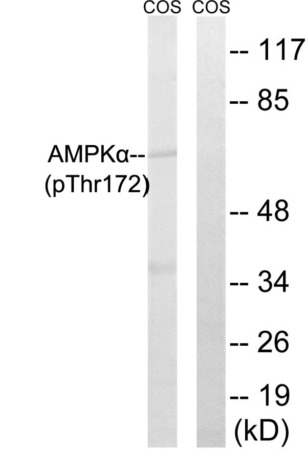
(Western blot analysis of lysates from COS7 cells, using AMPK alpha (Phospho-Thr172) Antibody. The lane on the right is blocked with the phospho peptide.)
WB (Western Blot)
(Western blot analysis of lysates from COS7 cells, using AMPK alpha (Phospho-Thr172) Antibody. The lane on the right is blocked with the phospho peptide.)
AMPK alpha, Polyclonal Antibody (Cat# AAA316054)
 WB (Western Blot)
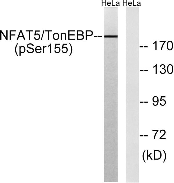
(Western blot analysis of lysates from HeLa cells treated with forskolin 40nM 30', using NFAT5/TonEBP (Phospho-Ser155) Antibody. The lane on the right is blocked with the phospho peptide.)
WB (Western Blot)
(Western blot analysis of lysates from HeLa cells treated with forskolin 40nM 30', using NFAT5/TonEBP (Phospho-Ser155) Antibody. The lane on the right is blocked with the phospho peptide.)
NFAT5, ELISA Kit (Cat# AAA319150)
 WB (Western Blot)
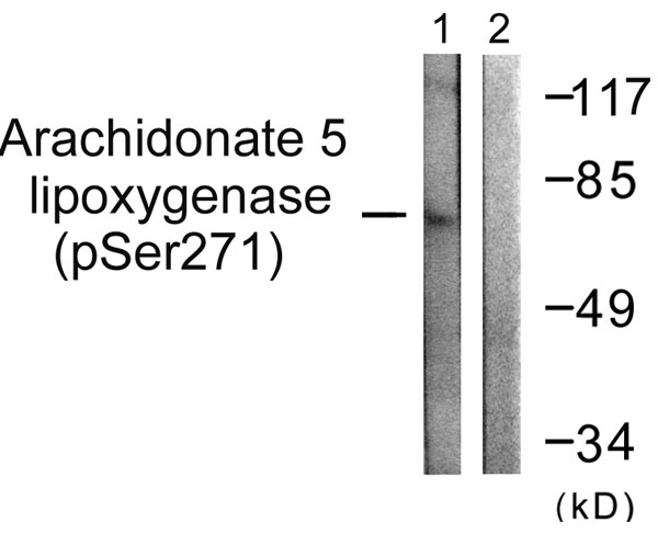
(Western blot analysis of lysates from HUVEC cells, using Arachidonate 5 Lipoxygenase (Phospho-Ser271) Antibody. The lane on the right is blocked with the phospho peptide.)
WB (Western Blot)
(Western blot analysis of lysates from HUVEC cells, using Arachidonate 5 Lipoxygenase (Phospho-Ser271) Antibody. The lane on the right is blocked with the phospho peptide.)
Arachidonate 5 Lipoxygenase, Polyclonal Antibody (Cat# AAA316290)
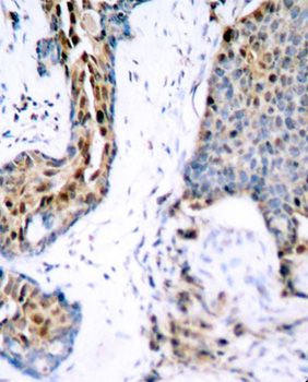
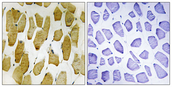
 IHC (Immunohiostchemistry)
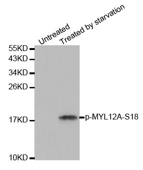
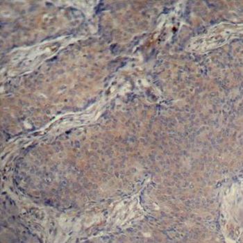
(Immunohistochemistry of paraffin-embedded human breast carcinoma using Phospho-MYL12A-S18 antibody.)
IHC (Immunohiostchemistry)
(Immunohistochemistry of paraffin-embedded human breast carcinoma using Phospho-MYL12A-S18 antibody.)
MYL12A-S18, Antibody (Cat# AAA37361)
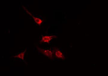
 IF (Immunofluorescence)
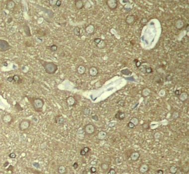
(Immunofluorescence staining of methanol-fixed HeLa cells using Phospho-MAPT-S404 antibody.)
IF (Immunofluorescence)
(Immunofluorescence staining of methanol-fixed HeLa cells using Phospho-MAPT-S404 antibody.)
MAPT-S404, Antibody (Cat# AAA37393)
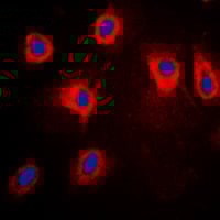
 IF (Immunofluorescence)
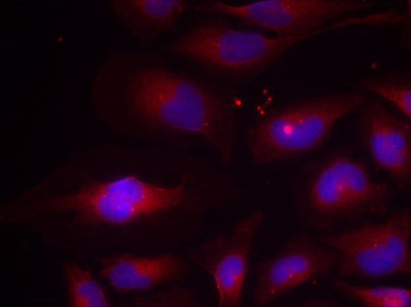
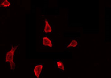
(AAA323708 staining SW626 cells by IF/ICC. The sample were fixed with PFA and permeabilized in 0.1% Triton X-100, then blocked in 10% serum for 45 minutes at 25 degree C. The primary antibody was diluted at 1/200 and incubated with the sample for 1 hour at 37 degree C. An Alexa Fluor 594 conjugated goat anti-rabbit IgG (H+L) antibody, diluted at 1/600, was used as secondary antibody.)
IF (Immunofluorescence)
(AAA323708 staining SW626 cells by IF/ICC. The sample were fixed with PFA and permeabilized in 0.1% Triton X-100, then blocked in 10% serum for 45 minutes at 25 degree C. The primary antibody was diluted at 1/200 and incubated with the sample for 1 hour at 37 degree C. An Alexa Fluor 594 conjugated goat anti-rabbit IgG (H+L) antibody, diluted at 1/600, was used as secondary antibody.)
beta-Catenin, Polyclonal Antibody (Cat# AAA323708)
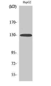
 WB (Western Blot)
(Western blot analysis of lysates from HepG2 cells treated with EGF 200ng/ml 30', using PDGFR beta (Phospho-Tyr740) Antibody.)
WB (Western Blot)
(Western blot analysis of lysates from HepG2 cells treated with EGF 200ng/ml 30', using PDGFR beta (Phospho-Tyr740) Antibody.)
PDGFR beta, Polyclonal Antibody (Cat# AAA318371)
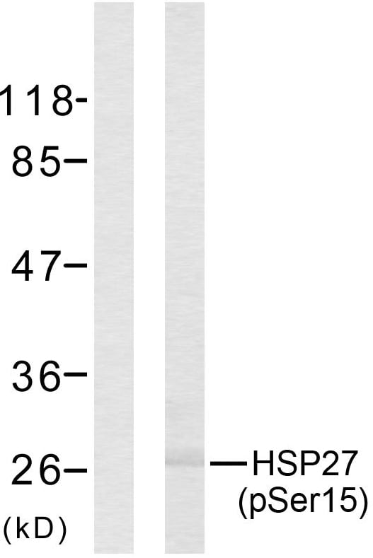 WB (Western Blot)
(Western blot analysis of lysates from HeLa cells treated with UV, using HSP27 (Phospho-Ser15) Antibody. The lane on the left is blocked with the phospho peptide.)
WB (Western Blot)
(Western blot analysis of lysates from HeLa cells treated with UV, using HSP27 (Phospho-Ser15) Antibody. The lane on the left is blocked with the phospho peptide.)
HSP27, Polyclonal Antibody (Cat# AAA316511)
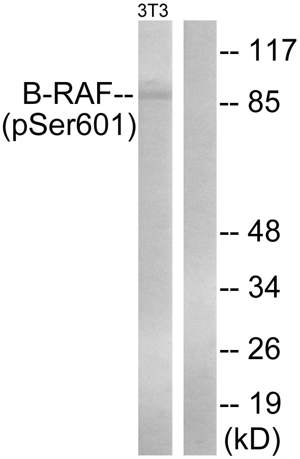 WB (Western Blot)
(Western blot analysis of lysates from NIH/3T3 cells treated with EGF 200ng/ml 30', using B-RAF (Phospho-Ser602) Antibody. The lane on the right is blocked with the phospho peptide.)
WB (Western Blot)
(Western blot analysis of lysates from NIH/3T3 cells treated with EGF 200ng/ml 30', using B-RAF (Phospho-Ser602) Antibody. The lane on the right is blocked with the phospho peptide.)
B-RAF, Polyclonal Antibody (Cat# AAA316297)
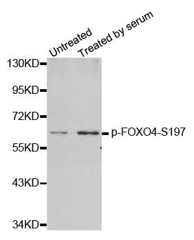
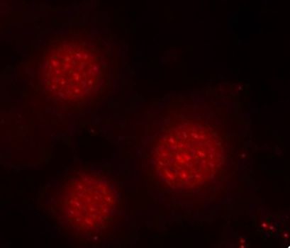 IF (Immunofluorescence)
(Immunofluorescence staining of methanol-fixed MCF-7 cells using Phospho-FOXO4-S197 antibody.)
IF (Immunofluorescence)
(Immunofluorescence staining of methanol-fixed MCF-7 cells using Phospho-FOXO4-S197 antibody.)
FOXO4-S197, Antibody (Cat# AAA37385)
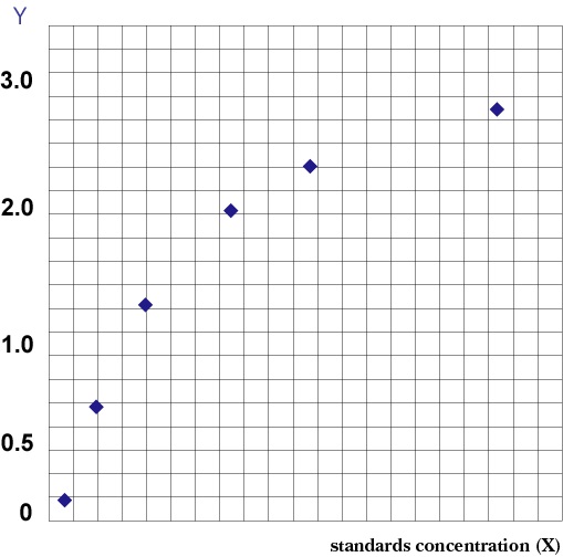 Standard Curve (Sample)
Phospho (637-DRP1), ELISA Kit (Cat# AAA206426)
 Application Data
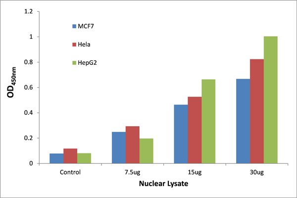
(The TFact p53 (Phospho-Ser376) DNA-Binding ELISA detects active p53 (Phospho-Ser376) in Hela, MCF7 and HepG2 Nuclear Extract. The Hela, MCF7 and HepG2 cells were grown 3 days in DMEM with 10% FBS and harvested for nuclear extract.)
Application Data
(The TFact p53 (Phospho-Ser376) DNA-Binding ELISA detects active p53 (Phospho-Ser376) in Hela, MCF7 and HepG2 Nuclear Extract. The Hela, MCF7 and HepG2 cells were grown 3 days in DMEM with 10% FBS and harvested for nuclear extract.)
p53, ELISA Kit (Cat# AAA315962)
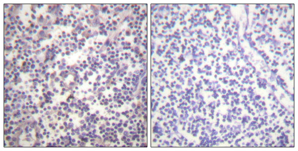
 IHC (Immunohiostchemistry)
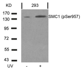
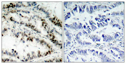
(Immunohistochemical analysis of paraffin-embedded human lung carcinoma tissue using SMC1(Phospho-Ser957) Antibody(left) or the same antibody preincubated with blocking peptide(right).)
IHC (Immunohiostchemistry)
(Immunohistochemical analysis of paraffin-embedded human lung carcinoma tissue using SMC1(Phospho-Ser957) Antibody(left) or the same antibody preincubated with blocking peptide(right).)
SMC1A, Polyclonal Antibody (Cat# AAA243112)

 WB (Western Blot)
(Western blot analysis of lysates from mouse brain, using Dynamin-1 (Phospho-Ser774) Antibody. The lane on the right is blocked with the phospho peptide.)
WB (Western Blot)
(Western blot analysis of lysates from mouse brain, using Dynamin-1 (Phospho-Ser774) Antibody. The lane on the right is blocked with the phospho peptide.)
Dynamin-1, Polyclonal Antibody (Cat# AAA316137)

 WB (Western Blot)
(All lanes use the Antibody at 1:1K dilution for 1 hour at room temperature.)
WB (Western Blot)
(All lanes use the Antibody at 1:1K dilution for 1 hour at room temperature.)
AMPA Receptor 1, Monoclonal Antibody (Cat# AAA128148)
 IHC (Immunohiostchemistry)
(Immunohistochemical analysis of paraffin-embedded Human Stomach Carcinoma Tissue using Phospho-Smad3 (S425) Mouse mAb diluted at 1:200)
IHC (Immunohiostchemistry)
(Immunohistochemical analysis of paraffin-embedded Human Stomach Carcinoma Tissue using Phospho-Smad3 (S425) Mouse mAb diluted at 1:200)
Smad3, Monoclonal Antibody (Cat# AAA309605)
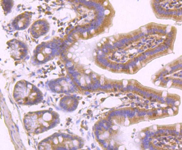
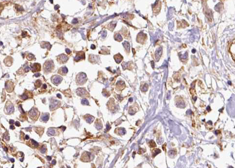
 IHC (Immunohiostchemistry)
(AAA321407 at 1/100 staining Human breast cancer tissue by IHC-P. The sample was formaldehyde fixed and a heat mediated antigen retrieval step in citrate buffer was performed. The sample was then blocked and incubated with the antibody for 1.5 hours at 22 degree C. An HRP conjugated goat anti-rabbit antibody was used as the secondary.)
IHC (Immunohiostchemistry)
(AAA321407 at 1/100 staining Human breast cancer tissue by IHC-P. The sample was formaldehyde fixed and a heat mediated antigen retrieval step in citrate buffer was performed. The sample was then blocked and incubated with the antibody for 1.5 hours at 22 degree C. An HRP conjugated goat anti-rabbit antibody was used as the secondary.)
TLK1, Polyclonal Antibody (Cat# AAA321407)
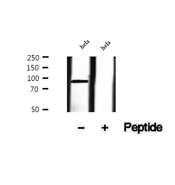
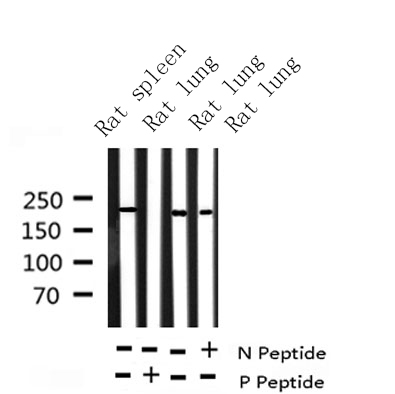 WB (Western Blot)
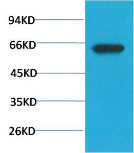
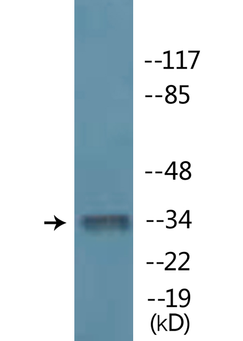
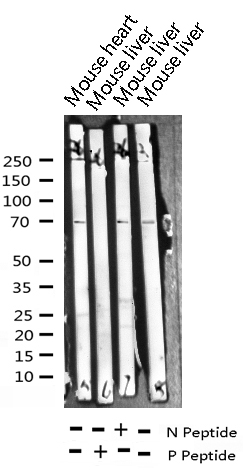
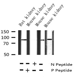
(Western blot analysis of BRCA1 phosphorylation expression in whole cell lysates, The lane on the left is treated with the antigen-specific peptide.)
WB (Western Blot)
(Western blot analysis of BRCA1 phosphorylation expression in whole cell lysates, The lane on the left is treated with the antigen-specific peptide.)
BRCA1, Polyclonal Antibody (Cat# AAA321610)
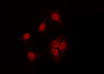
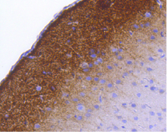
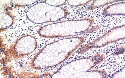
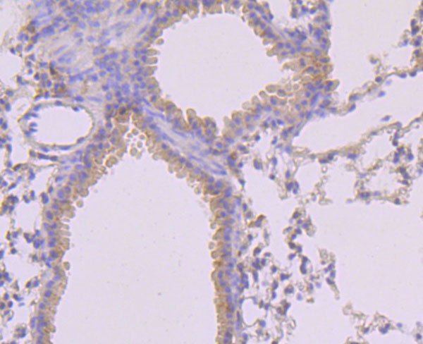
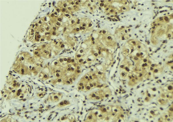
 IHC (Immunohistochemistry)
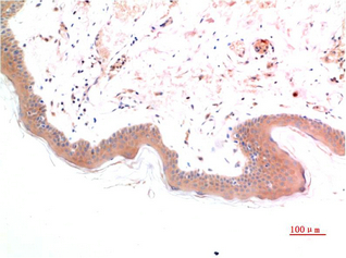
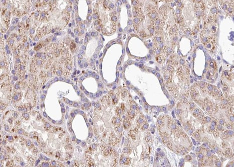
(AAA321636 at 1/100 staining human Kidney tissue sections by IHC-P. The tissue was formaldehyde fixed and a heat mediated antigen retrieval step in citrate buffer was performed. The tissue was then blocked and incubated with the antibody for 1.5 hours at 22 degree C. An HRP conjugated goat anti-rabbit antibody was used as the secondary.)
IHC (Immunohistochemistry)
(AAA321636 at 1/100 staining human Kidney tissue sections by IHC-P. The tissue was formaldehyde fixed and a heat mediated antigen retrieval step in citrate buffer was performed. The tissue was then blocked and incubated with the antibody for 1.5 hours at 22 degree C. An HRP conjugated goat anti-rabbit antibody was used as the secondary.)
SOX-9, Polyclonal Antibody (Cat# AAA321636)
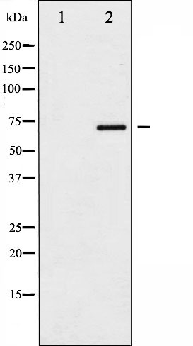
 WB (Western Blot)
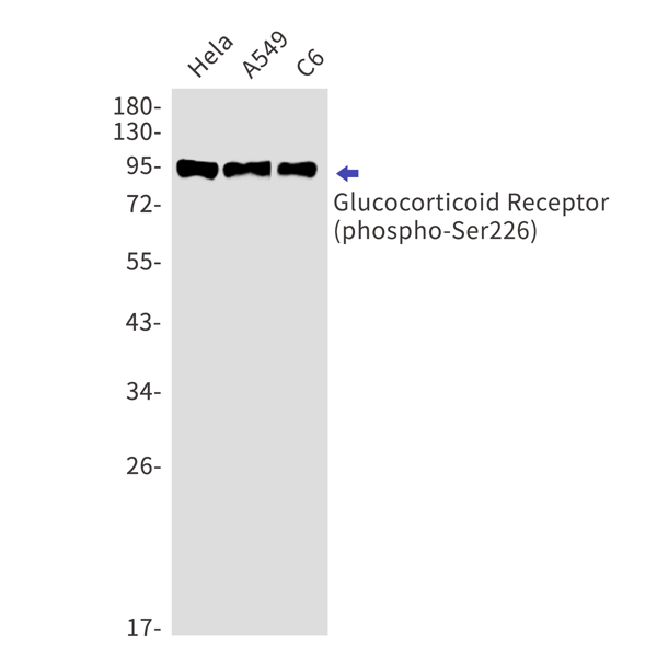
(Western blot analysis of Phospho-Cortactin (Tyr466) expression in various lysates)
WB (Western Blot)
(Western blot analysis of Phospho-Cortactin (Tyr466) expression in various lysates)
Cortactin, Polyclonal Antibody (Cat# AAA321727)
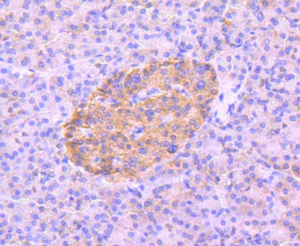
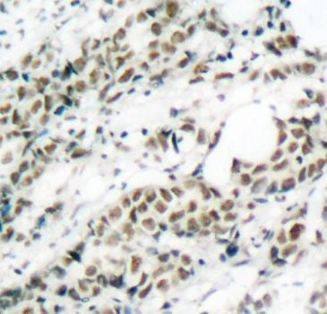
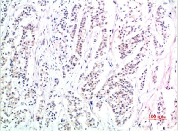
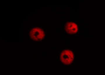
 IHC (Immunohiostchemistry)
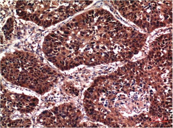
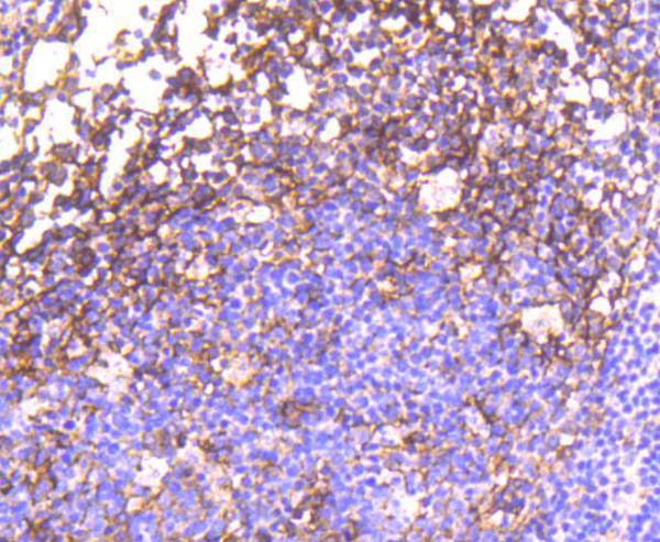
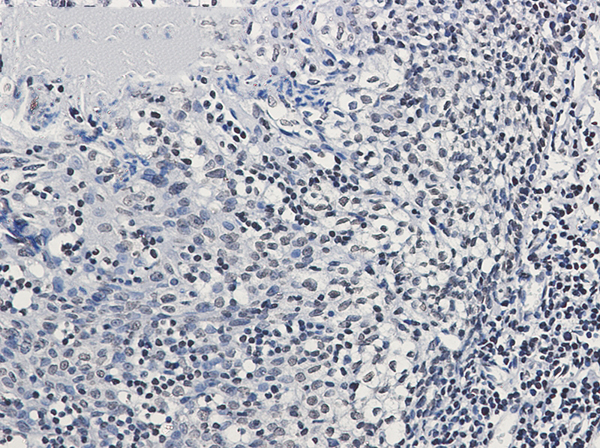
(Immunohistochemistry of Glucocorticoid Receptor (phospho-Ser226) in paraffin-embedded Human tonsil using Glucocorticoid Receptor (phospho-Ser226) Rabbit mAb at dilution 1/50)
IHC (Immunohiostchemistry)
(Immunohistochemistry of Glucocorticoid Receptor (phospho-Ser226) in paraffin-embedded Human tonsil using Glucocorticoid Receptor (phospho-Ser226) Rabbit mAb at dilution 1/50)
Glucocorticoid Receptor, Monoclonal Antibody (Cat# AAA314501)
 IHC (Immunohiostchemistry)
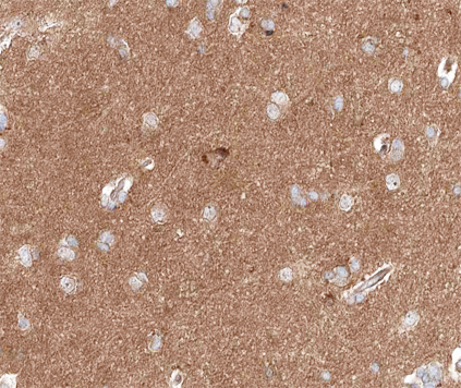
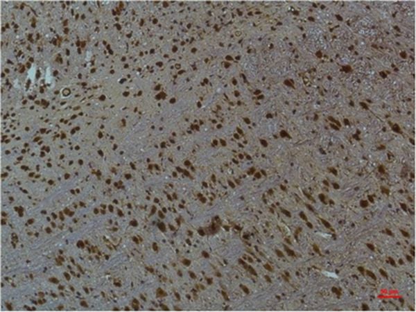
(Immunohistochemical analysis of paraffin-embedded Mouse Brain Tissue using CaMKIIb/ g /&d (Phospho Thr287) Mouse mAb diluted at 1:200.)
IHC (Immunohiostchemistry)
(Immunohistochemical analysis of paraffin-embedded Mouse Brain Tissue using CaMKIIb/ g /&d (Phospho Thr287) Mouse mAb diluted at 1:200.)
CaMKIIbeta/gamma/delta, Monoclonal Antibody (Cat# AAA309600)
What Are Phospho Antibodies?
Protein phosphorylation is a process where a phosphate group is added to certain amino acid residues of a protein – usually serine (S), threonine (T), or tyrosine (Y) - by enzymes called kinases. This process is integral in controlling cellular signaling, cellular growth, and other biological functions.
Our catalog includes a wide range of phospho-specific antibodies that can accurately detect this important marker. They perform strongly in widely-used laboratory applications such as Western blot, flow cytometry, immunohistochemistry, and immunofluorescence microscopy. We value your trust in us and are committed to providing top-quality products and services. All of our antibodies are guaranteed to work for the applications and species indicated on our website & associated product pages.
What Are The Key Applications of Phospho Antibodies?
1. Western Blotting
One of the first steps a researcher can take in utilizing these phospho-specific antibodies, is to check if the antibody works using a technique referred to as “Western blot”. For those unfamiliar, Western Blot aids in showing whether the protein that the antibody recognizes is appearing at the correct/expected size. These phospho-specific antibodies should also be able to detect changes in the target protein’s phosphorylation (on/off state) when cells are stimulated in certain ways.
2. Staining of Fixed Cells (Immunocytochemistry)
Another routine use of these phospho-specific antibodies, is to test if the antibody is able to demonstrate similar performance when used on fixed cells (intact cells that have been preserved) as it did in the Western blot tests. It is an important aspect in many cases to confirm that the antibody works in actual intact cell samples. Ideally, the method used for cellular fixation should be the same as what is used in pathology labs (like using 10% formalin). To check if the antibody works well in tissue sections (FFPE), researchers will often test it on fixed cells that are processed similar to tissue samples.
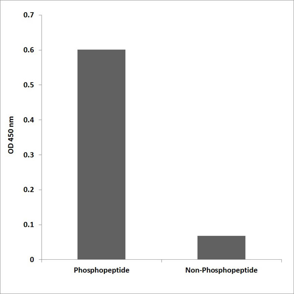
3. Specificity Tests Using Peptides
In order to make sure that the antibody is only binding to the right target:
- Laboratory technicians will mix the antibody with phospho-peptides (short segments of the protein containing the phosphate group modification).
- If the antibody signal disappears, it is confirmation that it is binding to the correct phosphorylated location.
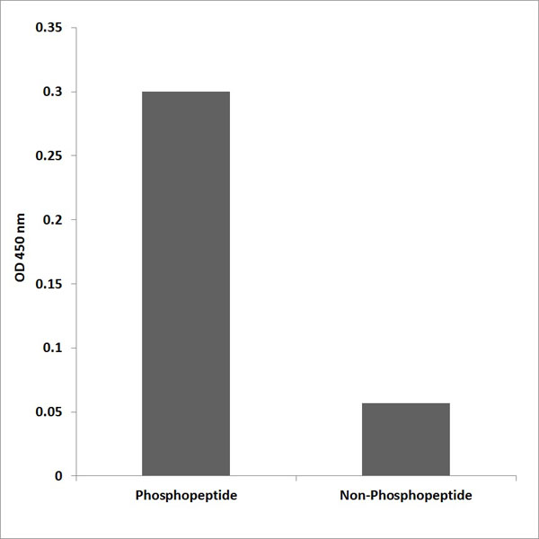
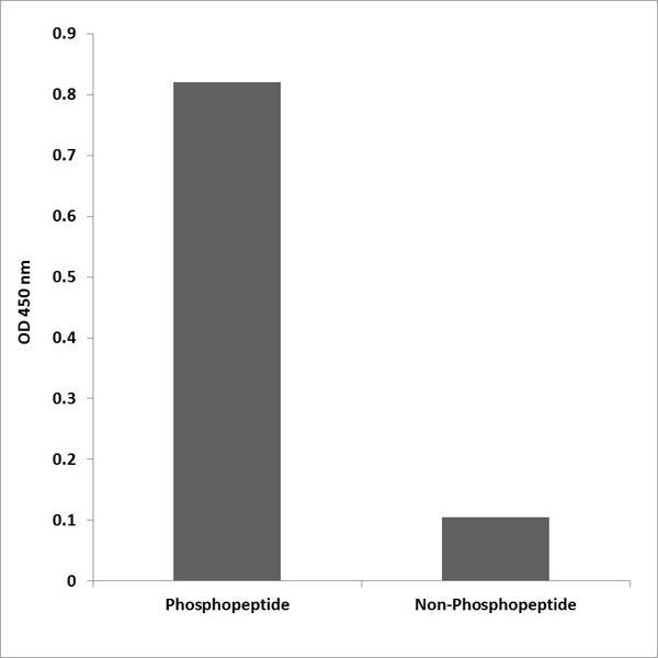
- A more robust test is to use both the phosphorylated and non-phosphorylated (dephosphorylated) versions of the protein. The antibody should react only with the phosphorylated one.
- Another method sometimes utilized is to treat the sample with an enzyme, such as alkaline phosphatase, that specifically removes phosphate groups. If the antibody signal disappears after this, it also confirms specificity.
4. Genetic Confirmation
As a final step, scientists can genetically manipulate the nucleotide sequence and alter the target protein by removing the exact site where phosphorylation happens. If the antibody no longer appears to detect the modified protein, it is strong evidence supporting the antibody being specific for that phosphorylated site.
Why Buy Phospho Antibodies Through Us?
- The production laboratory adheres to strict and consistent protocols prior to releasing any of these phospho-specific antibodies:
- Standard methods and proper controls in all tests to ensure high quality.
- These antibodies are tested and validated in different cell types and species.
- High quality control criterion to ensure each batch is consistent, so you will obtain reliable results every time.
FAQ
1. What Are Phospho-Specific Antibodies?
Phospho-specific antibodies are made to detect proteins only when they have a phosphate group linked to a specific amino acid residue. This empowers scientists understand if a protein is "turned on" or active, based on its phosphorylation state.
2. How to Detect Phosphorylated Proteins in a Western Blot?
To find out if a protein is phosphorylated using Western blot:
- Use a phospho-specific antibody that binds only to the phosphorylated form of the protein.
- You can also use a “regular” antibody for the same amino acid sequence of the protein that the phospho-specific antibody is binding to (but in this case, this antibody will not bind if there is a phosphate group present) in order to compare how much of it is phosphorylated versus how much is non-phosphorylated (or “total” protein, if the “normal” antibody’s epitopes are non-phospho-site-specific).
3. How to Choose the Best Antibody?
Here are some simple tips to help you pick the right antibody:
- Know your target
- Match your sample characteristics
- Confirm the intended use is appropriate
- Check “host” and “type”
- Check the “quality” of the presented data/images
- Appraise whether the available validation meets your needs