Filters
▼Clonality
▼Type
▼Reactivity
▼Gene Name
▼Isotype
▼Host
▼Application
▼Clone
▼Phospho Antibodies
Phospho-specific antibodies’ typical purpose is to enable researchers to detect changes in proteins. They will exclusively bind to the amino acid sequence on a protein that has been phosphorylated (which is both a physical & chemical change) and do not bind to the same amino acid sequence on said protein if it lacks said phosphorylation. This aids in being able to clearly see and understand the data produced from this particular protein modification.
Viewing 4850-4900 of 5298 product results
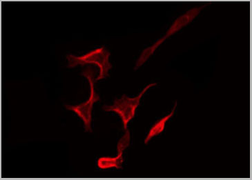
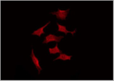
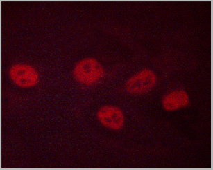
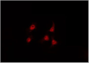
 IF (Immunofluorescence)
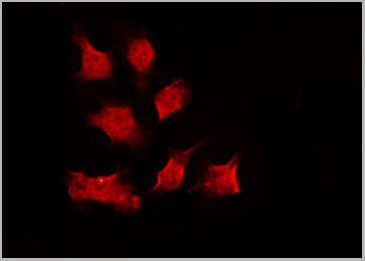
(AAA30940 staining HuvEc by IF/ICC. The sample were fixed with PFA and permeabilized in 0.1% Triton X-100, then blocked in 10% serum for 45 minutes at 25 degree C. The primary antibody was diluted at 1/200 and incubated with the sample for 1 hour at 37 degree C. An Alexa Fluor 594 conjugated goat anti-rabbit IgG (H+L) Ab, diluted at 1/600, was used as the secondary antibody.)
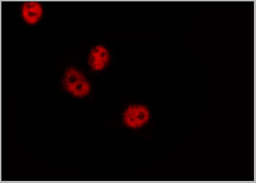
IF (Immunofluorescence)
(AAA30940 staining HuvEc by IF/ICC. The sample were fixed with PFA and permeabilized in 0.1% Triton X-100, then blocked in 10% serum for 45 minutes at 25 degree C. The primary antibody was diluted at 1/200 and incubated with the sample for 1 hour at 37 degree C. An Alexa Fluor 594 conjugated goat anti-rabbit IgG (H+L) Ab, diluted at 1/600, was used as the secondary antibody.)
GSK3 beta, Polyclonal Antibody (Cat# AAA30940)
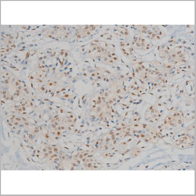
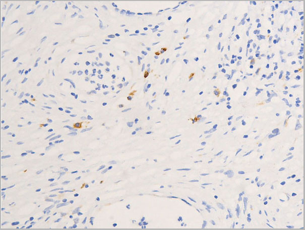
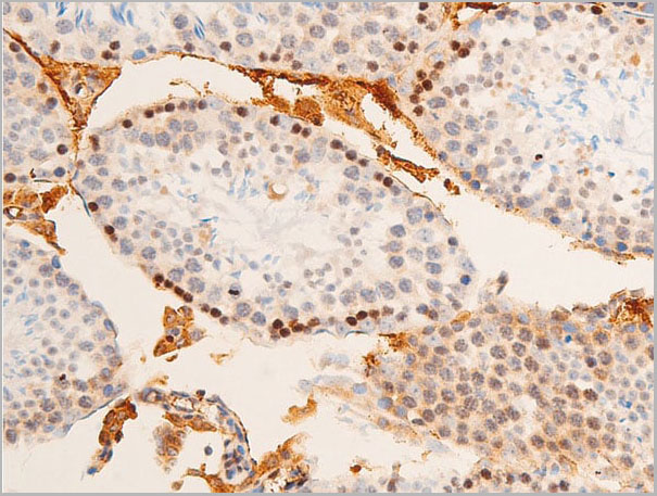
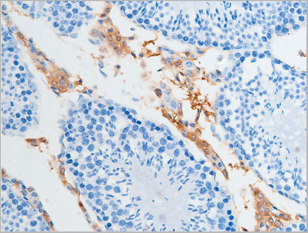
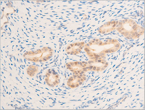
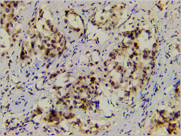
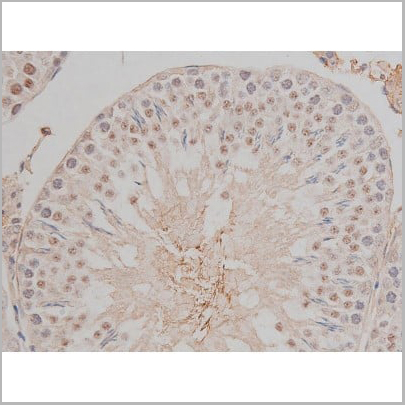
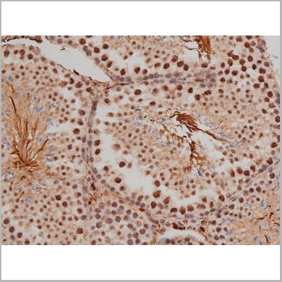
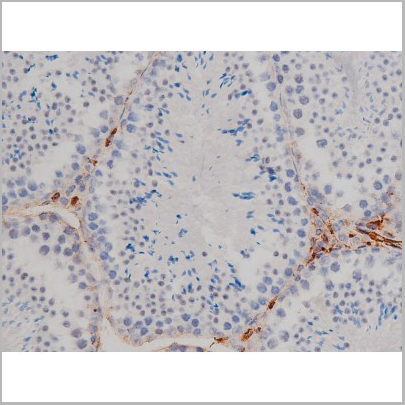
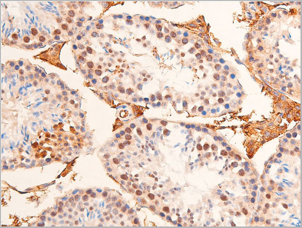
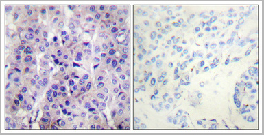
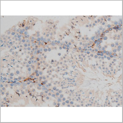
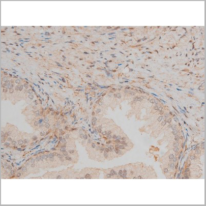
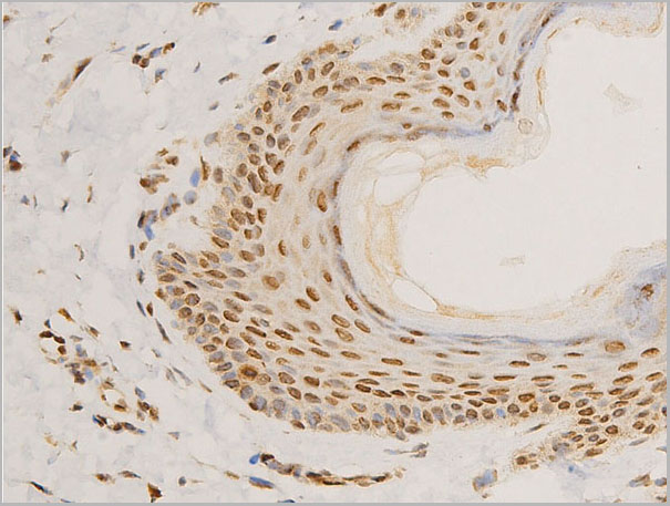
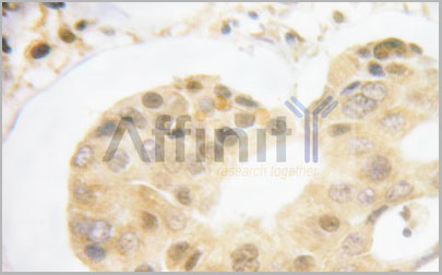
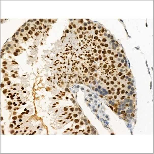
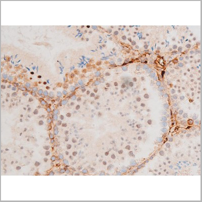
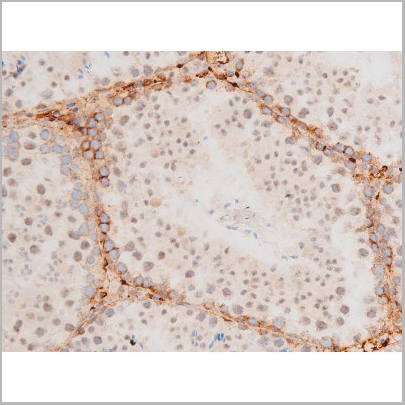
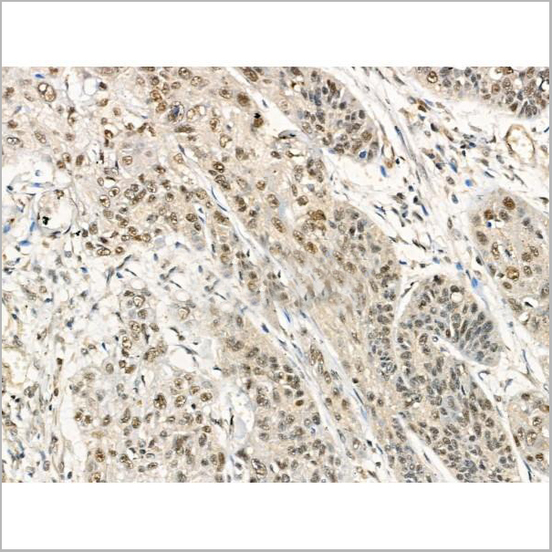
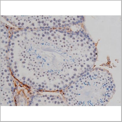
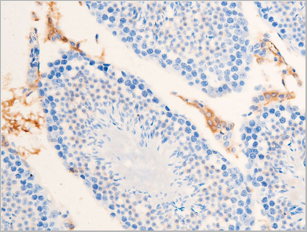
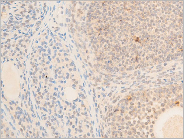
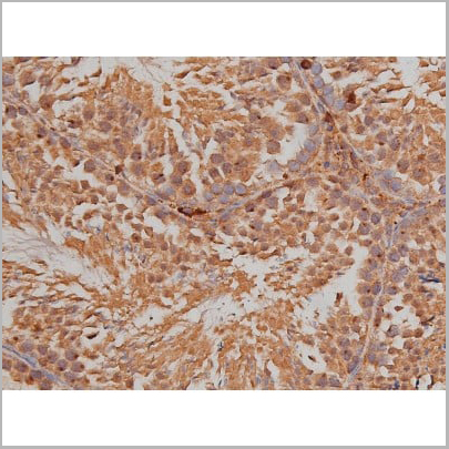
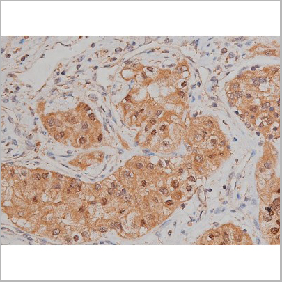
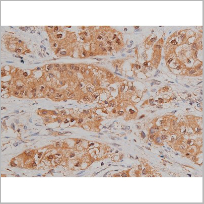
 IHC (Immunohistchemistry)
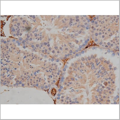
(Phospho-ERK1/2 (Thr202/Tyr204) Antibody for IHC in human testis)
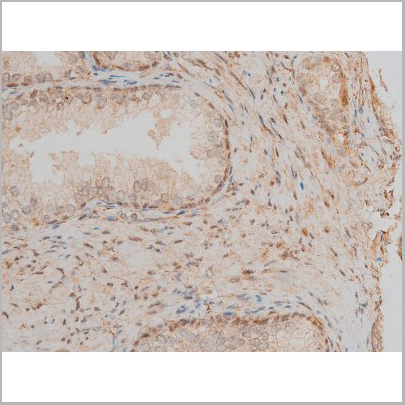
IHC (Immunohistchemistry)
(Phospho-ERK1/2 (Thr202/Tyr204) Antibody for IHC in human testis)
ERK1/2, Polyclonal Antibody (Cat# AAA30930)
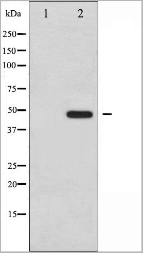
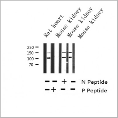
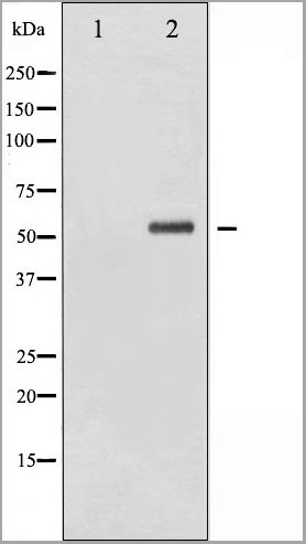
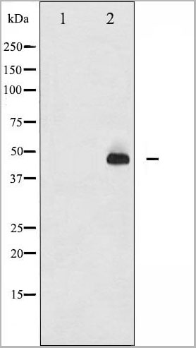
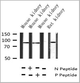
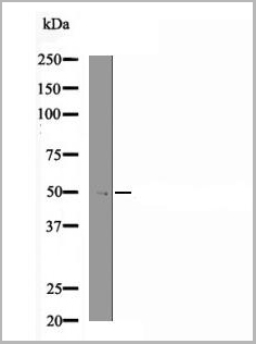
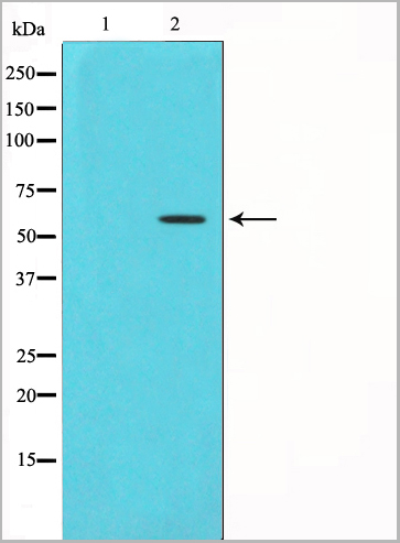
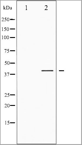
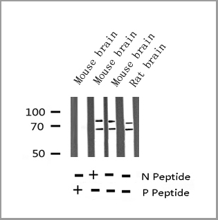
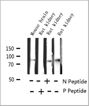
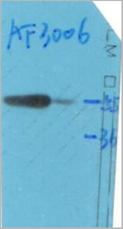
 WB (Western Blot)
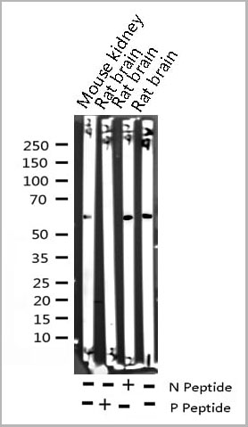
(Western blot analysis of Phospho-Tyrosine Hydroxylase (Ser19) expression in various lysates)
WB (Western Blot)
(Western blot analysis of Phospho-Tyrosine Hydroxylase (Ser19) expression in various lysates)
Tyrosine Hydroxylase, Polyclonal Antibody (Cat# AAA30988)
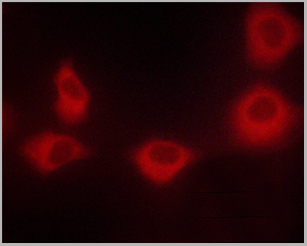
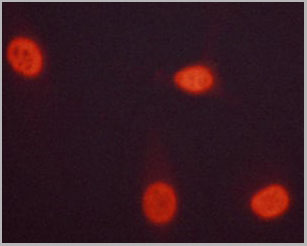
 IF (Immunofluorescence)
(AAA30921 staining Hela by IF/ICC. The sample were fixed with PFA and permeabilized in 0.1% Triton X-100, then blocked in 10% serum for 45 minutes at 25 degree C. The primary antibody was diluted at 1/200 and incubated with the sample for 1 hour at 37 degree C. An Alexa Fluor 594 conjugated goat anti-rabbit IgG (H+L) Ab, diluted at 1/600, was used as the secondary antibody.)
IF (Immunofluorescence)
(AAA30921 staining Hela by IF/ICC. The sample were fixed with PFA and permeabilized in 0.1% Triton X-100, then blocked in 10% serum for 45 minutes at 25 degree C. The primary antibody was diluted at 1/200 and incubated with the sample for 1 hour at 37 degree C. An Alexa Fluor 594 conjugated goat anti-rabbit IgG (H+L) Ab, diluted at 1/600, was used as the secondary antibody.)
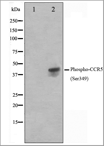
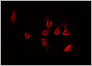
CCR5, Polyclonal Antibody (Cat# AAA30921)
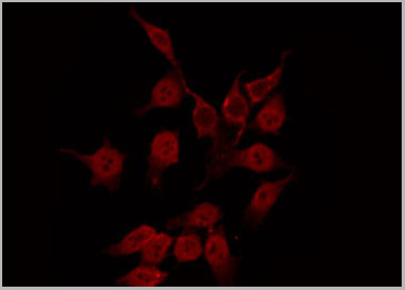
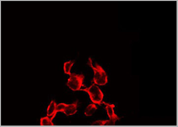
 IF (Immunofluorescence)
(AAA30935 staining HeLa by IF/ICC. The sample were fixed with PFA and permeabilized in 0.1% Triton X-100, then blocked in 10% serum for 45 minutes at 25 degree C. The primary antibody was diluted at 1/200 and incubated with the sample for 1 hour at 37 degree C. An Alexa Fluor 594 conjugated goat anti-rabbit IgG (H+L) Ab, diluted at 1/600, was used as the secondary antibody.)
IF (Immunofluorescence)
(AAA30935 staining HeLa by IF/ICC. The sample were fixed with PFA and permeabilized in 0.1% Triton X-100, then blocked in 10% serum for 45 minutes at 25 degree C. The primary antibody was diluted at 1/200 and incubated with the sample for 1 hour at 37 degree C. An Alexa Fluor 594 conjugated goat anti-rabbit IgG (H+L) Ab, diluted at 1/600, was used as the secondary antibody.)
GR, Polyclonal Antibody (Cat# AAA30935)

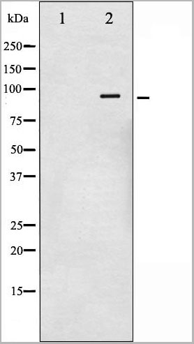
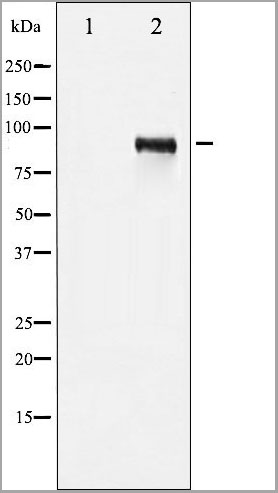
 WB (Western Blot)
(Western blot analysis of STAT3 phosphorylation expression in HeLa whole cell lysates, The lane on the left is treated with the antigen-specific peptide.)
WB (Western Blot)
(Western blot analysis of STAT3 phosphorylation expression in HeLa whole cell lysates, The lane on the left is treated with the antigen-specific peptide.)
STAT3, Polyclonal Antibody (Cat# AAA31042)
 WB (Western Blot)
(Western blot analysis of c-Jun phosphorylation expression in UV treated HeLa whole cell lysates, The lane on the left is treated with the antigen-specific peptide.)
WB (Western Blot)
(Western blot analysis of c-Jun phosphorylation expression in UV treated HeLa whole cell lysates, The lane on the left is treated with the antigen-specific peptide.)
c-Jun, Polyclonal Antibody (Cat# AAA30981)
 WB (Western Blot)
(Western blot analysis of c-Jun phosphorylation expression in UV treated HeLa whole cell lysates, The lane on the left is treated with the antigen-specific peptide.)
WB (Western Blot)
(Western blot analysis of c-Jun phosphorylation expression in UV treated HeLa whole cell lysates, The lane on the left is treated with the antigen-specific peptide.)
c-Jun, Polyclonal Antibody (Cat# AAA30984)
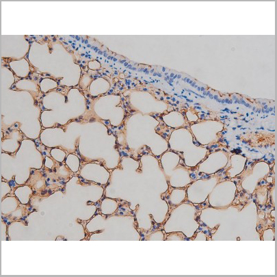
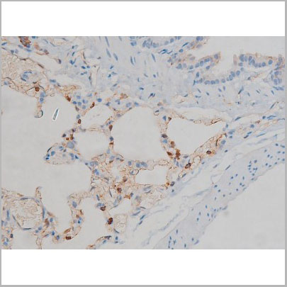
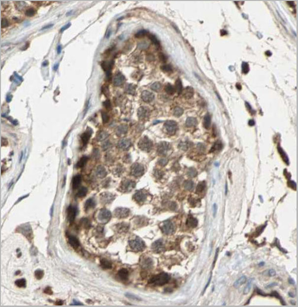
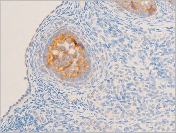
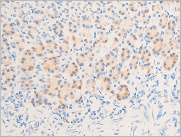
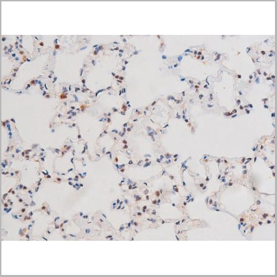
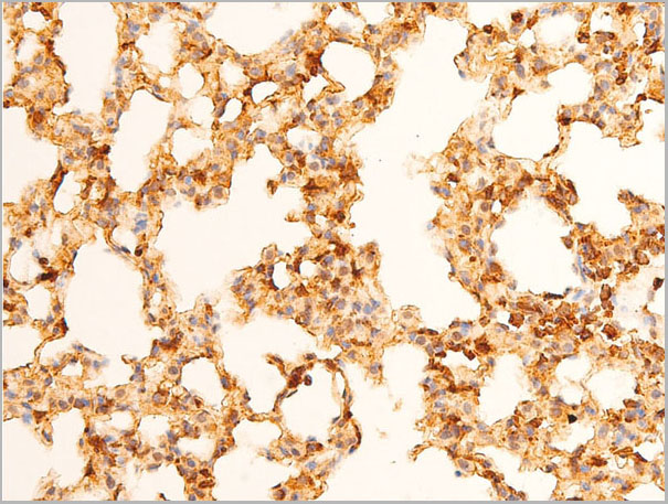
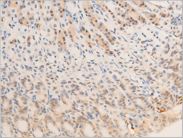
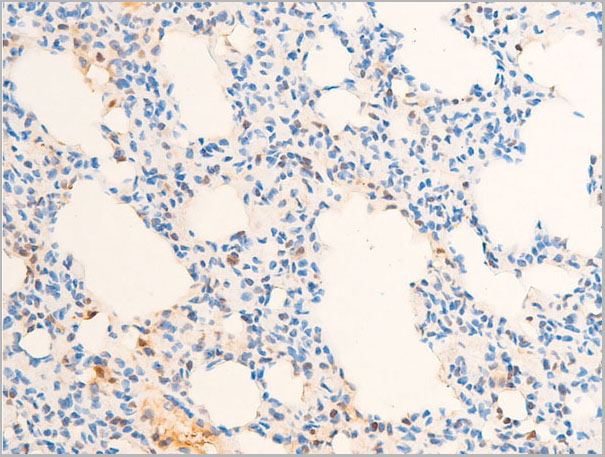
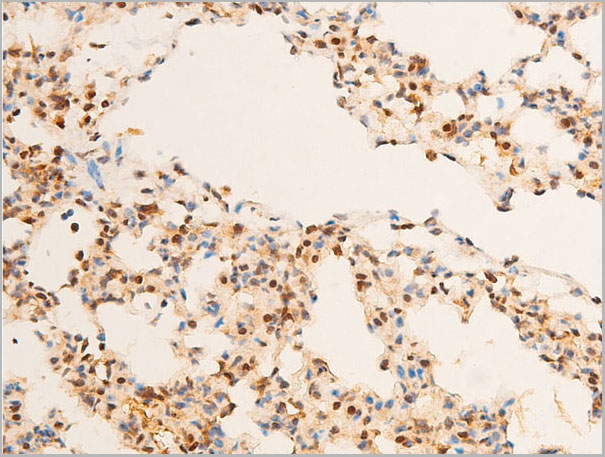
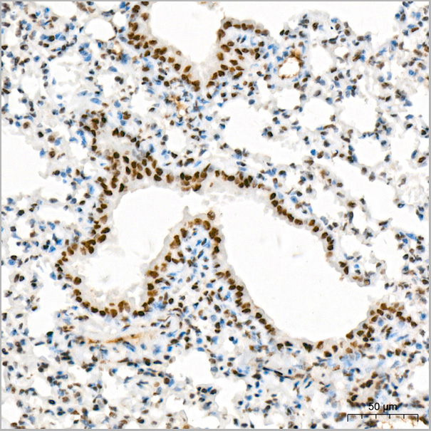
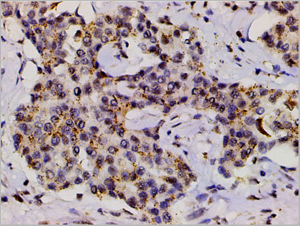
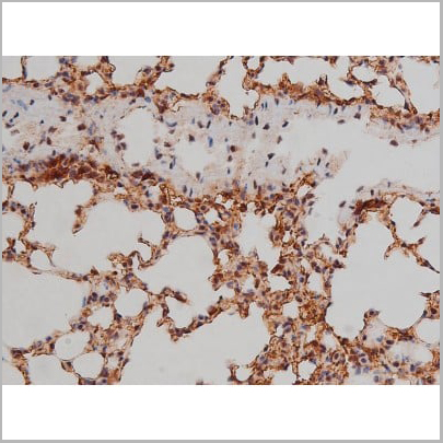
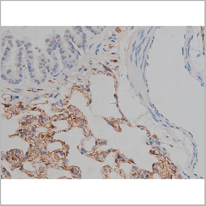
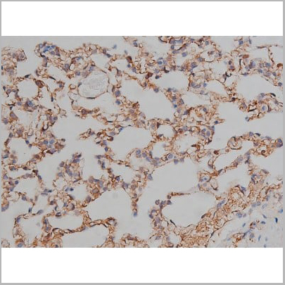
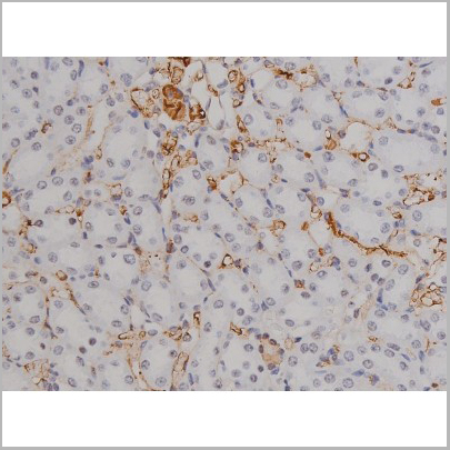
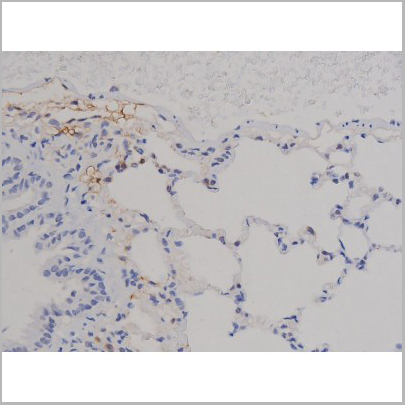
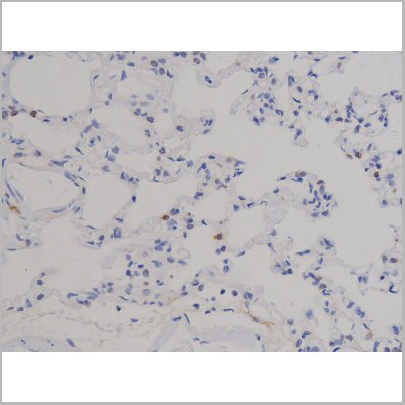
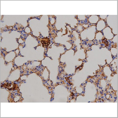
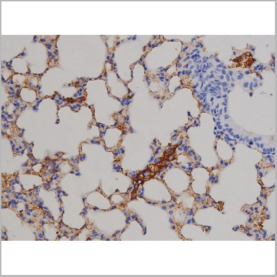
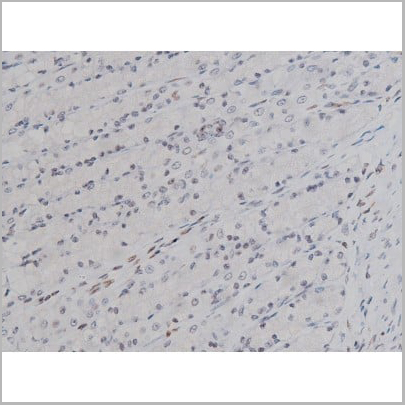
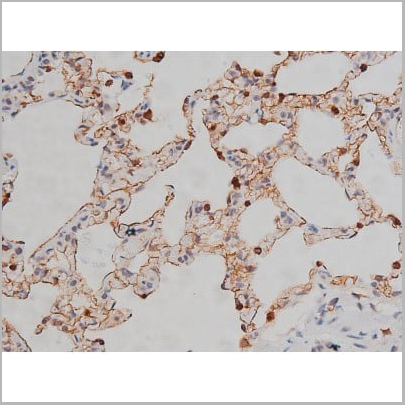
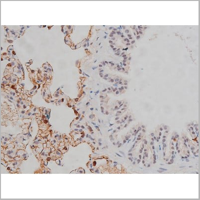
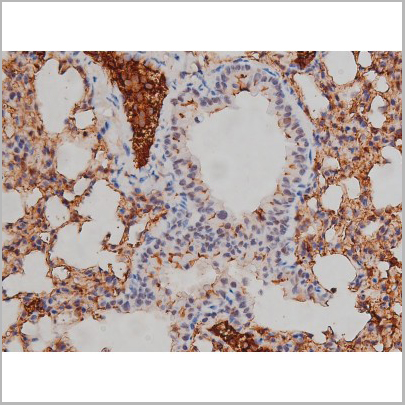
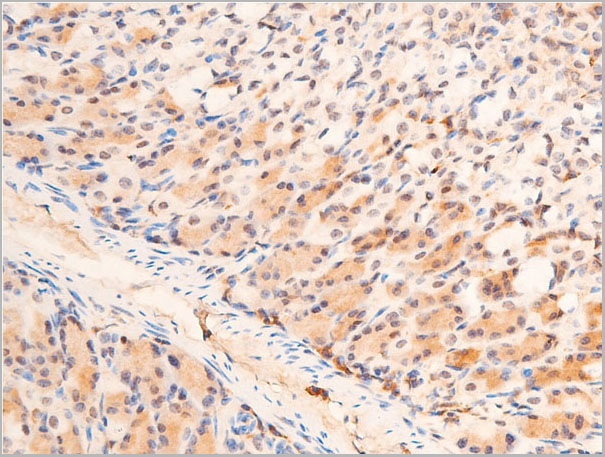
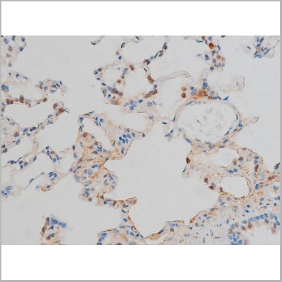
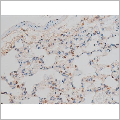
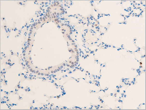
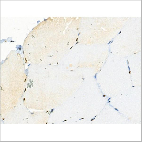
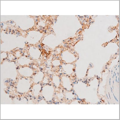
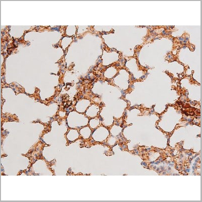
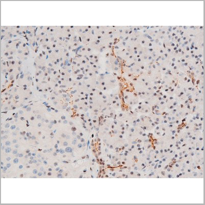
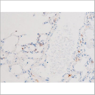
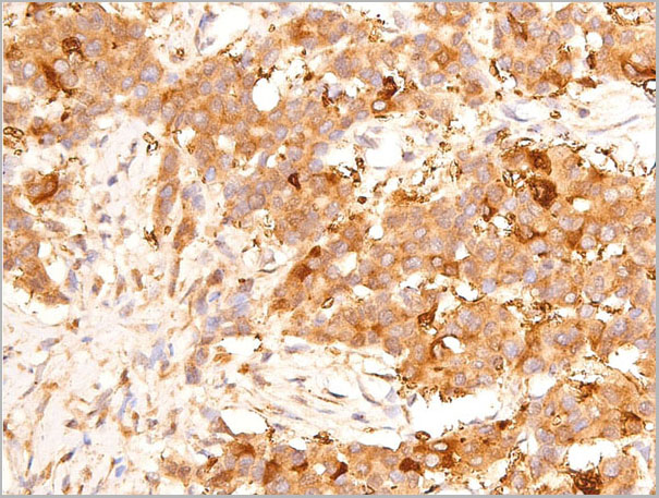
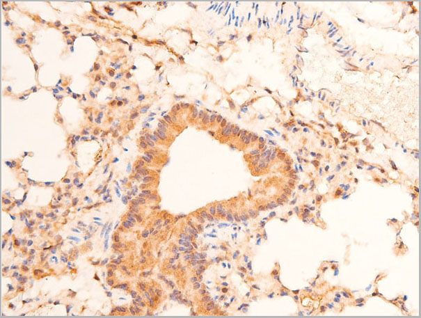
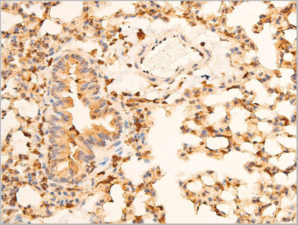
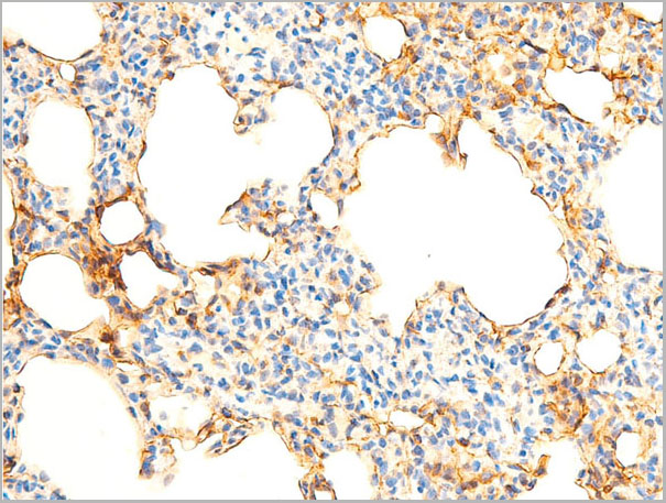
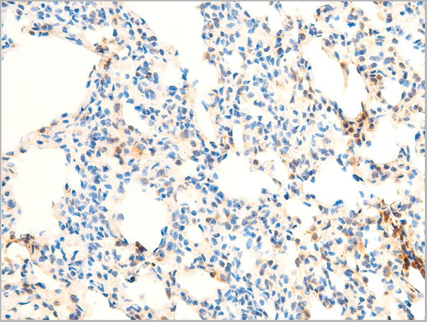
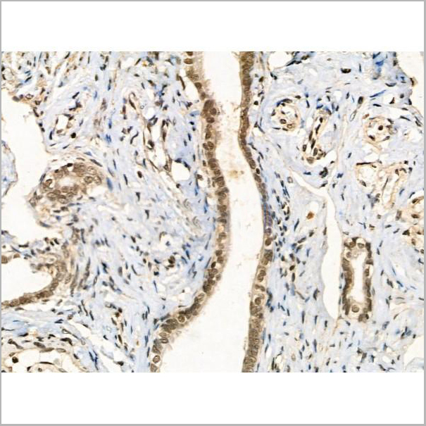
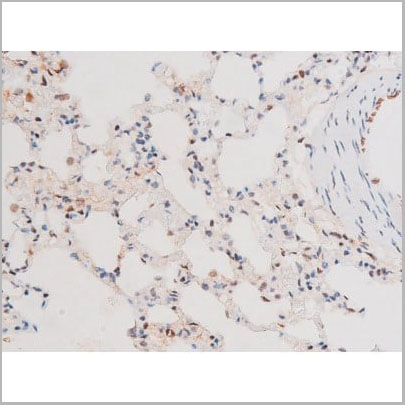
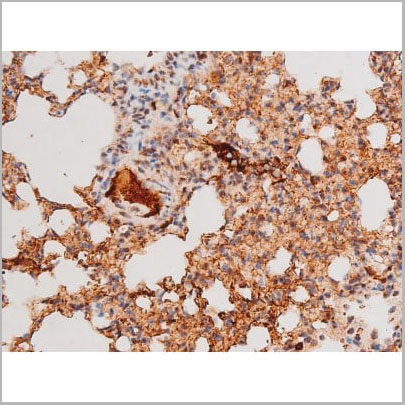
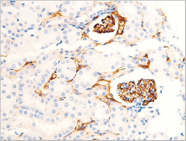
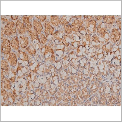
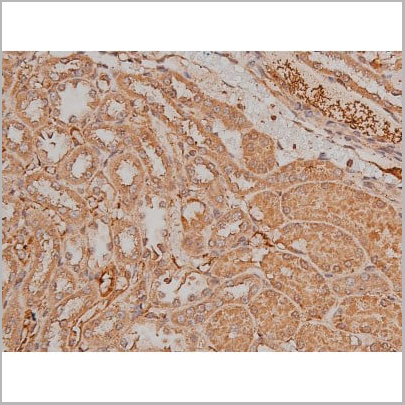
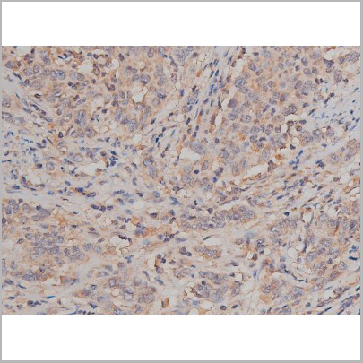
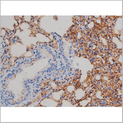 IHC (Immunohistchemistry)
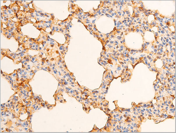
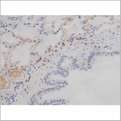
(AAA30994 at 1/200 staining Mouse lung tissue sections by IHC-P. The tissue was formaldehyde fixed and a heat mediated antigen retrieval step in citrate buffer was performed. The tissue was then blocked and incubated with the antibody for 1.5 hours at 22 degree C. An HRP conjugated goat anti-rabbit antibody was used as the secondary.)
IHC (Immunohistchemistry)
(AAA30994 at 1/200 staining Mouse lung tissue sections by IHC-P. The tissue was formaldehyde fixed and a heat mediated antigen retrieval step in citrate buffer was performed. The tissue was then blocked and incubated with the antibody for 1.5 hours at 22 degree C. An HRP conjugated goat anti-rabbit antibody was used as the secondary.)
IGF1R, Polyclonal Antibody (Cat# AAA30994)
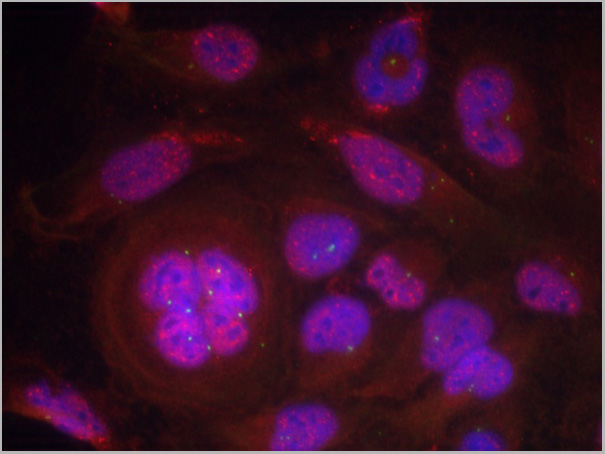
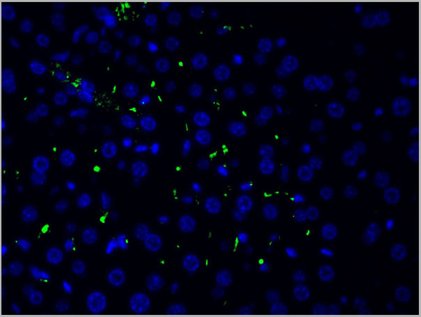
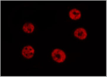
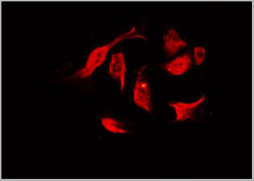
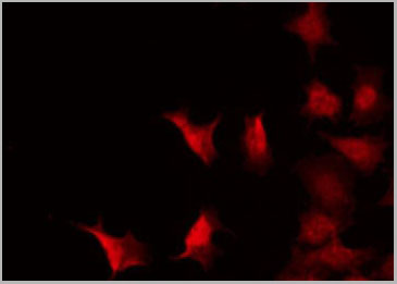
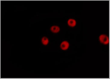
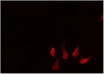
 IF (Immunofluorescence)
(Immunofluorescent analysis of Ryanodine Receptor 2 (phospho-Ser2808) staining in U251 cells. Formalin-fixed cells were permeabilized with 0.1% Triton X-100 in TBS for 5-10 minutes and blocked with 3% BSA-PBS for 30 minutes at room temperature. Cells were probed with the primary antibody in 3% BSA-PBS and incubated overnight at 4 C in a hidified chamber. Cells were washed with PBST and incubated with a DyLight 594-conjugated secondary antibody (red) in PBS at room temperature in the dark. DAPI was used to stain the cell nuclei (blue).)
IF (Immunofluorescence)
(Immunofluorescent analysis of Ryanodine Receptor 2 (phospho-Ser2808) staining in U251 cells. Formalin-fixed cells were permeabilized with 0.1% Triton X-100 in TBS for 5-10 minutes and blocked with 3% BSA-PBS for 30 minutes at room temperature. Cells were probed with the primary antibody in 3% BSA-PBS and incubated overnight at 4 C in a hidified chamber. Cells were washed with PBST and incubated with a DyLight 594-conjugated secondary antibody (red) in PBS at room temperature in the dark. DAPI was used to stain the cell nuclei (blue).)
Ryanodine Receptor 2 (phospho-Ser2808), Polyclonal Antibody (Cat# AAA29761)
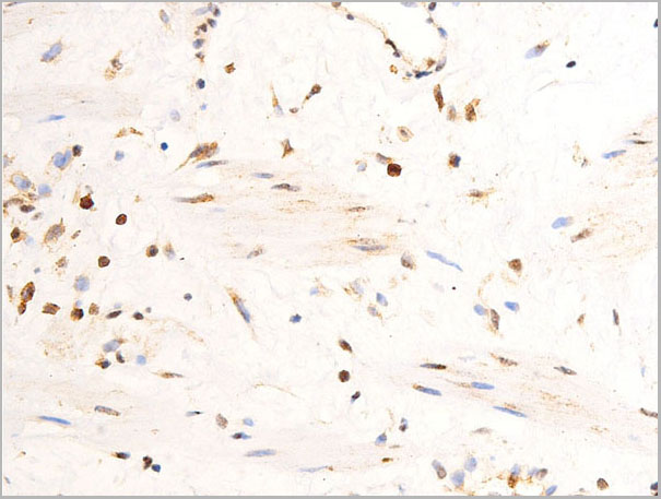
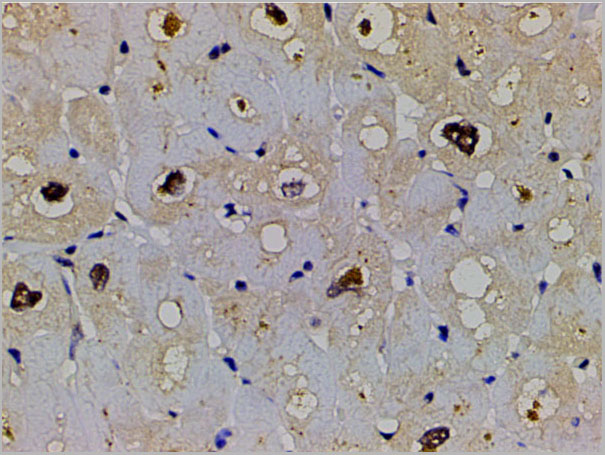
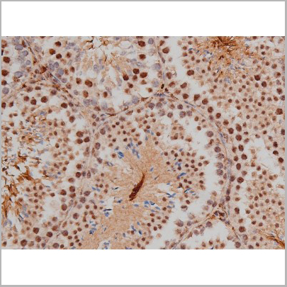
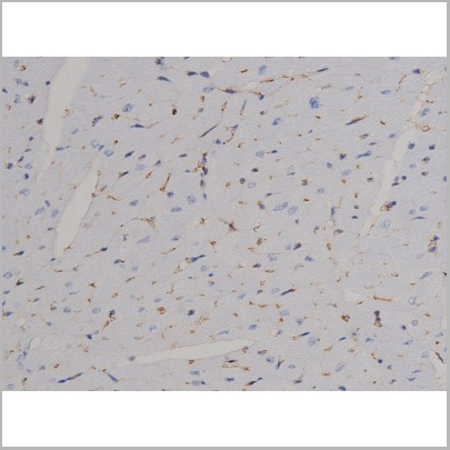
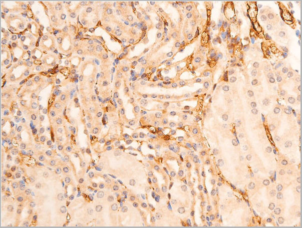
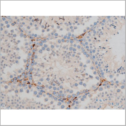
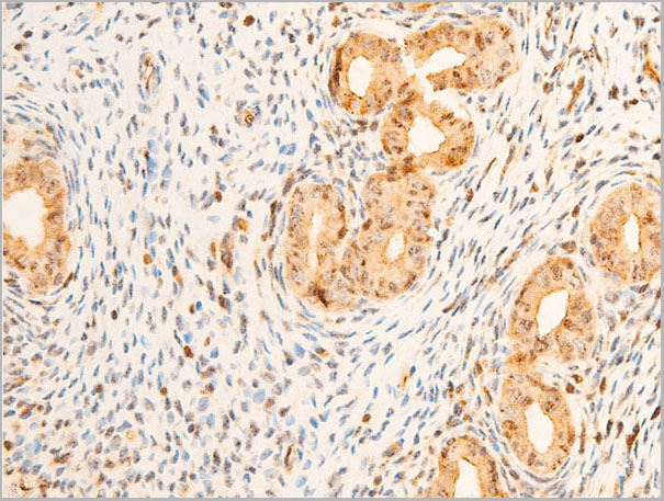
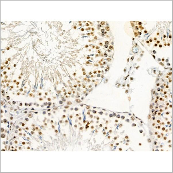
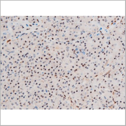
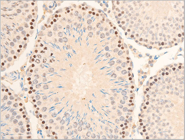
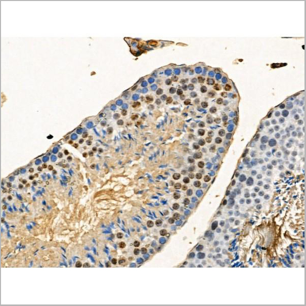
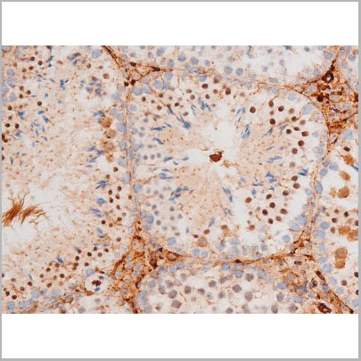
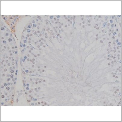
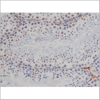
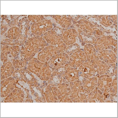
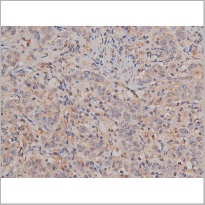
 IHC (Immunohistochemistry)
(AAA30959 at 1/100 staining rat testicular tissue sections by IHC-P. The tissue was formaldehyde fixed and a heat mediated antigen retrieval step in citrate buffer was performed. The tissue was then blocked and incubated with the antibody for 1.5 hours at 22 degree C. An HRP conjugated goat anti-rabbit antibody was used as the secondary.)
IHC (Immunohistochemistry)
(AAA30959 at 1/100 staining rat testicular tissue sections by IHC-P. The tissue was formaldehyde fixed and a heat mediated antigen retrieval step in citrate buffer was performed. The tissue was then blocked and incubated with the antibody for 1.5 hours at 22 degree C. An HRP conjugated goat anti-rabbit antibody was used as the secondary.)
Chk2, Polyclonal Antibody (Cat# AAA30959)
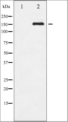
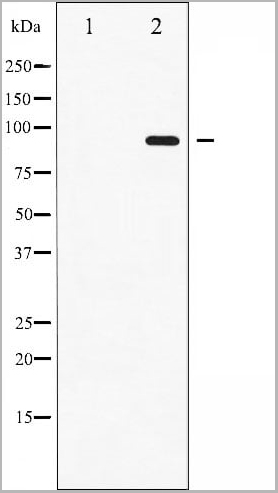
 WB (Western Blot)
(Western blot analysis of Phospho-EGFR (Ser1071) expression in various lysates)
WB (Western Blot)
(Western blot analysis of Phospho-EGFR (Ser1071) expression in various lysates)
EGFR, Polyclonal Antibody (Cat# AAA30964)
 WB (Western Blot)
(Western blot analysis of c-Jun phosphorylation expression in UV treated HeLa whole cell lysates, The lane on the left is treated with the antigen-specific peptide.)
WB (Western Blot)
(Western blot analysis of c-Jun phosphorylation expression in UV treated HeLa whole cell lysates, The lane on the left is treated with the antigen-specific peptide.)
c-Jun, Polyclonal Antibody (Cat# AAA30983)
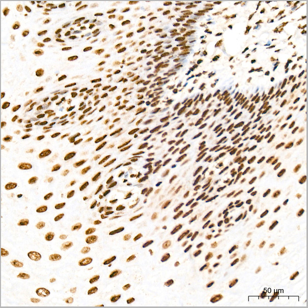
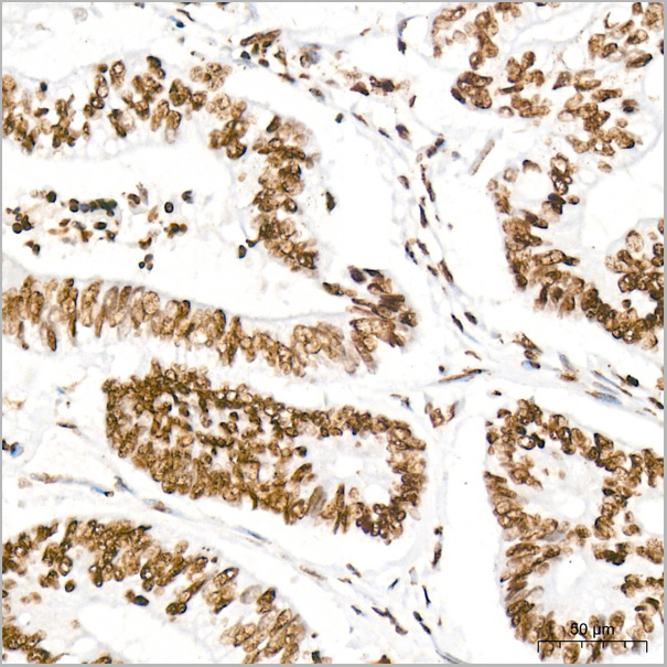
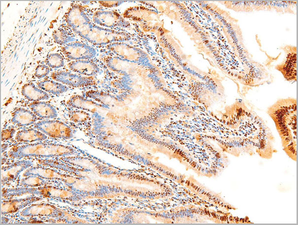
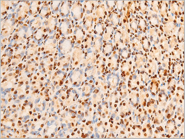
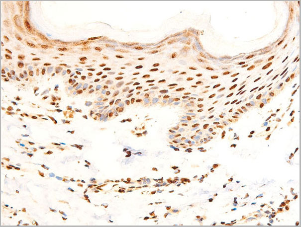
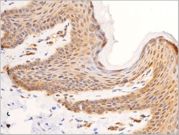
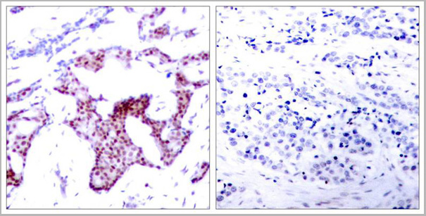
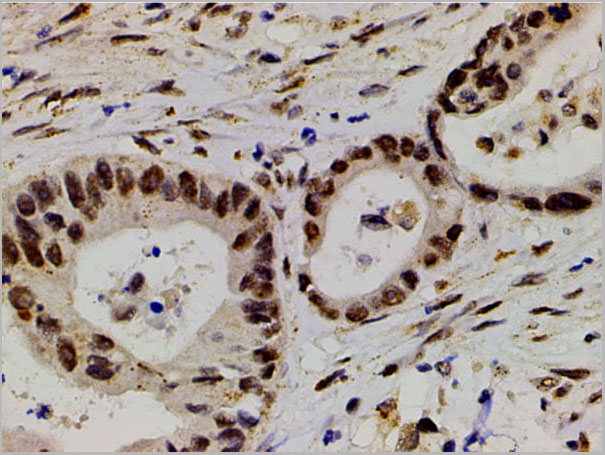
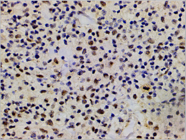
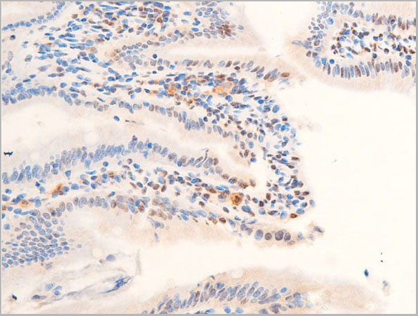
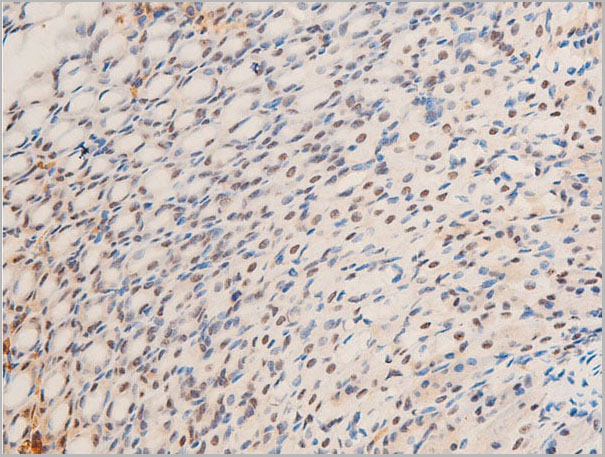
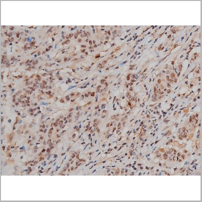
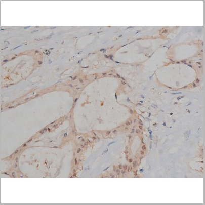
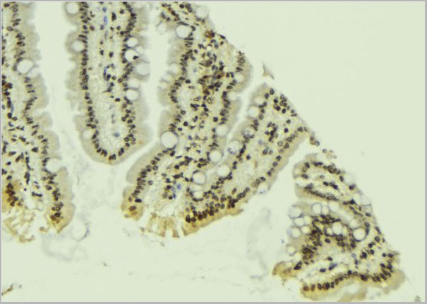
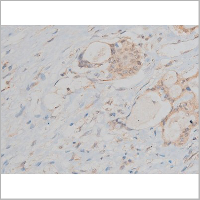
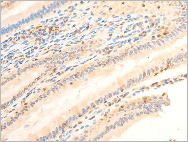
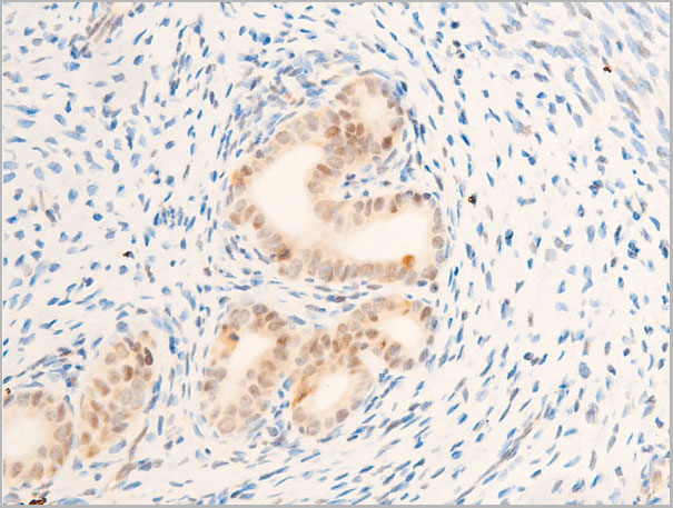
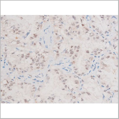
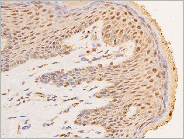
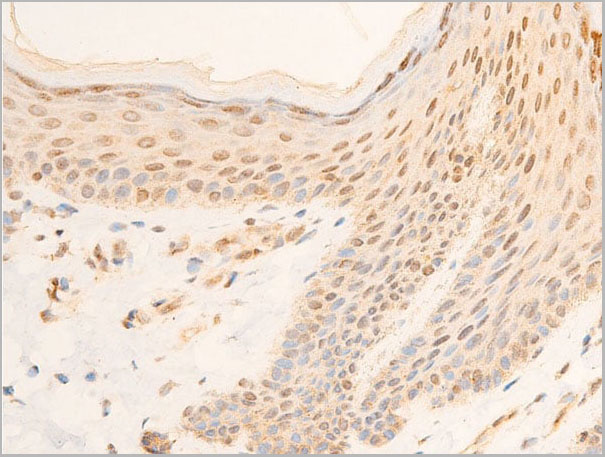
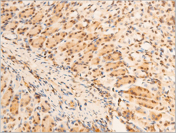
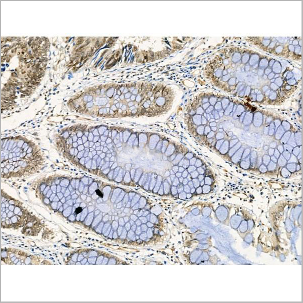
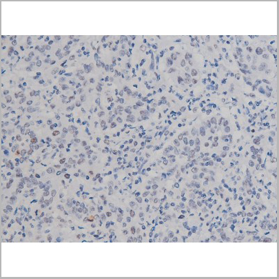
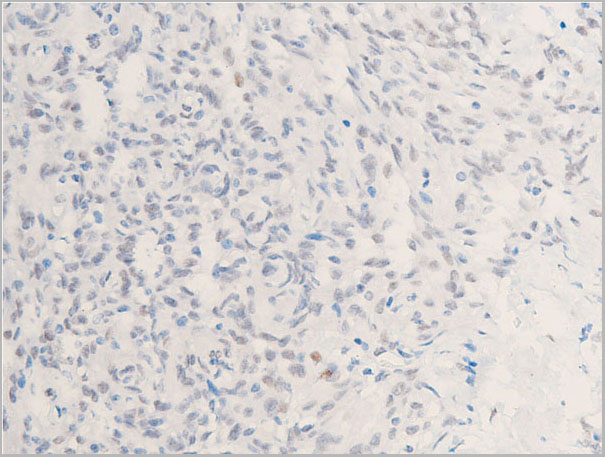
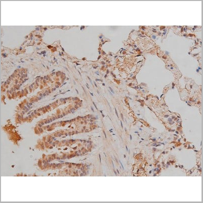
 IHC (Immunohistochemistry)
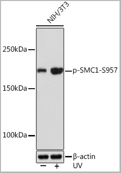
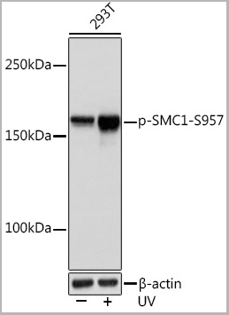
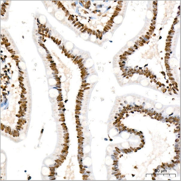
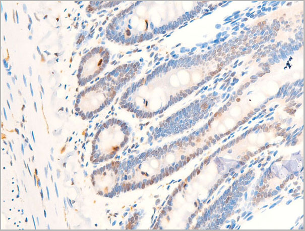
(Immunohistochemistry analysis of paraffin-embedded Human small intestine tissue using Phospho-SMC1-S957 Rabbit mAb (AAA28615) at a dilution of 1:200 (40x lens). High pressure antigen retrieval was performed with 0.01 M citrate buffer (pH 6.0) prior to IHC staining.)
IHC (Immunohistochemistry)
(Immunohistochemistry analysis of paraffin-embedded Human small intestine tissue using Phospho-SMC1-S957 Rabbit mAb (AAA28615) at a dilution of 1:200 (40x lens). High pressure antigen retrieval was performed with 0.01 M citrate buffer (pH 6.0) prior to IHC staining.)
SMC1-S957, Monoclonal Antibody (Cat# AAA28615)
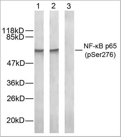
 IF (Immunofluorescence)
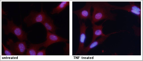
(Immunofluorescence staining of methanol-fixed MEF cells untreated or treated with TNF using NFkappaB-p65 (Phospho-Ser276) Antibody.)
IF (Immunofluorescence)
(Immunofluorescence staining of methanol-fixed MEF cells untreated or treated with TNF using NFkappaB-p65 (Phospho-Ser276) Antibody.)
NFkappaB-p65, Polyclonal Antibody (Cat# AAA29703)
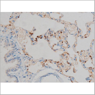
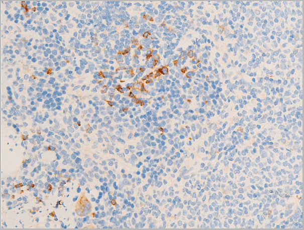
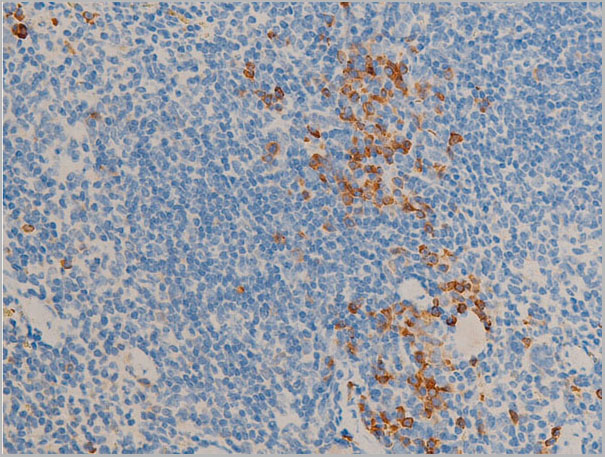
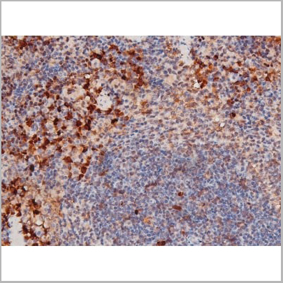
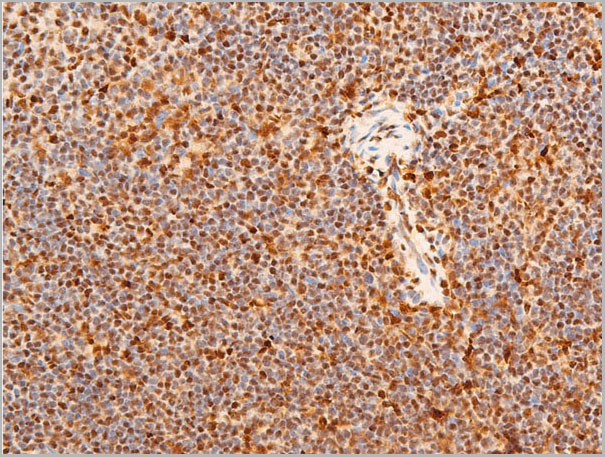
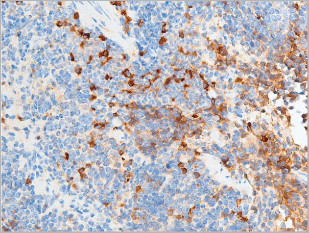
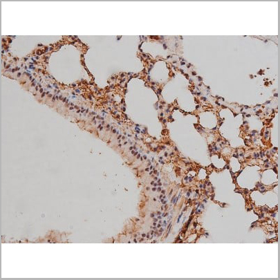
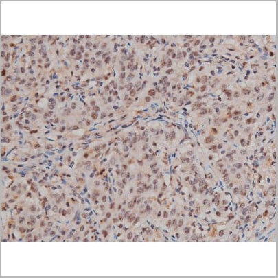
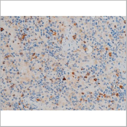
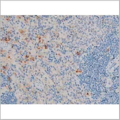
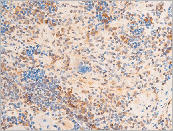
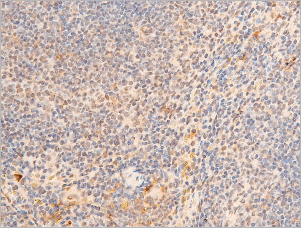
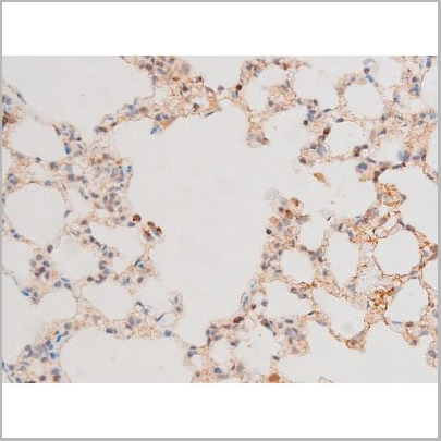
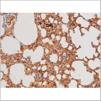
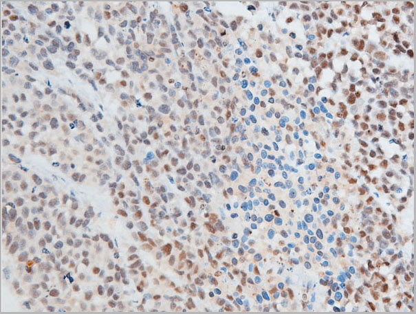
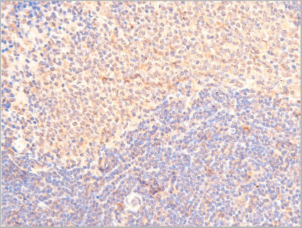
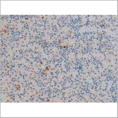
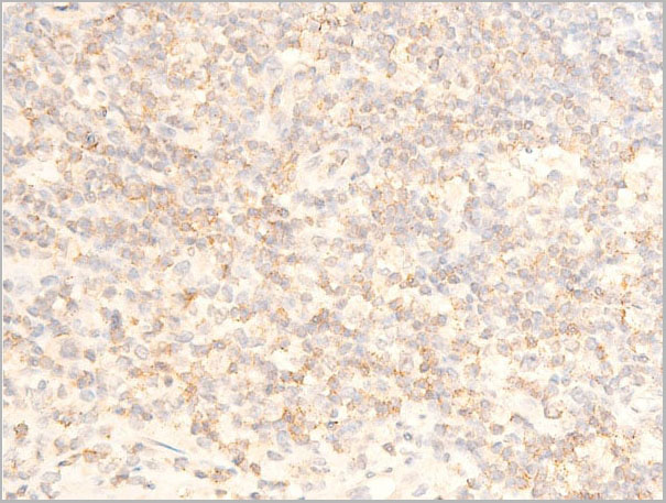
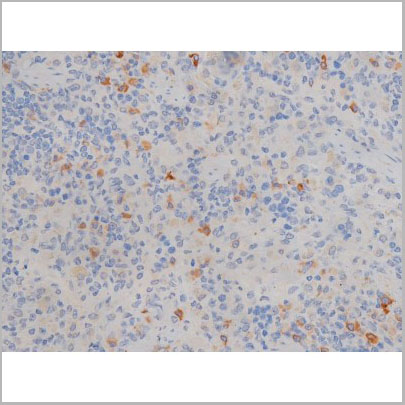
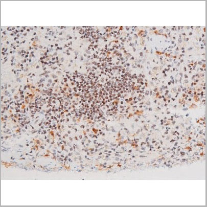
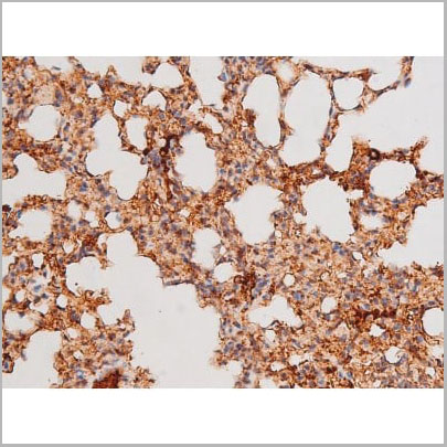
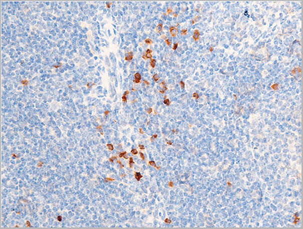
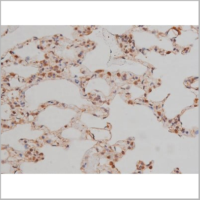
 IHC (Immunohistochemistry)
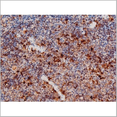
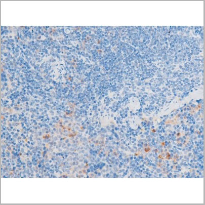
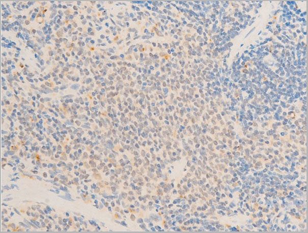
(AAA31055 at 1/200 staining Mouse spleen tissue sections by IHC-P. The tissue was formaldehyde fixed and a heat mediated antigen retrieval step in citrate buffer was performed. The tissue was then blocked and incubated with the antibody for 1.5 hours at 22 degree C. An HRP conjugated goat anti-rabbit antibody was used as the secondary.)
IHC (Immunohistochemistry)
(AAA31055 at 1/200 staining Mouse spleen tissue sections by IHC-P. The tissue was formaldehyde fixed and a heat mediated antigen retrieval step in citrate buffer was performed. The tissue was then blocked and incubated with the antibody for 1.5 hours at 22 degree C. An HRP conjugated goat anti-rabbit antibody was used as the secondary.)
LIMK1, Polyclonal Antibody (Cat# AAA31055)
 WB (Western Blot)
(Western blot analysis of c-Jun phosphorylation expression in UV treated HeLa whole cell lysates, The lane on the left is treated with the antigen-specific peptide.)
WB (Western Blot)
(Western blot analysis of c-Jun phosphorylation expression in UV treated HeLa whole cell lysates, The lane on the left is treated with the antigen-specific peptide.)
c-Jun, Polyclonal Antibody (Cat# AAA30982)
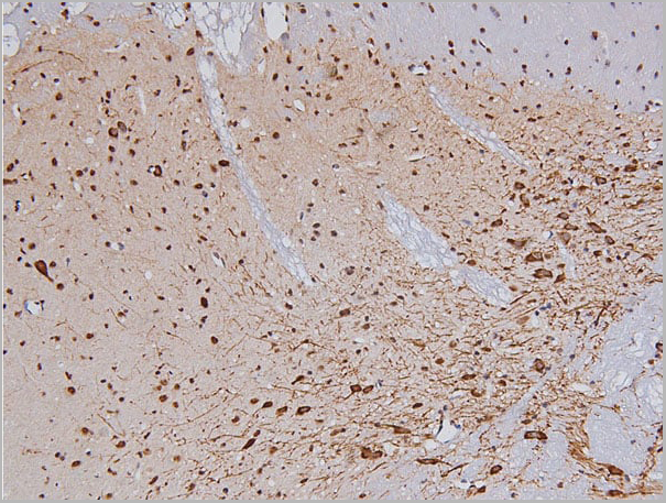
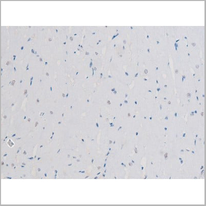
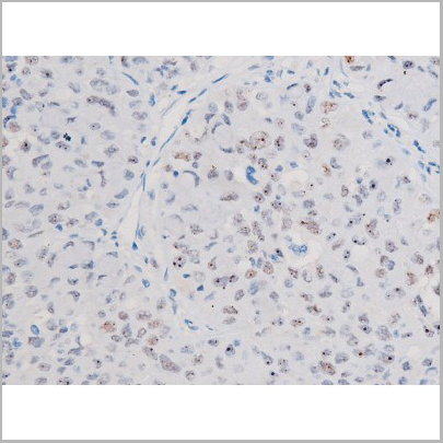
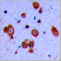
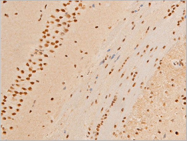
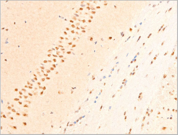
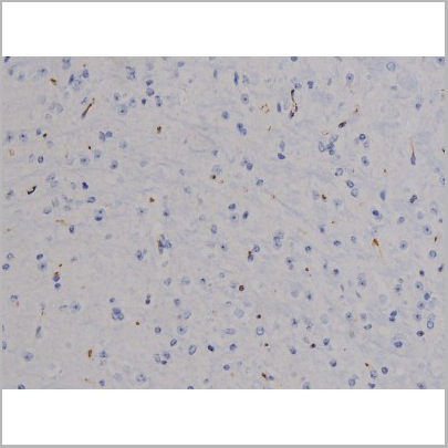
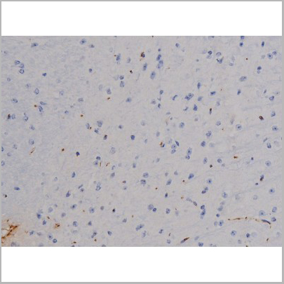
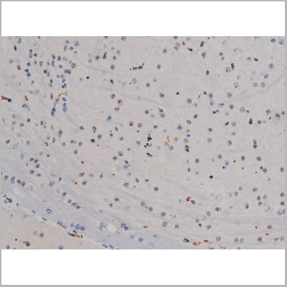
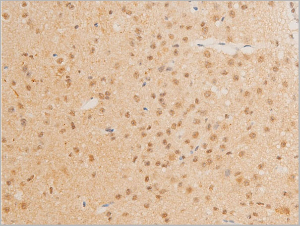
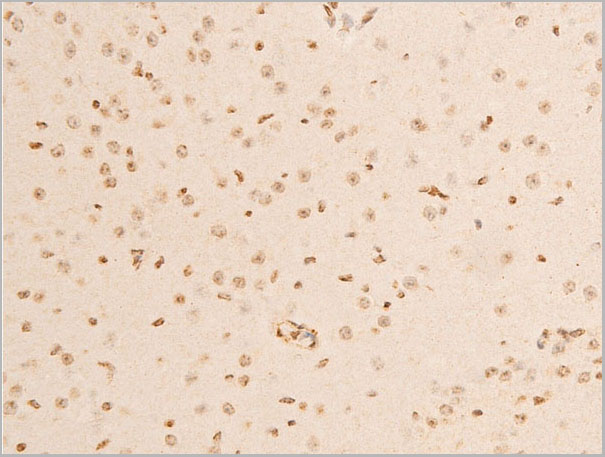
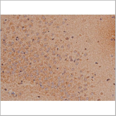
 IHC (Immunohistochemistry)
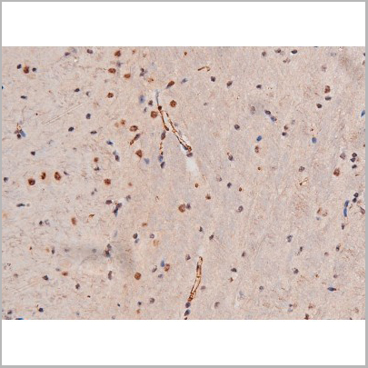
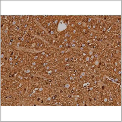
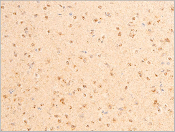
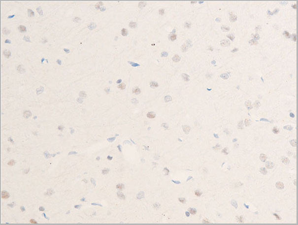
(AAA30991 at 1/200 staining Rat brain tissue sections by IHC-P. The tissue was formaldehyde fixed and a heat mediated antigen retrieval step in citrate buffer was performed. The tissue was then blocked and incubated with the Ab for 1.5 hours at 22°C. An HRP conjugated goat anti-rabbit Ab was used as the secondary Ab.)
IHC (Immunohistochemistry)
(AAA30991 at 1/200 staining Rat brain tissue sections by IHC-P. The tissue was formaldehyde fixed and a heat mediated antigen retrieval step in citrate buffer was performed. The tissue was then blocked and incubated with the Ab for 1.5 hours at 22°C. An HRP conjugated goat anti-rabbit Ab was used as the secondary Ab.)
SP1, Polyclonal Antibody (Cat# AAA30991)
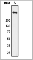
 WB (Western Blot)
(Western blot analysis of Phospho-Tau (Ser404) expression in various lysates)
WB (Western Blot)
(Western blot analysis of Phospho-Tau (Ser404) expression in various lysates)
Tau, Polyclonal Antibody (Cat# AAA30999)
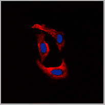
 IF (Immunofluorescence)
(AAA31046 staining HeLa by IF/ICC. The sample were fixed with PFA and permeabilized in 0.1% Triton X-100, then blocked in 10% serum for 45 minutes at 25 degree C. The primary antibody was diluted at 1/200 and incubated with the sample for 1 hour at 37 degree C. An Alexa Fluor 594 conjugated goat anti-rabbit IgG (H+L) Ab, diluted at 1/600, was used as the secondary antibody.)
IF (Immunofluorescence)
(AAA31046 staining HeLa by IF/ICC. The sample were fixed with PFA and permeabilized in 0.1% Triton X-100, then blocked in 10% serum for 45 minutes at 25 degree C. The primary antibody was diluted at 1/200 and incubated with the sample for 1 hour at 37 degree C. An Alexa Fluor 594 conjugated goat anti-rabbit IgG (H+L) Ab, diluted at 1/600, was used as the secondary antibody.)
STAT6, Polyclonal Antibody (Cat# AAA31046)
 IF (Immunofluorescence)
(AAA30955 staining A549 by IF/ICC. The sample were fixed with PFA and permeabilized in 0.1% Triton X-100, then blocked in 10% serum for 45 minutes at 25 degree C. The primary antibody was diluted at 1/200 and incubated with the sample for 1 hour at 37 degree C. An Alexa Fluor 594 conjugated goat anti-rabbit IgG (H+L) Ab, diluted at 1/600, was used as the secondary antibody.)
IF (Immunofluorescence)
(AAA30955 staining A549 by IF/ICC. The sample were fixed with PFA and permeabilized in 0.1% Triton X-100, then blocked in 10% serum for 45 minutes at 25 degree C. The primary antibody was diluted at 1/200 and incubated with the sample for 1 hour at 37 degree C. An Alexa Fluor 594 conjugated goat anti-rabbit IgG (H+L) Ab, diluted at 1/600, was used as the secondary antibody.)
JAK2, Polyclonal Antibody (Cat# AAA30955)

 IF (Immunofluorescence)
(AAA30990 staining K562 by IF/ICC. The sample were fixed with PFA and permeabilized in 0.1% Triton X-100, then blocked in 10% serum for 45 minutes at 25 degree C. The primary antibody was diluted at 1/200 and incubated with the sample for 1 hour at 37 degree C. An Alexa Fluor 594 conjugated goat anti-rabbit IgG (H+L) Ab, diluted at 1/600, was used as the secondary antibody.)
IF (Immunofluorescence)
(AAA30990 staining K562 by IF/ICC. The sample were fixed with PFA and permeabilized in 0.1% Triton X-100, then blocked in 10% serum for 45 minutes at 25 degree C. The primary antibody was diluted at 1/200 and incubated with the sample for 1 hour at 37 degree C. An Alexa Fluor 594 conjugated goat anti-rabbit IgG (H+L) Ab, diluted at 1/600, was used as the secondary antibody.)
Lyn, Polyclonal Antibody (Cat# AAA30990)
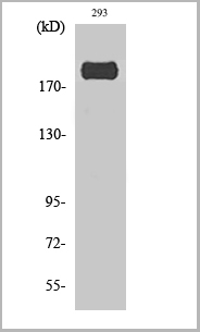
 WB (Western Blot)
(Western blot analysis Rpt6 (Phospho-Ser120) using 293 whole cell lysates)
WB (Western Blot)
(Western blot analysis Rpt6 (Phospho-Ser120) using 293 whole cell lysates)
Rpt6, Polyclonal Antibody (Cat# AAA29757)
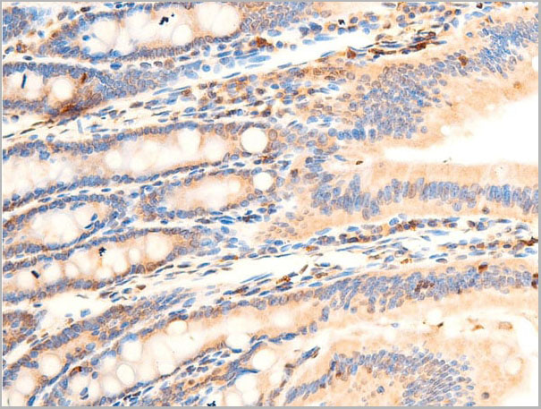
 IHC (Immunohistochemistry)
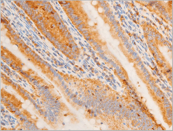
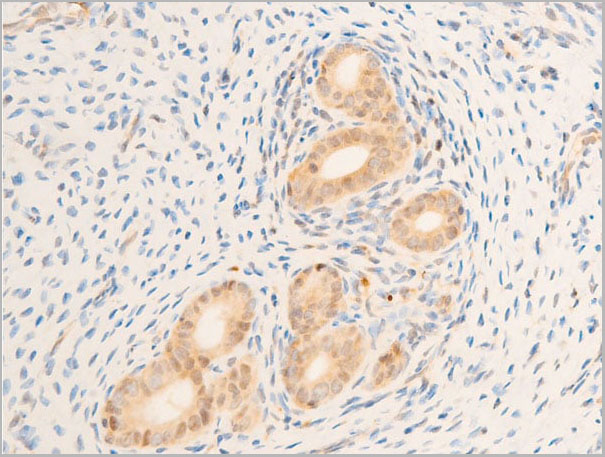
(AAA30939 at 1/100 staining rat appendiceal tissue sections by IHC-P. The tissue was formaldehyde fixed and a heat mediated antigen retrieval step in citrate buffer was performed. The tissue was then blocked and incubated with the antibody for 1.5 hours at 22 degree C. An HRP conjugated goat anti-rabbit antibody was used as the secondary.)
IHC (Immunohistochemistry)
(AAA30939 at 1/100 staining rat appendiceal tissue sections by IHC-P. The tissue was formaldehyde fixed and a heat mediated antigen retrieval step in citrate buffer was performed. The tissue was then blocked and incubated with the antibody for 1.5 hours at 22 degree C. An HRP conjugated goat anti-rabbit antibody was used as the secondary.)
Chk1, Polyclonal Antibody (Cat# AAA30939)


 IHC (Immunohistchemistry)
(Dilution: Western Blot: 1/500 - 1/2000. Immunohistochemistry: 1/100 - 1/300. ELISA: 1/5000. Not yet tested in other applications.)
IHC (Immunohistchemistry)
(Dilution: Western Blot: 1/500 - 1/2000. Immunohistochemistry: 1/100 - 1/300. ELISA: 1/5000. Not yet tested in other applications.)
IRS-1, Polyclonal Antibody (Cat# AAA28929)
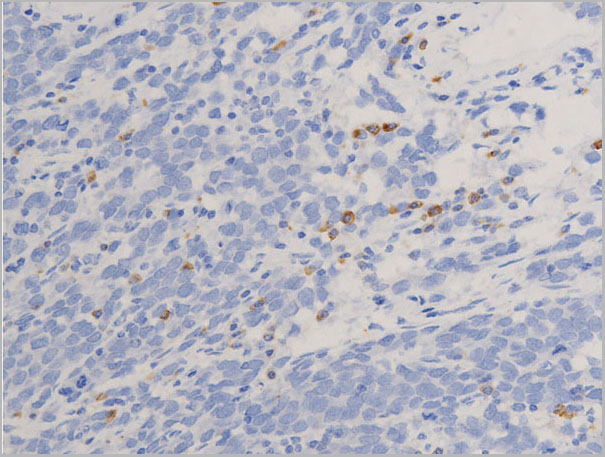
 IHC (Immunohistochemistry)
(AAA31015 at 1/200 staining Rat spleen tissue sections by IHC-P. The tissue was formaldehyde fixed and a heat mediated antigen retrieval step in citrate buffer was performed. The tissue was then blocked and incubated with the antibody for 1.5 hours at 22 degree C. An HRP conjugated goat anti-rabbit antibody was used as the secondary.)
IHC (Immunohistochemistry)
(AAA31015 at 1/200 staining Rat spleen tissue sections by IHC-P. The tissue was formaldehyde fixed and a heat mediated antigen retrieval step in citrate buffer was performed. The tissue was then blocked and incubated with the antibody for 1.5 hours at 22 degree C. An HRP conjugated goat anti-rabbit antibody was used as the secondary.)
CREB, Polyclonal Antibody (Cat# AAA31015)


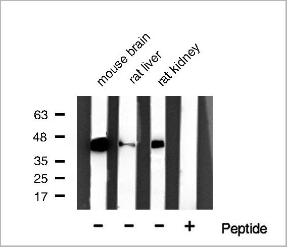
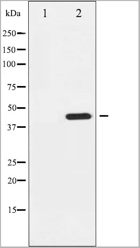
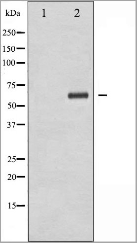
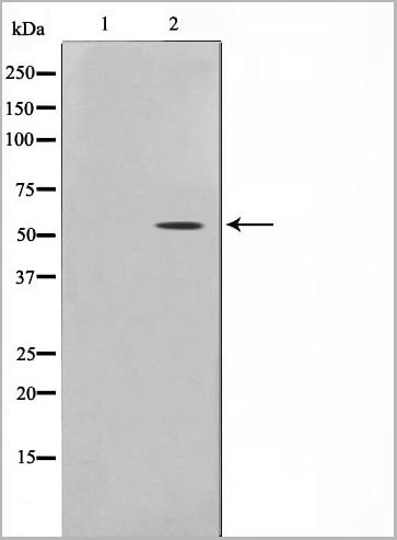
 WB (Western Blot)
(Western blot analysis of extracts from mouse brain, using Phospho-PDK1 (Ser241) Antibody.)
WB (Western Blot)
(Western blot analysis of extracts from mouse brain, using Phospho-PDK1 (Ser241) Antibody.)
PDK1, Polyclonal Antibody (Cat# AAA30952)
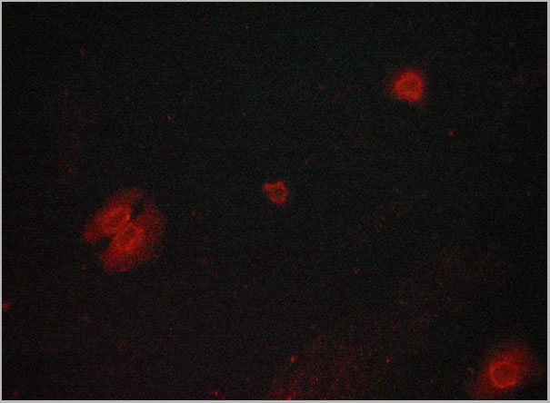
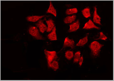
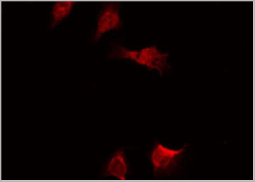
 IF (Immunofluorescence)
(AAA30966 staining 293 by IF/ICC. The sample were fixed with PFA and permeabilized in 0.1% Triton X-100, then blocked in 10% serum for 45 minutes at 25 degree C. The primary antibody was diluted at 1/200 and incubated with the sample for 1 hour at 37 degree C. An Alexa Fluor 594 conjugated goat anti-rabbit IgG (H+L) Ab, diluted at 1/600, was used as the secondary antibody.)
IF (Immunofluorescence)
(AAA30966 staining 293 by IF/ICC. The sample were fixed with PFA and permeabilized in 0.1% Triton X-100, then blocked in 10% serum for 45 minutes at 25 degree C. The primary antibody was diluted at 1/200 and incubated with the sample for 1 hour at 37 degree C. An Alexa Fluor 594 conjugated goat anti-rabbit IgG (H+L) Ab, diluted at 1/600, was used as the secondary antibody.)
Myc, Polyclonal Antibody (Cat# AAA30966)
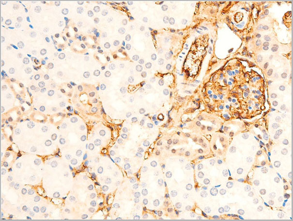
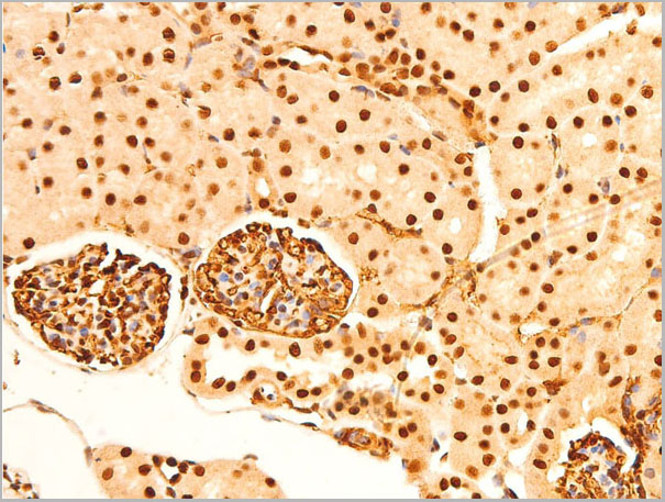
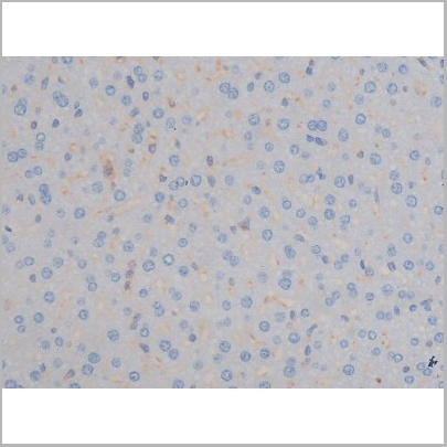
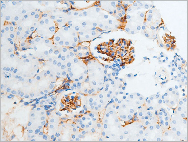
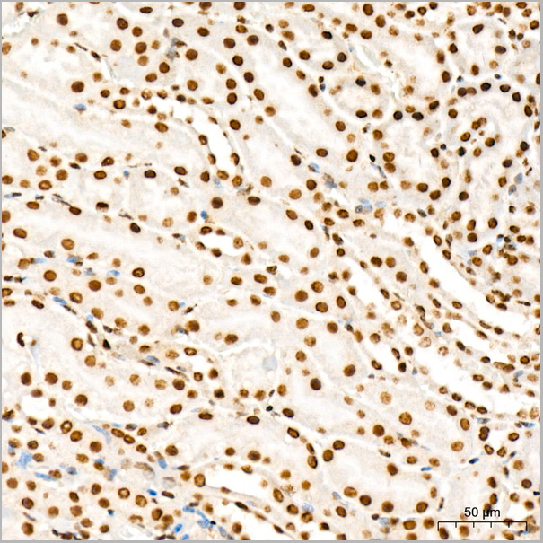
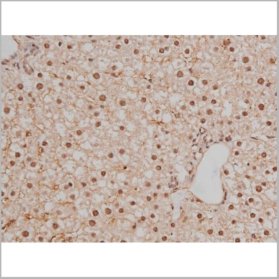
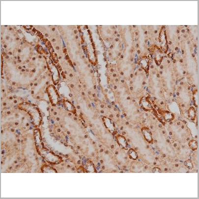
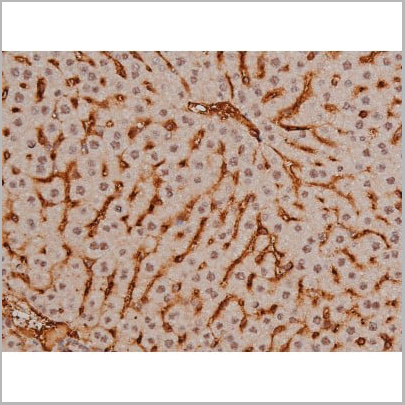
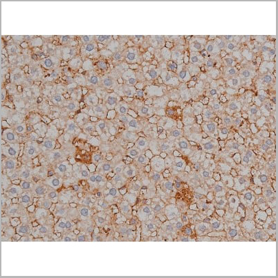
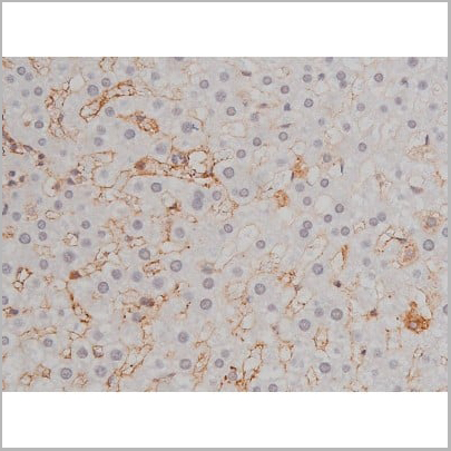
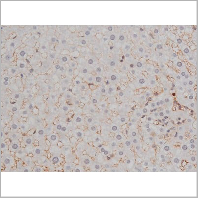
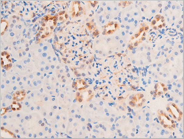
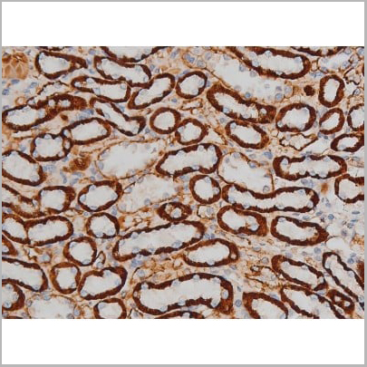
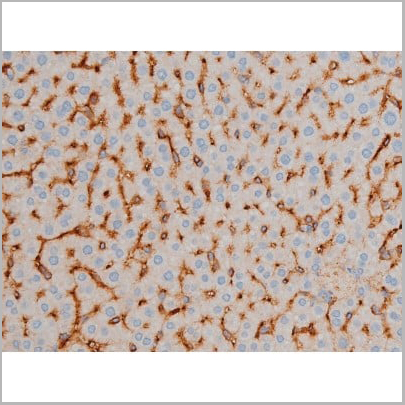
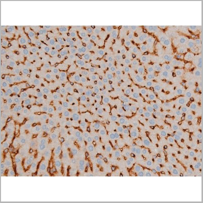
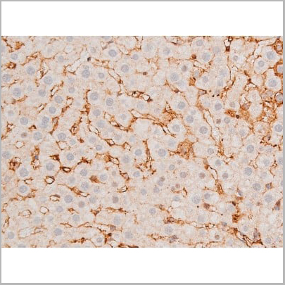
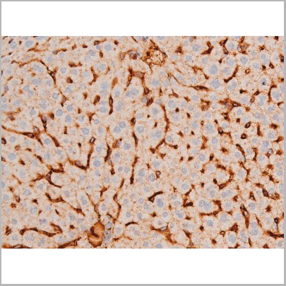
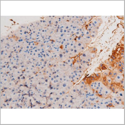
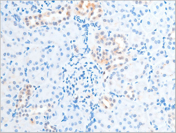
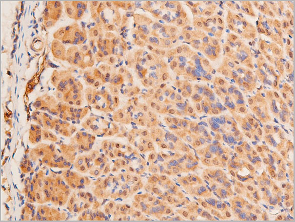
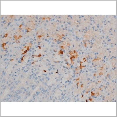
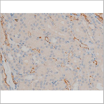
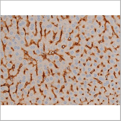
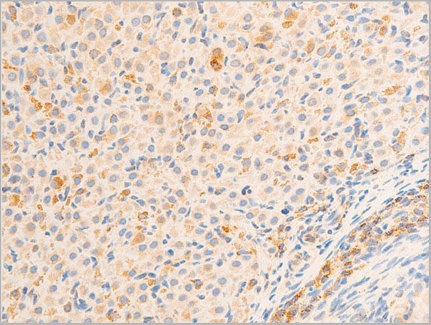
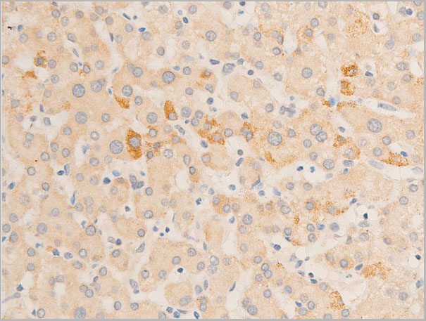
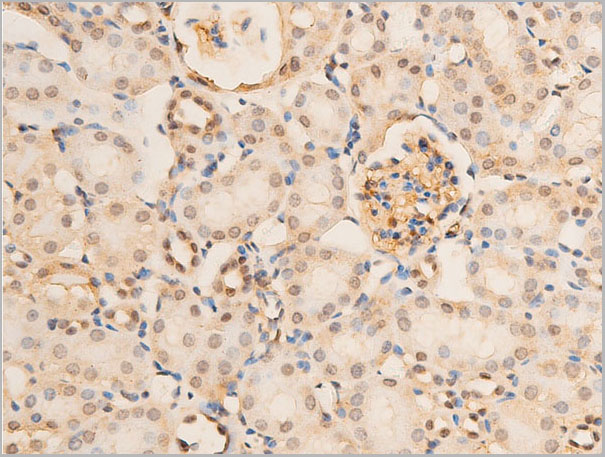
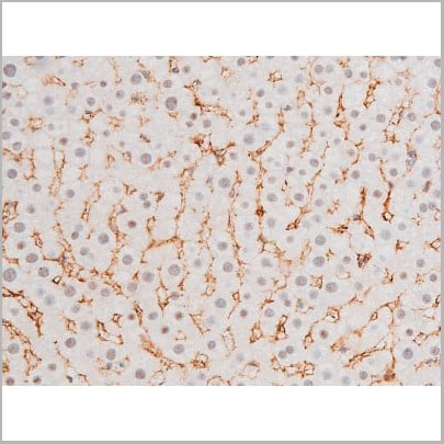
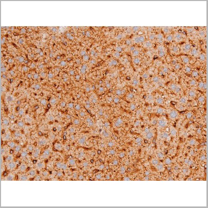
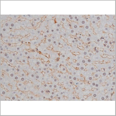
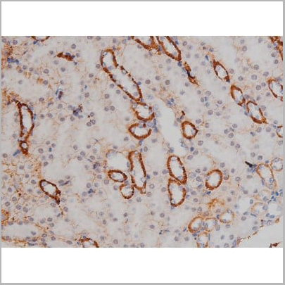
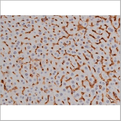
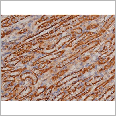
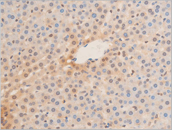 IHC (Immunohistochemistry)
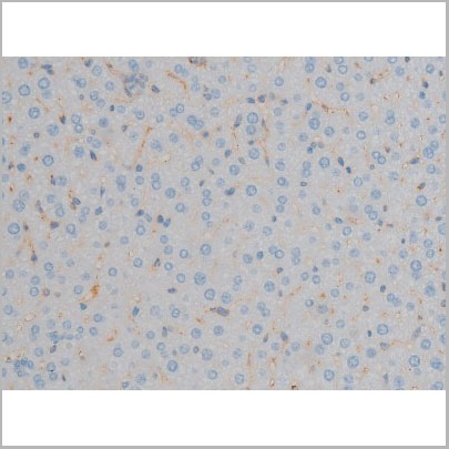
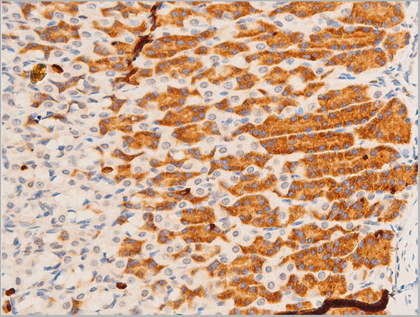
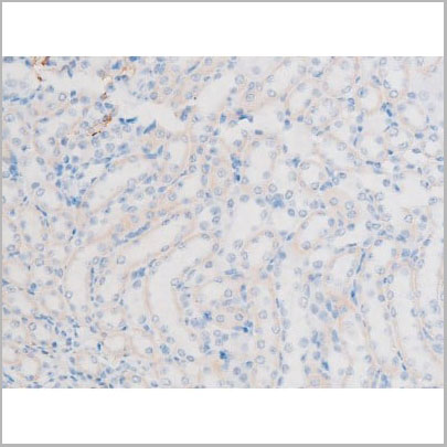
(AAA30967 at 1/100 staining rat liver tissue sections by IHC-P. The tissue was formaldehyde fixed and a heat mediated antigen retrieval step in citrate buffer was performed. The tissue was then blocked and incubated with the antibody for 1.5 hours at 22 degree C. An HRP conjugated goat anti-rabbit antibody was used as the secondary.)
IHC (Immunohistochemistry)
(AAA30967 at 1/100 staining rat liver tissue sections by IHC-P. The tissue was formaldehyde fixed and a heat mediated antigen retrieval step in citrate buffer was performed. The tissue was then blocked and incubated with the antibody for 1.5 hours at 22 degree C. An HRP conjugated goat anti-rabbit antibody was used as the secondary.)
Lamin A/C, Polyclonal Antibody (Cat# AAA30967)
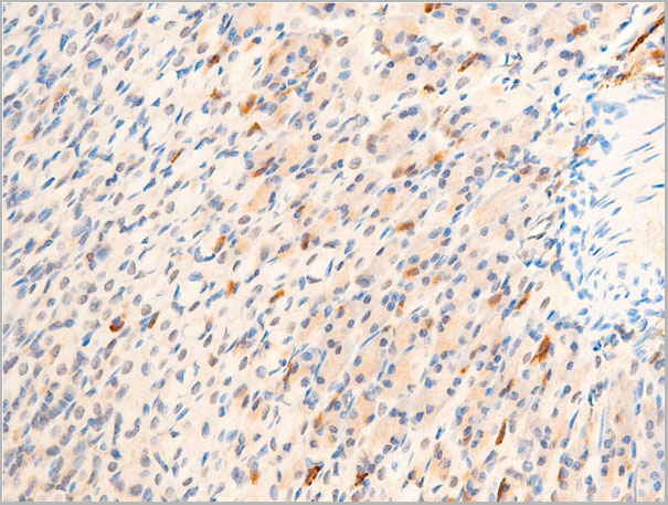
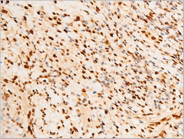
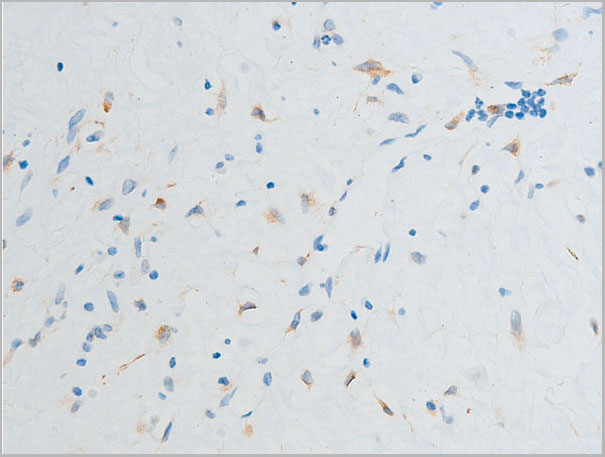
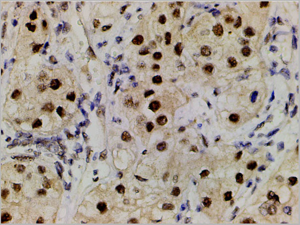
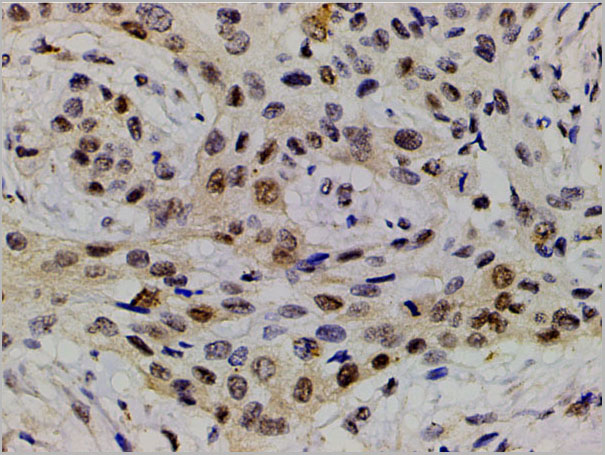
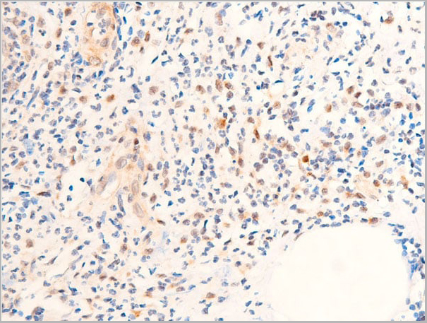
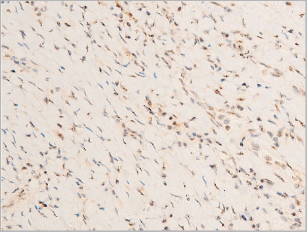
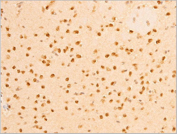
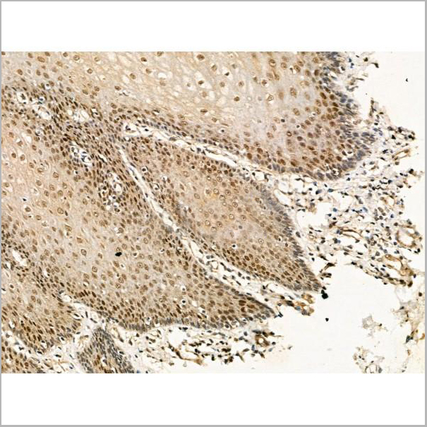
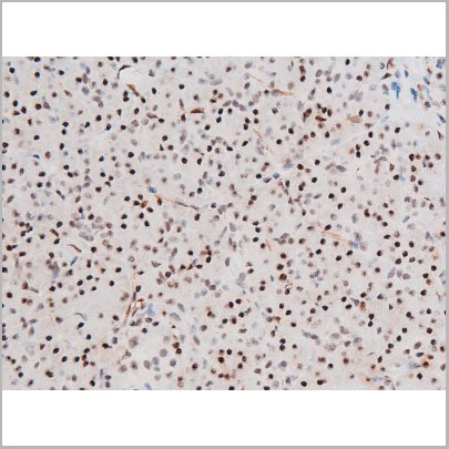
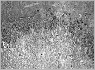 IHC (Immunohistochemistry)
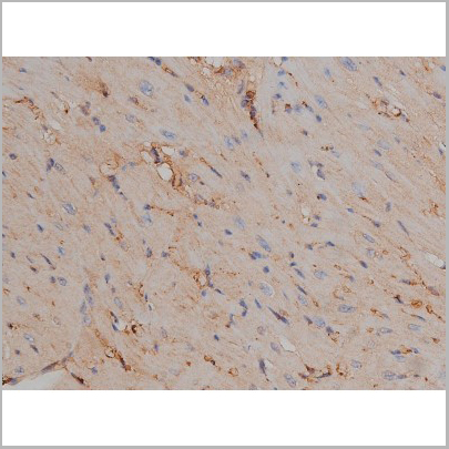
(Immunohistochemical analysis of paraffin-embedded rat hippocampal region tissue from a model with Alzheinmer's Disease useing Tau (phospho-Thr181) antibody)
IHC (Immunohistochemistry)
(Immunohistochemical analysis of paraffin-embedded rat hippocampal region tissue from a model with Alzheinmer's Disease useing Tau (phospho-Thr181) antibody)
Tau, Antibody (Cat# AAA17984)
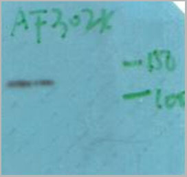
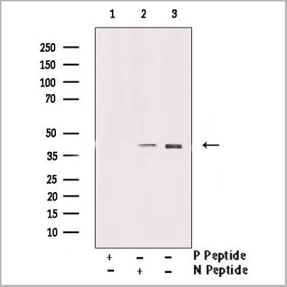
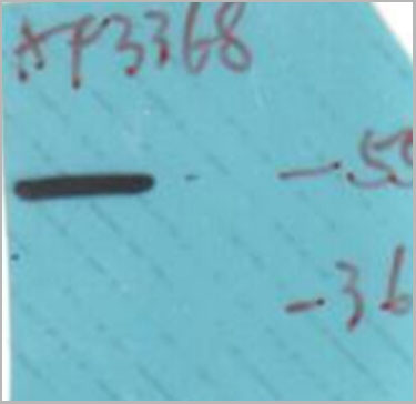
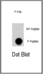 DB (Dot Blot)
(Dot blot analysis of anti-AKT3-pS472 Phospho-specific Pab on nitrocellulose membrane. 50ng of Phospho-peptide or Non Phospho-peptide per dot were adsorbed. Antibody working concentrations are 0.5ug per ml.)
DB (Dot Blot)
(Dot blot analysis of anti-AKT3-pS472 Phospho-specific Pab on nitrocellulose membrane. 50ng of Phospho-peptide or Non Phospho-peptide per dot were adsorbed. Antibody working concentrations are 0.5ug per ml.)
Phospho-AKT3 (S472), Polyclonal Antibody (Cat# AAA28777)
Predicted: Mouse, Rat.
 Application Data
(At 25 degree C. Samples were then incubated with primary Ab(At 37 degree C. An AlexaFluor594 conjugated goat anti-rabbit IgG(H+L) Ab(Red) and an AlexaFluor488 conjugated goat anti-mouse IgG(H+L) Ab(Green) were used as the secondary antibody.The nuclear counter stain is DAPI(blue).)
Application Data
(At 25 degree C. Samples were then incubated with primary Ab(At 37 degree C. An AlexaFluor594 conjugated goat anti-rabbit IgG(H+L) Ab(Red) and an AlexaFluor488 conjugated goat anti-mouse IgG(H+L) Ab(Green) were used as the secondary antibody.The nuclear counter stain is DAPI(blue).)
PAR4, Polyclonal Antibody (Cat# AAA31375)
Predicted Reactivity: Pig (100%), Zebrafish (92%), Rabbit (100%), Chicken (100%), Xenopus (92%)
 IF (Immunofluorescence)
(AAA31053 staining 293 by IF/ICC. The sample were fixed with PFA and permeabilized in 0.1% Triton X-100, then blocked in 10% serum for 45 minutes at 25 degree C. The primary antibody was diluted at 1/200 and incubated with the sample for 1 hour at 37 degree C. An Alexa Fluor 594 conjugated goat anti-rabbit IgG (H+L) Ab, diluted at 1/600, was used as the secondary antibody.)
IF (Immunofluorescence)
(AAA31053 staining 293 by IF/ICC. The sample were fixed with PFA and permeabilized in 0.1% Triton X-100, then blocked in 10% serum for 45 minutes at 25 degree C. The primary antibody was diluted at 1/200 and incubated with the sample for 1 hour at 37 degree C. An Alexa Fluor 594 conjugated goat anti-rabbit IgG (H+L) Ab, diluted at 1/600, was used as the secondary antibody.)
GATA4, Polyclonal Antibody (Cat# AAA31053)
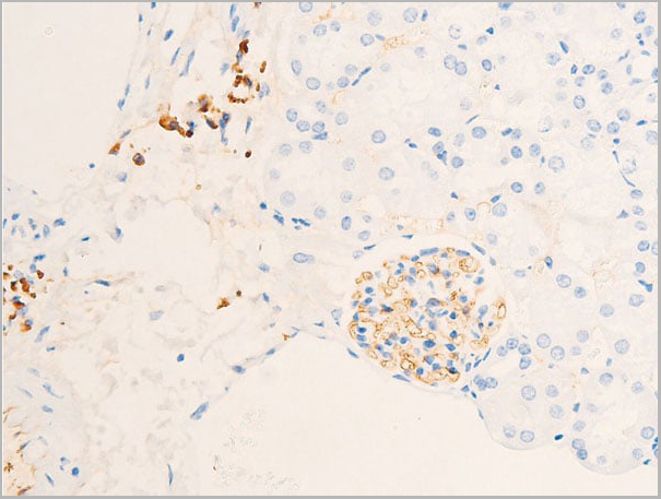
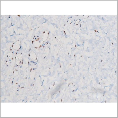
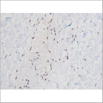
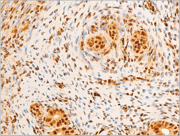
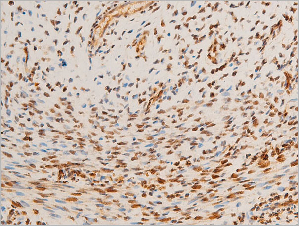
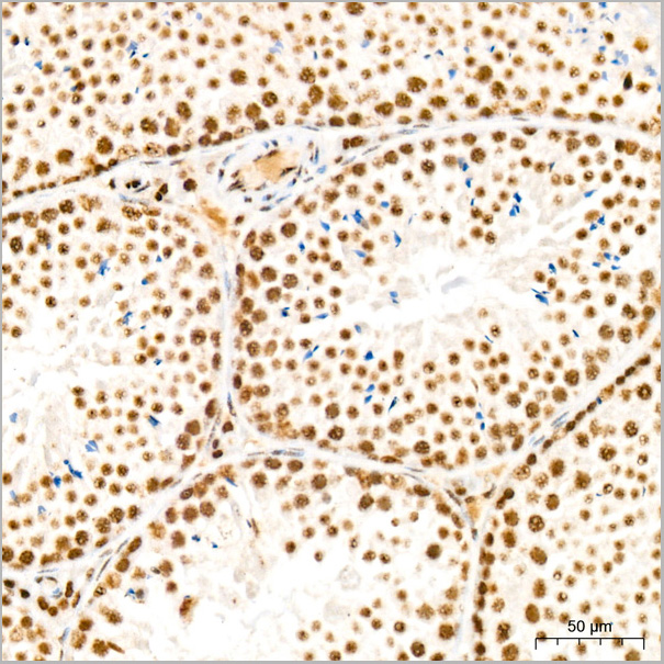
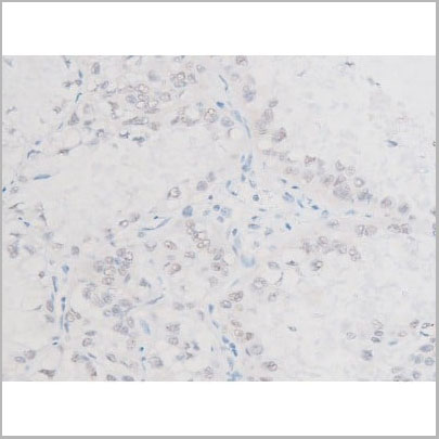
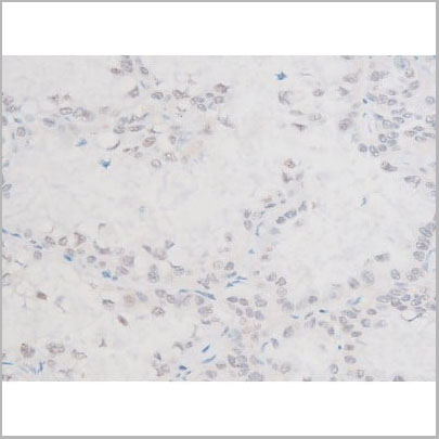
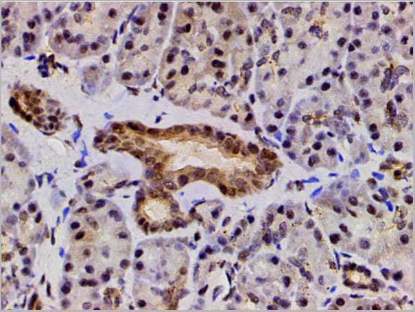
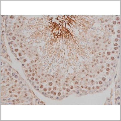
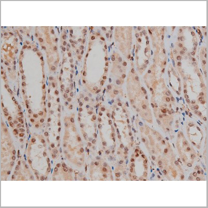
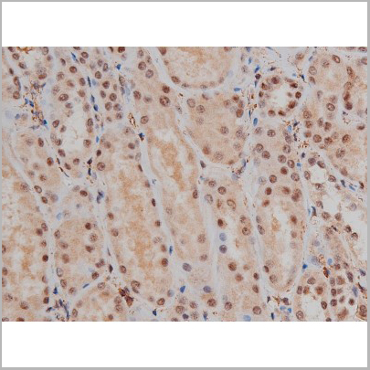
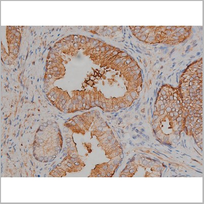
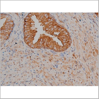
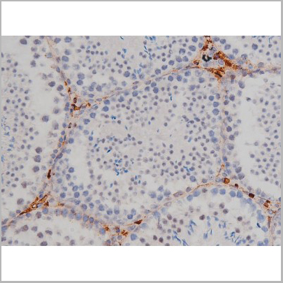
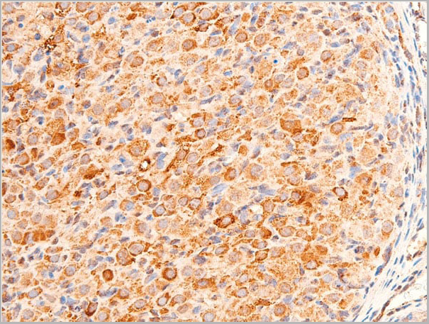
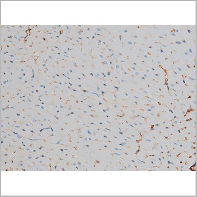
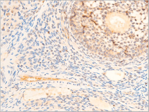
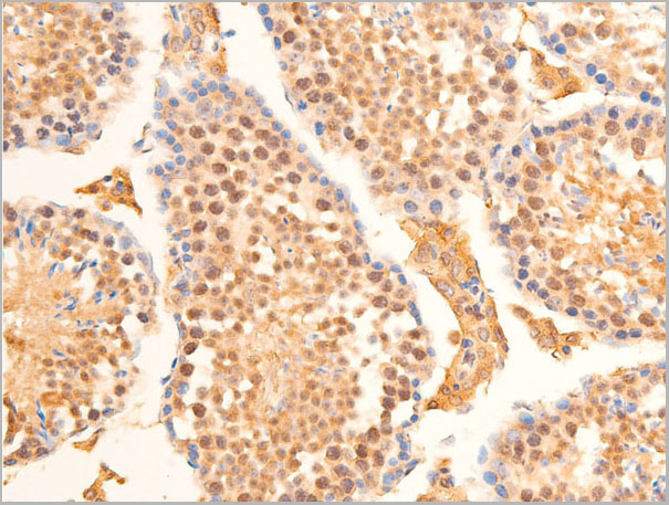
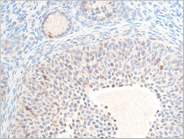
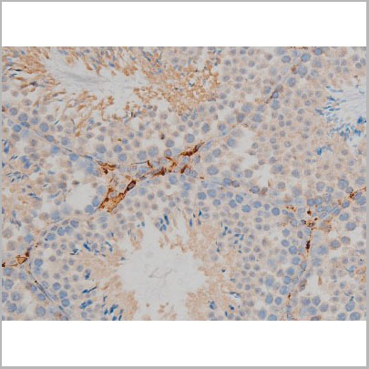
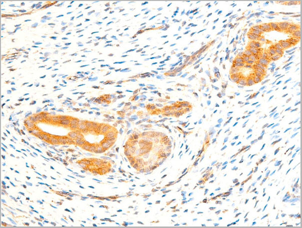
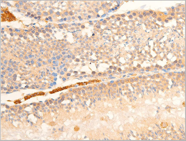
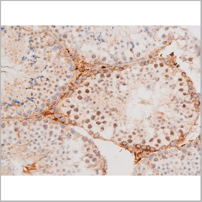
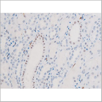
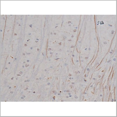
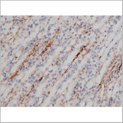
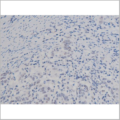
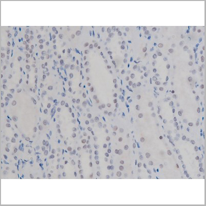
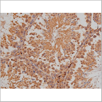
 IHC (Immunohistochemistry)
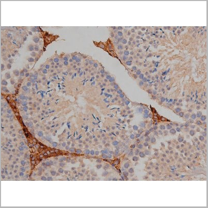
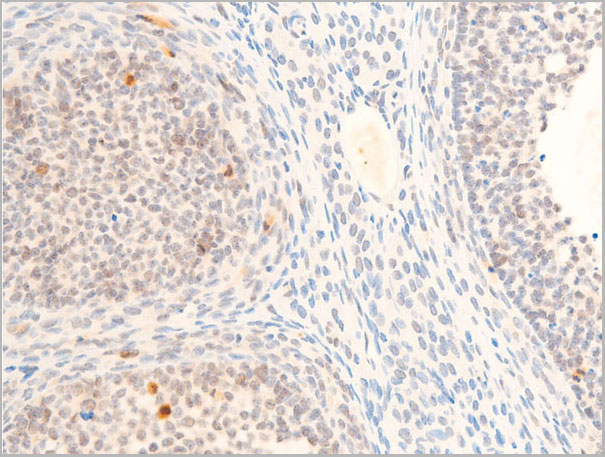
(AAA31059 at 1/100 staining Rat testis tissue by IHC-P. The sample was formaldehyde fixed and a heat mediated antigen retrieval step in citrate buffer was performed. The sample was then blocked and incubated with the primary antibody at 4°C overnight. An HRP conjugated anti-Rabbit antibody was used as the secondary antibody.)
IHC (Immunohistochemistry)
(AAA31059 at 1/100 staining Rat testis tissue by IHC-P. The sample was formaldehyde fixed and a heat mediated antigen retrieval step in citrate buffer was performed. The sample was then blocked and incubated with the primary antibody at 4°C overnight. An HRP conjugated anti-Rabbit antibody was used as the secondary antibody.)
Smad2/3, Polyclonal Antibody (Cat# AAA31059)
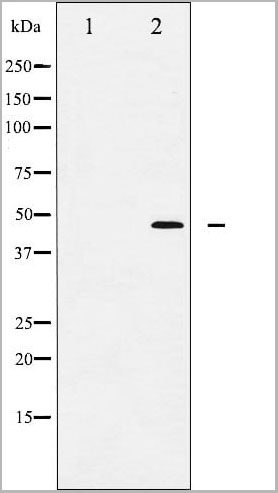
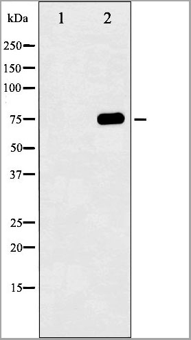
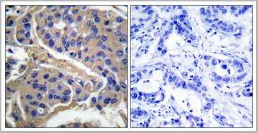
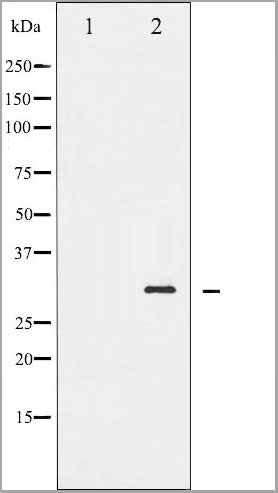
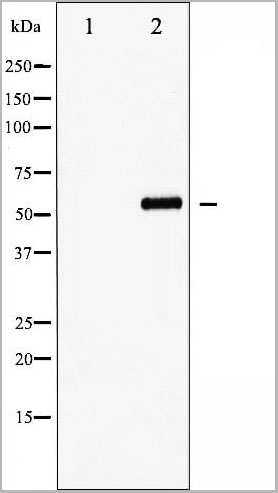
 WB (Western Blot)
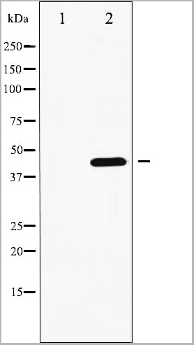
(Western blot analysis of XIAP phosphorylation expression in Anisomycin treated HepG2 whole cell lysates, The lane on the left is treated with the antigen-specific peptide.)
WB (Western Blot)
(Western blot analysis of XIAP phosphorylation expression in Anisomycin treated HepG2 whole cell lysates, The lane on the left is treated with the antigen-specific peptide.)
XIAP, Polyclonal Antibody (Cat# AAA31060)
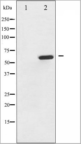
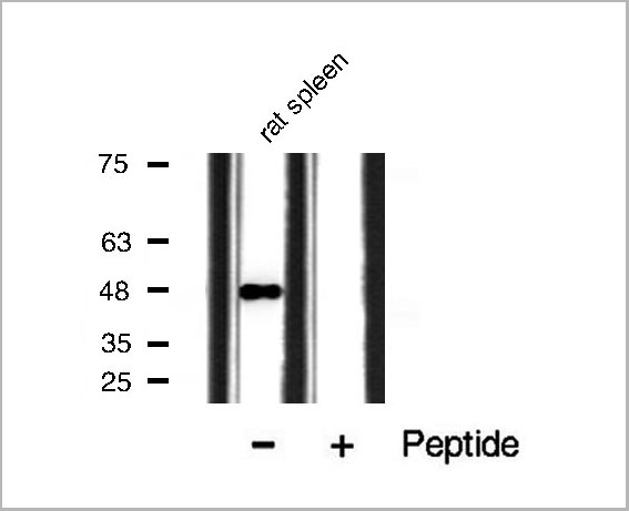
 WB (Western Blot)
(Western blot analysis of Chk2 phosphorylation expression in Jurkat whole cell lysates, The lane on the left is treated with the antigen-specific peptide.)
WB (Western Blot)
(Western blot analysis of Chk2 phosphorylation expression in Jurkat whole cell lysates, The lane on the left is treated with the antigen-specific peptide.)
Chk2, Polyclonal Antibody (Cat# AAA30957)
 WB (Western Blot)
(Western blot analysis of p53 phosphorylation expression in HU treated HeLa whole cell lysates, The lane on the left is treated with the antigen-specific peptide.)
WB (Western Blot)
(Western blot analysis of p53 phosphorylation expression in HU treated HeLa whole cell lysates, The lane on the left is treated with the antigen-specific peptide.)
p53, Polyclonal Antibody (Cat# AAA30977)

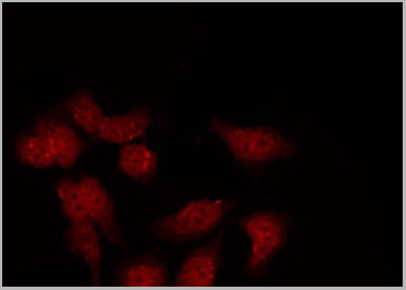
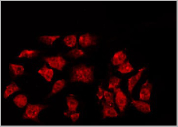
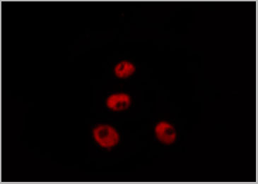
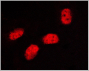
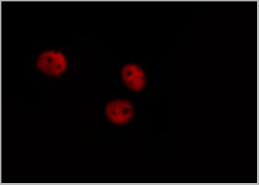
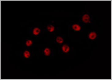
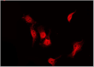
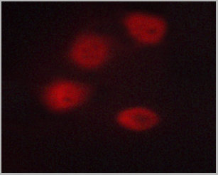
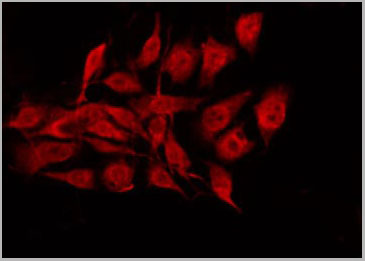 IF (Immunofluorescence)
(AAA30995 staining MCF7 by IF/ICC. The sample were fixed with PFA and permeabilized in 0.1% Triton X-100, then blocked in 10% serum for 45 minutes at 25 degree C. The primary antibody was diluted at 1/200 and incubated with the sample for 1 hour at 37 degree C. An Alexa Fluor 594 conjugated goat anti-rabbit IgG (H+L) Ab, diluted at 1/600, was used as the secondary antibody.)
IF (Immunofluorescence)
(AAA30995 staining MCF7 by IF/ICC. The sample were fixed with PFA and permeabilized in 0.1% Triton X-100, then blocked in 10% serum for 45 minutes at 25 degree C. The primary antibody was diluted at 1/200 and incubated with the sample for 1 hour at 37 degree C. An Alexa Fluor 594 conjugated goat anti-rabbit IgG (H+L) Ab, diluted at 1/600, was used as the secondary antibody.)
BCL-2, Polyclonal Antibody (Cat# AAA30995)
Tau, Polyclonal Antibody (Cat# AAA31002)
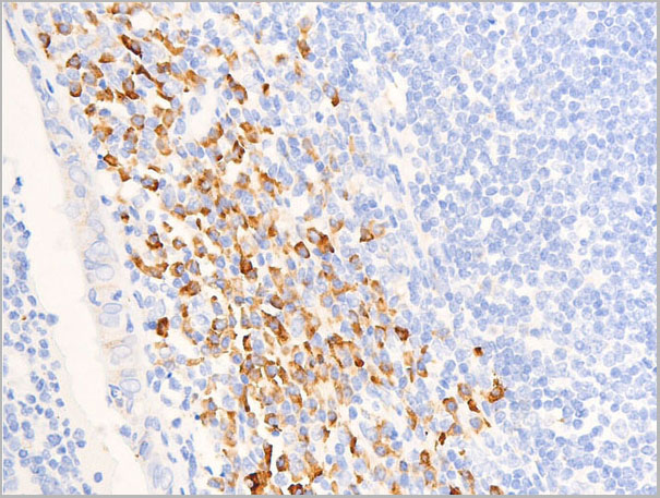
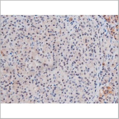
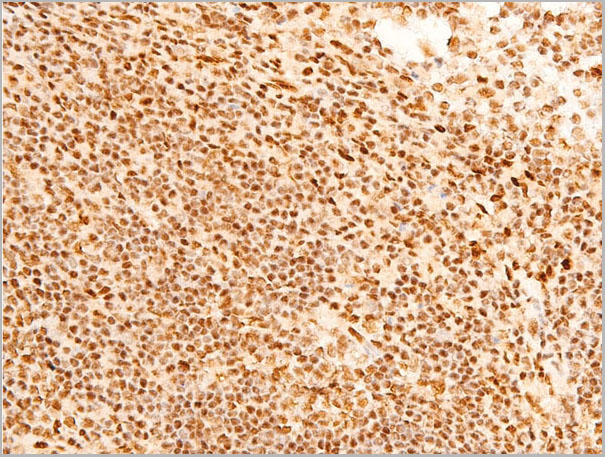
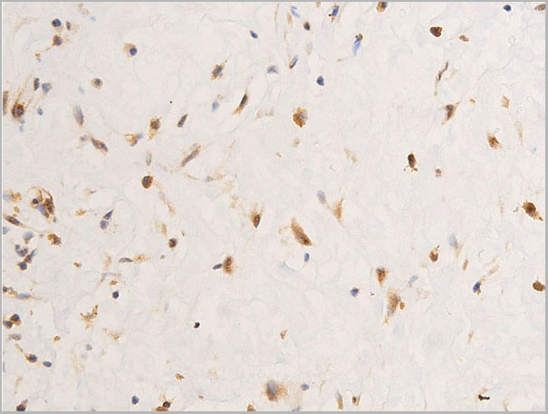
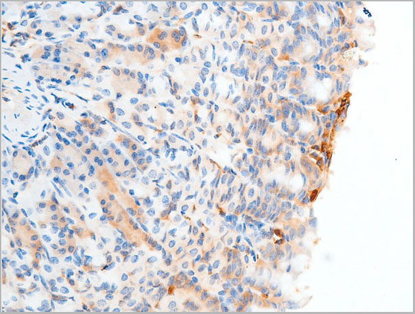
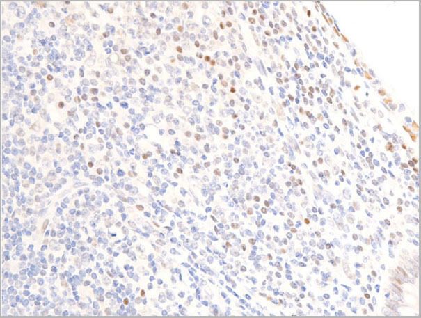
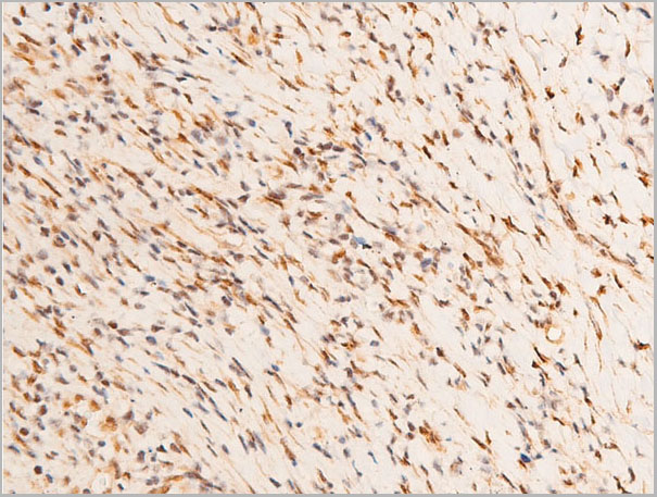
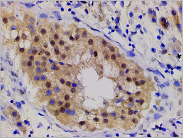
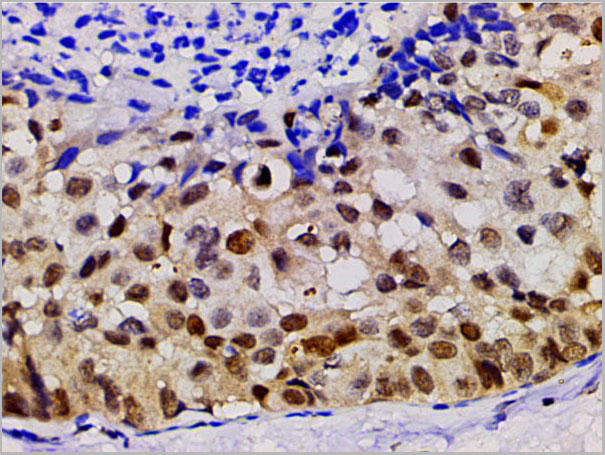
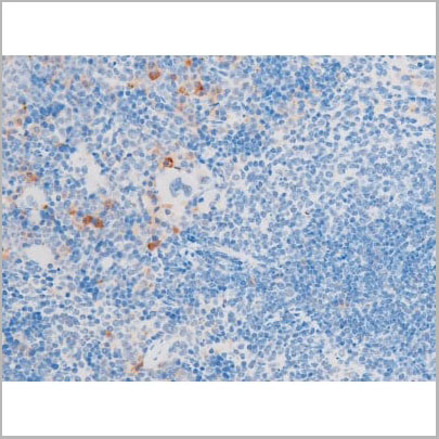
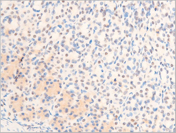
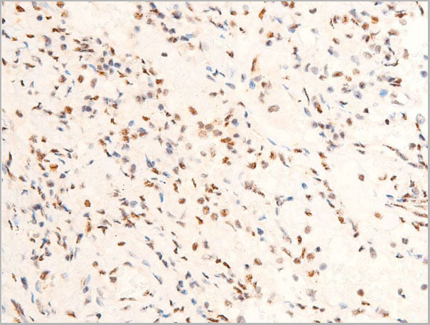
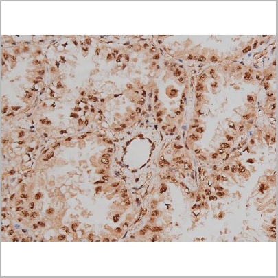
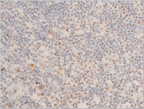
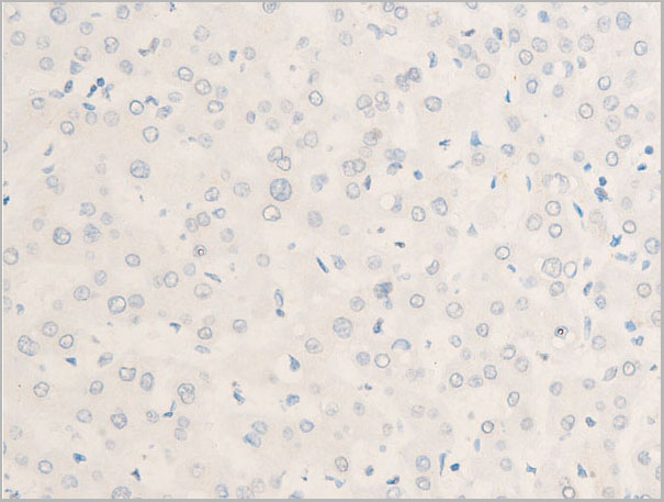
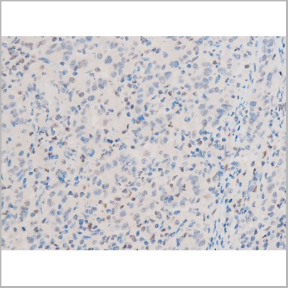
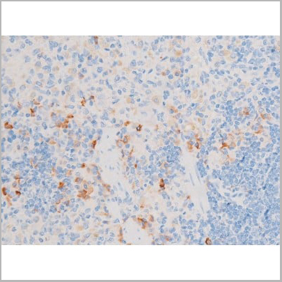
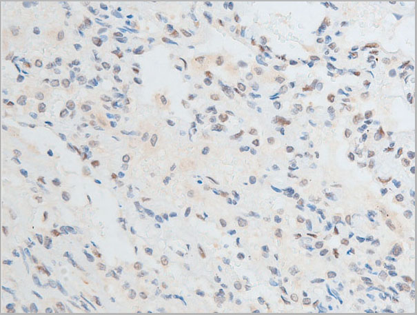
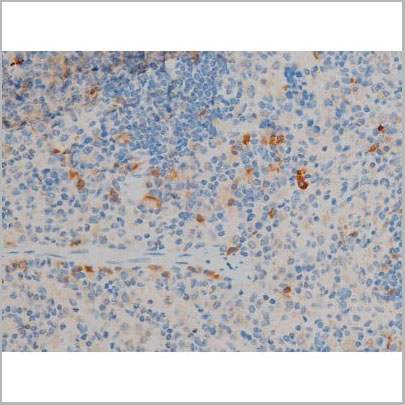
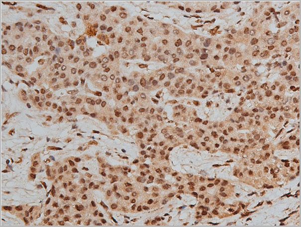
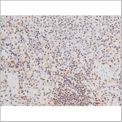
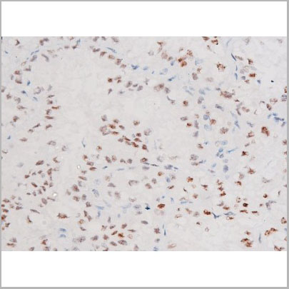
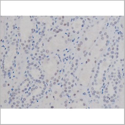
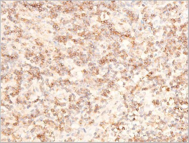
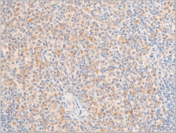
 IHC (Immunohistochemistry)
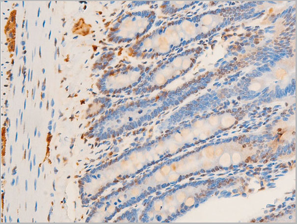
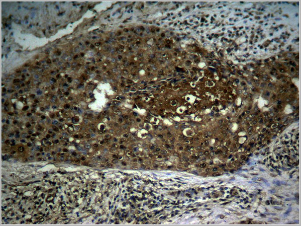
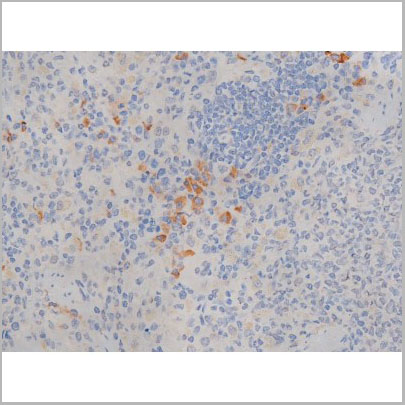
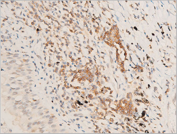
(AAA30934 at 1/100 staining human TB tissue sections by IHC-P. The tissue was formaldehyde fixed and a heat mediated antigen retrieval step in citrate buffer was performed. The tissue was then blocked and incubated with the antibody for 1.5 hours at 22 degree C. An HRP conjugated goat anti-rabbit antibody was used as the secondary.)
IHC (Immunohistochemistry)
(AAA30934 at 1/100 staining human TB tissue sections by IHC-P. The tissue was formaldehyde fixed and a heat mediated antigen retrieval step in citrate buffer was performed. The tissue was then blocked and incubated with the antibody for 1.5 hours at 22 degree C. An HRP conjugated goat anti-rabbit antibody was used as the secondary.)
I kappaB alpha, Polyclonal Antibody (Cat# AAA30934)
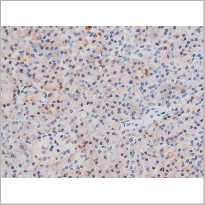
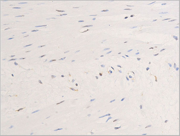
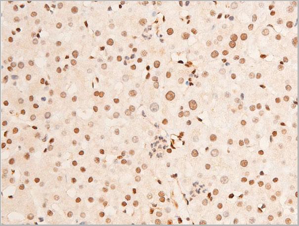
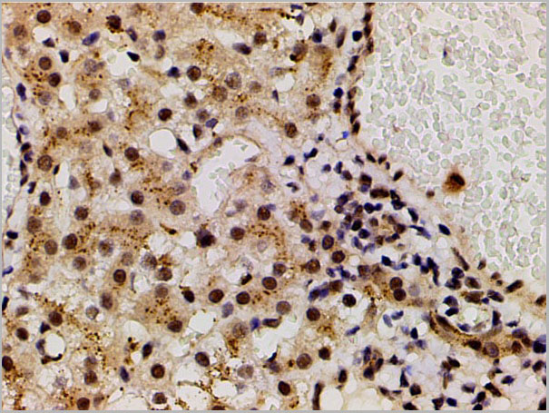
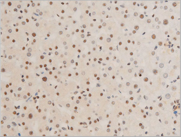
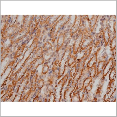
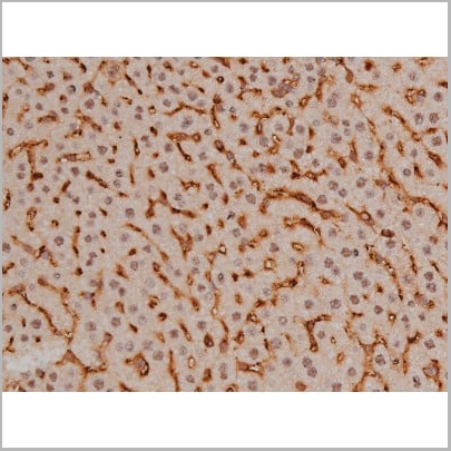
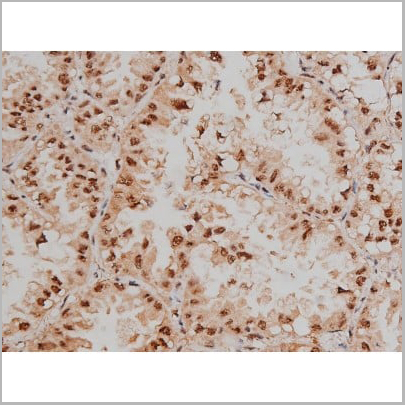
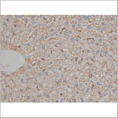
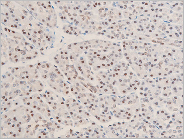
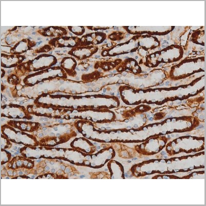
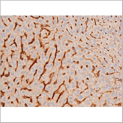
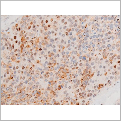
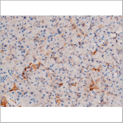
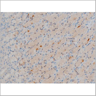
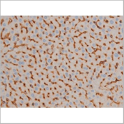
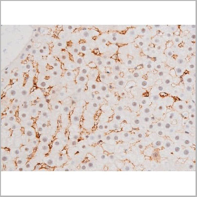
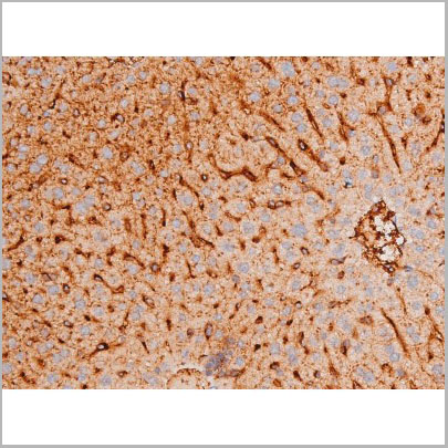
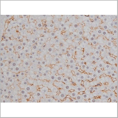
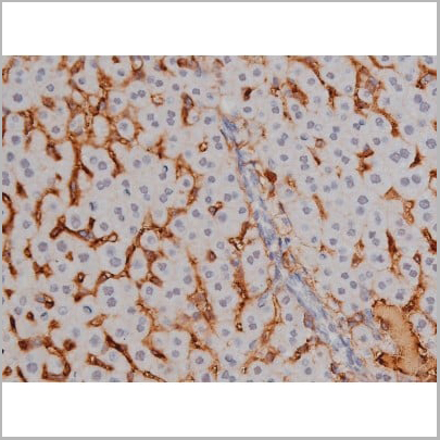
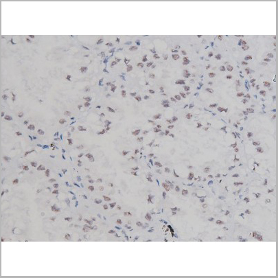
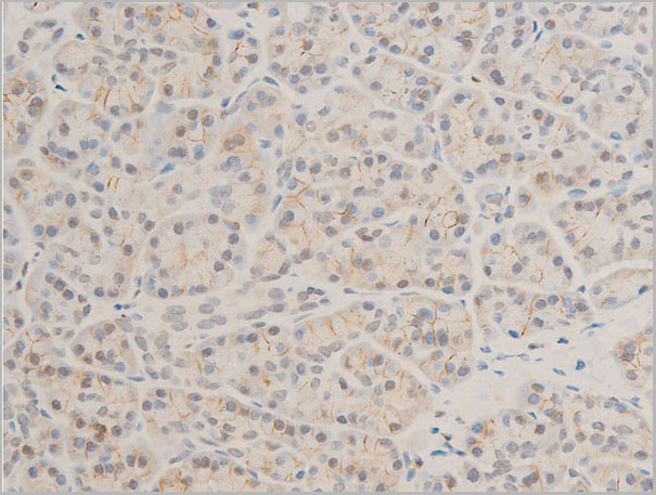
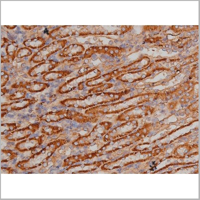
 IHC (Immunohistochemistry)
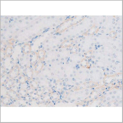
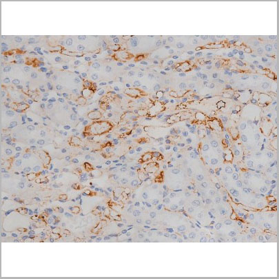
(AAA30948 at 1/100 staining rat liver tissue sections by IHC-P. The tissue was formaldehyde fixed and a heat mediated antigen retrieval step in citrate buffer was performed. The tissue was then blocked and incubated with the antibody for 1.5 hours at 22 degree C. An HRP conjugated goat anti-rabbit antibody was used as the secondary.)
IHC (Immunohistochemistry)
(AAA30948 at 1/100 staining rat liver tissue sections by IHC-P. The tissue was formaldehyde fixed and a heat mediated antigen retrieval step in citrate buffer was performed. The tissue was then blocked and incubated with the antibody for 1.5 hours at 22 degree C. An HRP conjugated goat anti-rabbit antibody was used as the secondary.)
Chk1, Polyclonal Antibody (Cat# AAA30948)
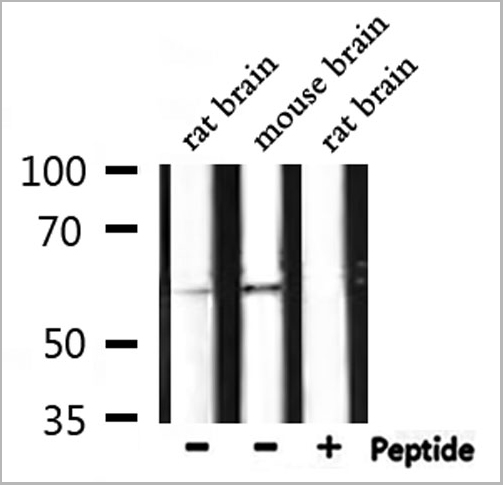
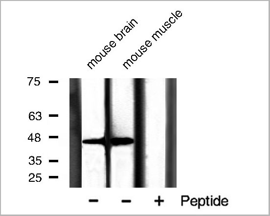
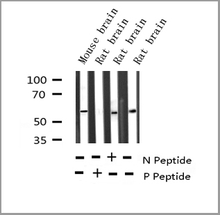
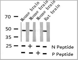
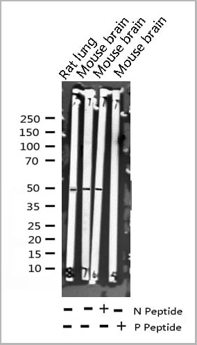
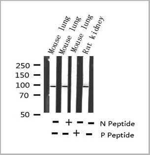
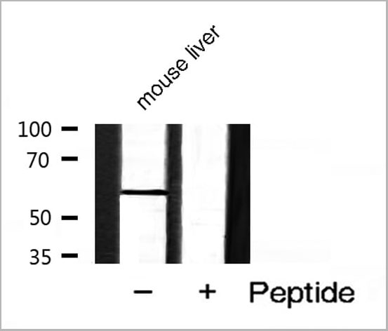
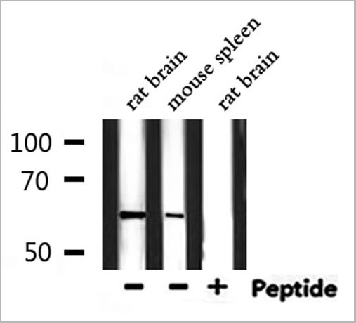
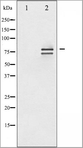
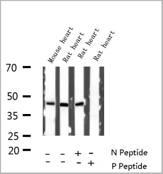
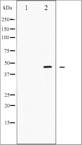
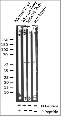
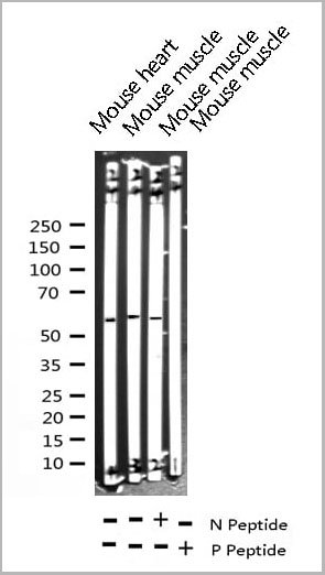
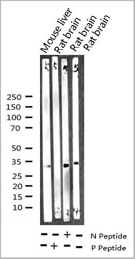
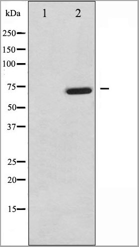
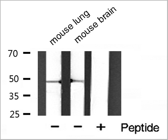 WB (Western Blot)
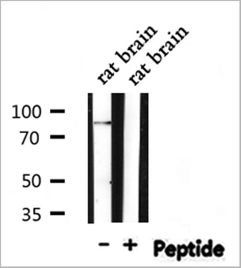
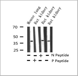
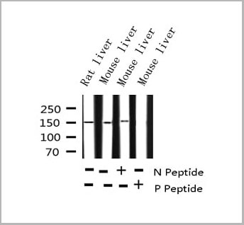
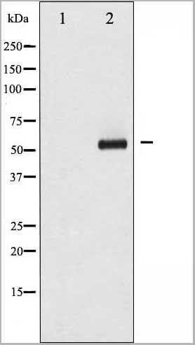
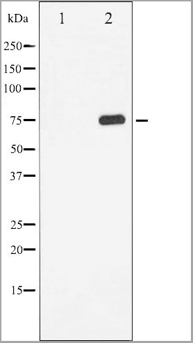
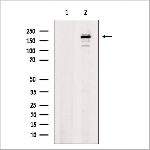
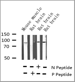
(Western blot analysis of Phospho-HDAC3 (Ser424) Antibody expression in mouse lung and mouse brain tissues lysates.The lane on the right is treated with the antigen-specific peptide.)
WB (Western Blot)
(Western blot analysis of Phospho-HDAC3 (Ser424) Antibody expression in mouse lung and mouse brain tissues lysates.The lane on the right is treated with the antigen-specific peptide.)
HDAC3, Polyclonal Antibody (Cat# AAA30951)

 WB (Western Blot)
(Western blot analysis of Phospho-Tau (Ser214) expression in various lysates)
WB (Western Blot)
(Western blot analysis of Phospho-Tau (Ser214) expression in various lysates)
Tau, Polyclonal Antibody (Cat# AAA30996)
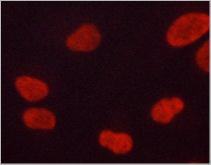
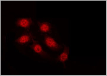
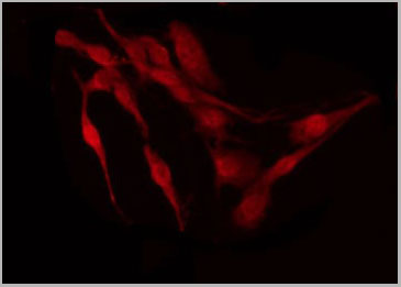
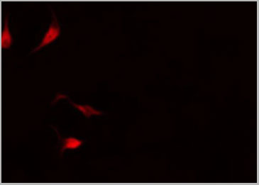
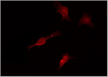
 IF (Immunofluorescence)
(AAA30936 staining HeLa by IF/ICC. The sample were fixed with PFA and permeabilized in 0.1% Triton X-100, then blocked in 10% serum for 45 minutes at 25 degree C. The primary antibody was diluted at 1/200 and incubated with the sample for 1 hour at 37 degree C. An Alexa Fluor 594 conjugated goat anti-rabbit IgG (H+L) Ab, diluted at 1/600, was used as the secondary antibody.)
IF (Immunofluorescence)
(AAA30936 staining HeLa by IF/ICC. The sample were fixed with PFA and permeabilized in 0.1% Triton X-100, then blocked in 10% serum for 45 minutes at 25 degree C. The primary antibody was diluted at 1/200 and incubated with the sample for 1 hour at 37 degree C. An Alexa Fluor 594 conjugated goat anti-rabbit IgG (H+L) Ab, diluted at 1/600, was used as the secondary antibody.)
NF kappaB p65, Polyclonal Antibody (Cat# AAA30936)

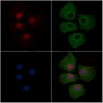
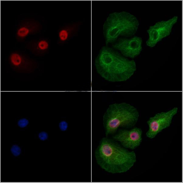 Application Data
(At 25 degree C. Samples were then incubated with primary Ab(At 37 degree C. An AlexaFluor594 conjugated goat anti-rabbit IgG(H+L) Ab(Red) and an AlexaFluor488 conjugated goat anti-mouse IgG(H+L) Ab(Green) were used as the secondary antibody.The nuclear counter stain is DAPI (blue).)
Application Data
(At 25 degree C. Samples were then incubated with primary Ab(At 37 degree C. An AlexaFluor594 conjugated goat anti-rabbit IgG(H+L) Ab(Red) and an AlexaFluor488 conjugated goat anti-mouse IgG(H+L) Ab(Green) were used as the secondary antibody.The nuclear counter stain is DAPI (blue).)
Dnmt1, Polyclonal Antibody (Cat# AAA31301)
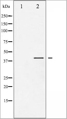
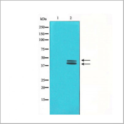
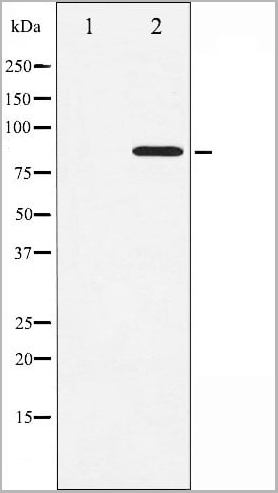
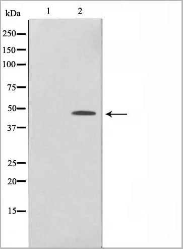
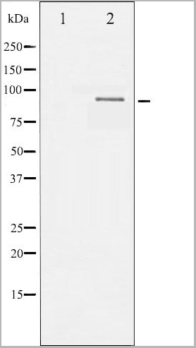
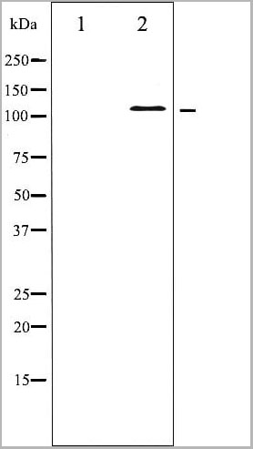
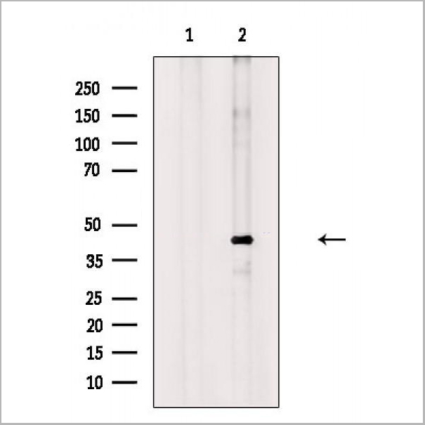
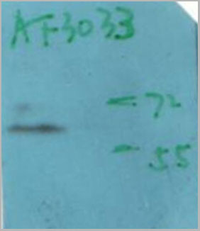
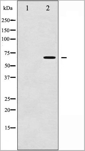
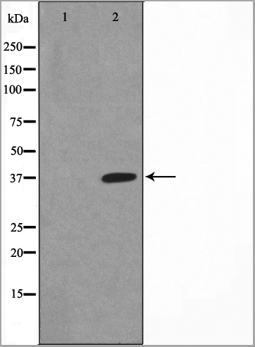
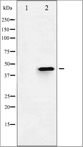
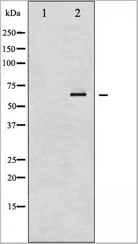
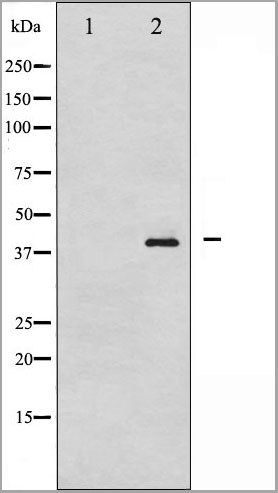
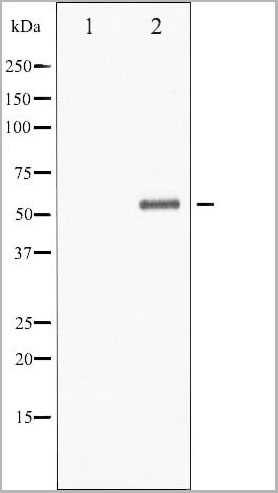
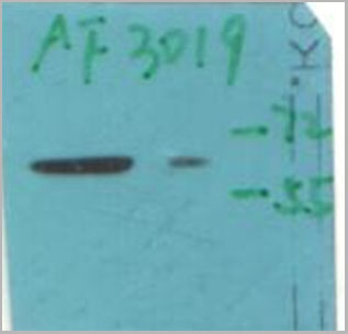
 WB (Western Blot)
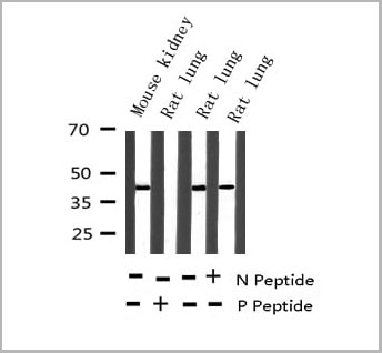
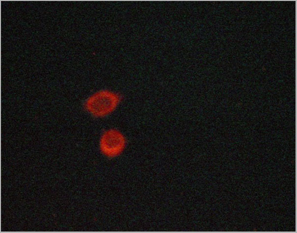
(Western blot analysis of FOXO4 phosphorylation expression in EGF treated HuvEc whole cell lysates, The lane on the left is treated with the antigen-specific peptide.)
WB (Western Blot)
(Western blot analysis of FOXO4 phosphorylation expression in EGF treated HuvEc whole cell lysates, The lane on the left is treated with the antigen-specific peptide.)
FOXO4, Polyclonal Antibody (Cat# AAA31061)
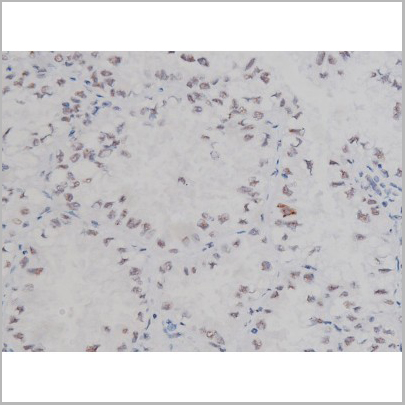
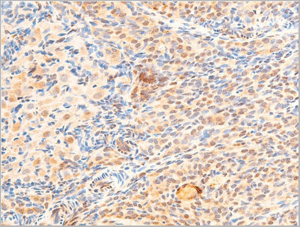
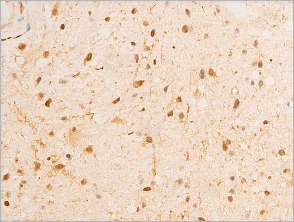
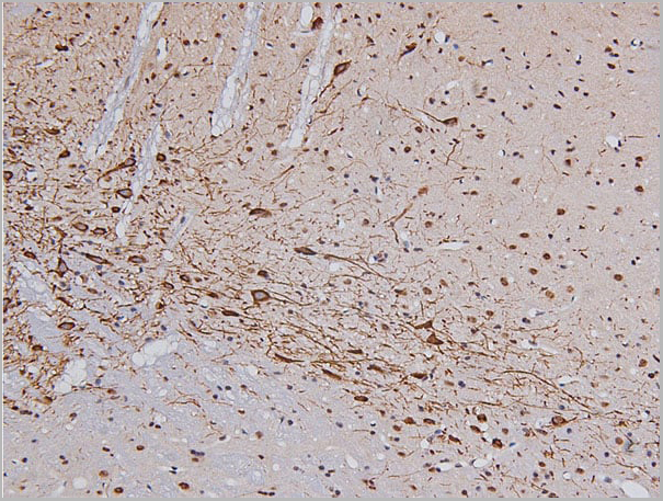
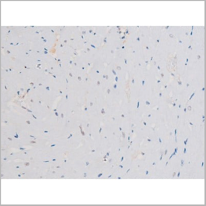
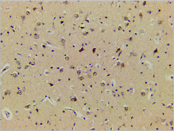
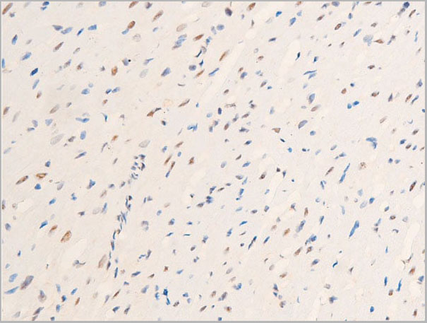
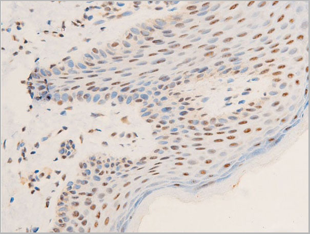
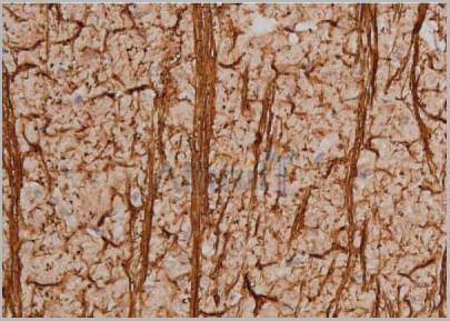
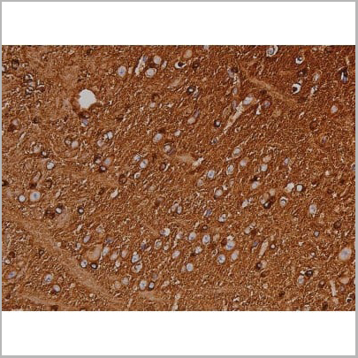
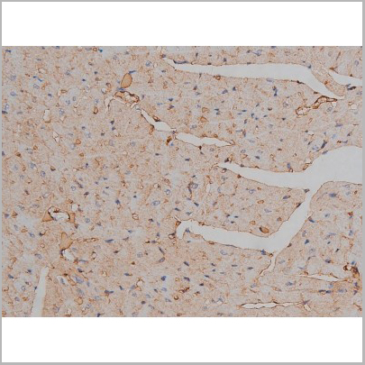
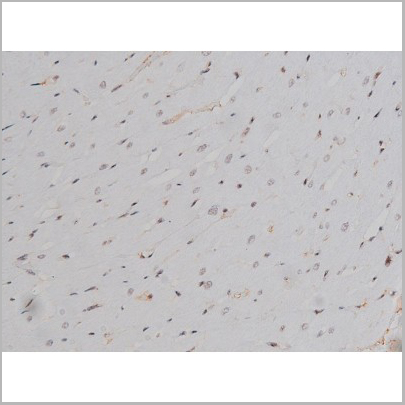
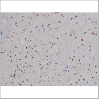
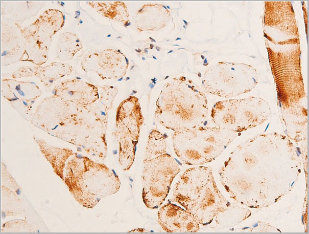
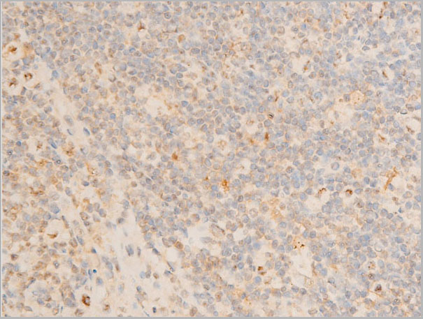
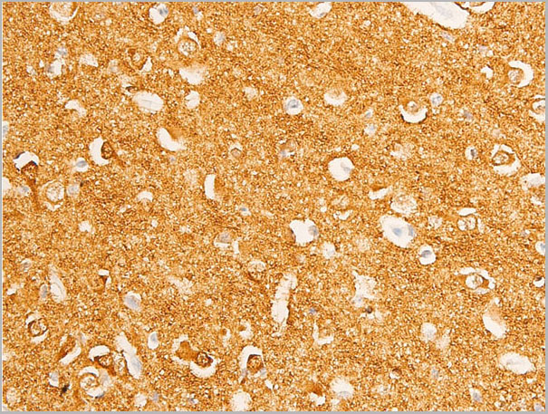
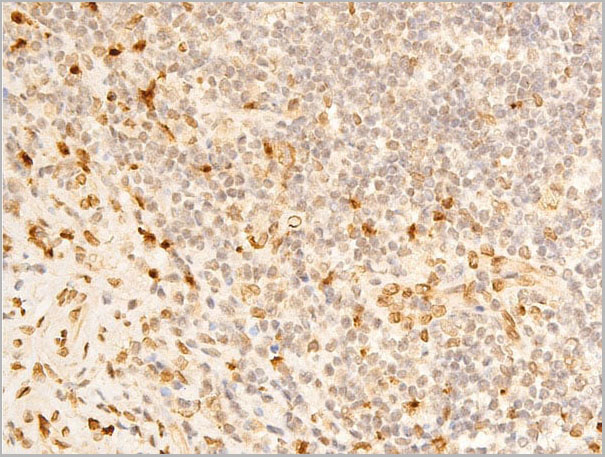
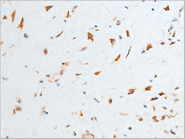
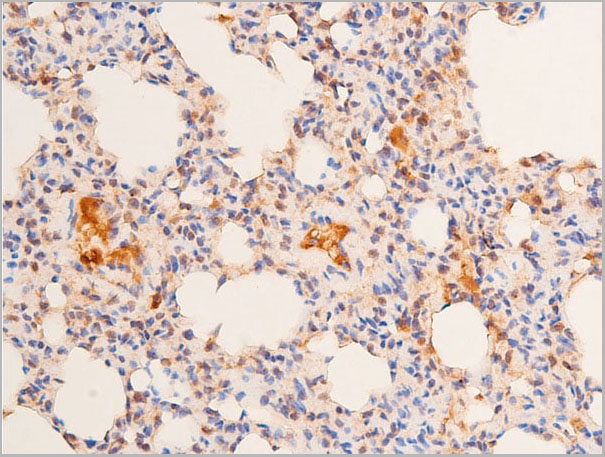
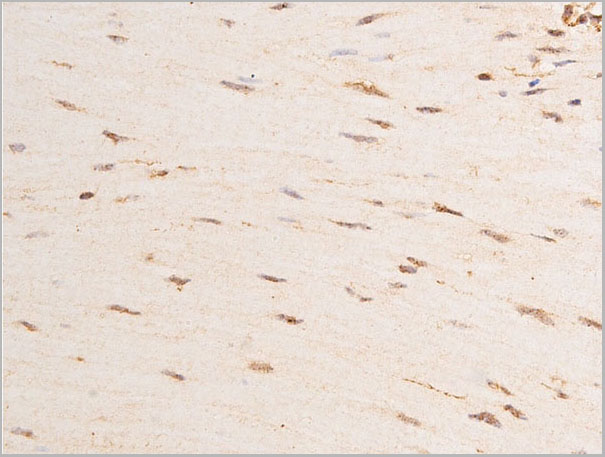
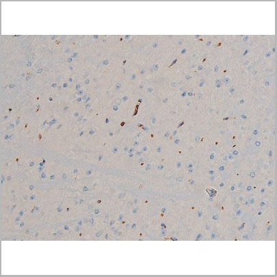
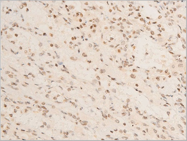
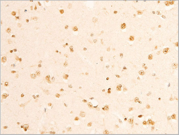
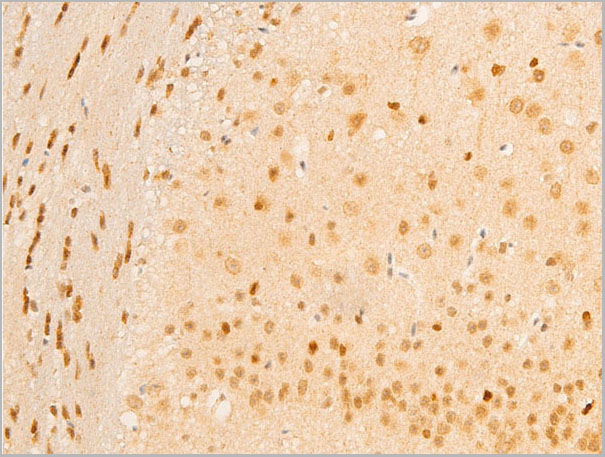
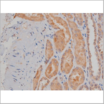
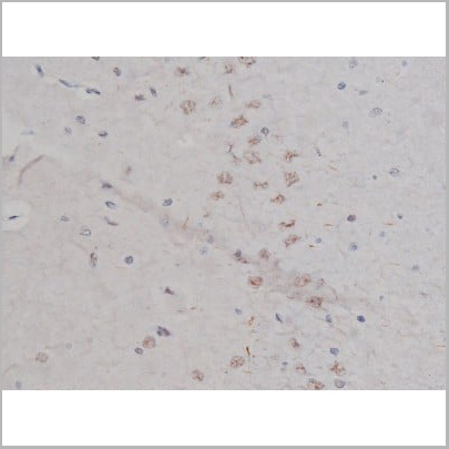 IHC (Immunohistochemistry)
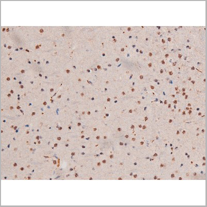
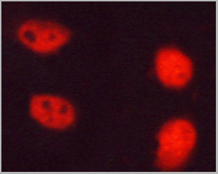
(AAA30987 at 1/200 staining Rat brain tissue sections by IHC-P. The tissue was formaldehyde fixed and a heat mediated antigen retrieval step in citrate buffer was performed. The tissue was then blocked and incubated with the antibody for 1.5 hours at 22 degree C. An HRP conjugated goat anti-rabbit antibody was used as the secondary.)
IHC (Immunohistochemistry)
(AAA30987 at 1/200 staining Rat brain tissue sections by IHC-P. The tissue was formaldehyde fixed and a heat mediated antigen retrieval step in citrate buffer was performed. The tissue was then blocked and incubated with the antibody for 1.5 hours at 22 degree C. An HRP conjugated goat anti-rabbit antibody was used as the secondary.)
NPM, Polyclonal Antibody (Cat# AAA30987)
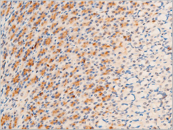
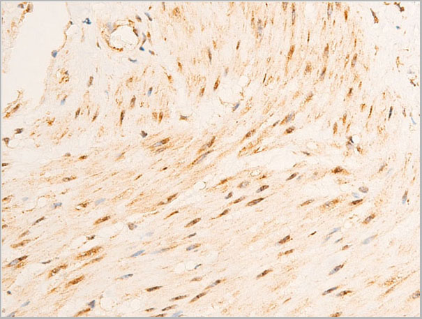
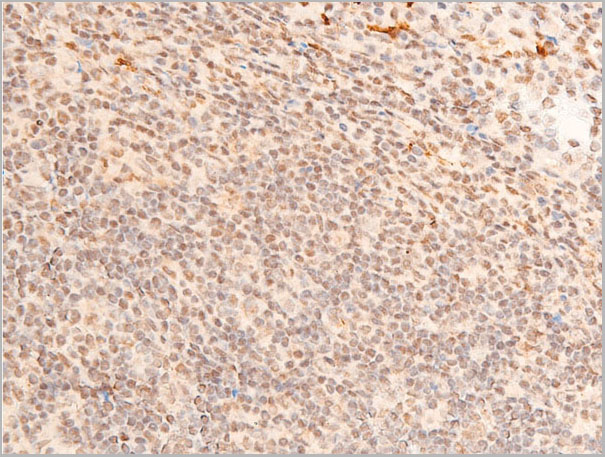
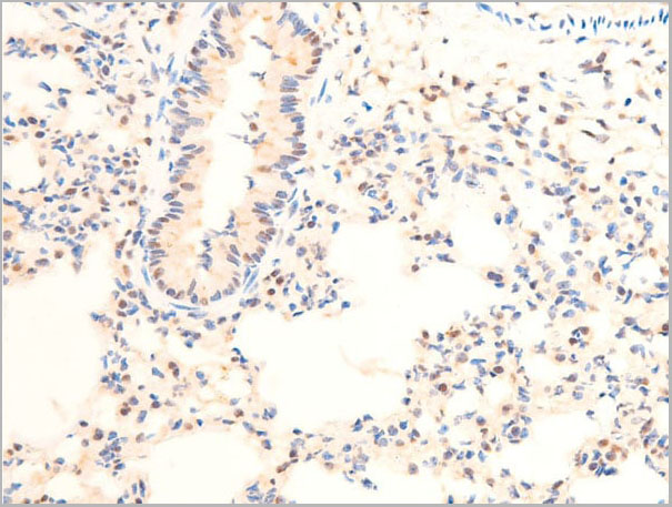
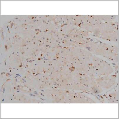
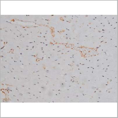
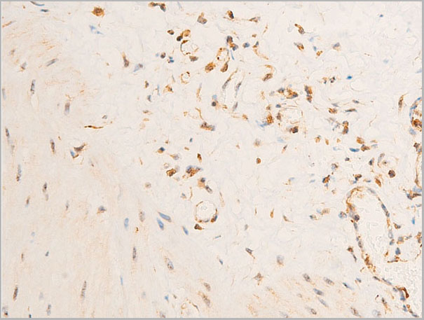
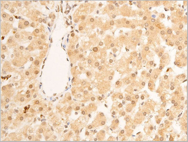
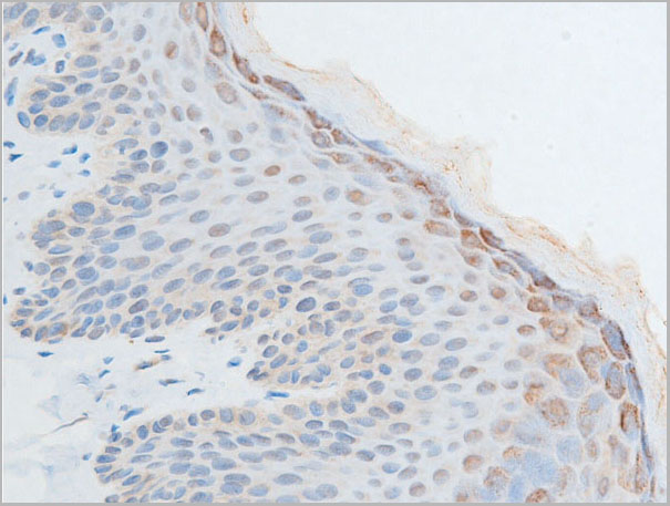
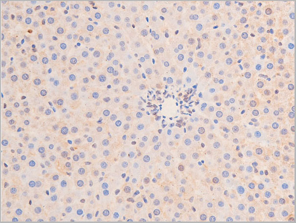
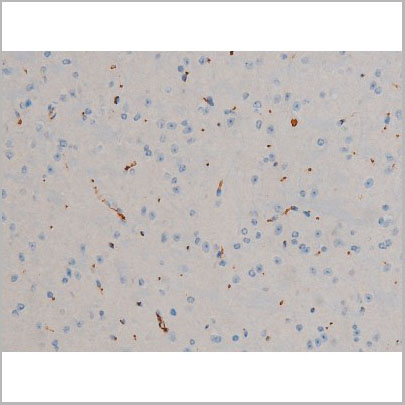
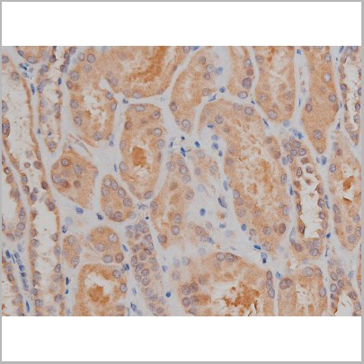
 IHC (Immunohistchemistry)
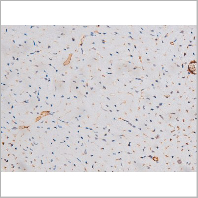
(AAA30947 at 1/100 staining rat brain tissue sections by IHC-P. The tissue was formaldehyde fixed and a heat mediated antigen retrieval step in citrate buffer was performed. The tissue was then blocked and incubated with the antibody for 1.5 hours at 22 degree C. An HRP conjugated goat anti-rabbit antibody was used as the secondary.)
IHC (Immunohistchemistry)
(AAA30947 at 1/100 staining rat brain tissue sections by IHC-P. The tissue was formaldehyde fixed and a heat mediated antigen retrieval step in citrate buffer was performed. The tissue was then blocked and incubated with the antibody for 1.5 hours at 22 degree C. An HRP conjugated goat anti-rabbit antibody was used as the secondary.)
Chk1, Polyclonal Antibody (Cat# AAA30947)
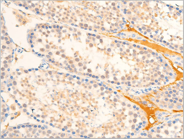
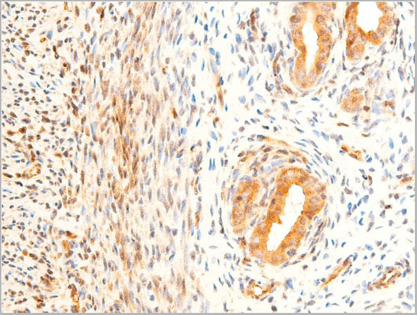
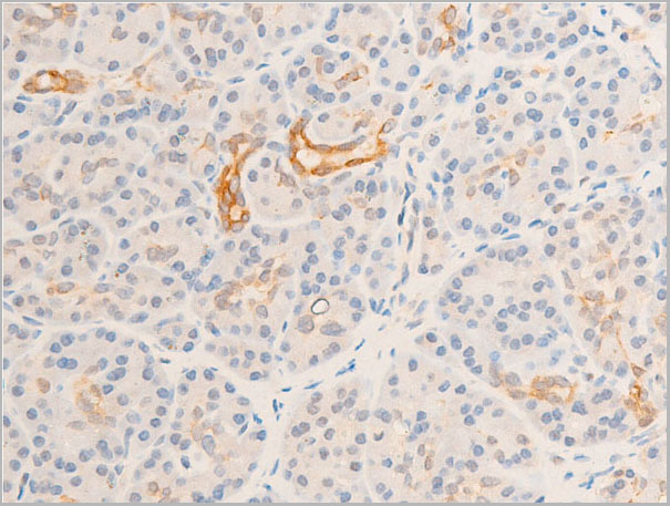
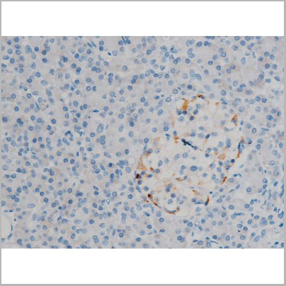
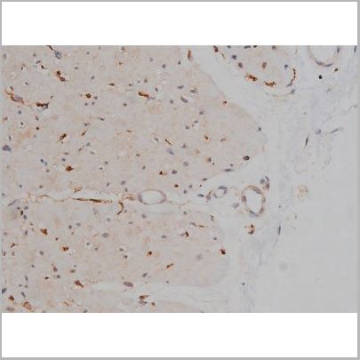
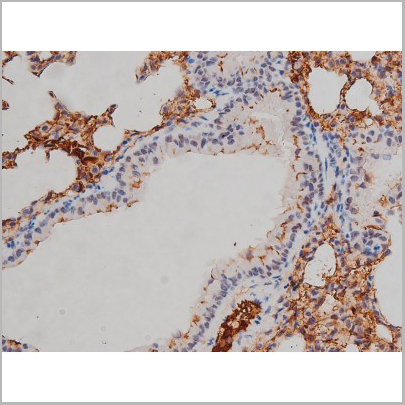
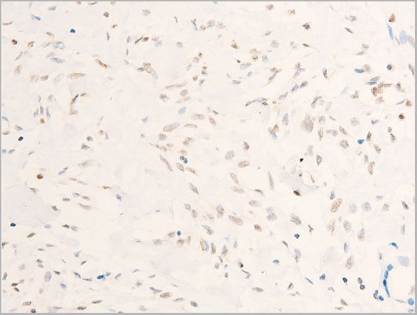
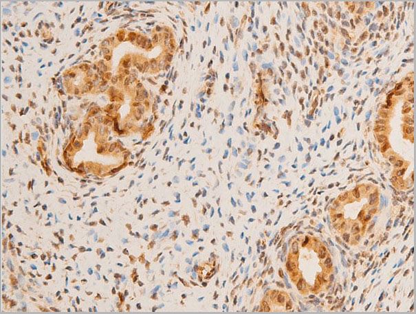
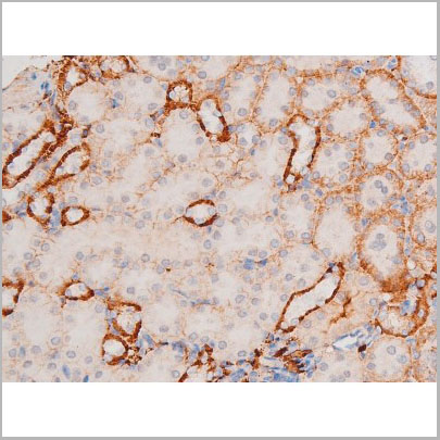
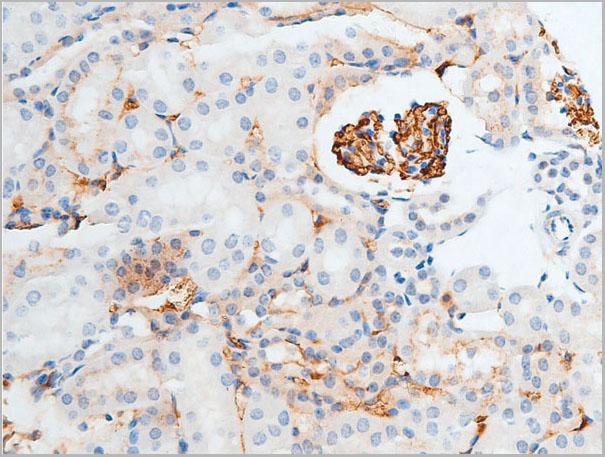
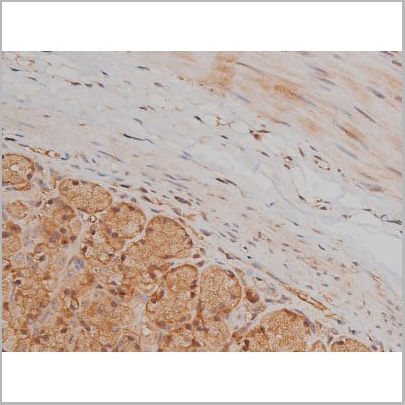
 IHC (Immunohistochemistry)
(AAA30953 at 1/100 staining rat ovarian tissue sections by IHC-P. The tissue was formaldehyde fixed and a heat mediated antigen retrieval step in citrate buffer was performed. The tissue was then blocked and incubated with the antibody for 1.5 hours at 22 degree C. An HRP conjugated goat anti-rabbit antibody was used as the secondary.)
IHC (Immunohistochemistry)
(AAA30953 at 1/100 staining rat ovarian tissue sections by IHC-P. The tissue was formaldehyde fixed and a heat mediated antigen retrieval step in citrate buffer was performed. The tissue was then blocked and incubated with the antibody for 1.5 hours at 22 degree C. An HRP conjugated goat anti-rabbit antibody was used as the secondary.)
MAP3K7, Polyclonal Antibody (Cat# AAA30953)

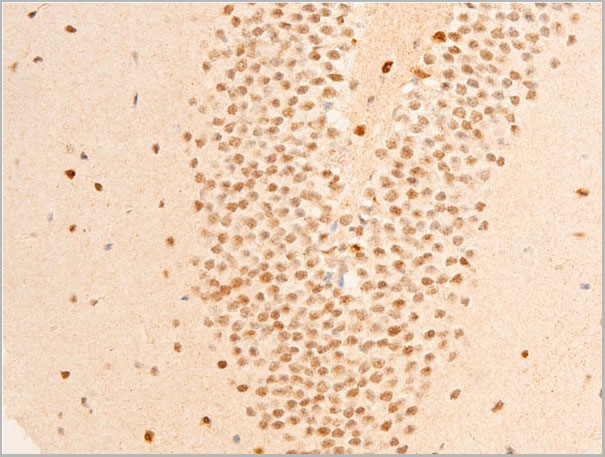
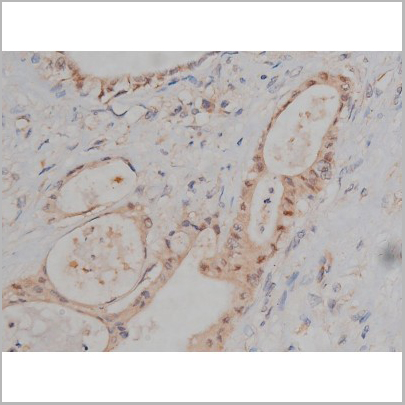
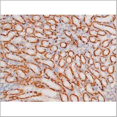
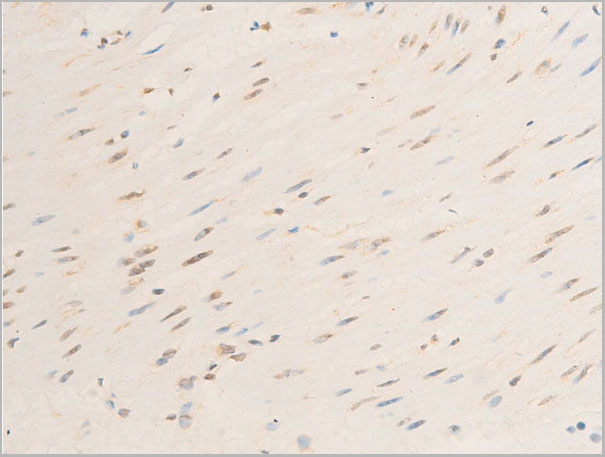
 IHC (Immunohistochemistry)
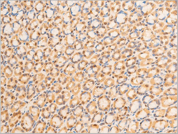
(AAA30992 at 1/200 staining Rat ganstric tissue sections by IHC-P. The tissue was formaldehyde fixed and a heat mediated antigen retrieval step in citrate buffer was performed. The tissue was then blocked and incubated with the antibody for 1.5 hours at 22 degree C. An HRP conjugated goat anti-rabbit antibody was used as the secondary.)
IHC (Immunohistochemistry)
(AAA30992 at 1/200 staining Rat ganstric tissue sections by IHC-P. The tissue was formaldehyde fixed and a heat mediated antigen retrieval step in citrate buffer was performed. The tissue was then blocked and incubated with the antibody for 1.5 hours at 22 degree C. An HRP conjugated goat anti-rabbit antibody was used as the secondary.)
SP1, Polyclonal Antibody (Cat# AAA30992)
What Are Phospho Antibodies?
Protein phosphorylation is a process where a phosphate group is added to certain amino acid residues of a protein – usually serine (S), threonine (T), or tyrosine (Y) - by enzymes called kinases. This process is integral in controlling cellular signaling, cellular growth, and other biological functions.
Our catalog includes a wide range of phospho-specific antibodies that can accurately detect this important marker. They perform strongly in widely-used laboratory applications such as Western blot, flow cytometry, immunohistochemistry, and immunofluorescence microscopy. We value your trust in us and are committed to providing top-quality products and services. All of our antibodies are guaranteed to work for the applications and species indicated on our website & associated product pages.
What Are The Key Applications of Phospho Antibodies?
1. Western Blotting
One of the first steps a researcher can take in utilizing these phospho-specific antibodies, is to check if the antibody works using a technique referred to as “Western blot”. For those unfamiliar, Western Blot aids in showing whether the protein that the antibody recognizes is appearing at the correct/expected size. These phospho-specific antibodies should also be able to detect changes in the target protein’s phosphorylation (on/off state) when cells are stimulated in certain ways.
2. Staining of Fixed Cells (Immunocytochemistry)
Another routine use of these phospho-specific antibodies, is to test if the antibody is able to demonstrate similar performance when used on fixed cells (intact cells that have been preserved) as it did in the Western blot tests. It is an important aspect in many cases to confirm that the antibody works in actual intact cell samples. Ideally, the method used for cellular fixation should be the same as what is used in pathology labs (like using 10% formalin). To check if the antibody works well in tissue sections (FFPE), researchers will often test it on fixed cells that are processed similar to tissue samples.
3. Specificity Tests Using Peptides
In order to make sure that the antibody is only binding to the right target:
- Laboratory technicians will mix the antibody with phospho-peptides (short segments of the protein containing the phosphate group modification).
- If the antibody signal disappears, it is confirmation that it is binding to the correct phosphorylated location.
- A more robust test is to use both the phosphorylated and non-phosphorylated (dephosphorylated) versions of the protein. The antibody should react only with the phosphorylated one.
- Another method sometimes utilized is to treat the sample with an enzyme, such as alkaline phosphatase, that specifically removes phosphate groups. If the antibody signal disappears after this, it also confirms specificity.
4. Genetic Confirmation
As a final step, scientists can genetically manipulate the nucleotide sequence and alter the target protein by removing the exact site where phosphorylation happens. If the antibody no longer appears to detect the modified protein, it is strong evidence supporting the antibody being specific for that phosphorylated site.
Why Buy Phospho Antibodies Through Us?
- The production laboratory adheres to strict and consistent protocols prior to releasing any of these phospho-specific antibodies:
- Standard methods and proper controls in all tests to ensure high quality.
- These antibodies are tested and validated in different cell types and species.
- High quality control criterion to ensure each batch is consistent, so you will obtain reliable results every time.
FAQ
1. What Are Phospho-Specific Antibodies?
Phospho-specific antibodies are made to detect proteins only when they have a phosphate group linked to a specific amino acid residue. This empowers scientists understand if a protein is "turned on" or active, based on its phosphorylation state.
2. How to Detect Phosphorylated Proteins in a Western Blot?
To find out if a protein is phosphorylated using Western blot:
- Use a phospho-specific antibody that binds only to the phosphorylated form of the protein.
- You can also use a “regular” antibody for the same amino acid sequence of the protein that the phospho-specific antibody is binding to (but in this case, this antibody will not bind if there is a phosphate group present) in order to compare how much of it is phosphorylated versus how much is non-phosphorylated (or “total” protein, if the “normal” antibody’s epitopes are non-phospho-site-specific).
3. How to Choose the Best Antibody?
Here are some simple tips to help you pick the right antibody:
- Know your target
- Match your sample characteristics
- Confirm the intended use is appropriate
- Check “host” and “type”
- Check the “quality” of the presented data/images
- Appraise whether the available validation meets your needs