Filters
▼Clonality
▼Type
▼Reactivity
▼Gene Name
▼Isotype
▼Host
▼Application
▼Clone
▼Phospho Antibodies
Phospho-specific antibodies’ typical purpose is to enable researchers to detect changes in proteins. They will exclusively bind to the amino acid sequence on a protein that has been phosphorylated (which is both a physical & chemical change) and do not bind to the same amino acid sequence on said protein if it lacks said phosphorylation. This aids in being able to clearly see and understand the data produced from this particular protein modification.
Viewing 5050-5100 of 5298 product results
 IF (Immunofluorescence)
(P-peptide-+ Immunofluorescence analysis of HeLa cells, using TGF beta Receptor I (Phospho-Ser165) antibody.)
IF (Immunofluorescence)
(P-peptide-+ Immunofluorescence analysis of HeLa cells, using TGF beta Receptor I (Phospho-Ser165) antibody.)
TGF beta Receptor I, Antibody (Cat# AAA18002)
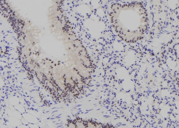
 IHC (Immunohistochemistry)
(Immunohistochemistry analysis of paraffin-embedded human brain tissue, using Kv4.2/KCND2 (Phospho-Ser616) antibody.)
IHC (Immunohistochemistry)
(Immunohistochemistry analysis of paraffin-embedded human brain tissue, using Kv4.2/KCND2 (Phospho-Ser616) antibody.)
Kv4.2/KCND2, Antibody (Cat# AAA18001)


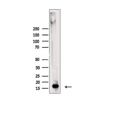
 WB (Western Blot)
(Detection of human Phospho KAP-1 (S824) by western blot. Samples: Whole cell lysate (25 ug) from HEK293T cells treated with 100 uM etoposide (+) or mock treated (-) prepared using NETN lysis buffer. Antibody: Rabbit anti-Phospho KAP-1 (S824) recombinant monoclonal antibody [BL-246-7B5] (AAA23791 lot 4) used at 1:1000. Secondary: HRP-conjugated goat anti-rabbit IgG . Chemiluminescence with an exposure time of 3 seconds. Lower Panel: Rabbit anti-KAP1 recombinant monoclonal antibody [BL-248-2G6] .)
WB (Western Blot)
(Detection of human Phospho KAP-1 (S824) by western blot. Samples: Whole cell lysate (25 ug) from HEK293T cells treated with 100 uM etoposide (+) or mock treated (-) prepared using NETN lysis buffer. Antibody: Rabbit anti-Phospho KAP-1 (S824) recombinant monoclonal antibody [BL-246-7B5] (AAA23791 lot 4) used at 1:1000. Secondary: HRP-conjugated goat anti-rabbit IgG . Chemiluminescence with an exposure time of 3 seconds. Lower Panel: Rabbit anti-KAP1 recombinant monoclonal antibody [BL-248-2G6] .)
KAP-1, Monoclonal Recombinant Antibody (Cat# AAA23791)


 IHC (Immunohistochemistry)
(Dilution: Western Blot: 1/500 - 1/2000. Immunohistochemistry: 1/100 - 1/300. Immunofluorescence: 1/200 - 1/1000. ELISA: 1/20000. Not yet tested in other applications.)
IHC (Immunohistochemistry)
(Dilution: Western Blot: 1/500 - 1/2000. Immunohistochemistry: 1/100 - 1/300. Immunofluorescence: 1/200 - 1/1000. ELISA: 1/20000. Not yet tested in other applications.)
FoxO3A, Polyclonal Antibody (Cat# AAA28927)


 IHC (Immunohistochemistry)
(Dilution: IF: 1:50-200 WB 1:500-2000, IHC 1:50-300 IHC 1:50-300)
IHC (Immunohistochemistry)
(Dilution: IF: 1:50-200 WB 1:500-2000, IHC 1:50-300 IHC 1:50-300)
ERK 1/2, Polyclonal Antibody (Cat# AAA28925)
 Standard Curve (Sample)
phospho Tau Protein, ELISA Kit (Cat# AAA17648)

 WB (Western Blot)
(Western blot analysis of extracts from 293 cells using Src (Ab-529) antibody and Src (phospho-Tyr529) antibody (AAA17990, Lane 3 and 4).)
WB (Western Blot)
(Western blot analysis of extracts from 293 cells using Src (Ab-529) antibody and Src (phospho-Tyr529) antibody (AAA17990, Lane 3 and 4).)
Src, Antibody (Cat# AAA17990)


 WB (Western Blot)
(Western blot analysis of ENO3 over-expressed 293 cell line, cotransfected with ENO3 Validated Chimera RNAi (Lane 2) or non-transfected control (Lane 1). Blot probed with ENO3 monoclonal antibody. GAPDH (36.1kD) used as specificity and loading control.)
WB (Western Blot)
(Western blot analysis of ENO3 over-expressed 293 cell line, cotransfected with ENO3 Validated Chimera RNAi (Lane 2) or non-transfected control (Lane 1). Blot probed with ENO3 monoclonal antibody. GAPDH (36.1kD) used as specificity and loading control.)
ENO3, Monoclonal Antibody (Cat# AAA24498)


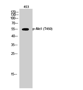

 IHC (Immunohistchemistry)
(Dilution: Western Blot: 1/500 - 1/2000. Immunohistochemistry: 1/100 - 1/300. ELISA: 1/5000. Not yet tested in other applications.)
IHC (Immunohistchemistry)
(Dilution: Western Blot: 1/500 - 1/2000. Immunohistochemistry: 1/100 - 1/300. ELISA: 1/5000. Not yet tested in other applications.)
Akt1, Polyclonal Antibody (Cat# AAA28920)
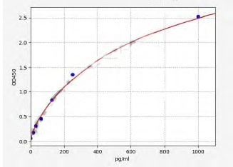
 Standard Curve (Sample)
Phospho Tau (P181), ELISA Kit (Cat# AAA27564)


 IHC (Immunohistochemistry)
(Dilution: IF: 1:50-200 Western Blot: 1/500 - 1/2000. Immunohistochemistry: 1/100 - 1/300. ELISA: 1/20000. Not yet tested in other applications.)
IHC (Immunohistochemistry)
(Dilution: IF: 1:50-200 Western Blot: 1/500 - 1/2000. Immunohistochemistry: 1/100 - 1/300. ELISA: 1/20000. Not yet tested in other applications.)
AP-1, Polyclonal Antibody (Cat# AAA28921)


 IHC (Immunohistochemistry)
(Dilution: IF: 1:50-200 Western Blot: 1/500 - 1/2000. Immunohistochemistry: 1/100 - 1/300. ELISA: 1/40000. Not yet tested in other applications.)
IHC (Immunohistochemistry)
(Dilution: IF: 1:50-200 Western Blot: 1/500 - 1/2000. Immunohistochemistry: 1/100 - 1/300. ELISA: 1/40000. Not yet tested in other applications.)
Akt, Polyclonal Antibody (Cat# AAA28919)


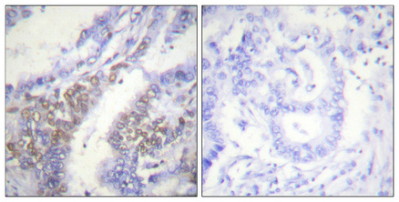
 IHC (Immunohistchemistry)
(At 1/200 staining Human lung cancer tissue sections by IHC-P. The tissue was formaldehyde fixed and a heat mediated antigen retrieval step in citrate buffer was performed. The tissue was then blocked and incubated with the antibody for 1.5 hours at 22 degree C. An HRP conjugated goat anti-rabbit antibody was used as the secondary antibody.)
IHC (Immunohistchemistry)
(At 1/200 staining Human lung cancer tissue sections by IHC-P. The tissue was formaldehyde fixed and a heat mediated antigen retrieval step in citrate buffer was performed. The tissue was then blocked and incubated with the antibody for 1.5 hours at 22 degree C. An HRP conjugated goat anti-rabbit antibody was used as the secondary antibody.)
CUTL1, Polyclonal Antibody (Cat# AAA31424)
Predicted Reactivity: Pig (100%), Sheep (100%), Dog (100%), Xenopus (100%)
 Standard Curve (Sample)
Phospho-CAMP Response Element Binding Protein (pCREB), ELISA Kit (Cat# AAA18969)


 IHC (Immunohistochemistry)
(At 1/100 staining Mouse brain tissue by IHC-P. The sample was formaldehyde fixed and a heat mediated antigen retrieval step in citrate buffer was performed. The sample was then blocked and incubated with the primary antibody at 4 degree C overnight. An HRP conjugated anti-Rabbit antibody was used as the secondary antibody.)
IHC (Immunohistochemistry)
(At 1/100 staining Mouse brain tissue by IHC-P. The sample was formaldehyde fixed and a heat mediated antigen retrieval step in citrate buffer was performed. The sample was then blocked and incubated with the primary antibody at 4 degree C overnight. An HRP conjugated anti-Rabbit antibody was used as the secondary antibody.)
HBP1, Polyclonal Antibody (Cat# AAA31326)


 Application Data
(At 25 degree C. Samples were then incubated with primary Ab(At 37 degree C. An AlexaFluor594 conjugated goat anti-rabbit IgG(H+L) Ab(Red) and an AlexaFluor488 conjugated goat anti-mouse IgG(H+L) Ab(Green) were used as the secondary antibody.The nuclear counter stain is DAPI(blue).)
Application Data
(At 25 degree C. Samples were then incubated with primary Ab(At 37 degree C. An AlexaFluor594 conjugated goat anti-rabbit IgG(H+L) Ab(Red) and an AlexaFluor488 conjugated goat anti-mouse IgG(H+L) Ab(Green) were used as the secondary antibody.The nuclear counter stain is DAPI(blue).)
FRS2, Polyclonal Antibody (Cat# AAA31412)
Predicted Reactivity: Pig (100%), Bovine (100%), Horse (100%), Sheep (100%), Rabbit (100%), Dog (100%), Chicken (100%)


 WB (Western Blot)
(Western blot analysis of Phospho-AML1 (Ser397) in lysates of Jurkat, using Phospho-AML1 (Ser397) Antibody.)
WB (Western Blot)
(Western blot analysis of Phospho-AML1 (Ser397) in lysates of Jurkat, using Phospho-AML1 (Ser397) Antibody.)
AML1, Polyclonal Antibody (Cat# AAA31153)
Predicted: Pig, Zebrafish, Bovine, Horse, Rabbit, Dog, Chicken, Xenopus


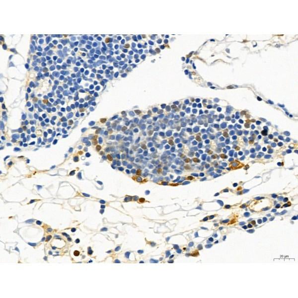
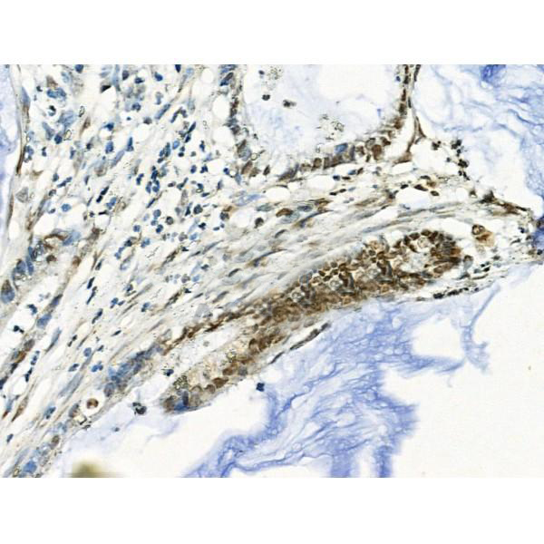
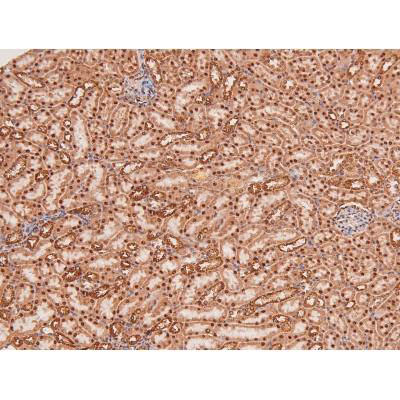
 IHC (Immunohistochemistry)
(At 1/100 staining Human kidney cancer and adjacent normal tissues by IHC-P. The sample was formaldehyde fixed and a heat mediated antigen retrieval step in citrate buffer was performed. The sample was then blocked and incubated with the primary antibody at 4 degree C overnight. An HRP conjugated anti-Rabbit antibody was used as the secondary antibody.)
IHC (Immunohistochemistry)
(At 1/100 staining Human kidney cancer and adjacent normal tissues by IHC-P. The sample was formaldehyde fixed and a heat mediated antigen retrieval step in citrate buffer was performed. The sample was then blocked and incubated with the primary antibody at 4 degree C overnight. An HRP conjugated anti-Rabbit antibody was used as the secondary antibody.)
Smad5, Polyclonal Antibody (Cat# AAA31331)


 WB (Western Blot)
(Western blot analysis of ENO3 over-expressed 293 cell line, cotransfected with ENO3 Validated Chimera RNAi (Lane 2) or non-transfected control (Lane 1). Blot probed with ENO3 monoclonal antibody. GAPDH (36.1kD) used as specificity and loading control.)
WB (Western Blot)
(Western blot analysis of ENO3 over-expressed 293 cell line, cotransfected with ENO3 Validated Chimera RNAi (Lane 2) or non-transfected control (Lane 1). Blot probed with ENO3 monoclonal antibody. GAPDH (36.1kD) used as specificity and loading control.)
ENO3, Monoclonal Antibody (Cat# AAA25384)


 Application Data
(At 25 degree C. Samples were then incubated with primary Ab(At 37 degree C. An AlexaFluor594 conjugated goat anti-rabbit IgG(H+L) Ab(Red) and an AlexaFluor488 conjugated goat anti-mouse IgG(H+L) Ab(Green) were used as the secondary antibody.The nuclear counter stain is DAPI(blue).)
Application Data
(At 25 degree C. Samples were then incubated with primary Ab(At 37 degree C. An AlexaFluor594 conjugated goat anti-rabbit IgG(H+L) Ab(Red) and an AlexaFluor488 conjugated goat anti-mouse IgG(H+L) Ab(Green) were used as the secondary antibody.The nuclear counter stain is DAPI(blue).)
MLC2, Polyclonal Antibody (Cat# AAA31471)
Predicted Reactivity: Horse (100%), Sheep (100%), Rabbit (100%), Dog (100%), Chicken (100%)


 Application Data
(At 25 degree C. The primary antibody was diluted at 1/200 and incubated with the sample for 1 hour at 37 degree C. An Alexa Fluor 594 conjugated goat anti-rabbit IgG (H+L) antibody(Red), diluted at 1/600, was used as secondary antibody.)
Application Data
(At 25 degree C. The primary antibody was diluted at 1/200 and incubated with the sample for 1 hour at 37 degree C. An Alexa Fluor 594 conjugated goat anti-rabbit IgG (H+L) antibody(Red), diluted at 1/600, was used as secondary antibody.)
JAK3, Polyclonal Antibody (Cat# AAA31405)
Predicted Reactivity: Pig (100%), Bovine (91%), Horse (100%), Sheep (100%), Dog (100%)


 IF (Immunofluorescence)
(Immunofluorescence analysis of U-2 OS cells using Phospho-Histone H3-S10/T11 Rabbit pAb at dilution of 100 (40x lens). Blue: DAPI for nuclear staining.)
IF (Immunofluorescence)
(Immunofluorescence analysis of U-2 OS cells using Phospho-Histone H3-S10/T11 Rabbit pAb at dilution of 100 (40x lens). Blue: DAPI for nuclear staining.)
Phospho-Histone H3-S10/T11, Polyclonal Antibody (Cat# AAA28391)


 IHC (Immunohistchemistry)
(Dilution: Western Blot: 1/500 - 1/2000. Immunohistochemistry: 1/100 - 1/300. ELISA: 1/5000. Not yet tested in other applications.)
IHC (Immunohistchemistry)
(Dilution: Western Blot: 1/500 - 1/2000. Immunohistochemistry: 1/100 - 1/300. ELISA: 1/5000. Not yet tested in other applications.)
FAK, Polyclonal Antibody (Cat# AAA28953)


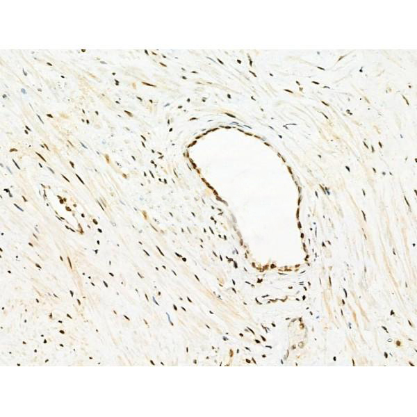
 IHC (Immunohistchemistry)
(At 1/100 staining Human gastric cancer by IHC-P. The sample was formaldehyde fixed and a heat mediated antigen retrieval step in citrate buffer was performed. The sample was then blocked and incubated with the primary antibody at 4 degree C overnight. An HRP conjugated anti-Rabbit antibody was used as the secondary antibody.)
IHC (Immunohistchemistry)
(At 1/100 staining Human gastric cancer by IHC-P. The sample was formaldehyde fixed and a heat mediated antigen retrieval step in citrate buffer was performed. The sample was then blocked and incubated with the primary antibody at 4 degree C overnight. An HRP conjugated anti-Rabbit antibody was used as the secondary antibody.)
SRPK2, Polyclonal Antibody (Cat# AAA31293)


 IHC (Immunohistochemistry)
(Dilution: Western Blot: 1/500 - 1/2000. Immunohistochemistry: 1/100 - 1/300. Immunofluorescence: 1/200 - 1/1000. ELISA: 1/10000. Not yet tested in other applications.)
IHC (Immunohistochemistry)
(Dilution: Western Blot: 1/500 - 1/2000. Immunohistochemistry: 1/100 - 1/300. Immunofluorescence: 1/200 - 1/1000. ELISA: 1/10000. Not yet tested in other applications.)
ERK 1/2, Polyclonal Antibody (Cat# AAA28926)


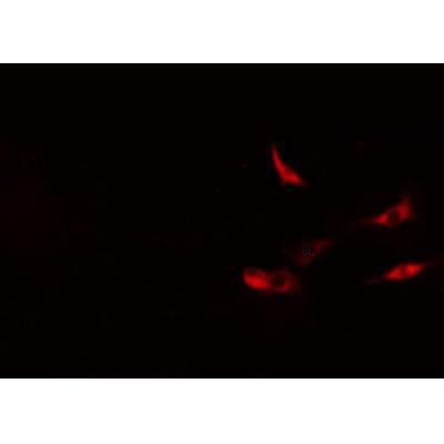
 ICC (Immunocytochemistry)
(ICC staining Phospho-JunD (S255) in MCF-7 cells (green). The nuclear counter stain is DAPI (blue). Cells were fixed in paraformaldehyde, permeabilised with 0.25% Triton X100/PBS.)
ICC (Immunocytochemistry)
(ICC staining Phospho-JunD (S255) in MCF-7 cells (green). The nuclear counter stain is DAPI (blue). Cells were fixed in paraformaldehyde, permeabilised with 0.25% Triton X100/PBS.)
JunD, Monoclonal Antibody (Cat# AAA29784)


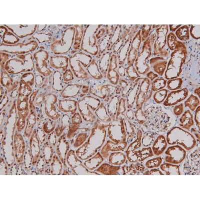
 IHC (Immunohistochemistry)
(At 1/100 staining Mouse kidney tissue by IHC-P. The sample was formaldehyde fixed and a heat mediated antigen retrieval step in citrate buffer was performed. The sample was then blocked and incubated with the primary antibody at 4 degree C overnight. An HRP conjugated anti-Rabbit antibody was used as the secondary antibody.)
IHC (Immunohistochemistry)
(At 1/100 staining Mouse kidney tissue by IHC-P. The sample was formaldehyde fixed and a heat mediated antigen retrieval step in citrate buffer was performed. The sample was then blocked and incubated with the primary antibody at 4 degree C overnight. An HRP conjugated anti-Rabbit antibody was used as the secondary antibody.)
YB1, Polyclonal Antibody (Cat# AAA31294)


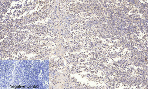
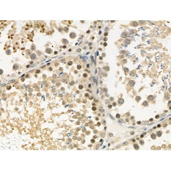
 IHC (Immunohistchemistry)
(Immunohistochemical analysis of (AAA28653) on paraffin-embedded Human breastcarcinoma tissue. Tissue was fixed withformaldehyde at room temperature. Heatinduced epitope retrieval was performed byEDTA buffer (pH9. 0). Samples wereincubated with primary antibody(1:100) for 1hour at room temperature. Undiluted CRFAnti-Polyvalent HRP Polymer antibody wasused as the secondary antibody.)
IHC (Immunohistchemistry)
(Immunohistochemical analysis of (AAA28653) on paraffin-embedded Human breastcarcinoma tissue. Tissue was fixed withformaldehyde at room temperature. Heatinduced epitope retrieval was performed byEDTA buffer (pH9. 0). Samples wereincubated with primary antibody(1:100) for 1hour at room temperature. Undiluted CRFAnti-Polyvalent HRP Polymer antibody wasused as the secondary antibody.)
Phospho-CDC25A (S124), Polyclonal Antibody (Cat# AAA28653)


 Application Data
(At 25 degree C. Samples were then incubated with primary Ab(At 37 degree C. An AlexaFluor594 conjugated goat anti-rabbit IgG(H+L) Ab(Red) and an AlexaFluor488 conjugated goat anti-mouse IgG(H+L) Ab(Green) were used as the secondary antibody.The nuclear counter stain is DAPI (blue).)
Application Data
(At 25 degree C. Samples were then incubated with primary Ab(At 37 degree C. An AlexaFluor594 conjugated goat anti-rabbit IgG(H+L) Ab(Red) and an AlexaFluor488 conjugated goat anti-mouse IgG(H+L) Ab(Green) were used as the secondary antibody.The nuclear counter stain is DAPI (blue).)
JIP1, Polyclonal Antibody (Cat# AAA31318)


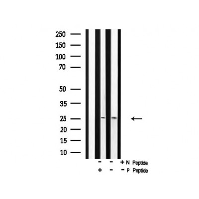
 WB (Western Blot)
(Detection of human Phospho-MCM2 (S53) by western blot. Samples: Whole cell lysate (10 ug) from HDLM-2, HeLa, Jurkat, MCF-7, MOLT-4, Malme-3M, HEK293T, and Raji cells prepared using NETN lysis buffer. Antibody: Rabbit anti-Phospho-MCM2 (S53) recombinant monoclonal antibody (AAA23835 lot 1) used at 1:1000. Secondary: HRP-conjugated goat anti-rabbit IgG . Detection: Chemiluminescence with an exposure time of 30 seconds. Lower Panel: Rabbit anti-Actin recombinant monoclonal .)
WB (Western Blot)
(Detection of human Phospho-MCM2 (S53) by western blot. Samples: Whole cell lysate (10 ug) from HDLM-2, HeLa, Jurkat, MCF-7, MOLT-4, Malme-3M, HEK293T, and Raji cells prepared using NETN lysis buffer. Antibody: Rabbit anti-Phospho-MCM2 (S53) recombinant monoclonal antibody (AAA23835 lot 1) used at 1:1000. Secondary: HRP-conjugated goat anti-rabbit IgG . Detection: Chemiluminescence with an exposure time of 30 seconds. Lower Panel: Rabbit anti-Actin recombinant monoclonal .)
MCM2, Monoclonal Recombinant Antibody (Cat# AAA23835)


 IHC (Immunohistchemistry)
(At 1/100 staining Human prostate cancer and adjacent normal tissues by IHC-P. The sample was formaldehyde fixed and a heat mediated antigen retrieval step in citrate buffer was performed. The sample was then blocked and incubated with the primary antibody at 4 degree C overnight. An HRP conjugated anti-Rabbit antibody was used as the secondary antibody.)
IHC (Immunohistchemistry)
(At 1/100 staining Human prostate cancer and adjacent normal tissues by IHC-P. The sample was formaldehyde fixed and a heat mediated antigen retrieval step in citrate buffer was performed. The sample was then blocked and incubated with the primary antibody at 4 degree C overnight. An HRP conjugated anti-Rabbit antibody was used as the secondary antibody.)
TOPBP1, Polyclonal Antibody (Cat# AAA31467)
Predicted Reactivity: Pig (100%), Zebrafish (80%), Bovine (100%), Horse (100%), Sheep (100%), Rabbit (100%), Dog (100%), Chicken (80%), Xenopus (80%)


 ICC (Immunocytochemistry)
(ICC staining Phospho-eIF4E (S209) in 293 cells (green). The nuclear counter stain is DAPI (blue). Cells were fixed in paraformaldehyde, permeabilised with 0.25% Triton X100/PBS.)
ICC (Immunocytochemistry)
(ICC staining Phospho-eIF4E (S209) in 293 cells (green). The nuclear counter stain is DAPI (blue). Cells were fixed in paraformaldehyde, permeabilised with 0.25% Triton X100/PBS.)
eIF4E, Monoclonal Antibody (Cat# AAA29779)


 Application Data
(At 25 degree C. The primary antibody was diluted at 1/200 and incubated with the sample for 1 hour at 37 degree C. An Alexa Fluor 594 conjugated goat anti-rabbit IgG (H+L) Ab, diluted at 1/600, was used as the secondary antibody.)
Application Data
(At 25 degree C. The primary antibody was diluted at 1/200 and incubated with the sample for 1 hour at 37 degree C. An Alexa Fluor 594 conjugated goat anti-rabbit IgG (H+L) Ab, diluted at 1/600, was used as the secondary antibody.)
p27 Kip1, Polyclonal Antibody (Cat# AAA31387)
Predicted Reactivity: Bovine (89%), Sheep (89%), Rabbit (100%), Dog (100%), Chicken (89%)


 Application Data
(At 25 degree C. Samples were then incubated with primary Ab(At 37 degree C. An AlexaFluor594 conjugated goat anti-rabbit IgG(H+L) Ab(Red) and an AlexaFluor488 conjugated goat anti-mouse IgG(H+L) Ab(Green) were used as the secondary antibody.The nuclear counter stain is DAPI (blue).)
Application Data
(At 25 degree C. Samples were then incubated with primary Ab(At 37 degree C. An AlexaFluor594 conjugated goat anti-rabbit IgG(H+L) Ab(Red) and an AlexaFluor488 conjugated goat anti-mouse IgG(H+L) Ab(Green) were used as the secondary antibody.The nuclear counter stain is DAPI (blue).)
eIF4B, Polyclonal Antibody (Cat# AAA31302)


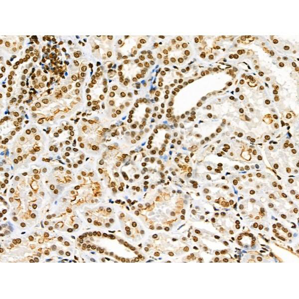
 IHC (Immunohistochemistry)
(At 1/100 staining Human esophageal cancer and adjacent normal tissues by IHC-P. The sample was formaldehyde fixed and a heat mediated antigen retrieval step in citrate buffer was performed. The sample was then blocked and incubated with the primary antibody at 4 degree C overnight. An HRP conjugated anti-Rabbit antibody was used as the secondary antibody.)
IHC (Immunohistochemistry)
(At 1/100 staining Human esophageal cancer and adjacent normal tissues by IHC-P. The sample was formaldehyde fixed and a heat mediated antigen retrieval step in citrate buffer was performed. The sample was then blocked and incubated with the primary antibody at 4 degree C overnight. An HRP conjugated anti-Rabbit antibody was used as the secondary antibody.)
EIF2S2, Polyclonal Antibody (Cat# AAA31456)
Predicted Reactivity: Pig (100%), Zebrafish (100%), Bovine (100%), Horse (100%), Sheep (83%), Rabbit (100%), Dog (83%), Xenopus (100%)


 IHC (Immunohistochemistry)
(At 1/100 staining Human prostate cancer and adjacent normal tissues by IHC-P. The sample was formaldehyde fixed and a heat mediated antigen retrieval step in citrate buffer was performed. The sample was then blocked and incubated with the primary antibody at 4 degree C overnight. An HRP conjugated anti-Rabbit antibody was used as the secondary antibody.)
IHC (Immunohistochemistry)
(At 1/100 staining Human prostate cancer and adjacent normal tissues by IHC-P. The sample was formaldehyde fixed and a heat mediated antigen retrieval step in citrate buffer was performed. The sample was then blocked and incubated with the primary antibody at 4 degree C overnight. An HRP conjugated anti-Rabbit antibody was used as the secondary antibody.)
EEF1A2, Polyclonal Antibody (Cat# AAA31289)


 Application Data
(At 25 degree C. The primary antibody was diluted at 1/200 and incubated with the sample for 1 hour at 37 degree C. An Alexa Fluor 594 conjugated goat anti-rabbit IgG (H+L) antibody(Red), diluted at 1/600, was used as secondary antibody.)
Application Data
(At 25 degree C. The primary antibody was diluted at 1/200 and incubated with the sample for 1 hour at 37 degree C. An Alexa Fluor 594 conjugated goat anti-rabbit IgG (H+L) antibody(Red), diluted at 1/600, was used as secondary antibody.)
JAK3, Polyclonal Antibody (Cat# AAA31404)
Predicted Reactivity: Bovine (100%), Sheep (100%), Dog (100%)


 WB (Western Blot)
(Western blot analysis of Phospho-MOB1A (Tyr26) in lysates of 293 IFN 2500U/ml 30', using Phospho-MOB1A (Tyr26) Antibody.)
WB (Western Blot)
(Western blot analysis of Phospho-MOB1A (Tyr26) in lysates of 293 IFN 2500U/ml 30', using Phospho-MOB1A (Tyr26) Antibody.)
MOB1A, Polyclonal Antibody (Cat# AAA31161)
Predicted: Bovine, Horse, Sheep, Rabbit, Dog, Xenopus


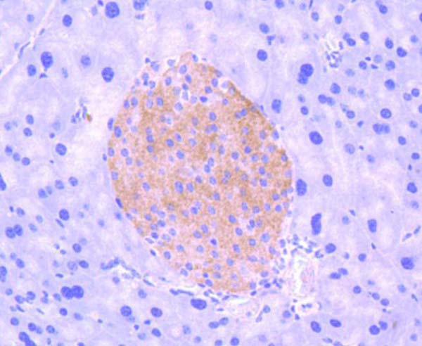
 IHC (Immunohistochemistry)
(At 1/100 staining Human gastric cancer and adjacent normal tissues by IHC-P. The sample was formaldehyde fixed and a heat mediated antigen retrieval step in citrate buffer was performed. The sample was then blocked and incubated with the primary antibody at 4 degree C overnight. An HRP conjugated anti-Rabbit antibody was used as the secondary antibody.)
IHC (Immunohistochemistry)
(At 1/100 staining Human gastric cancer and adjacent normal tissues by IHC-P. The sample was formaldehyde fixed and a heat mediated antigen retrieval step in citrate buffer was performed. The sample was then blocked and incubated with the primary antibody at 4 degree C overnight. An HRP conjugated anti-Rabbit antibody was used as the secondary antibody.)
CAMKK1/2, Polyclonal Antibody (Cat# AAA31402)
Predicted Reactivity: Pig (100%), Bovine (100%), Sheep (100%), Rabbit (100%), Dog (100%), Chicken (100%), Xenopus (100%)


 IHC (Immunohistochemistry)
(Dilution: WB 1:500-2000, IF 1:50-300, IHC 1:50-300 IHC 1:50-300)
IHC (Immunohistochemistry)
(Dilution: WB 1:500-2000, IF 1:50-300, IHC 1:50-300 IHC 1:50-300)
p38, Polyclonal Antibody (Cat# AAA28937)


 ICC (Immunocytochemistry)
(ICC staining phospho -TrkB (Y817) in SH-SY-5Y cells (green). The nuclear counter stain is DAPI (blue). Cells were fixed in paraformaldehyde, permeabilised with 0.25% Triton X100/PBS.)
ICC (Immunocytochemistry)
(ICC staining phospho -TrkB (Y817) in SH-SY-5Y cells (green). The nuclear counter stain is DAPI (blue). Cells were fixed in paraformaldehyde, permeabilised with 0.25% Triton X100/PBS.)
TrkB, Monoclonal Antibody (Cat# AAA29783)


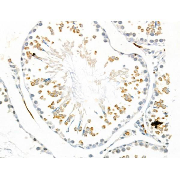
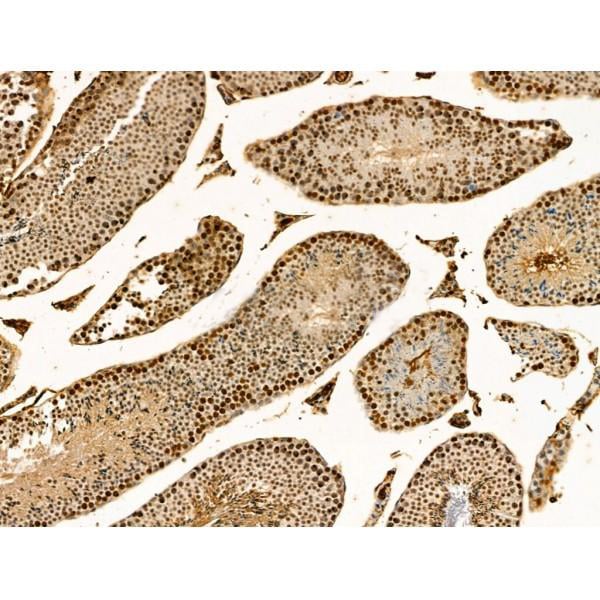
 IHC (Immunohistochemistry)
(At 1/100 staining Mouse testis tissue by IHC-P. The sample was formaldehyde fixed and a heat mediated antigen retrieval step in citrate buffer was performed. The sample was then blocked and incubated with the primary antibody at 4 degree C overnight. An HRP conjugated anti-Rabbit antibody was used as the secondary antibody.)
IHC (Immunohistochemistry)
(At 1/100 staining Mouse testis tissue by IHC-P. The sample was formaldehyde fixed and a heat mediated antigen retrieval step in citrate buffer was performed. The sample was then blocked and incubated with the primary antibody at 4 degree C overnight. An HRP conjugated anti-Rabbit antibody was used as the secondary antibody.)
IRS1, Polyclonal Antibody (Cat# AAA31366)
Predicted Reactivity: Pig (100%), Zebrafish (100%), Bovine (100%), Horse (100%), Sheep (100%), Rabbit (100%), Dog (100%)


 Application Data
(At 25 degree C. Samples were then incubated with primary Ab(At 37 degree C. An AlexaFluor594 conjugated goat anti-rabbit IgG(H+L) Ab(Red) and an AlexaFluor488 conjugated goat anti-mouse IgG(H+L) Ab(Green) were used as the secondary antibody.The nuclear counter stain is DAPI(blue).)
Application Data
(At 25 degree C. Samples were then incubated with primary Ab(At 37 degree C. An AlexaFluor594 conjugated goat anti-rabbit IgG(H+L) Ab(Red) and an AlexaFluor488 conjugated goat anti-mouse IgG(H+L) Ab(Green) were used as the secondary antibody.The nuclear counter stain is DAPI(blue).)
Bcl-2, Polyclonal Antibody (Cat# AAA31360)
Predicted Reactivity: Pig (86%), Bovine (83%)


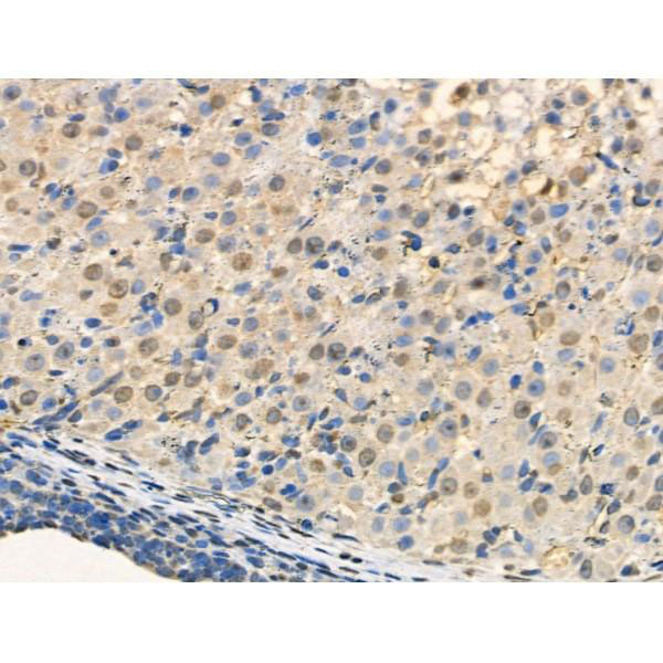
 IHC (Immunohistchemistry)
(At 1/100 staining Mouse liver tissue by IHC-P. The sample was formaldehyde fixed and a heat mediated antigen retrieval step in citrate buffer was performed. The sample was then blocked and incubated with the primary antibody at 4 degree C overnight. An HRP conjugated anti-Rabbit antibody was used as the secondary antibody.)
IHC (Immunohistchemistry)
(At 1/100 staining Mouse liver tissue by IHC-P. The sample was formaldehyde fixed and a heat mediated antigen retrieval step in citrate buffer was performed. The sample was then blocked and incubated with the primary antibody at 4 degree C overnight. An HRP conjugated anti-Rabbit antibody was used as the secondary antibody.)
NDEL1, Polyclonal Antibody (Cat# AAA31462)
Predicted Reactivity: Pig (100%), Bovine (100%), Horse (100%), Sheep (100%), Rabbit (100%), Dog (100%), Chicken (100%), Xenopus (100%)


 Application Data
(At 25 degree C. Samples were then incubated with primary Ab(At 37 degree C. An AlexaFluor594 conjugated goat anti-rabbit IgG(H+L) Ab(Red) and an AlexaFluor488 conjugated goat anti-mouse IgG(H+L) Ab(Green) were used as the secondary antibody.The nuclear counter stain is DAPI(blue).)
Application Data
(At 25 degree C. Samples were then incubated with primary Ab(At 37 degree C. An AlexaFluor594 conjugated goat anti-rabbit IgG(H+L) Ab(Red) and an AlexaFluor488 conjugated goat anti-mouse IgG(H+L) Ab(Green) were used as the secondary antibody.The nuclear counter stain is DAPI(blue).)
CDK5, Polyclonal Antibody (Cat# AAA31445)
Predicted Reactivity: Zebrafish (100%), Bovine (100%), Horse (100%), Sheep (100%), Rabbit (100%), Dog (100%), Chicken (100%), Xenopus (100%)


 Application Data
(At 25 degree C. Samples were then incubated with primary Ab(At 37 degree C. An AlexaFluor594 conjugated goat anti-rabbit IgG(H+L) Ab(Red) and an AlexaFluor488 conjugated goat anti-mouse IgG(H+L) Ab(Green) were used as the secondary antibody.The nuclear counter stain is DAPI (blue).)
Application Data
(At 25 degree C. Samples were then incubated with primary Ab(At 37 degree C. An AlexaFluor594 conjugated goat anti-rabbit IgG(H+L) Ab(Red) and an AlexaFluor488 conjugated goat anti-mouse IgG(H+L) Ab(Green) were used as the secondary antibody.The nuclear counter stain is DAPI (blue).)
MCM2, Polyclonal Antibody (Cat# AAA31328)


 WB (Western Blot)
(Western blot analysis of ENO3 over-expressed 293 cell line, cotransfected with ENO3 Validated Chimera RNAi (Lane 2) or non-transfected control (Lane 1). Blot probed with ENO3 monoclonal antibody. GAPDH (36.1kD) used as specificity and loading control.)
WB (Western Blot)
(Western blot analysis of ENO3 over-expressed 293 cell line, cotransfected with ENO3 Validated Chimera RNAi (Lane 2) or non-transfected control (Lane 1). Blot probed with ENO3 monoclonal antibody. GAPDH (36.1kD) used as specificity and loading control.)
ENO3, Monoclonal Antibody (Cat# AAA25679)


 ICC (Immunocytochemistry)
(ICC staining Phospho-RSK1 p90 (T359+S363) in 293 cells (green). The nuclear counter stain is DAPI (blue). Cells were fixed in paraformaldehyde, permeabilised with 0.25% Triton X100/PBS.)
ICC (Immunocytochemistry)
(ICC staining Phospho-RSK1 p90 (T359+S363) in 293 cells (green). The nuclear counter stain is DAPI (blue). Cells were fixed in paraformaldehyde, permeabilised with 0.25% Triton X100/PBS.)
RSK1, Monoclonal Antibody (Cat# AAA29777)


 Application Data
(At 25 degree C. The primary antibody was diluted at 1/200 and incubated with the sample for 1 hour at 37 degree C. An Alexa Fluor 594 conjugated goat anti-rabbit IgG (H+L) Ab, diluted at 1/600, was used as the secondary antibody.)
Application Data
(At 25 degree C. The primary antibody was diluted at 1/200 and incubated with the sample for 1 hour at 37 degree C. An Alexa Fluor 594 conjugated goat anti-rabbit IgG (H+L) Ab, diluted at 1/600, was used as the secondary antibody.)
Mnk1, Polyclonal Antibody (Cat# AAA31406)
Predicted Reactivity: Pig (100%), Zebrafish (100%), Bovine (100%), Horse (100%), Sheep (100%), Rabbit (100%), Dog (100%), Chicken (100%), Xenopus (100%)


 Application Data
(At 25 degree C. Samples were then incubated with primary Ab(At 37 degree C. An AlexaFluor594 conjugated goat anti-rabbit IgG(H+L) Ab(Red) and an AlexaFluor488 conjugated goat anti-mouse IgG(H+L) Ab(Green) were used as the secondary antibody.The nuclear counter stain is DAPI (blue).)
Application Data
(At 25 degree C. Samples were then incubated with primary Ab(At 37 degree C. An AlexaFluor594 conjugated goat anti-rabbit IgG(H+L) Ab(Red) and an AlexaFluor488 conjugated goat anti-mouse IgG(H+L) Ab(Green) were used as the secondary antibody.The nuclear counter stain is DAPI (blue).)
RPA32/RPA2, Polyclonal Antibody (Cat# AAA31304)
What Are Phospho Antibodies?
Protein phosphorylation is a process where a phosphate group is added to certain amino acid residues of a protein – usually serine (S), threonine (T), or tyrosine (Y) - by enzymes called kinases. This process is integral in controlling cellular signaling, cellular growth, and other biological functions.
Our catalog includes a wide range of phospho-specific antibodies that can accurately detect this important marker. They perform strongly in widely-used laboratory applications such as Western blot, flow cytometry, immunohistochemistry, and immunofluorescence microscopy. We value your trust in us and are committed to providing top-quality products and services. All of our antibodies are guaranteed to work for the applications and species indicated on our website & associated product pages.
What Are The Key Applications of Phospho Antibodies?
1. Western Blotting
One of the first steps a researcher can take in utilizing these phospho-specific antibodies, is to check if the antibody works using a technique referred to as “Western blot”. For those unfamiliar, Western Blot aids in showing whether the protein that the antibody recognizes is appearing at the correct/expected size. These phospho-specific antibodies should also be able to detect changes in the target protein’s phosphorylation (on/off state) when cells are stimulated in certain ways.
2. Staining of Fixed Cells (Immunocytochemistry)
Another routine use of these phospho-specific antibodies, is to test if the antibody is able to demonstrate similar performance when used on fixed cells (intact cells that have been preserved) as it did in the Western blot tests. It is an important aspect in many cases to confirm that the antibody works in actual intact cell samples. Ideally, the method used for cellular fixation should be the same as what is used in pathology labs (like using 10% formalin). To check if the antibody works well in tissue sections (FFPE), researchers will often test it on fixed cells that are processed similar to tissue samples.
3. Specificity Tests Using Peptides
In order to make sure that the antibody is only binding to the right target:
- Laboratory technicians will mix the antibody with phospho-peptides (short segments of the protein containing the phosphate group modification).
- If the antibody signal disappears, it is confirmation that it is binding to the correct phosphorylated location.
- A more robust test is to use both the phosphorylated and non-phosphorylated (dephosphorylated) versions of the protein. The antibody should react only with the phosphorylated one.
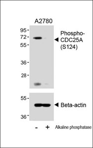
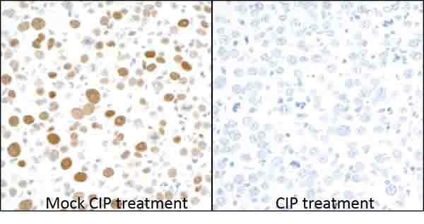
- Another method sometimes utilized is to treat the sample with an enzyme, such as alkaline phosphatase, that specifically removes phosphate groups. If the antibody signal disappears after this, it also confirms specificity.
4. Genetic Confirmation
As a final step, scientists can genetically manipulate the nucleotide sequence and alter the target protein by removing the exact site where phosphorylation happens. If the antibody no longer appears to detect the modified protein, it is strong evidence supporting the antibody being specific for that phosphorylated site.
Why Buy Phospho Antibodies Through Us?
- The production laboratory adheres to strict and consistent protocols prior to releasing any of these phospho-specific antibodies:
- Standard methods and proper controls in all tests to ensure high quality.
- These antibodies are tested and validated in different cell types and species.
- High quality control criterion to ensure each batch is consistent, so you will obtain reliable results every time.
FAQ
1. What Are Phospho-Specific Antibodies?
Phospho-specific antibodies are made to detect proteins only when they have a phosphate group linked to a specific amino acid residue. This empowers scientists understand if a protein is "turned on" or active, based on its phosphorylation state.
2. How to Detect Phosphorylated Proteins in a Western Blot?
To find out if a protein is phosphorylated using Western blot:
- Use a phospho-specific antibody that binds only to the phosphorylated form of the protein.
- You can also use a “regular” antibody for the same amino acid sequence of the protein that the phospho-specific antibody is binding to (but in this case, this antibody will not bind if there is a phosphate group present) in order to compare how much of it is phosphorylated versus how much is non-phosphorylated (or “total” protein, if the “normal” antibody’s epitopes are non-phospho-site-specific).
3. How to Choose the Best Antibody?
Here are some simple tips to help you pick the right antibody:
- Know your target
- Match your sample characteristics
- Confirm the intended use is appropriate
- Check “host” and “type”
- Check the “quality” of the presented data/images
- Appraise whether the available validation meets your needs