Filters
▼Clonality
▼Type
▼Reactivity
▼Gene Name
▼Isotype
▼Host
▼Application
▼Clone
▼Phospho Antibodies
Phospho-specific antibodies’ typical purpose is to enable researchers to detect changes in proteins. They will exclusively bind to the amino acid sequence on a protein that has been phosphorylated (which is both a physical & chemical change) and do not bind to the same amino acid sequence on said protein if it lacks said phosphorylation. This aids in being able to clearly see and understand the data produced from this particular protein modification.
Viewing 5000-5050 of 5298 product results
 Application Data
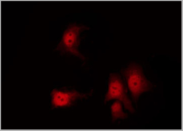
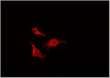
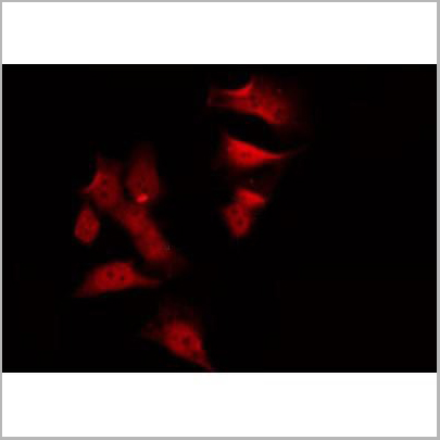
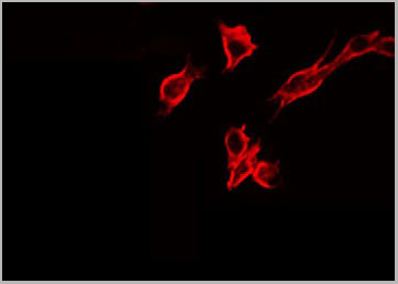
(AAA31369 staining Hela cells(4h of LPS treatment) by IF/ICC. The samples were fixed with PFA and permeabilized in 0.1% Triton X-100,then blocked in 10% serum for 45 minutes at 25°C. Samples were then incubated with primary Ab(AAA31369 1:200) and mouse anti-beta tubulin Ab(T0023 1:200) for 1 hour at 37°C. An AlexaFluor594 conjugated goat anti-rabbit IgG(H+L) Ab(Red) and an AlexaFluor488 conjugated goat anti-mouse IgG(H+L) Ab(Green) were used as the secondary Ab. The nuclear counter stain is DAPI(blue).)
Application Data
(AAA31369 staining Hela cells(4h of LPS treatment) by IF/ICC. The samples were fixed with PFA and permeabilized in 0.1% Triton X-100,then blocked in 10% serum for 45 minutes at 25°C. Samples were then incubated with primary Ab(AAA31369 1:200) and mouse anti-beta tubulin Ab(T0023 1:200) for 1 hour at 37°C. An AlexaFluor594 conjugated goat anti-rabbit IgG(H+L) Ab(Red) and an AlexaFluor488 conjugated goat anti-mouse IgG(H+L) Ab(Green) were used as the secondary Ab. The nuclear counter stain is DAPI(blue).)
FOXM1, Polyclonal Antibody (Cat# AAA31369)
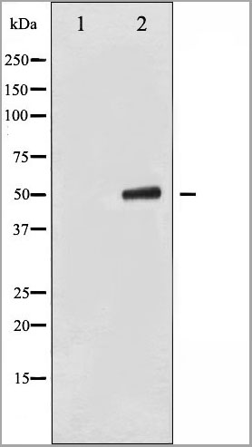
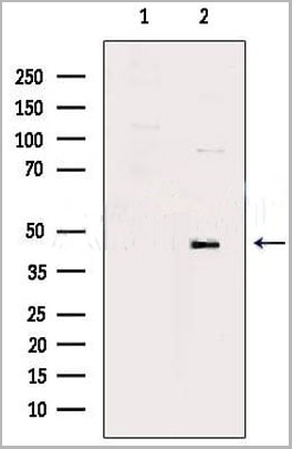
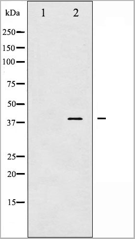
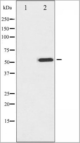
ENO3, Monoclonal Antibody (Cat# AAA24202)

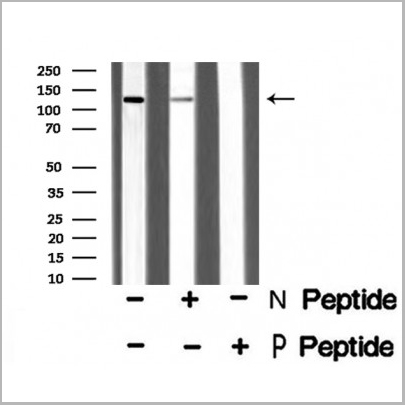
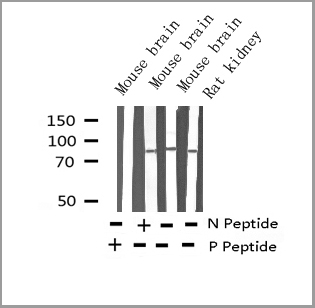
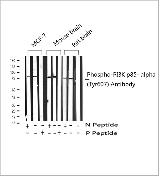 WB (Western Blot)
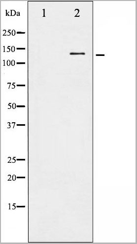
(Western blot analysis of Phospho-PI3K p85 alpha (Tyr607) expression in various lysates)
WB (Western Blot)
(Western blot analysis of Phospho-PI3K p85 alpha (Tyr607) expression in various lysates)
PI3K p85 alpha, Polyclonal Antibody (Cat# AAA31027)
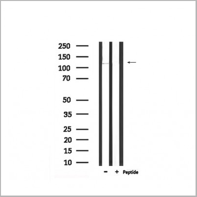
 WB (Western Blot)
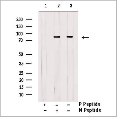
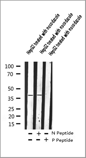
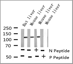
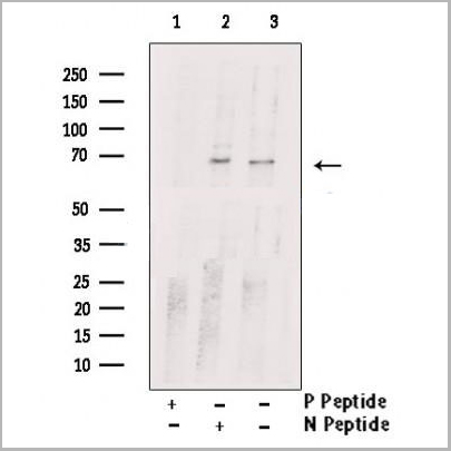
(Western blot analysis of Adrenergic Receptor beta2 phosphorylation expression in nocodazole treated HepG2 whole cell lysates, The lane on the left is treated with the antigen-specific peptide.)
WB (Western Blot)
(Western blot analysis of Adrenergic Receptor beta2 phosphorylation expression in nocodazole treated HepG2 whole cell lysates, The lane on the left is treated with the antigen-specific peptide.)
Adrenergic Receptor beta2, Polyclonal Antibody (Cat# AAA30989)
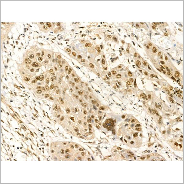
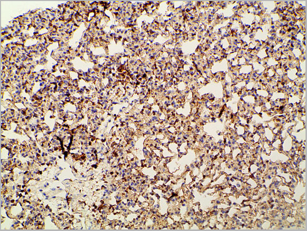
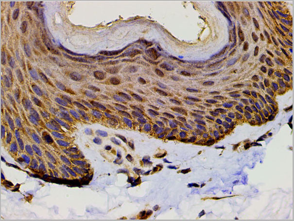
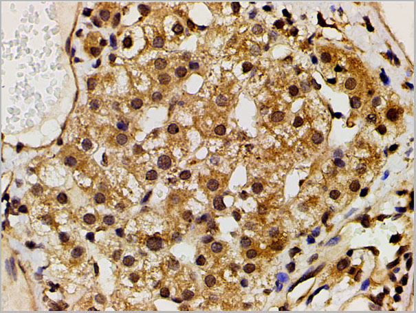
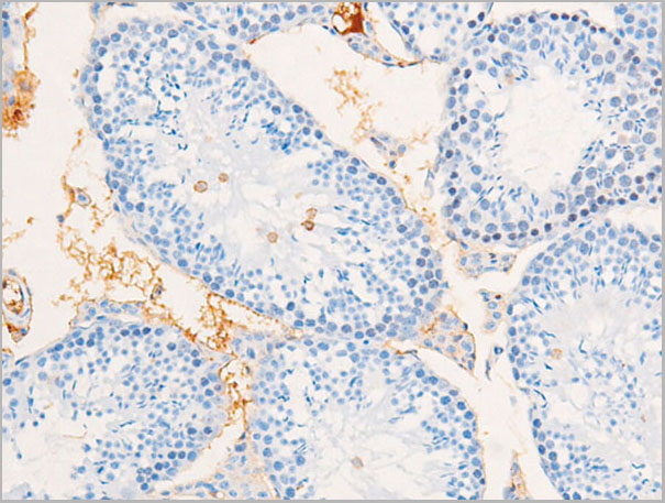
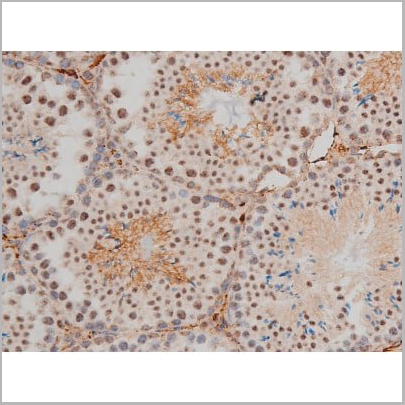
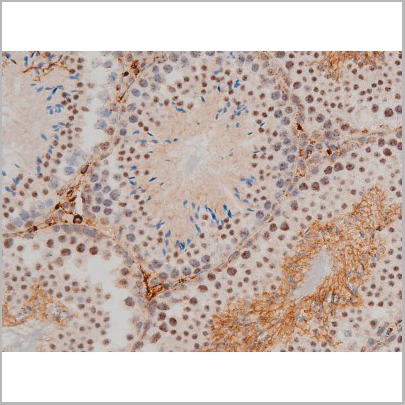
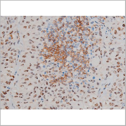
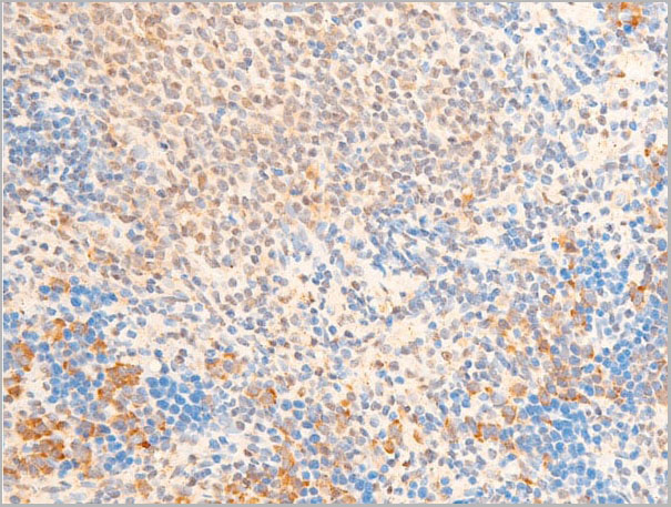
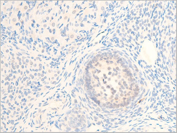
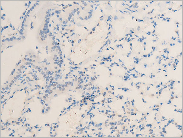
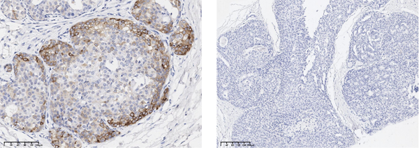
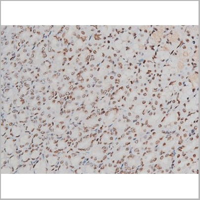
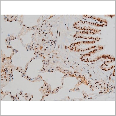
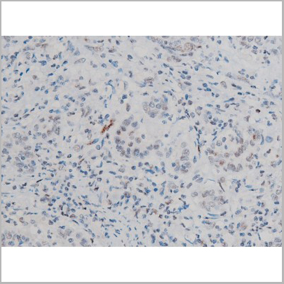
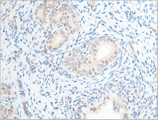
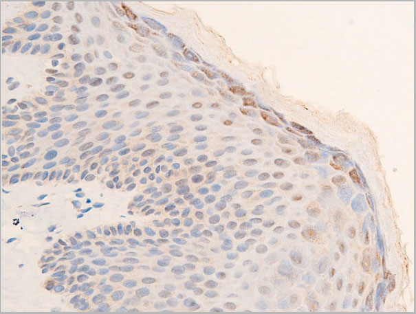
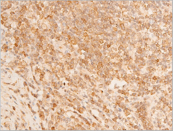
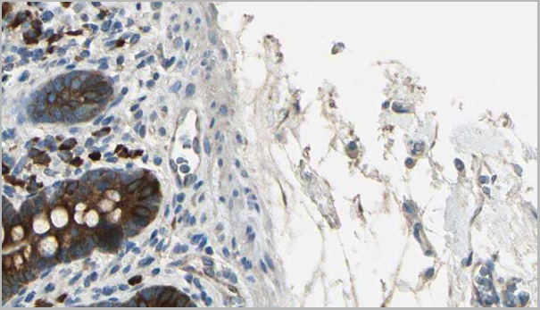
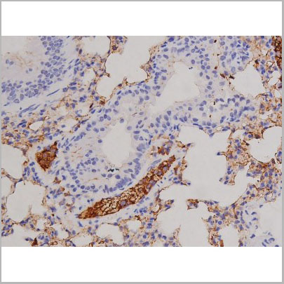
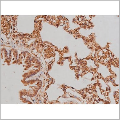
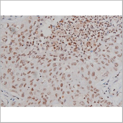
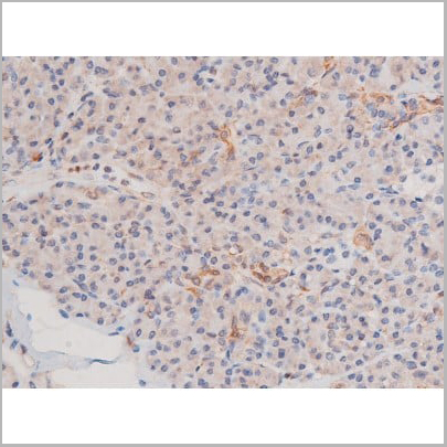
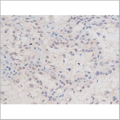
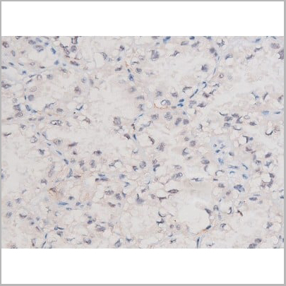
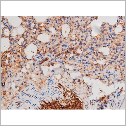
 IHC (Immunohistochemistry)
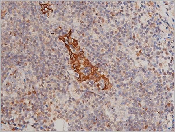
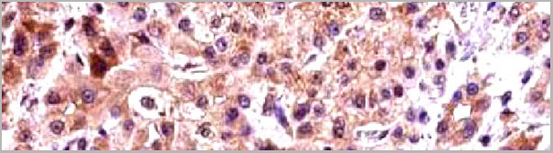
(AAA30985 at 1/50 staining human breast cancer tissue sections by IHC-P. The tissue was formaldehyde fixed and a heat mediated antigen retrieval step in citrate buffer was performed. The tissue was then blocked and incubated with the antibody for 1.5 hours at 22 degree C. An HRP conjugated goat anti-rabbit antibody was used as the secondary.)
IHC (Immunohistochemistry)
(AAA30985 at 1/50 staining human breast cancer tissue sections by IHC-P. The tissue was formaldehyde fixed and a heat mediated antigen retrieval step in citrate buffer was performed. The tissue was then blocked and incubated with the antibody for 1.5 hours at 22 degree C. An HRP conjugated goat anti-rabbit antibody was used as the secondary.)
Keratin 8, Polyclonal Antibody (Cat# AAA30985)
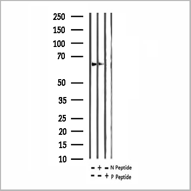
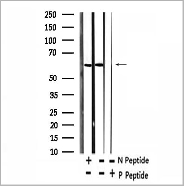
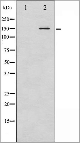
 WB (Western Blot)
(Western blot analysis of EGFR phosphorylation expression in 293 whole cell lysates, The lane on the left is treated with the antigen-specific peptide.)
WB (Western Blot)
(Western blot analysis of EGFR phosphorylation expression in 293 whole cell lysates, The lane on the left is treated with the antigen-specific peptide.)
EGFR, Polyclonal Antibody (Cat# AAA30962)
 WB (Western Blot)
(K-562 cells were subjected to SDS PAGE followed by western blot with AAA27491 (phospho (403/404) -TDP43 Antibody) at dilution of 1:3000)
WB (Western Blot)
(K-562 cells were subjected to SDS PAGE followed by western blot with AAA27491 (phospho (403/404) -TDP43 Antibody) at dilution of 1:3000)
phospho(403/404)-TDP43, Monoclonal Antibody (Cat# AAA27491)
Purification: Protein A+G purification
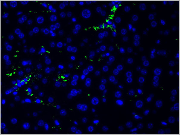
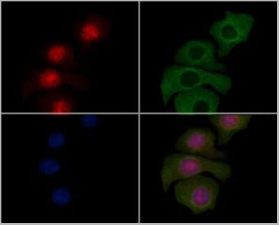
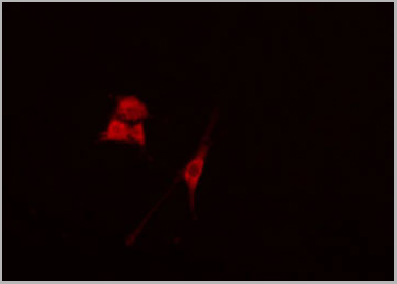
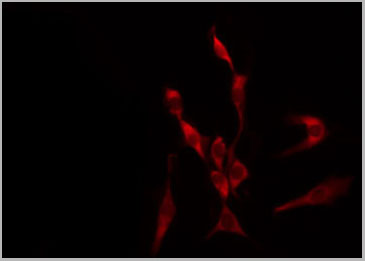
 IF (Immunofluorescence)
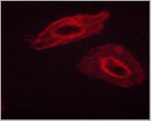
(AAA29758 staining HepG2 cells(4h of LPS treatment) by IF/ICC. The samples were fixed with PFA and permeabilized in 0.1% Triton X-100,then blocked in 10% serum for 45 minutes at 25°C. Samples were then incubated with primary Ab(1:200) and mouse anti-beta tubulin Ab(1:200) for 1 hour at 37°C. An AlexaFluor594 conjugated goat anti-rabbit IgG(H+L) Ab(Red) and an AlexaFluor488 conjugated goat anti-mouse IgG(H+L) Ab(Green) were used as the secondary antibody. The nuclear counter stain is DAPI(blue).)
IF (Immunofluorescence)
(AAA29758 staining HepG2 cells(4h of LPS treatment) by IF/ICC. The samples were fixed with PFA and permeabilized in 0.1% Triton X-100,then blocked in 10% serum for 45 minutes at 25°C. Samples were then incubated with primary Ab(1:200) and mouse anti-beta tubulin Ab(1:200) for 1 hour at 37°C. An AlexaFluor594 conjugated goat anti-rabbit IgG(H+L) Ab(Red) and an AlexaFluor488 conjugated goat anti-mouse IgG(H+L) Ab(Green) were used as the secondary antibody. The nuclear counter stain is DAPI(blue).)
STING, Polyclonal Antibody (Cat# AAA29758)
 WB (Western Blot)
(Western blot analysis of JunD phosphorylation expression in 293 whole cell lysates, The lane on the left is treated with the antigen-specific peptide.)
WB (Western Blot)
(Western blot analysis of JunD phosphorylation expression in 293 whole cell lysates, The lane on the left is treated with the antigen-specific peptide.)
JunD, Polyclonal Antibody (Cat# AAA31019)
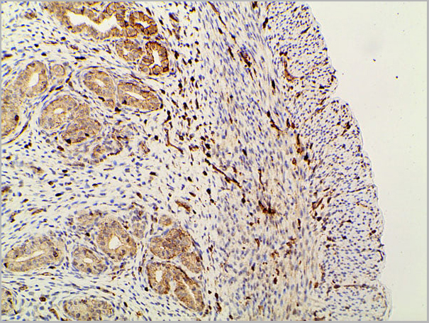
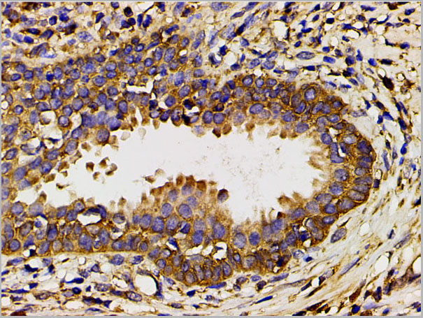
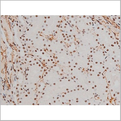
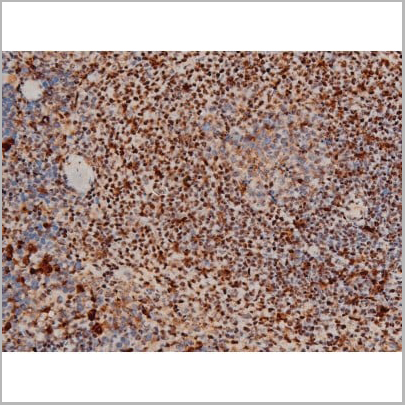
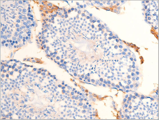
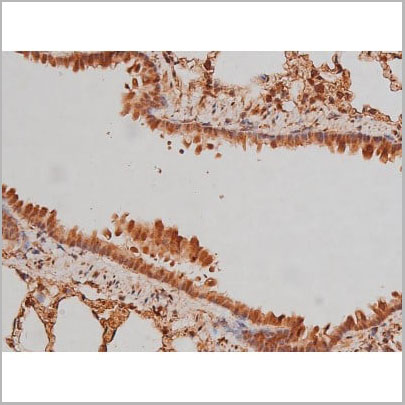
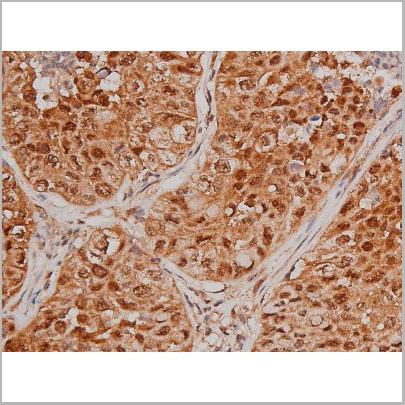
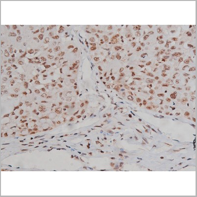
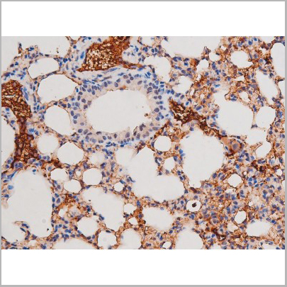
 IHC (Immunohistochemistry)
(AAA31014 at 1/200 staining Rat ganstric tissue sections by IHC-P. The tissue was formaldehyde fixed and a heat mediated antigen retrieval step in citrate buffer was performed. The tissue was then blocked and incubated with the antibody for 1.5 hours at 22 degree C. An HRP conjugated goat anti-rabbit antibody was used as the secondary.)
IHC (Immunohistochemistry)
(AAA31014 at 1/200 staining Rat ganstric tissue sections by IHC-P. The tissue was formaldehyde fixed and a heat mediated antigen retrieval step in citrate buffer was performed. The tissue was then blocked and incubated with the antibody for 1.5 hours at 22 degree C. An HRP conjugated goat anti-rabbit antibody was used as the secondary.)
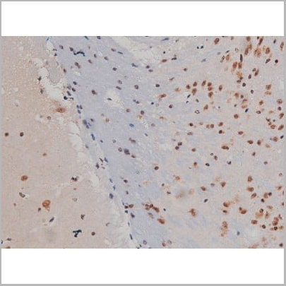
Histone H2A.X, Polyclonal Antibody (Cat# AAA31014)
 Application Data
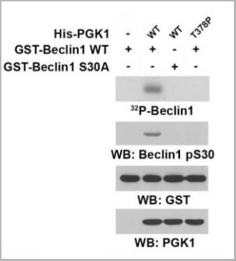
(The indicated His-PGK1 proteins immobilized on Ni-NTAagarose beads was incubated with DYKDDDDK-ARD1 and Ac-CoA, followed by incubation with purified GST-Beclin1 or GST-Beclin1 S30A, inthe presence of [γ-32P] ATP. Autoradiography was performed.)
Application Data
(The indicated His-PGK1 proteins immobilized on Ni-NTAagarose beads was incubated with DYKDDDDK-ARD1 and Ac-CoA, followed by incubation with purified GST-Beclin1 or GST-Beclin1 S30A, inthe presence of [γ-32P] ATP. Autoradiography was performed.)
Beclin1 (Phospho-Ser30), Polyclonal Antibody (Cat# AAA29760)
 WB (Western Blot)
(Western blot analysis of FKHRL1 phosphorylation expression in serum treated NIH-3T3 whole cell lysates, The lane on the left is treated with the antigen-specific peptide.)
WB (Western Blot)
(Western blot analysis of FKHRL1 phosphorylation expression in serum treated NIH-3T3 whole cell lysates, The lane on the left is treated with the antigen-specific peptide.)
FKHRL1, Polyclonal Antibody (Cat# AAA30954)
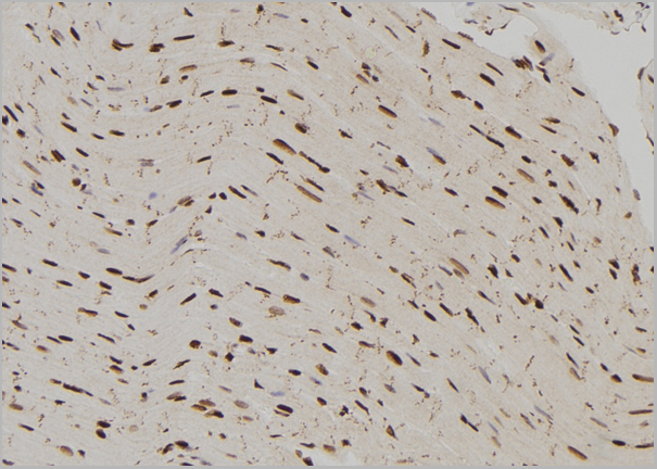
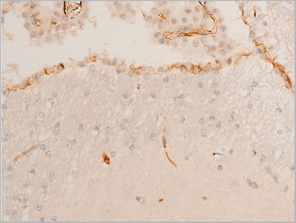
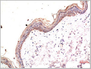 IHC (Immunohistochemistry)
(Immunohistochemical analysis of paraffin-embedded Human Skin Tissue using Phospho-MLKL S358 Mouse mAb diluted at 1:200.)
IHC (Immunohistochemistry)
(Immunohistochemical analysis of paraffin-embedded Human Skin Tissue using Phospho-MLKL S358 Mouse mAb diluted at 1:200.)
phospho-MLKL, Monoclonal Antibody (Cat# AAA29732)

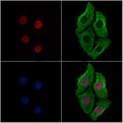
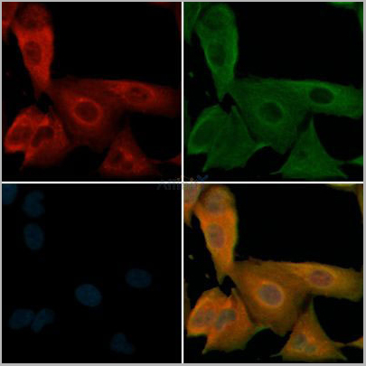
 IF (Immunofluorescence)
(AAA30956 staining HepG2 cells(4h of LPS treatment) by IF/ICC. The samples were fixed with PFA and permeabilized in 0.1% Triton X-100,then blocked in 10% serum for 45 minutes at 25°C. Samples were then incubated with primary Ab(AAA30956 1:200) and mouse anti-beta tubulin Ab( 1:200) for 1 hour at 37°C. An AlexaFluor594 conjugated goat anti-rabbit IgG(H+L) Ab(Red) and an AlexaFluor488 conjugated goat anti-mouse IgG(H+L) Ab(Green) were used as the secondary Ab. The nuclear counter stain is DAPI(blue).)
IF (Immunofluorescence)
(AAA30956 staining HepG2 cells(4h of LPS treatment) by IF/ICC. The samples were fixed with PFA and permeabilized in 0.1% Triton X-100,then blocked in 10% serum for 45 minutes at 25°C. Samples were then incubated with primary Ab(AAA30956 1:200) and mouse anti-beta tubulin Ab( 1:200) for 1 hour at 37°C. An AlexaFluor594 conjugated goat anti-rabbit IgG(H+L) Ab(Red) and an AlexaFluor488 conjugated goat anti-mouse IgG(H+L) Ab(Green) were used as the secondary Ab. The nuclear counter stain is DAPI(blue).)
MITF, Polyclonal Antibody (Cat# AAA30956)


 WB (Western Blot)
(Western blot analysis of extracts from serum-starved NIH-3T3, untreated (line A); treated with PDGFA (5 ug/mL, 5 min), without peptide (line B) or antigen-specific phosphopeptide (line C) or antigen-specific peptide (line D) using Phospho-AKT (Ser473) rabbit monoclonal Antibody (#AAA27779) at 1:2000 dilution.)
WB (Western Blot)
(Western blot analysis of extracts from serum-starved NIH-3T3, untreated (line A); treated with PDGFA (5 ug/mL, 5 min), without peptide (line B) or antigen-specific phosphopeptide (line C) or antigen-specific peptide (line D) using Phospho-AKT (Ser473) rabbit monoclonal Antibody (#AAA27779) at 1:2000 dilution.)
AKT, Monoclonal Recombinant Antibody (Cat# AAA27779)
 WB (Western Blot)
(Western blot analysis of extracts from A431, using Phospho-SHP-2 (Tyr62) Antibody. Lane1 was treated with phospho-blocking peptide, Lane2 was treated with non-phospho-blocking peptide.)
WB (Western Blot)
(Western blot analysis of extracts from A431, using Phospho-SHP-2 (Tyr62) Antibody. Lane1 was treated with phospho-blocking peptide, Lane2 was treated with non-phospho-blocking peptide.)
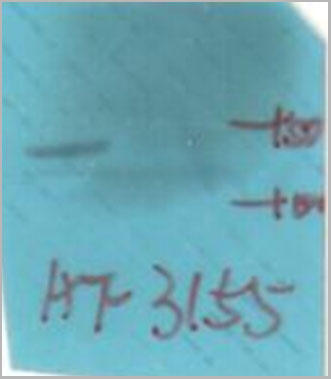
SHP-2, Polyclonal Antibody (Cat# AAA31155)
Predicted: Bovine, Horse, Sheep, Rabbit, Dog, Chicken, Xenopus
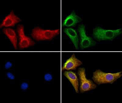
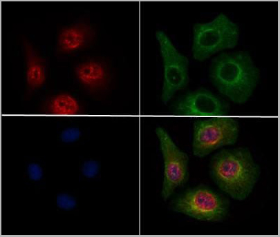 IF (Immunofluorescence)
(AAA31156 staining HepG2 cells(4h of LPS treatment) by IF/ICC. The samples were fixed with PFA and permeabilized in 0.1% Triton X-100,then blocked in 10% serum for 45 minutes at 25°C. Samples were then incubated with primary Ab(AAA31156 1:200) and mouse anti-beta tubulin Ab( 1:200) for 1 hour at 37°C. An AlexaFluor594 conjugated goat anti-rabbit IgG(H+L) Ab(Red) and an AlexaFluor488 conjugated goat anti-mouse IgG(H+L) Ab(Green) were used as the secondary Ab. The nuclear counter stain is DAPI(blue).)
IF (Immunofluorescence)
(AAA31156 staining HepG2 cells(4h of LPS treatment) by IF/ICC. The samples were fixed with PFA and permeabilized in 0.1% Triton X-100,then blocked in 10% serum for 45 minutes at 25°C. Samples were then incubated with primary Ab(AAA31156 1:200) and mouse anti-beta tubulin Ab( 1:200) for 1 hour at 37°C. An AlexaFluor594 conjugated goat anti-rabbit IgG(H+L) Ab(Red) and an AlexaFluor488 conjugated goat anti-mouse IgG(H+L) Ab(Green) were used as the secondary Ab. The nuclear counter stain is DAPI(blue).)
Beclin-1, Polyclonal Antibody (Cat# AAA31156)
Predicted: Pig, Bovine, Horse, Sheep, Rabbit, Dog, Chicken, Xenopus

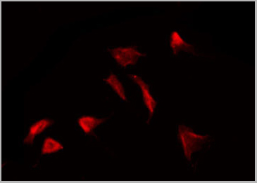
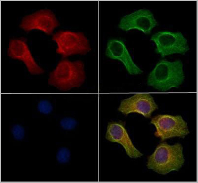
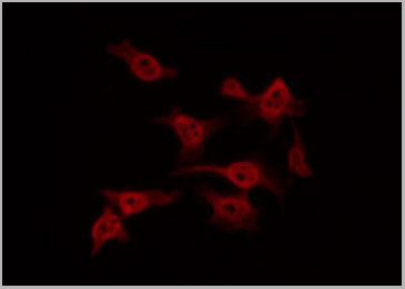
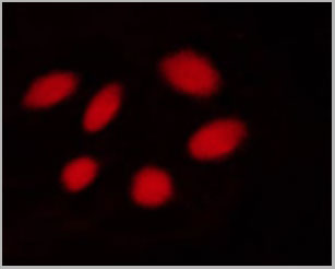
 IF (Immunoflouorescence)
(AAA31011 staining Hela by IF/ICC. The sample were fixed with PFA and permeabilized in 0.1% Triton X-100, then blocked in 10% serum for 45 minutes at 25 degree C. The primary antibody was diluted at 1/200 and incubated with the sample for 1 hour at 37 degree C. An Alexa Fluor 594 conjugated goat anti-rabbit IgG (H+L) Ab, diluted at 1/600, was used as the secondary antibody.)
IF (Immunoflouorescence)
(AAA31011 staining Hela by IF/ICC. The sample were fixed with PFA and permeabilized in 0.1% Triton X-100, then blocked in 10% serum for 45 minutes at 25 degree C. The primary antibody was diluted at 1/200 and incubated with the sample for 1 hour at 37 degree C. An Alexa Fluor 594 conjugated goat anti-rabbit IgG (H+L) Ab, diluted at 1/600, was used as the secondary antibody.)
ATF2, Polyclonal Antibody (Cat# AAA31011)
 IF (Immunofluorescence)
(AAA31033 staining NIH-3T3 by IF/ICC. The sample were fixed with PFA and permeabilized in 0.1% Triton X-100, then blocked in 10% serum for 45 minutes at 25 degree C. The primary antibody was diluted at 1/200 and incubated with the sample for 1 hour at 37 degree C. An Alexa Fluor 594 conjugated goat anti-rabbit IgG (H+L) Ab, diluted at 1/600, was used as the secondary antibody.)
IF (Immunofluorescence)
(AAA31033 staining NIH-3T3 by IF/ICC. The sample were fixed with PFA and permeabilized in 0.1% Triton X-100, then blocked in 10% serum for 45 minutes at 25 degree C. The primary antibody was diluted at 1/200 and incubated with the sample for 1 hour at 37 degree C. An Alexa Fluor 594 conjugated goat anti-rabbit IgG (H+L) Ab, diluted at 1/600, was used as the secondary antibody.)
CDC25B, Polyclonal Antibody (Cat# AAA31033)
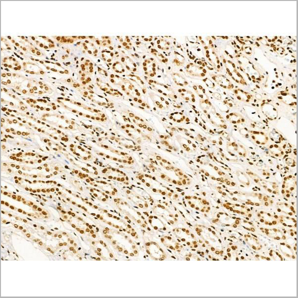
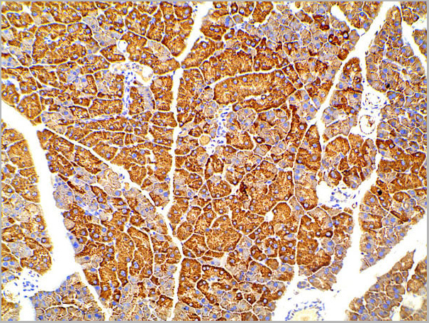
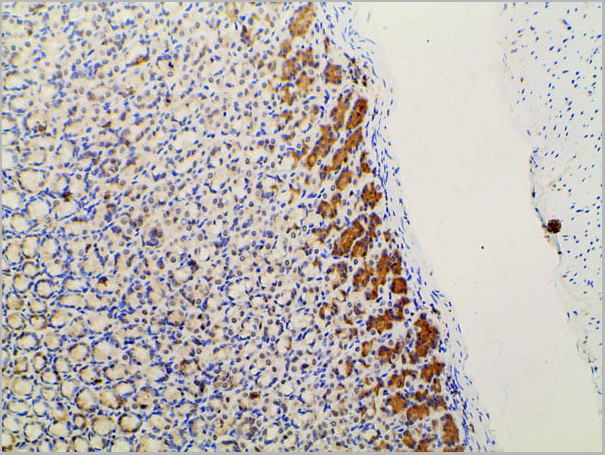
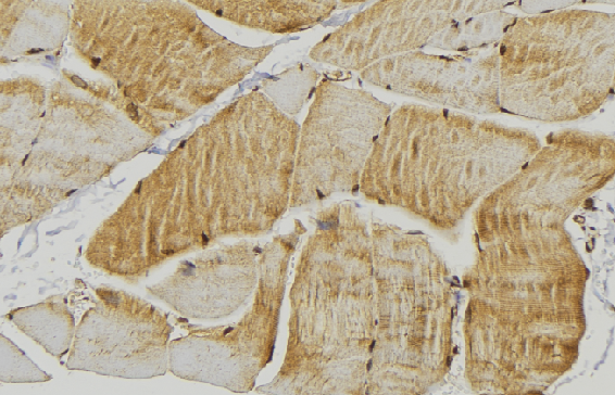
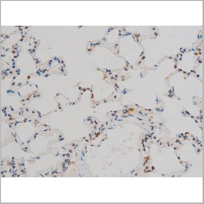
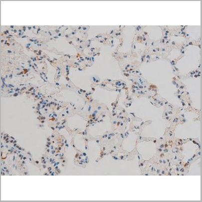
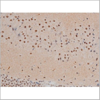
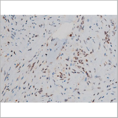
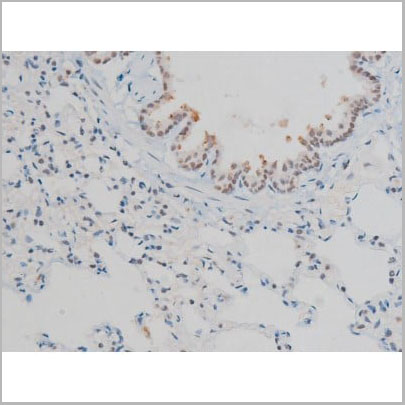
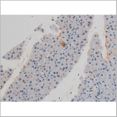
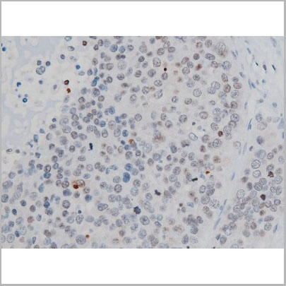
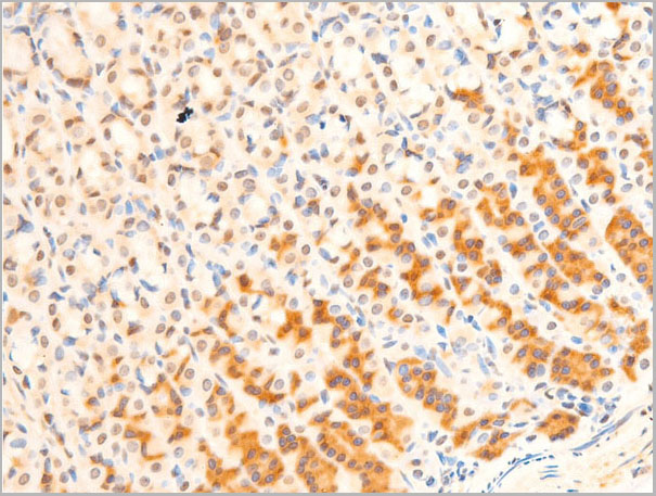
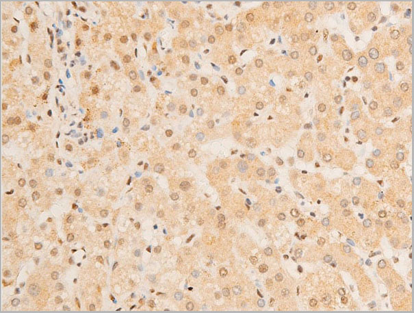
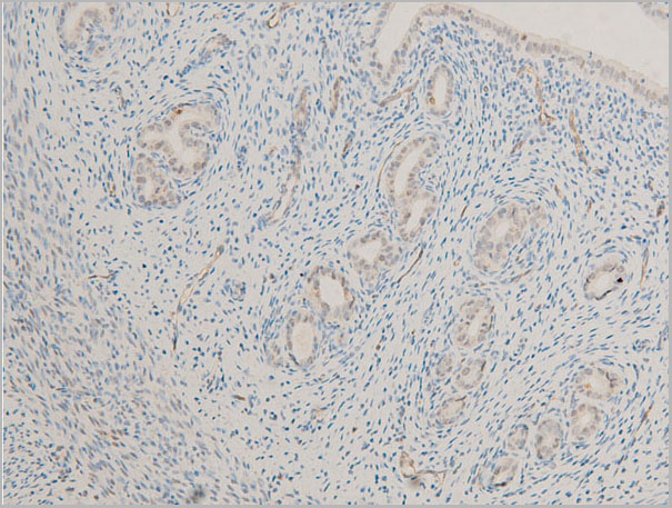
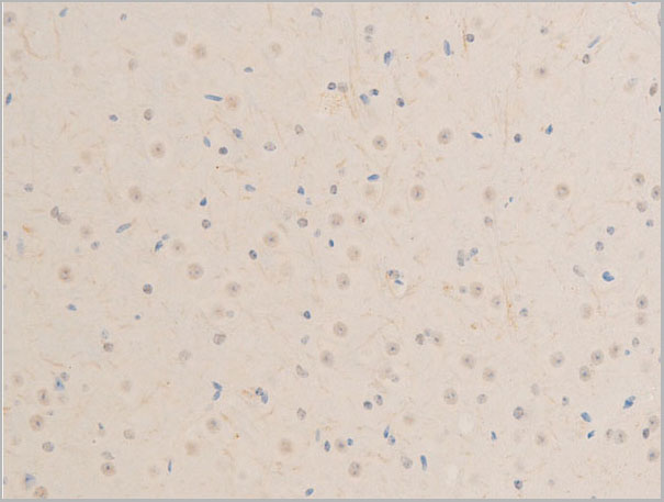
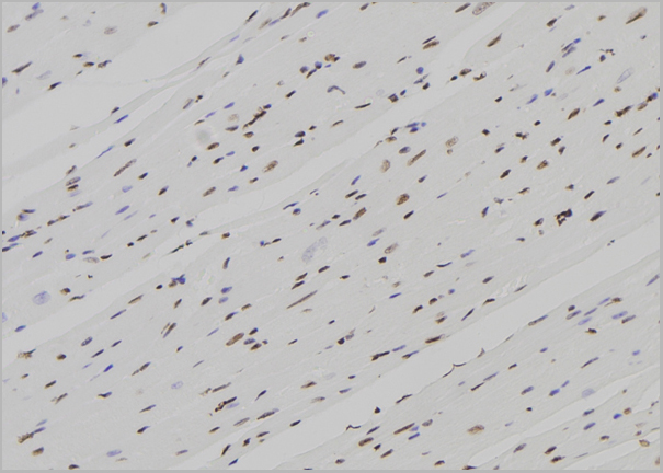
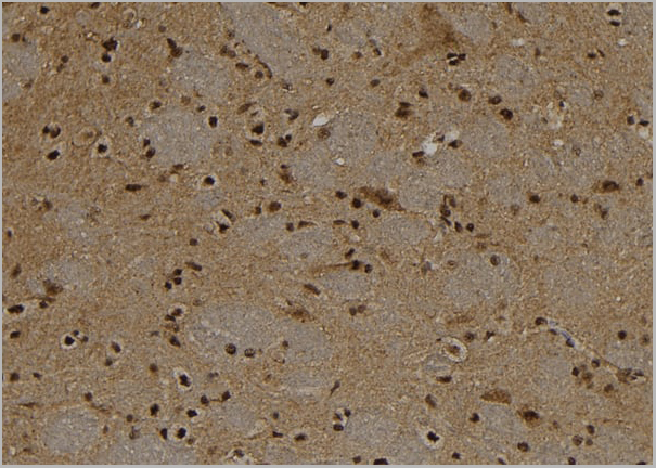
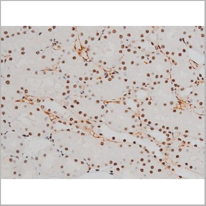
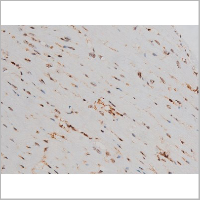
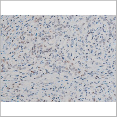
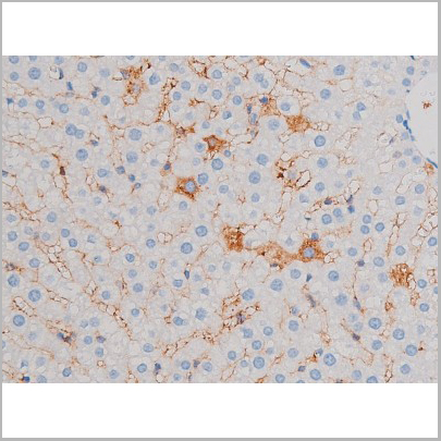
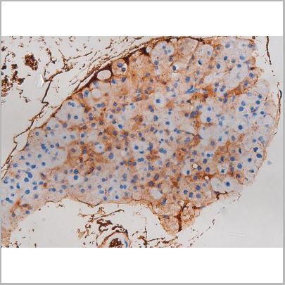
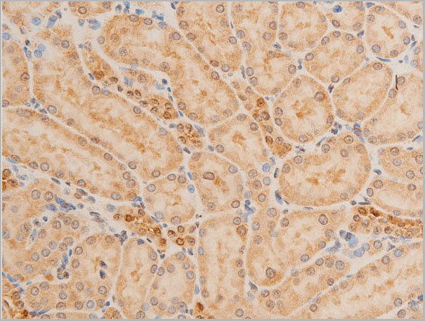
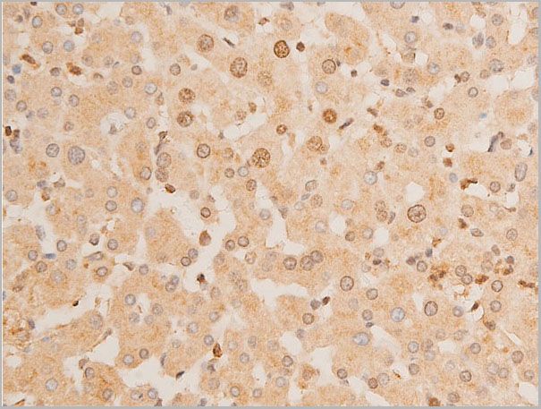
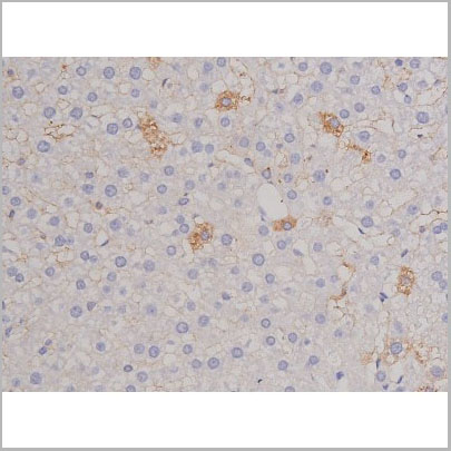
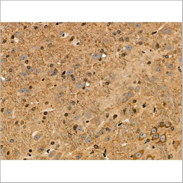
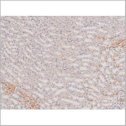
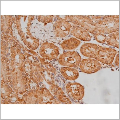
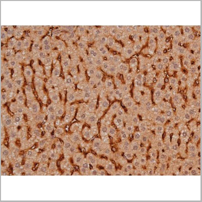
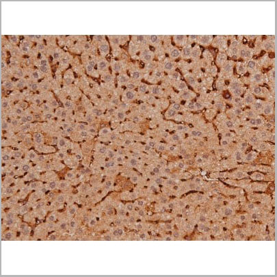
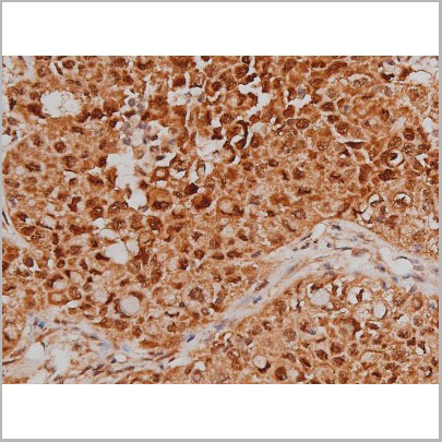
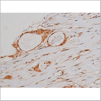
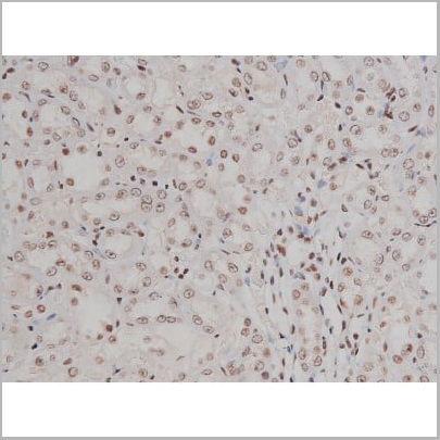
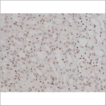
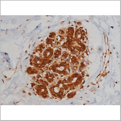
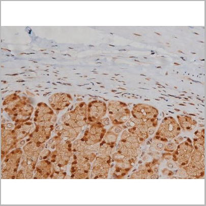
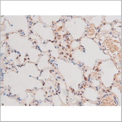
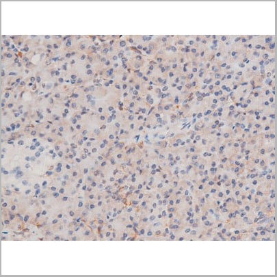
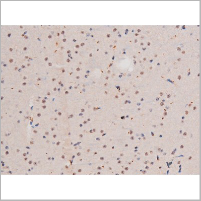
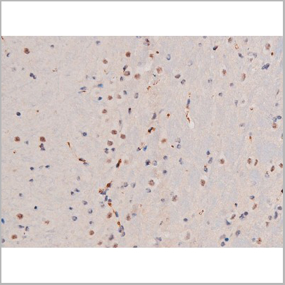
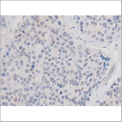
 IHC (Immunohistochemistry)
(AAA30960 at 1/100 staining rat liver tissue sections by IHC-P. The tissue was formaldehyde fixed and a heat mediated antigen retrieval step in citrate buffer was performed. The tissue was then blocked and incubated with the antibody for 1.5 hours at 22 degree C. An HRP conjugated goat anti-rabbit antibody was used as the secondary.)
IHC (Immunohistochemistry)
(AAA30960 at 1/100 staining rat liver tissue sections by IHC-P. The tissue was formaldehyde fixed and a heat mediated antigen retrieval step in citrate buffer was performed. The tissue was then blocked and incubated with the antibody for 1.5 hours at 22 degree C. An HRP conjugated goat anti-rabbit antibody was used as the secondary.)
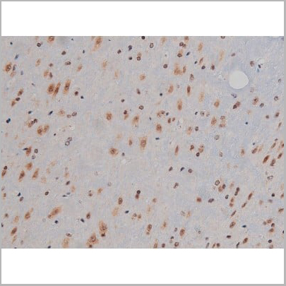
Chk2, Polyclonal Antibody (Cat# AAA30960)
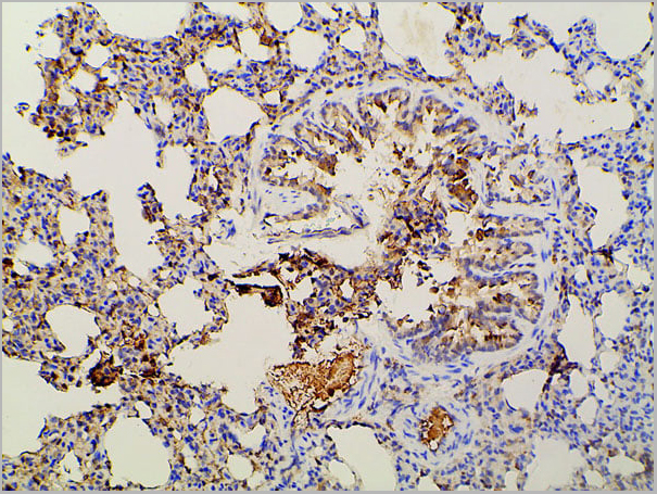
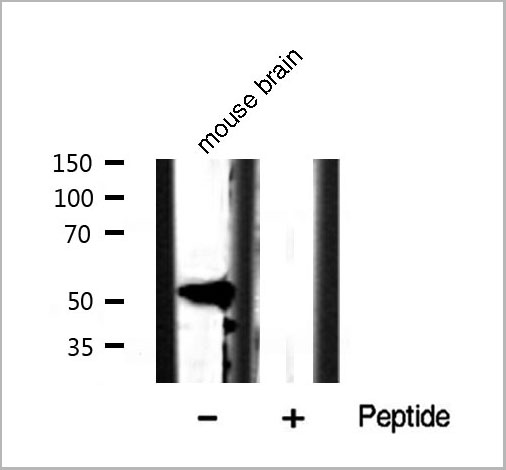
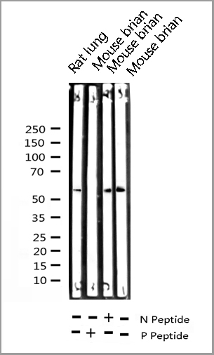
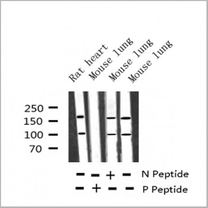
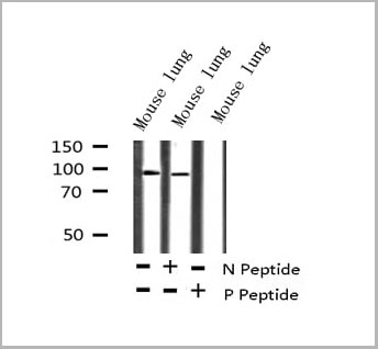 WB (Western Blot)
(Western blot analysis of Phospho-p73 (Tyr99) expression in Mouse lung lysate)
WB (Western Blot)
(Western blot analysis of Phospho-p73 (Tyr99) expression in Mouse lung lysate)
p73, Polyclonal Antibody (Cat# AAA30950)
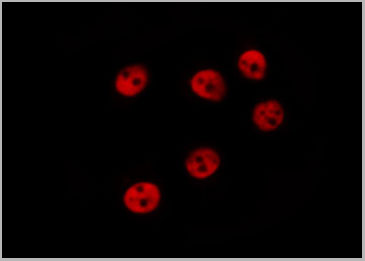
 IF (Immunofluorescence)
(AAA31008 staining Hela cells by IF/ICC. The sample were fixed with PFA and permeabilized in 0.1% Triton X-100, then blocked in 10% serum for 45 minutes at 25 degree C. The primary antibody was diluted at 1/200 and incubated with the sample for 1 hour at 37 degree C. An Alexa Fluor 594 conjugated goat anti-rabbit IgG (H+L) antibody, diluted at 1/600, was used as secondary antibody.)
IF (Immunofluorescence)
(AAA31008 staining Hela cells by IF/ICC. The sample were fixed with PFA and permeabilized in 0.1% Triton X-100, then blocked in 10% serum for 45 minutes at 25 degree C. The primary antibody was diluted at 1/200 and incubated with the sample for 1 hour at 37 degree C. An Alexa Fluor 594 conjugated goat anti-rabbit IgG (H+L) antibody, diluted at 1/600, was used as secondary antibody.)
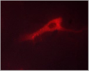
Bcr, Polyclonal Antibody (Cat# AAA31008)
 IF (Immunofluorescence)
(AAA31134 staining HepG2 cells(30min of 4uM Forskolin treatment) by IF/ICC. The samples were fixed with PFA and permeabilized in 0.1% Triton X-100,then blocked in 10% serum for 45 minutes at 25°C. Samples were then incubated with primary Ab (AAA31134) and mouse anti-beta tubulin Ab for 1 hour at 37°C. An AlexaFluor594 conjugated goat anti-rabbit IgG(H+L) Ab(Red) and an AlexaFluor488 conjugated goat anti-mouse IgG(H+L) Ab(Green) were used as the secondary antibody. The nuclear counter stain is DAPI(blue).)
IF (Immunofluorescence)
(AAA31134 staining HepG2 cells(30min of 4uM Forskolin treatment) by IF/ICC. The samples were fixed with PFA and permeabilized in 0.1% Triton X-100,then blocked in 10% serum for 45 minutes at 25°C. Samples were then incubated with primary Ab (AAA31134) and mouse anti-beta tubulin Ab for 1 hour at 37°C. An AlexaFluor594 conjugated goat anti-rabbit IgG(H+L) Ab(Red) and an AlexaFluor488 conjugated goat anti-mouse IgG(H+L) Ab(Green) were used as the secondary antibody. The nuclear counter stain is DAPI(blue).)
PERK, Polyclonal Antibody (Cat# AAA31134)
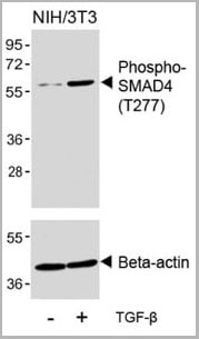 WB (Western Blot)
(Western blot analysis of lysates from NIH/3T3 cell line, untreated or treated with TGF-beta (100ng/ml, 30 min), using Phospho-SMAD4 Antibody(upper) or Beta-actin (lower).)
WB (Western Blot)
(Western blot analysis of lysates from NIH/3T3 cell line, untreated or treated with TGF-beta (100ng/ml, 30 min), using Phospho-SMAD4 Antibody(upper) or Beta-actin (lower).)
Phospho-SMAD4 (T277), Polyclonal Antibody (Cat# AAA28715)
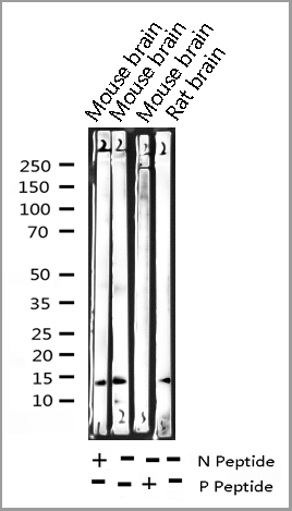
Predicted Species Reactivity: Rat
 WB (Western Blot)
(Western blot analysis of Phospho-FAK (Tyr576) Antibody expression in NIH-3T3 cells lysates.The lane on the right is treated with the antigen-specific peptide.)
WB (Western Blot)
(Western blot analysis of Phospho-FAK (Tyr576) Antibody expression in NIH-3T3 cells lysates.The lane on the right is treated with the antigen-specific peptide.)
FAK, Polyclonal Antibody (Cat# AAA31066)

 Application Data
(At 25 degree C. The primary antibody was diluted at 1/200 and incubated with the sample for 1 hour at 37 degree C. An Alexa Fluor 594 conjugated goat anti-rabbit IgG (H+L) Ab, diluted at 1/600, was used as the secondary antibody.)
Application Data
(At 25 degree C. The primary antibody was diluted at 1/200 and incubated with the sample for 1 hour at 37 degree C. An Alexa Fluor 594 conjugated goat anti-rabbit IgG (H+L) Ab, diluted at 1/600, was used as the secondary antibody.)
FAK, Polyclonal Antibody (Cat# AAA31446)


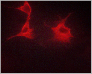
 IF (Immunofluorescence)
(AAA31063 staining HeLa by IF/ICC. The sample were fixed with PFA and permeabilized in 0.1% Triton X-100, then blocked in 10% serum for 45 minutes at 25 degree C. The primary antibody was diluted at 1/200 and incubated with the sample for 1 hour at 37 degree C. An Alexa Fluor 594 conjugated goat anti-rabbit IgG (H+L) Ab, diluted at 1/600, was used as the secondary antibody.)
IF (Immunofluorescence)
(AAA31063 staining HeLa by IF/ICC. The sample were fixed with PFA and permeabilized in 0.1% Triton X-100, then blocked in 10% serum for 45 minutes at 25 degree C. The primary antibody was diluted at 1/200 and incubated with the sample for 1 hour at 37 degree C. An Alexa Fluor 594 conjugated goat anti-rabbit IgG (H+L) Ab, diluted at 1/600, was used as the secondary antibody.)
NF kappaB p65, Polyclonal Antibody (Cat# AAA31063)
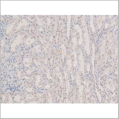

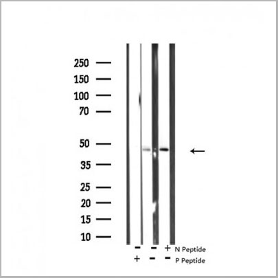
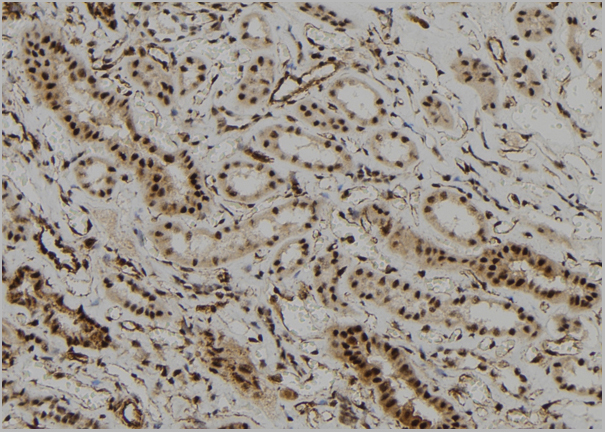
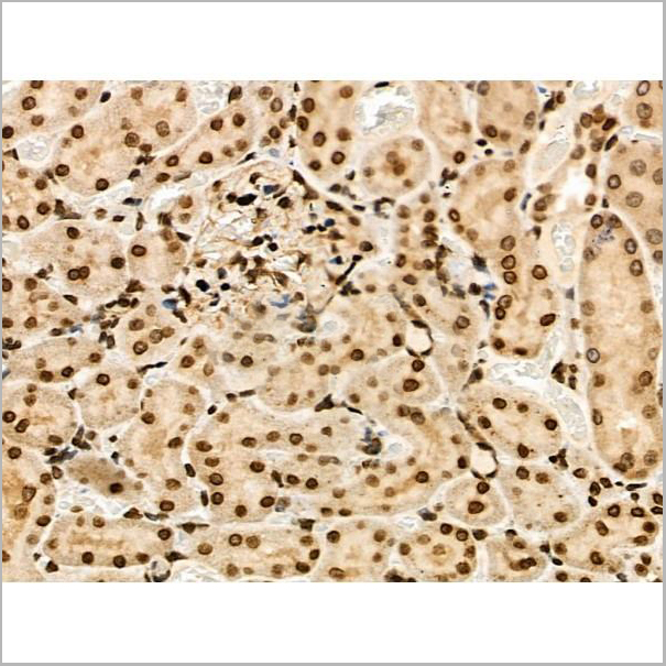
 IHC (Immunohistchemistry)
(At 1/200 staining Mouse kidney tissue sections by IHC-P. The tissue was formaldehyde fixed and a heat mediated antigen retrieval step in citrate buffer was performed. The tissue was then blocked and incubated with the antibody for 1.5 hours at 22 degree C. An HRP conjugated goat anti-rabbit antibody was used as the secondary antibody.)
IHC (Immunohistchemistry)
(At 1/200 staining Mouse kidney tissue sections by IHC-P. The tissue was formaldehyde fixed and a heat mediated antigen retrieval step in citrate buffer was performed. The tissue was then blocked and incubated with the antibody for 1.5 hours at 22 degree C. An HRP conjugated goat anti-rabbit antibody was used as the secondary antibody.)
p38 gamma/delta, Polyclonal Antibody (Cat# AAA31407)
Predicted Reactivity: Pig (100%), Zebrafish (100%), Bovine (100%), Horse (100%), Rabbit (100%), Dog (100%), Xenopus (100%)
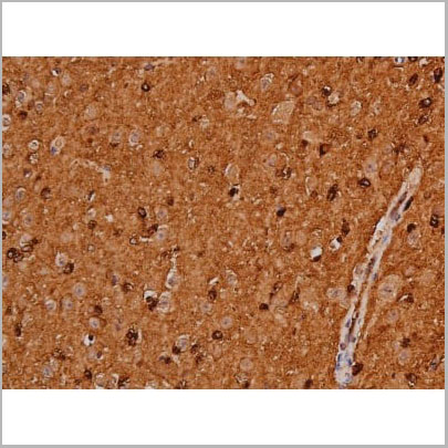
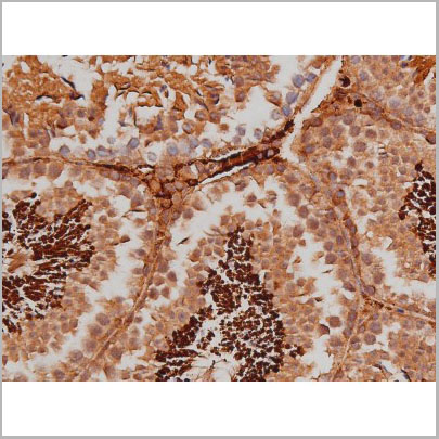
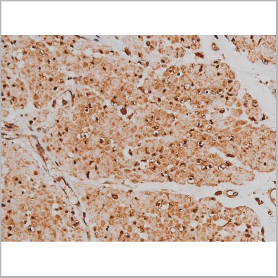
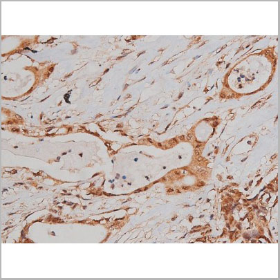
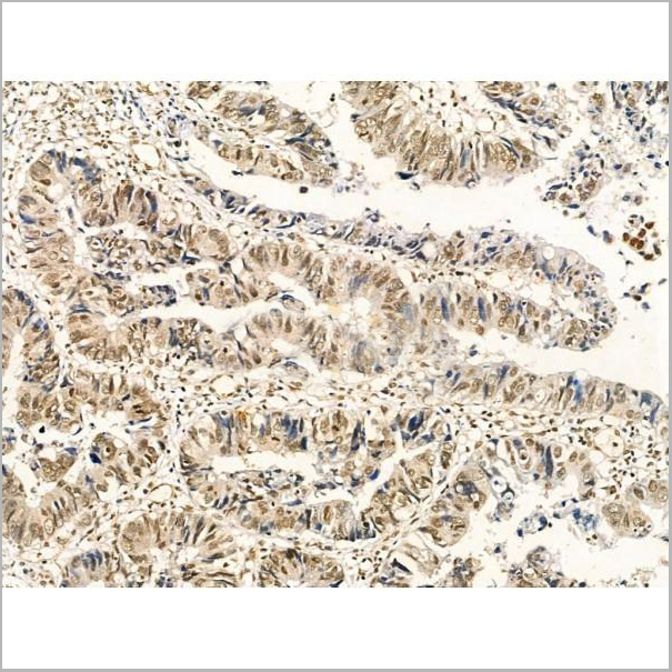
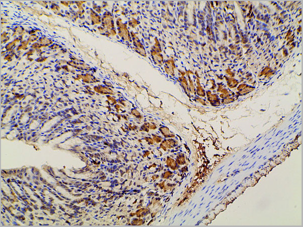
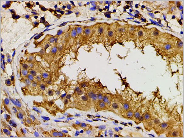
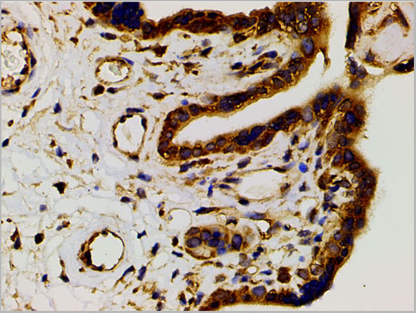
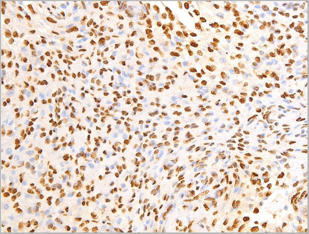
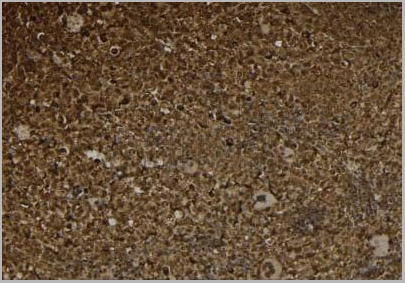
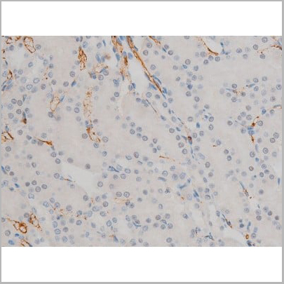
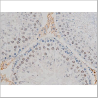
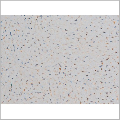
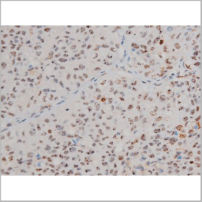
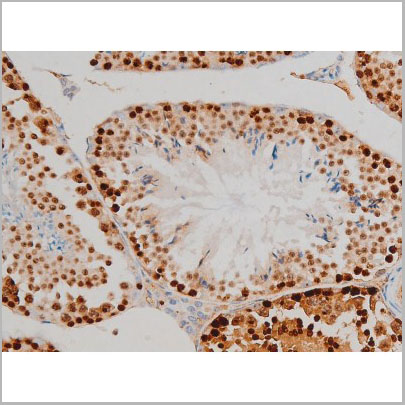
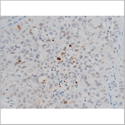
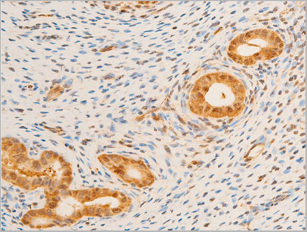
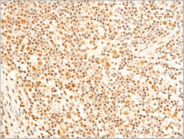
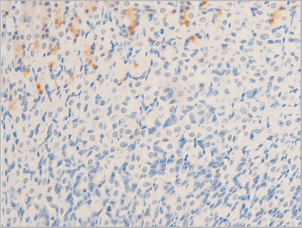
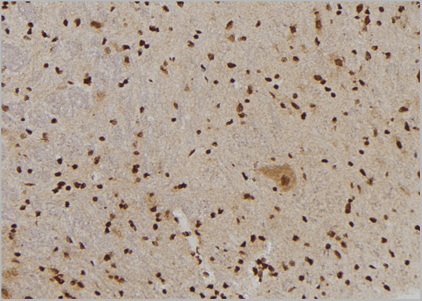
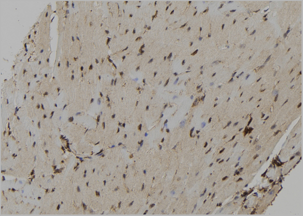
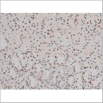
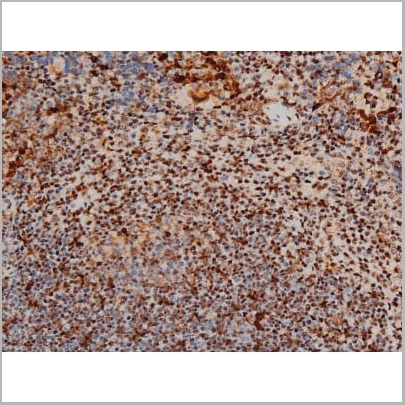
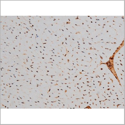
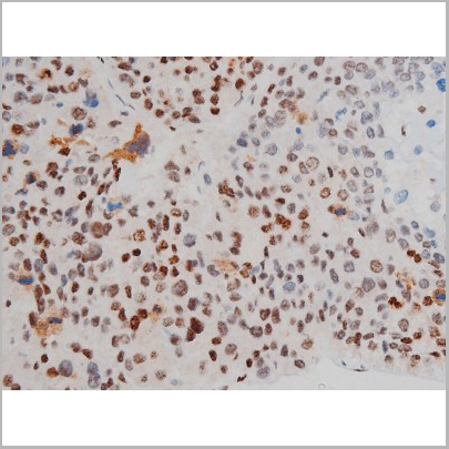
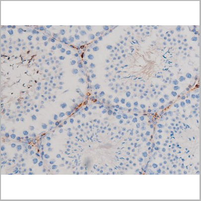
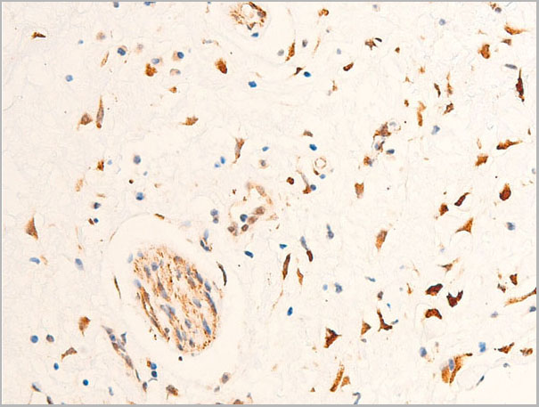
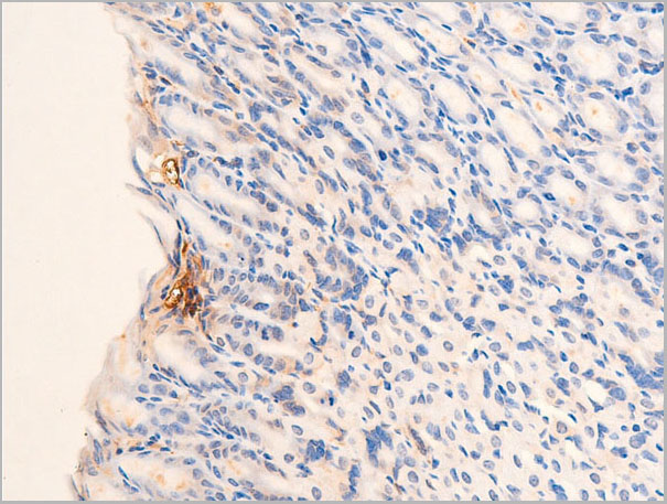
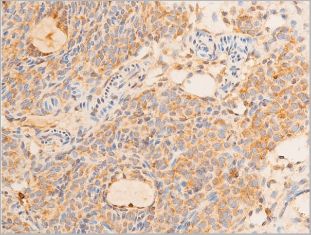
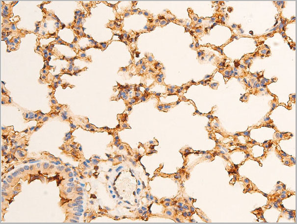
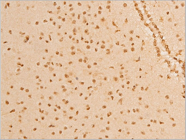
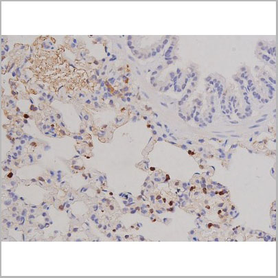
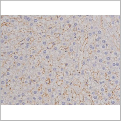
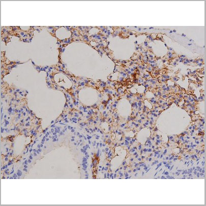
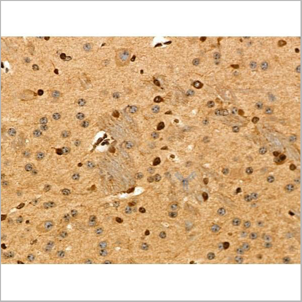
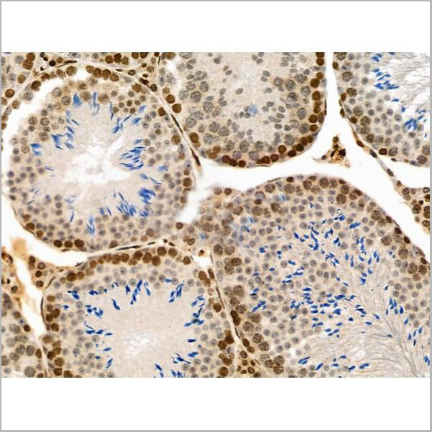
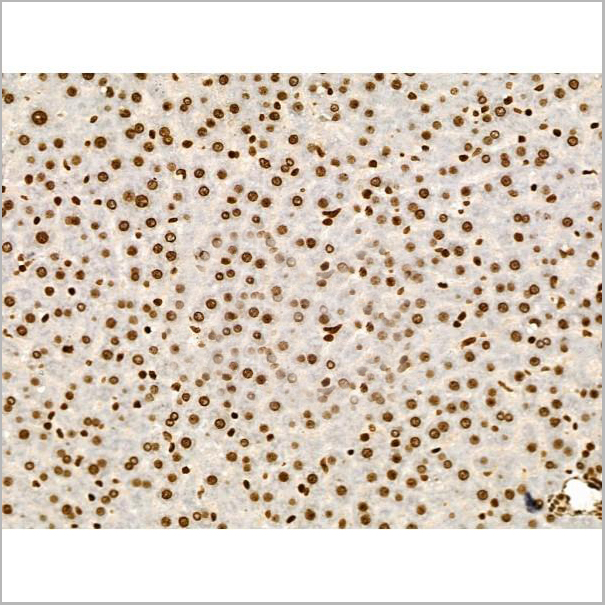
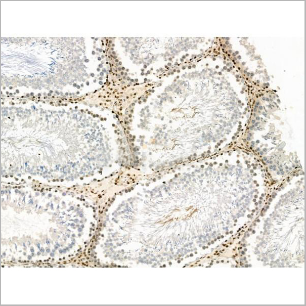
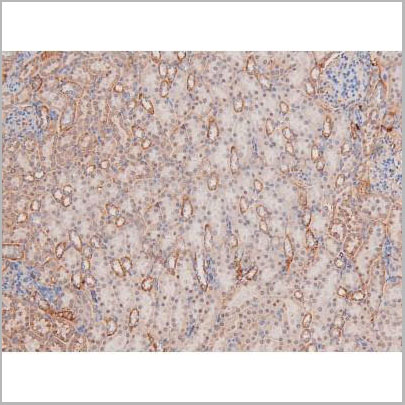
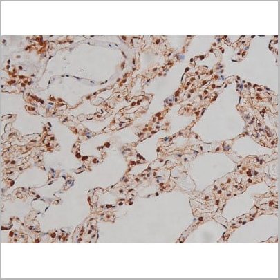
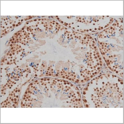
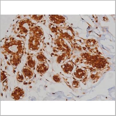
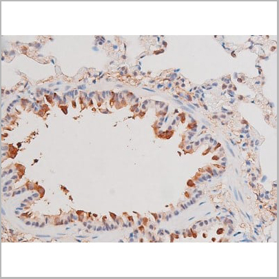
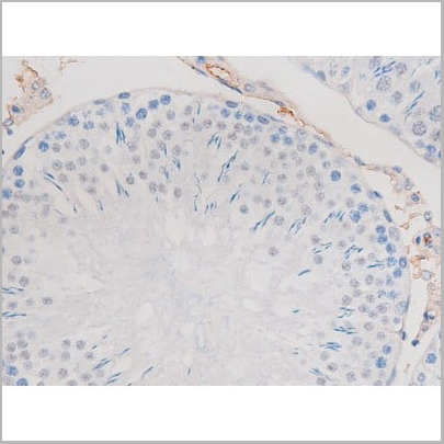
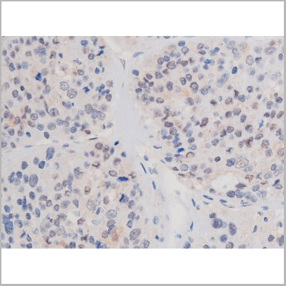
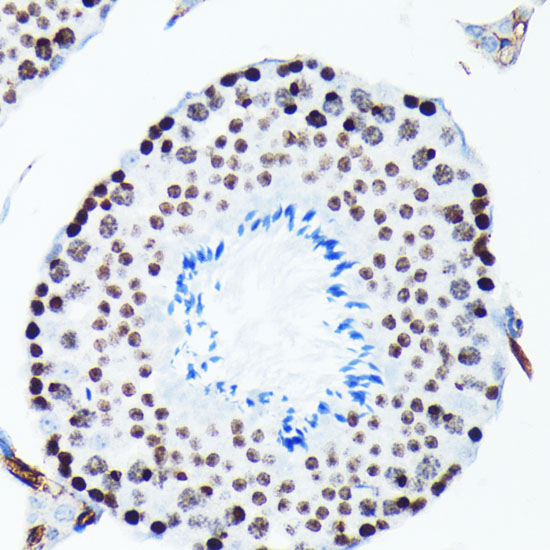
 IHC (Immunohistochemistry)
(AAA31005 at 1/200 staining Rat ganstric tissue sections by IHC-P. The tissue was formaldehyde fixed and a heat mediated antigen retrieval step in citrate buffer was performed. The tissue was then blocked and incubated with the antibody for 1.5 hours at 22 degree C. An HRP conjugated goat anti-rabbit antibody was used as the secondary.)
IHC (Immunohistochemistry)
(AAA31005 at 1/200 staining Rat ganstric tissue sections by IHC-P. The tissue was formaldehyde fixed and a heat mediated antigen retrieval step in citrate buffer was performed. The tissue was then blocked and incubated with the antibody for 1.5 hours at 22 degree C. An HRP conjugated goat anti-rabbit antibody was used as the secondary.)
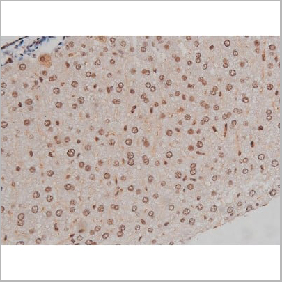
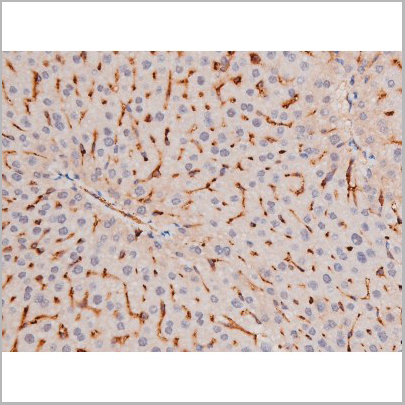
Tau, Polyclonal Antibody (Cat# AAA31005)
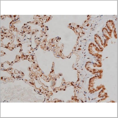
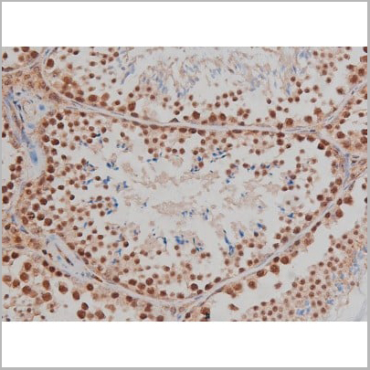

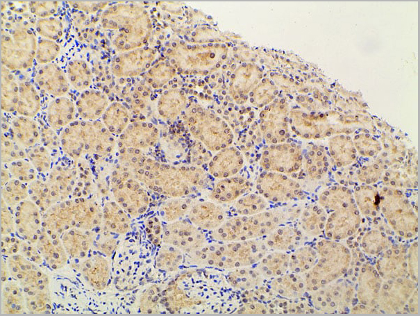
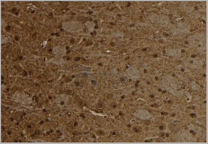
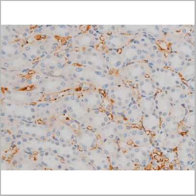
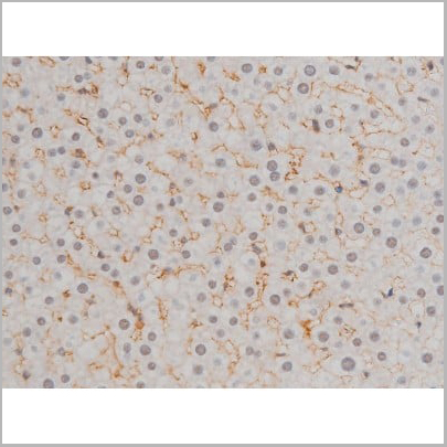
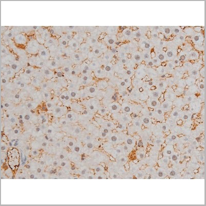
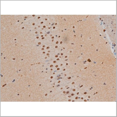
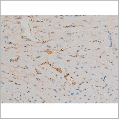
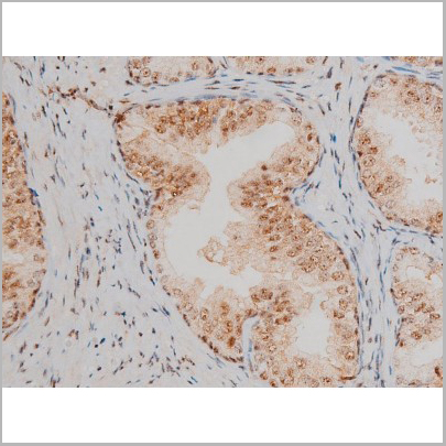
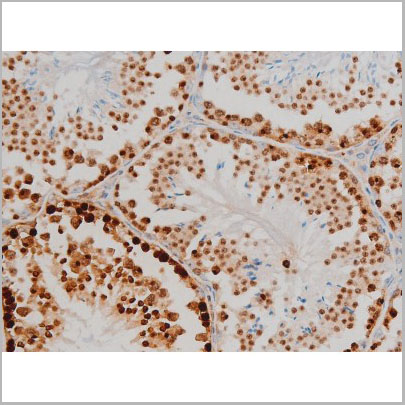
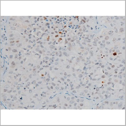
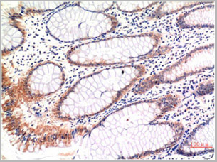
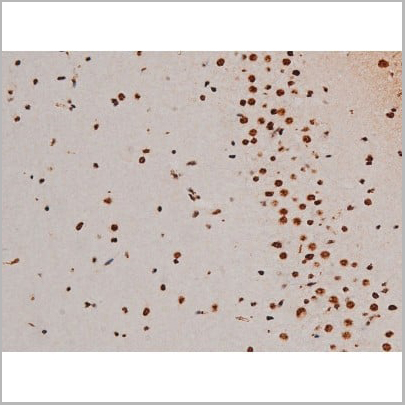
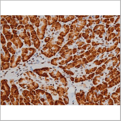
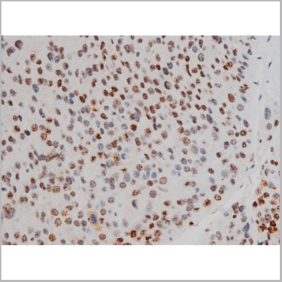
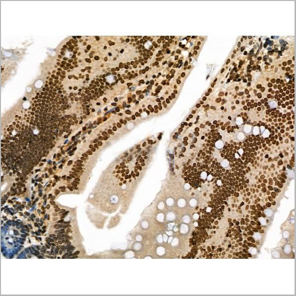
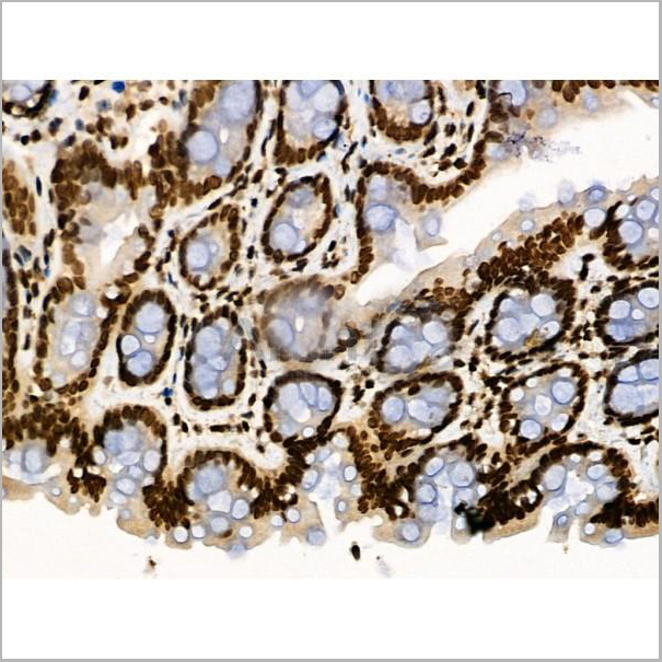
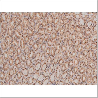
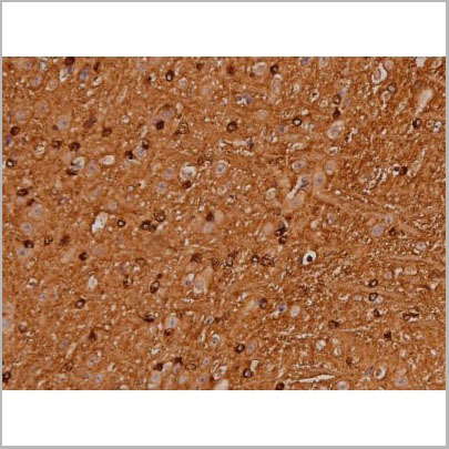
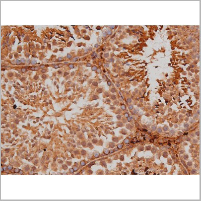
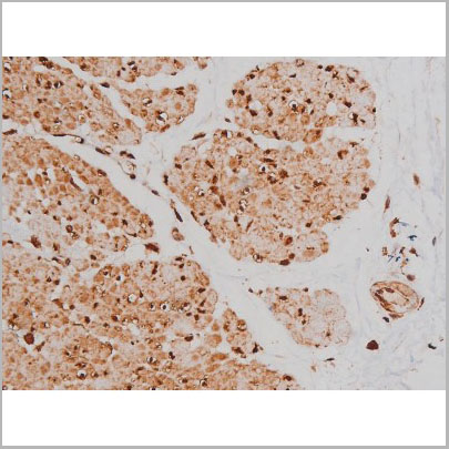
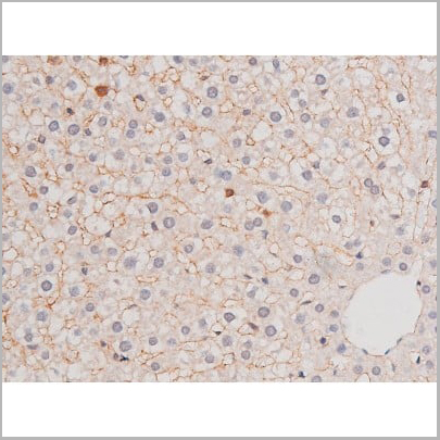
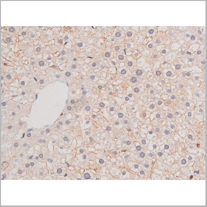
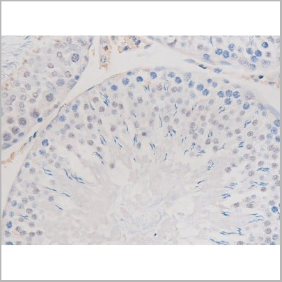
 IHC (Immunohistochemistry)
(AAA30993 at 1/200 staining Rat ganstric tissue sections by IHC-P. The tissue was formaldehyde fixed and a heat mediated antigen retrieval step in citrate buffer was performed. The tissue was then blocked and incubated with the antibody for 1.5 hours at 22 degree C. An HRP conjugated goat anti-rabbit antibody was used as the secondary.)
IHC (Immunohistochemistry)
(AAA30993 at 1/200 staining Rat ganstric tissue sections by IHC-P. The tissue was formaldehyde fixed and a heat mediated antigen retrieval step in citrate buffer was performed. The tissue was then blocked and incubated with the antibody for 1.5 hours at 22 degree C. An HRP conjugated goat anti-rabbit antibody was used as the secondary.)
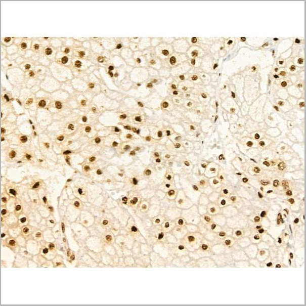
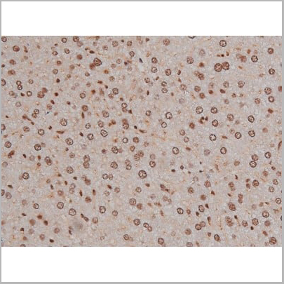
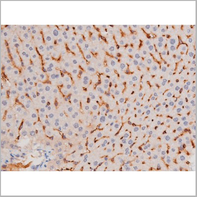
IGF1R, Polyclonal Antibody (Cat# AAA30993)
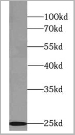
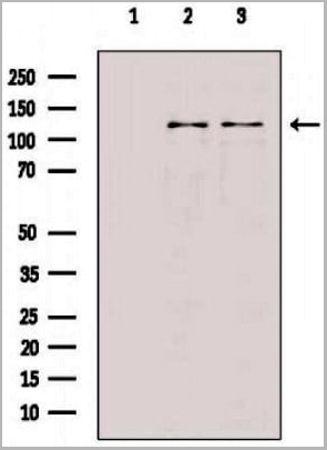
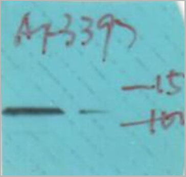
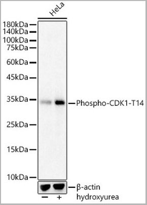 WB (Western Blot)
(Western blot analysis of Lysates HeLa, using Phospho-CDK1-T14 Rabbit pAb (AAA10633) at 1:500 dilution.HeLa cells were treated by Hydroxyurea (4 mM) at 37 degree C for 20 hours.Secondary antibody: HRP Goat Anti-Rabbit IgG (H+L) at 1:10000 dilution.Lysates/proteins: 25ug per lane.Blocking buffer: 3% nonfat dry milk in TBST.Detection: ECL Basic Kit.Exposure time: 3s.)
WB (Western Blot)
(Western blot analysis of Lysates HeLa, using Phospho-CDK1-T14 Rabbit pAb (AAA10633) at 1:500 dilution.HeLa cells were treated by Hydroxyurea (4 mM) at 37 degree C for 20 hours.Secondary antibody: HRP Goat Anti-Rabbit IgG (H+L) at 1:10000 dilution.Lysates/proteins: 25ug per lane.Blocking buffer: 3% nonfat dry milk in TBST.Detection: ECL Basic Kit.Exposure time: 3s.)
CDK1-T14, Polyclonal Antibody (Cat# AAA10633)
 IF (Immunoflouorescence)
(AAA31039 staining HeLa cells by ICC/IF. Cells were fixed with PFA and permeabilized in 0.1% saponin prior to blocking in 10% serum for 45 minutes at 37 degree C. The primary antibody was diluted 1/400 and incubated with the sample for 1 hour at 37 degree C. A Alexa Fluor 594 conjugated goat polyclonal to rabbit IgG (H+L), diluted 1/600 was used as secondary antibody.)
IF (Immunoflouorescence)
(AAA31039 staining HeLa cells by ICC/IF. Cells were fixed with PFA and permeabilized in 0.1% saponin prior to blocking in 10% serum for 45 minutes at 37 degree C. The primary antibody was diluted 1/400 and incubated with the sample for 1 hour at 37 degree C. A Alexa Fluor 594 conjugated goat polyclonal to rabbit IgG (H+L), diluted 1/600 was used as secondary antibody.)
ADD1, Polyclonal Antibody (Cat# AAA31039)
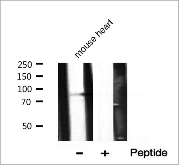
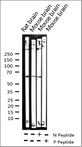
 WB (Western Blot)
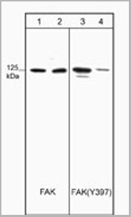
(Western blot analysis of HUVECs untreated (lanes 1 & 3) or treated with alkaline phosphatase (lanes 2 & 4). Blots were probed with mouse monoclonal anti-FAK (lanes 1 & 2) and anti-FAK (Tyr-397) (lanes 3 & 4).)
WB (Western Blot)
(Western blot analysis of HUVECs untreated (lanes 1 & 3) or treated with alkaline phosphatase (lanes 2 & 4). Blots were probed with mouse monoclonal anti-FAK (lanes 1 & 2) and anti-FAK (Tyr-397) (lanes 3 & 4).)
FAK (Tyr-397), Monoclonal Antibody (Cat# AAA14099)

 IF (Immunofluorescence)
(AAA31072 staining HeLa by IF/ICC. The sample were fixed with PFA and permeabilized in 0.1% Triton X-100, then blocked in 10% serum for 45 minutes at 25 degree C. The primary antibody was diluted at 1/200 and incubated with the sample for 1 hour at 37 degree C. An Alexa Fluor 594 conjugated goat anti-rabbit IgG (H+L) Ab, diluted at 1/600, was used as the secondary antibody.)
IF (Immunofluorescence)
(AAA31072 staining HeLa by IF/ICC. The sample were fixed with PFA and permeabilized in 0.1% Triton X-100, then blocked in 10% serum for 45 minutes at 25 degree C. The primary antibody was diluted at 1/200 and incubated with the sample for 1 hour at 37 degree C. An Alexa Fluor 594 conjugated goat anti-rabbit IgG (H+L) Ab, diluted at 1/600, was used as the secondary antibody.)
LKB1, Polyclonal Antibody (Cat# AAA31072)
RIPK2, ELISA Kit (Cat# AAA30733)

 WB (Western Blot)
(Western blot analysis of extracts from COLO205 cells using Src (Ab-418) antibody and Src (phospho-Tyr418) antibody.)
WB (Western Blot)
(Western blot analysis of extracts from COLO205 cells using Src (Ab-418) antibody and Src (phospho-Tyr418) antibody.)
Src, Antibody (Cat# AAA17987)


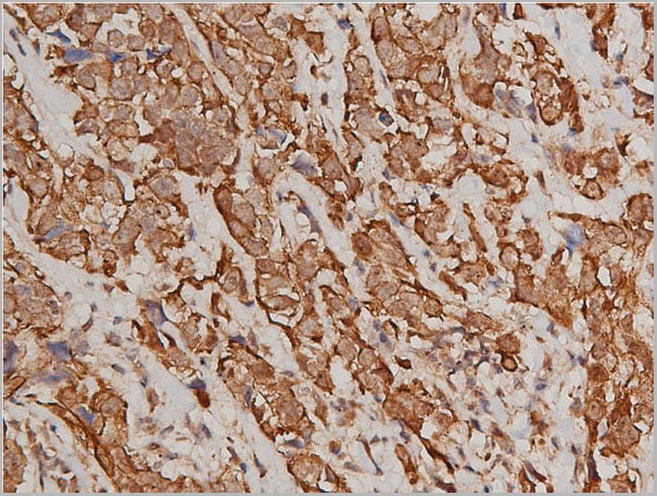
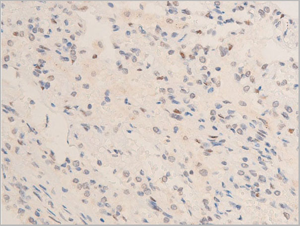
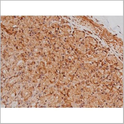
 DB (Dot Blot)
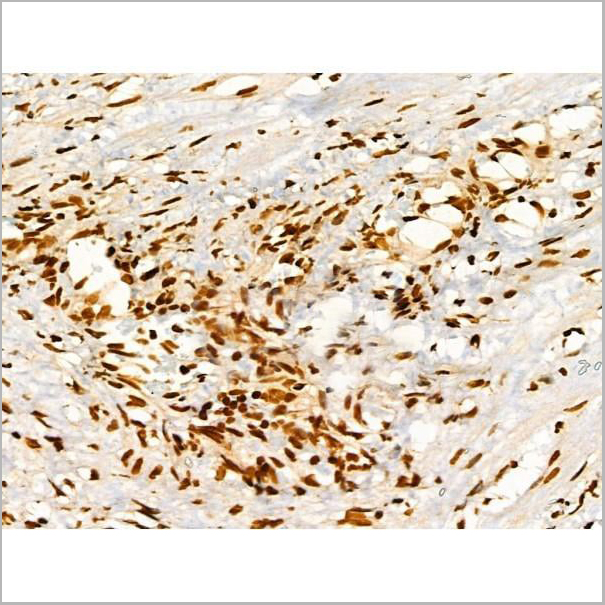
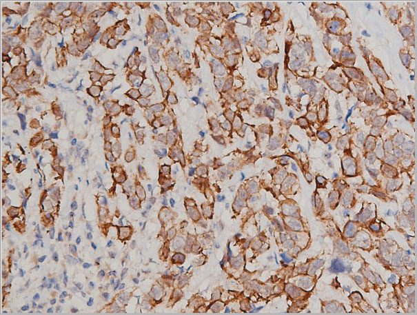
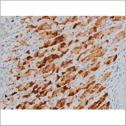
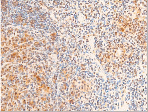
(Formalin-fixed and paraffin-embedded human cancer tissue reacted with the primary antibody, which was peroxidase-conjugated to the secondary antibody, followed by AEC staining. This data demonstrates the use of this antibody for immunohistochemistry; clinical relevance has not been evaluated. BC = breast carcinoma; HC = hepatocarcinoma.)
DB (Dot Blot)
(Formalin-fixed and paraffin-embedded human cancer tissue reacted with the primary antibody, which was peroxidase-conjugated to the secondary antibody, followed by AEC staining. This data demonstrates the use of this antibody for immunohistochemistry; clinical relevance has not been evaluated. BC = breast carcinoma; HC = hepatocarcinoma.)
Phospho-HIST1H3B3 (S10), Polyclonal Antibody (Cat# AAA28670)
 Standard Curve (Sample)
phospho inhibitory subunit of NF kappaB alpha (pIKBalpha), ELISA Kit (Cat# AAA22345)


 WB (Western Blot)
(Dilution: IF: 1:50-200 Western Blot: 1/500 - 1/2000. Immunohistochemistry: 1/100 - 1/300. ELISA: 1/20000. Not yet tested in other applications.)
WB (Western Blot)
(Dilution: IF: 1:50-200 Western Blot: 1/500 - 1/2000. Immunohistochemistry: 1/100 - 1/300. ELISA: 1/20000. Not yet tested in other applications.)
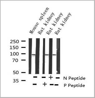
Chk2, Polyclonal Antibody (Cat# AAA28922)
 Standard Curve (Sample)
Phospho Spleen tyosine kinase, ELISA Kit (Cat# AAA16702)


 Application Data
(Dilution: IF: 1:50-200 IHC 1:100-200)
Application Data
(Dilution: IF: 1:50-200 IHC 1:100-200)
phospho-MLKL, Monoclonal Antibody (Cat# AAA28911)
 Standard Curve (Sample)
Phospho-Tau (Thr217), P-tau217, ELISA Kit (Cat# AAA19109)


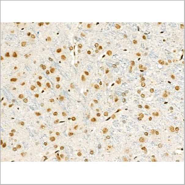
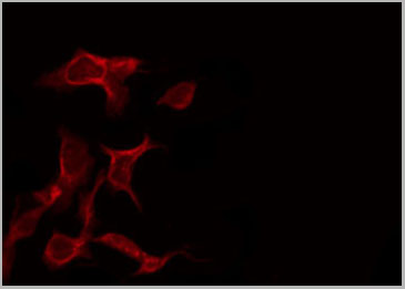
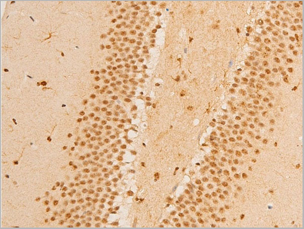
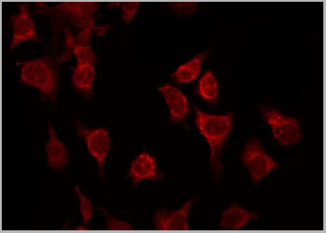
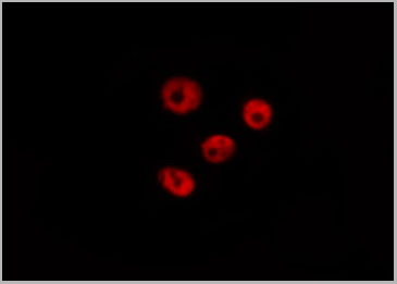
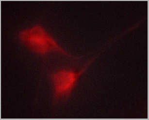
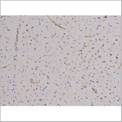
 IF (Immunofluorescence)
(Immunofluorescence analysis of NIH-3T3 cells using Phospho-Histone H2AX-S139 antibody . Blue: DAPI for nuclear staining.)
IF (Immunofluorescence)
(Immunofluorescence analysis of NIH-3T3 cells using Phospho-Histone H2AX-S139 antibody . Blue: DAPI for nuclear staining.)
Phospho-Histone H2AX-S139, Monoclonal Antibody (Cat# AAA28390)
 Standard Curve (Sample)
Phospho-AKT (S473), ELISA Kit (Cat# AAA11391)


 WB (Western Blot)
(Detection of mouse Phospho RNA Polymerase 2 (S2) by western blot. Samples: Whole cell lysate (10 ug) from NIH 3T3, CT26, CH27, TCMK-1, and BW5147.3 cells prepared using NETN lysis buffer. Antibody: Affinity purified rabbit anti-Phospho RNA Polymerase 2 (S2) antibody (AAA23772 lot 4) used for WB at 0.04 ug/ml. Detection: Chemiluminescence with an exposure time of 3 seconds.)
WB (Western Blot)
(Detection of mouse Phospho RNA Polymerase 2 (S2) by western blot. Samples: Whole cell lysate (10 ug) from NIH 3T3, CT26, CH27, TCMK-1, and BW5147.3 cells prepared using NETN lysis buffer. Antibody: Affinity purified rabbit anti-Phospho RNA Polymerase 2 (S2) antibody (AAA23772 lot 4) used for WB at 0.04 ug/ml. Detection: Chemiluminescence with an exposure time of 3 seconds.)
RNA Polymerase II, Polyclonal Antibody (Cat# AAA23772)


 IHC (Immunohistchemistry)
(Dilution: Western Blot: 1/500 - 1/2000. Immunohistochemistry: 1/100 - 1/300. ELISA: 1/40000. Not yet tested in other applications.)
IHC (Immunohistchemistry)
(Dilution: Western Blot: 1/500 - 1/2000. Immunohistochemistry: 1/100 - 1/300. ELISA: 1/40000. Not yet tested in other applications.)
Cytokeratin 8, Polyclonal Antibody (Cat# AAA28924)


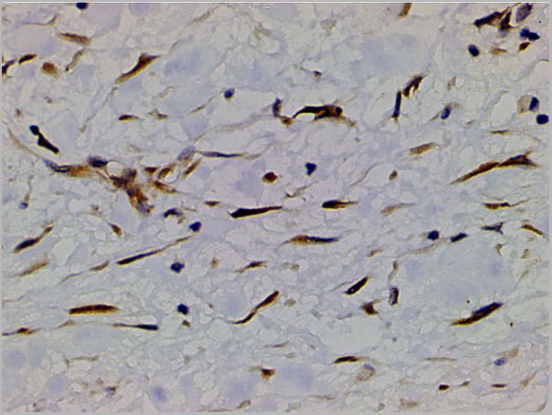
 IF (Immunofluorescence)
(Immunofluorescence analysis of U-2 OS cells using Phospho-Histone H3-T6 Rabbit pAb (AAA28403) at dilution of 100 (40x lens). Blue: DAPI for nuclear staining.)
IF (Immunofluorescence)
(Immunofluorescence analysis of U-2 OS cells using Phospho-Histone H3-T6 Rabbit pAb (AAA28403) at dilution of 100 (40x lens). Blue: DAPI for nuclear staining.)
Histone H3-T6, Polyclonal Antibody (Cat# AAA28403)


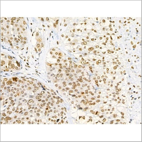
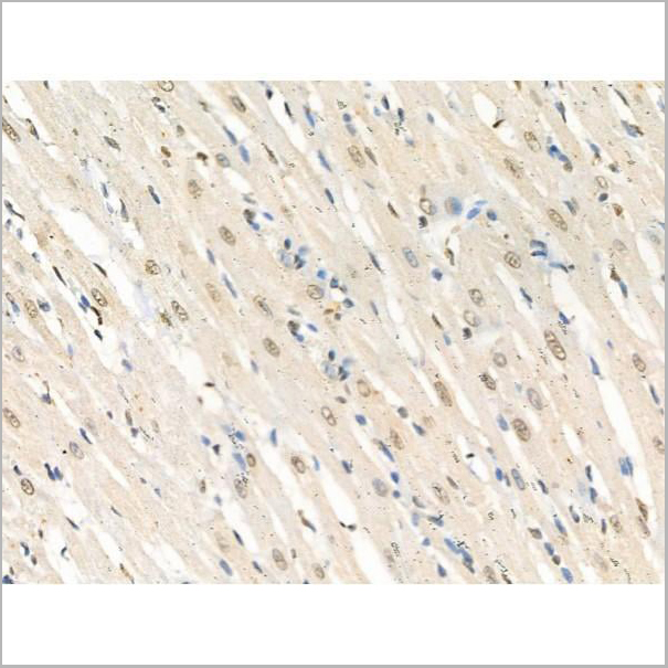
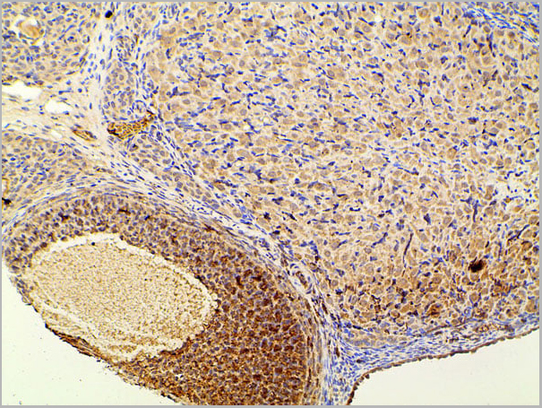
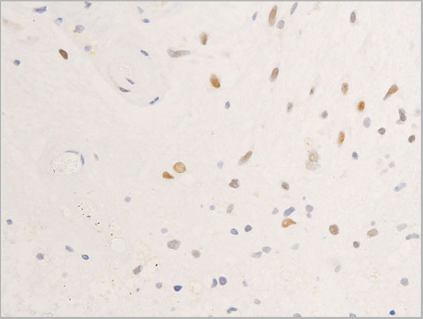
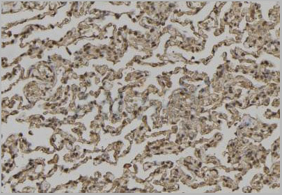
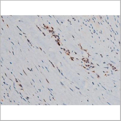
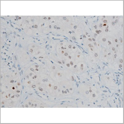
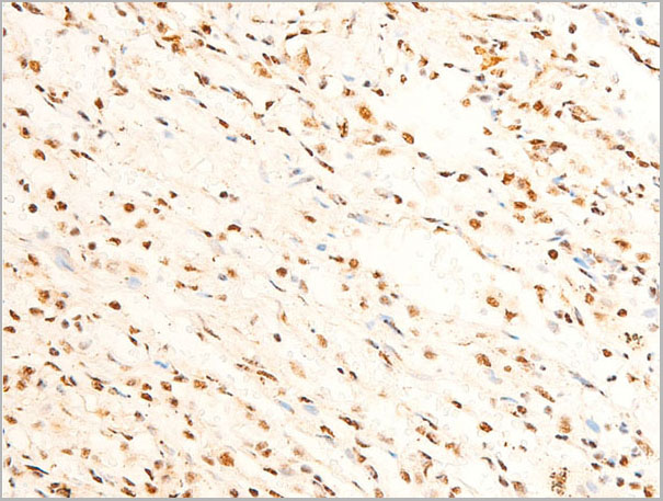
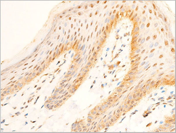
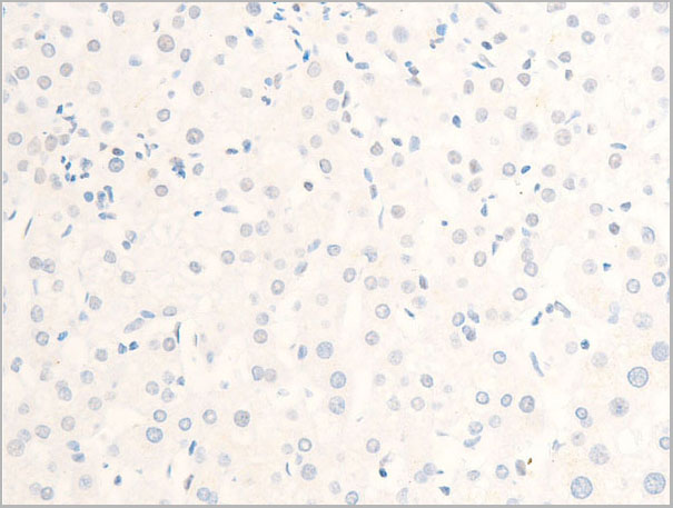
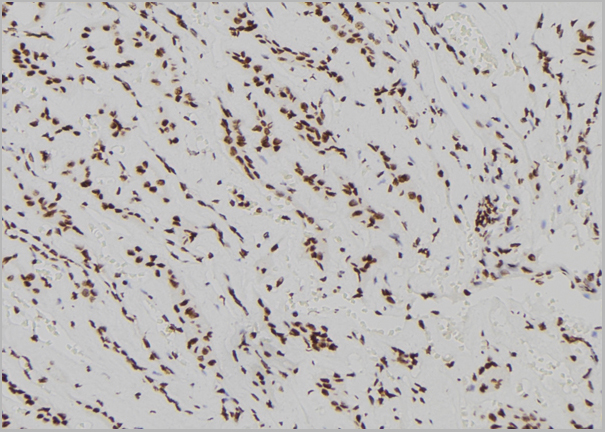
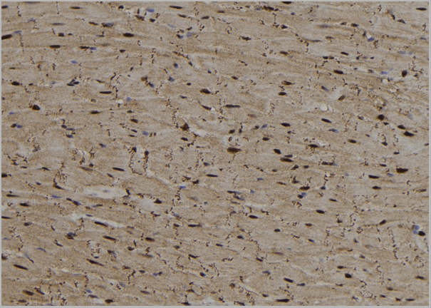
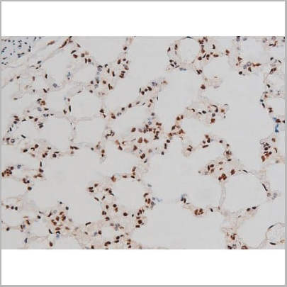
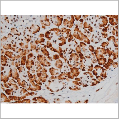
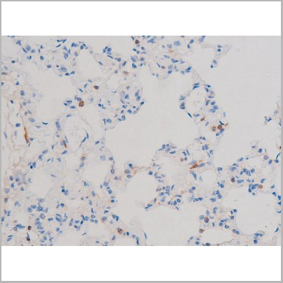
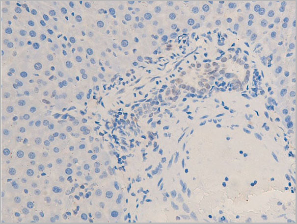
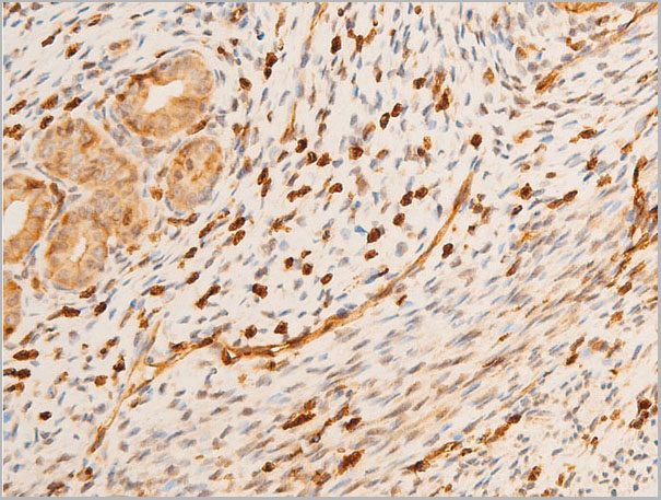
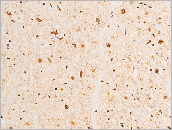
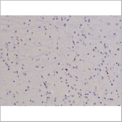
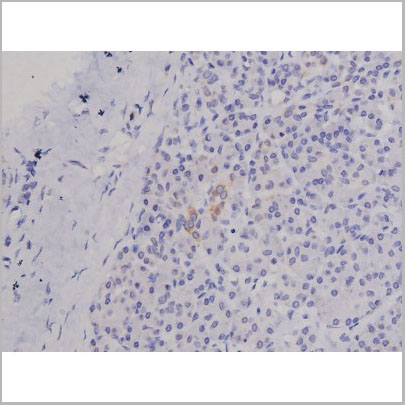
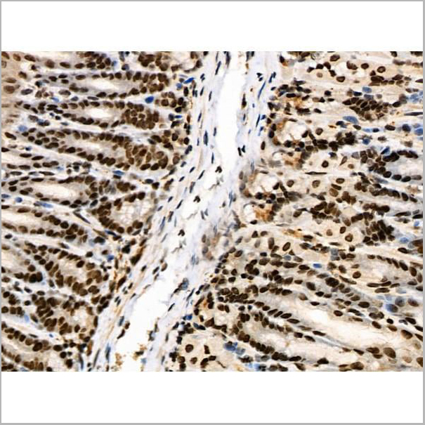
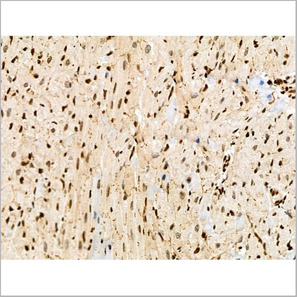
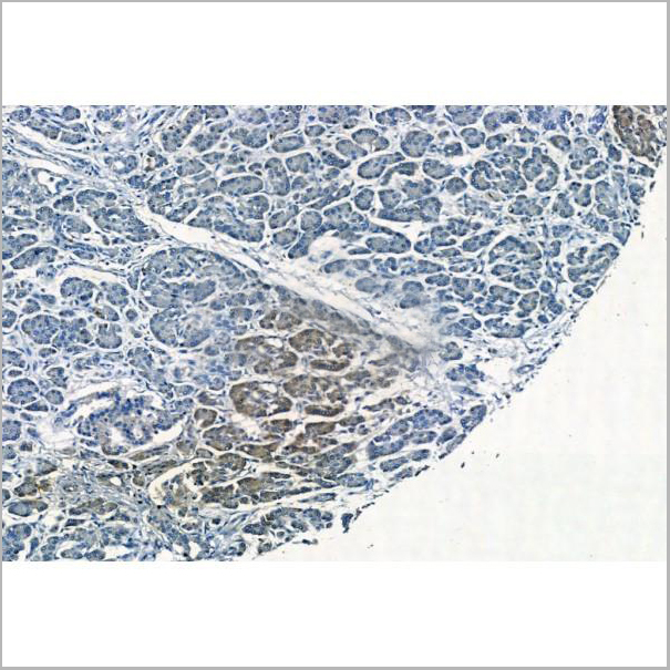
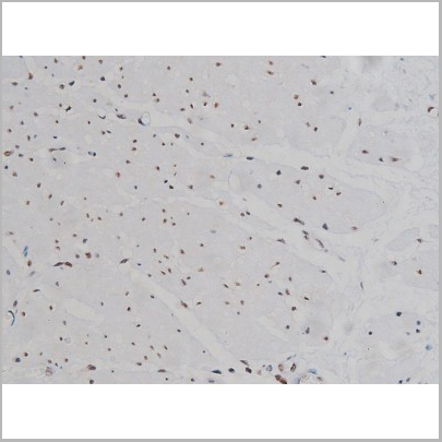
 IHC (Immunohistochemistry)
(At 1/100 staining Mouse heart tissue by IHC-P. The sample was formaldehyde fixed and a heat mediated antigen retrieval step in citrate buffer was performed. The sample was then blocked and incubated with the primary antibody at 4 degree C overnight. An HRP conjugated anti-Rabbit antibody was used as the secondary antibody.)
IHC (Immunohistochemistry)
(At 1/100 staining Mouse heart tissue by IHC-P. The sample was formaldehyde fixed and a heat mediated antigen retrieval step in citrate buffer was performed. The sample was then blocked and incubated with the primary antibody at 4 degree C overnight. An HRP conjugated anti-Rabbit antibody was used as the secondary antibody.)
ID2, Polyclonal Antibody (Cat# AAA31291)


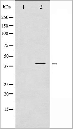
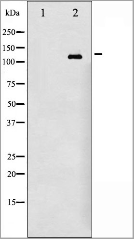
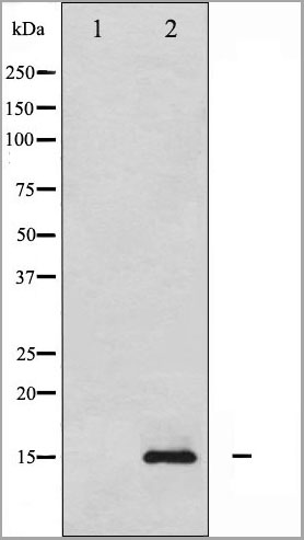
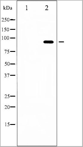
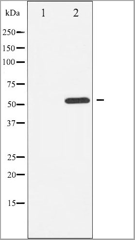
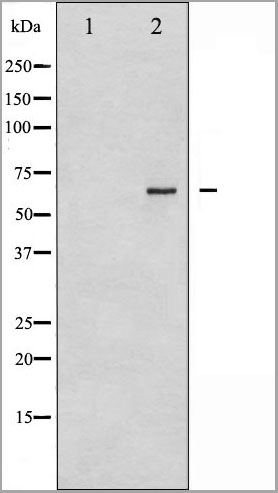
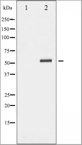
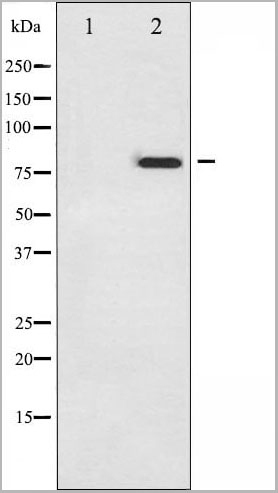
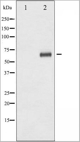
 WB (Western Blot)
(Western blot analysis of ENO3 over-expressed 293 cell line, cotransfected with ENO3 Validated Chimera RNAi (Lane 2) or non-transfected control (Lane 1). Blot probed with ENO3 monoclonal antibody. GAPDH (36.1kD) used as specificity and loading control.)
WB (Western Blot)
(Western blot analysis of ENO3 over-expressed 293 cell line, cotransfected with ENO3 Validated Chimera RNAi (Lane 2) or non-transfected control (Lane 1). Blot probed with ENO3 monoclonal antibody. GAPDH (36.1kD) used as specificity and loading control.)
ENO3, Monoclonal Antibody (Cat# AAA24793)


 IHC (Immunohistochemistry)
(Dilution: IF: 1:50-200 Western Blot: 1/500 - 1/2000. Immunohistochemistry: 1/100 - 1/300. Immunoprecipitation: 2-5 ug/mg lysate. ELISA: 1/10000. Not yet tested in other applications.)
IHC (Immunohistochemistry)
(Dilution: IF: 1:50-200 Western Blot: 1/500 - 1/2000. Immunohistochemistry: 1/100 - 1/300. Immunoprecipitation: 2-5 ug/mg lysate. ELISA: 1/10000. Not yet tested in other applications.)
CREB-1, Polyclonal Antibody (Cat# AAA28923)
What Are Phospho Antibodies?
Protein phosphorylation is a process where a phosphate group is added to certain amino acid residues of a protein – usually serine (S), threonine (T), or tyrosine (Y) - by enzymes called kinases. This process is integral in controlling cellular signaling, cellular growth, and other biological functions.
Our catalog includes a wide range of phospho-specific antibodies that can accurately detect this important marker. They perform strongly in widely-used laboratory applications such as Western blot, flow cytometry, immunohistochemistry, and immunofluorescence microscopy. We value your trust in us and are committed to providing top-quality products and services. All of our antibodies are guaranteed to work for the applications and species indicated on our website & associated product pages.
What Are The Key Applications of Phospho Antibodies?
1. Western Blotting
One of the first steps a researcher can take in utilizing these phospho-specific antibodies, is to check if the antibody works using a technique referred to as “Western blot”. For those unfamiliar, Western Blot aids in showing whether the protein that the antibody recognizes is appearing at the correct/expected size. These phospho-specific antibodies should also be able to detect changes in the target protein’s phosphorylation (on/off state) when cells are stimulated in certain ways.
2. Staining of Fixed Cells (Immunocytochemistry)
Another routine use of these phospho-specific antibodies, is to test if the antibody is able to demonstrate similar performance when used on fixed cells (intact cells that have been preserved) as it did in the Western blot tests. It is an important aspect in many cases to confirm that the antibody works in actual intact cell samples. Ideally, the method used for cellular fixation should be the same as what is used in pathology labs (like using 10% formalin). To check if the antibody works well in tissue sections (FFPE), researchers will often test it on fixed cells that are processed similar to tissue samples.
3. Specificity Tests Using Peptides
In order to make sure that the antibody is only binding to the right target:
- Laboratory technicians will mix the antibody with phospho-peptides (short segments of the protein containing the phosphate group modification).
- If the antibody signal disappears, it is confirmation that it is binding to the correct phosphorylated location.
- A more robust test is to use both the phosphorylated and non-phosphorylated (dephosphorylated) versions of the protein. The antibody should react only with the phosphorylated one.
- Another method sometimes utilized is to treat the sample with an enzyme, such as alkaline phosphatase, that specifically removes phosphate groups. If the antibody signal disappears after this, it also confirms specificity.
4. Genetic Confirmation
As a final step, scientists can genetically manipulate the nucleotide sequence and alter the target protein by removing the exact site where phosphorylation happens. If the antibody no longer appears to detect the modified protein, it is strong evidence supporting the antibody being specific for that phosphorylated site.
Why Buy Phospho Antibodies Through Us?
- The production laboratory adheres to strict and consistent protocols prior to releasing any of these phospho-specific antibodies:
- Standard methods and proper controls in all tests to ensure high quality.
- These antibodies are tested and validated in different cell types and species.
- High quality control criterion to ensure each batch is consistent, so you will obtain reliable results every time.
FAQ
1. What Are Phospho-Specific Antibodies?
Phospho-specific antibodies are made to detect proteins only when they have a phosphate group linked to a specific amino acid residue. This empowers scientists understand if a protein is "turned on" or active, based on its phosphorylation state.
2. How to Detect Phosphorylated Proteins in a Western Blot?
To find out if a protein is phosphorylated using Western blot:
- Use a phospho-specific antibody that binds only to the phosphorylated form of the protein.
- You can also use a “regular” antibody for the same amino acid sequence of the protein that the phospho-specific antibody is binding to (but in this case, this antibody will not bind if there is a phosphate group present) in order to compare how much of it is phosphorylated versus how much is non-phosphorylated (or “total” protein, if the “normal” antibody’s epitopes are non-phospho-site-specific).
3. How to Choose the Best Antibody?
Here are some simple tips to help you pick the right antibody:
- Know your target
- Match your sample characteristics
- Confirm the intended use is appropriate
- Check “host” and “type”
- Check the “quality” of the presented data/images
- Appraise whether the available validation meets your needs