Filters
▼Clonality
▼Type
▼Reactivity
▼Gene Name
▼Isotype
▼Host
▼Application
▼Clone
▼Phospho Antibodies
Phospho-specific antibodies’ typical purpose is to enable researchers to detect changes in proteins. They will exclusively bind to the amino acid sequence on a protein that has been phosphorylated (which is both a physical & chemical change) and do not bind to the same amino acid sequence on said protein if it lacks said phosphorylation. This aids in being able to clearly see and understand the data produced from this particular protein modification.
Viewing 4900-4950 of 5298 product results
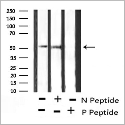
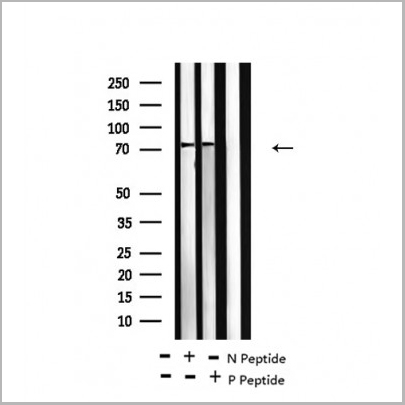
 WB (Western Blot)
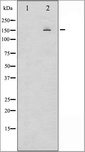
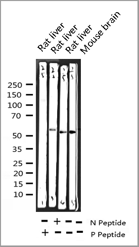
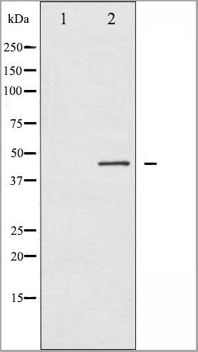
(Western blot analysis of Ezrin phosphorylation expression in Na3VO4 treated K562 whole cell lysates, The lane on the left is treated with the antigen-specific peptide.)
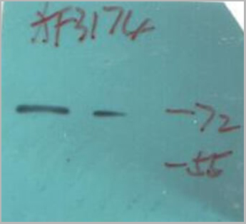
WB (Western Blot)
(Western blot analysis of Ezrin phosphorylation expression in Na3VO4 treated K562 whole cell lysates, The lane on the left is treated with the antigen-specific peptide.)
Ezrin, Polyclonal Antibody (Cat# AAA31010)
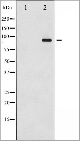
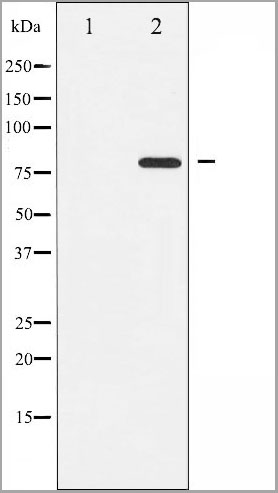
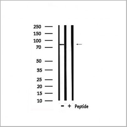
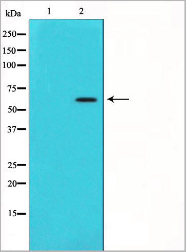
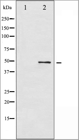
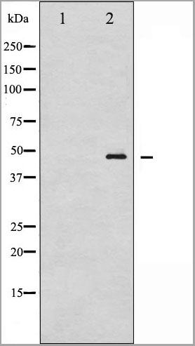
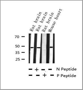
 WB (Western Blot)
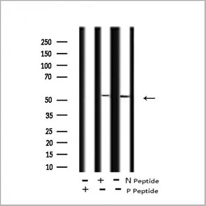
(Western blot analysis of extracts of various tissue sample, using Phospho-PLCG1 (Tyr771) Antibody.)
WB (Western Blot)
(Western blot analysis of extracts of various tissue sample, using Phospho-PLCG1 (Tyr771) Antibody.)
PLCG1, Polyclonal Antibody (Cat# AAA31021)

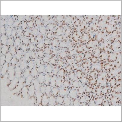
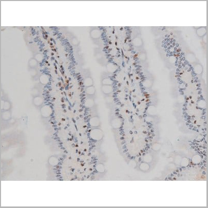
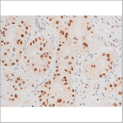
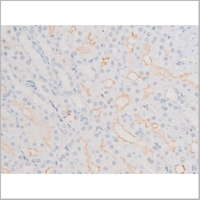
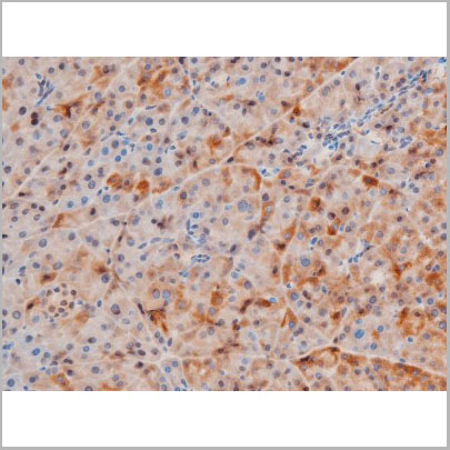
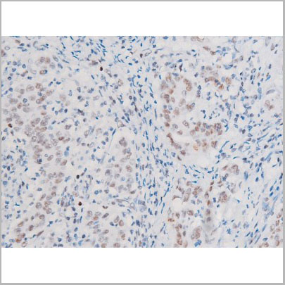
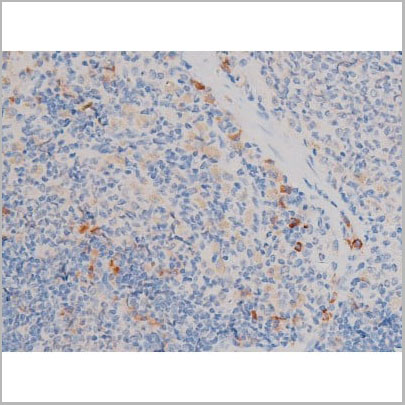
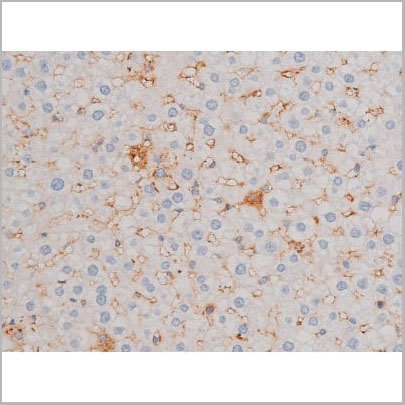
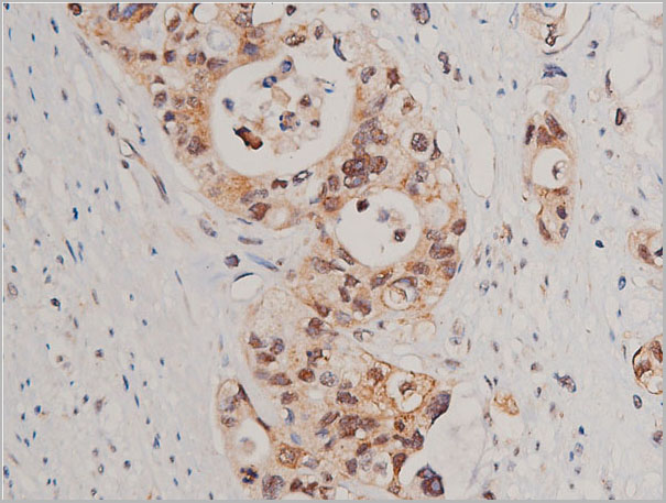
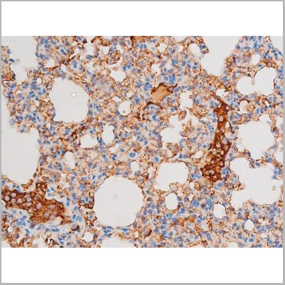
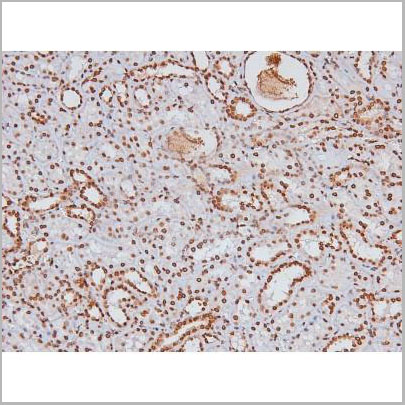
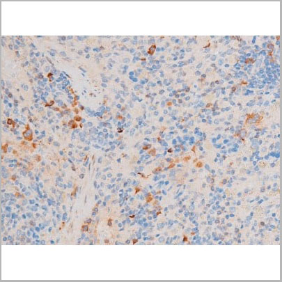
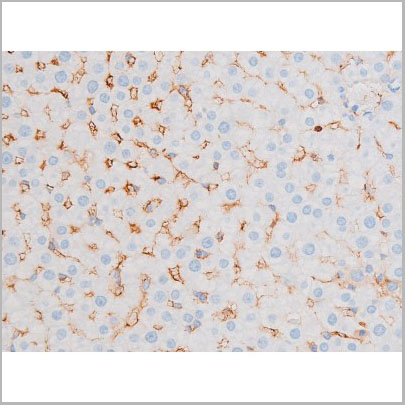
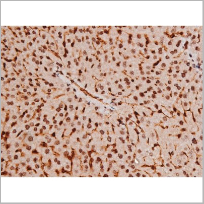
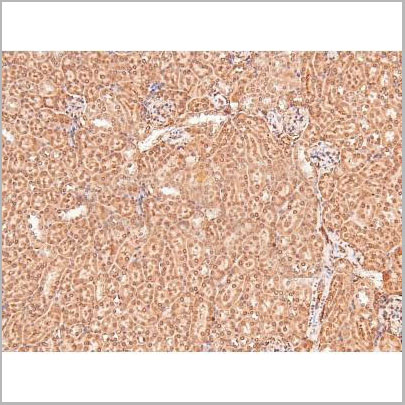
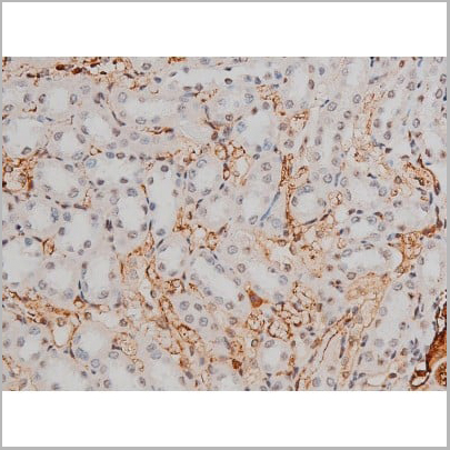
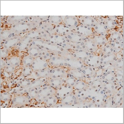
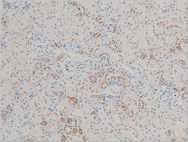
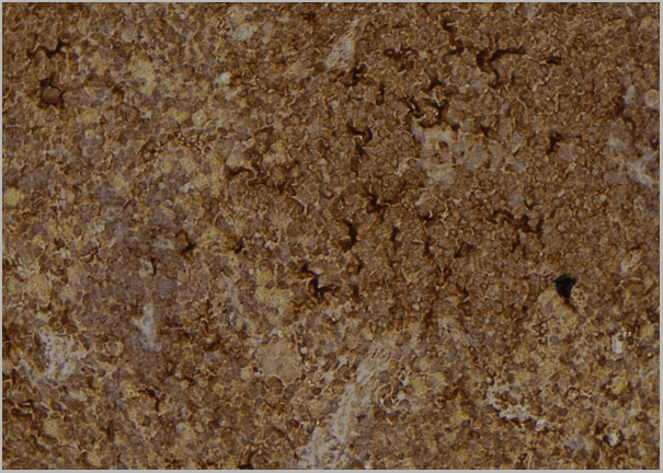
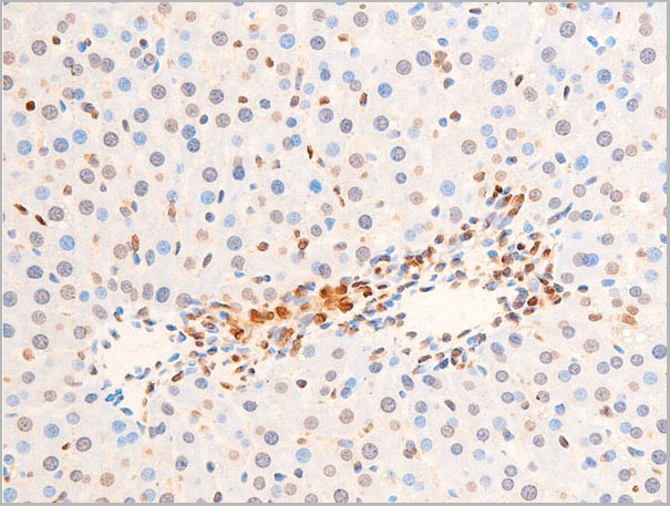
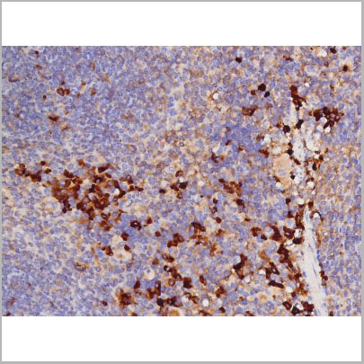
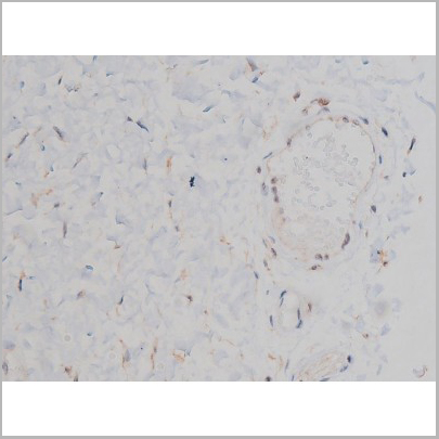
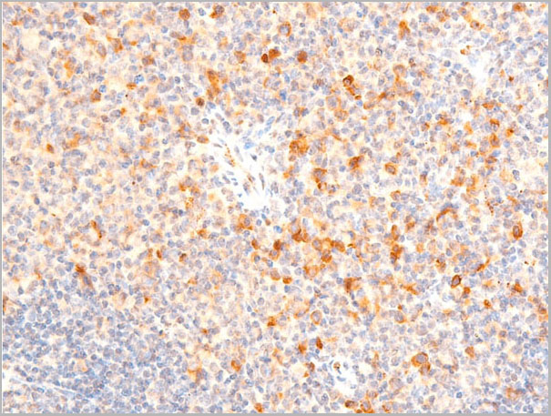
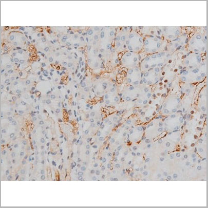
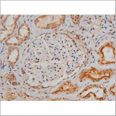
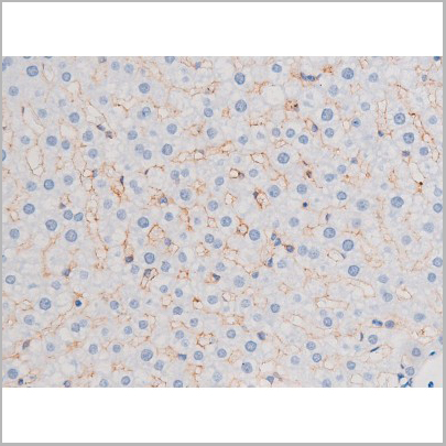
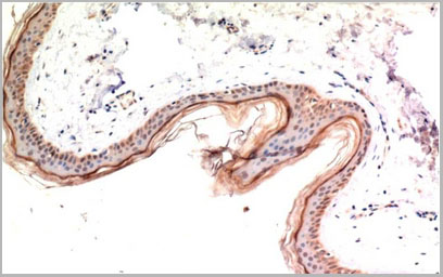
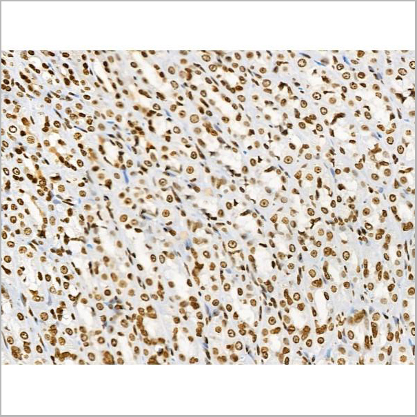
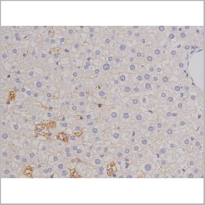
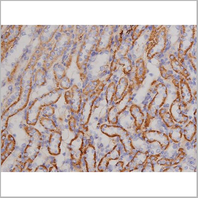
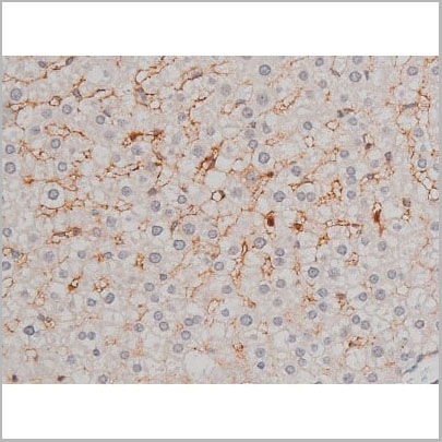
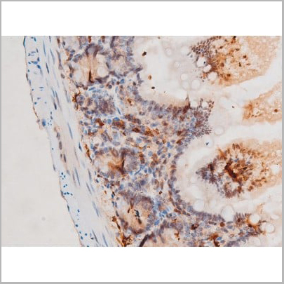
 IHC (Immunohistochemistry)
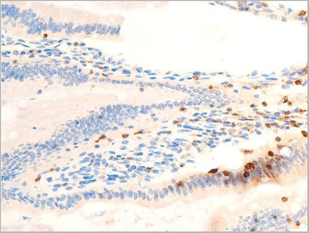
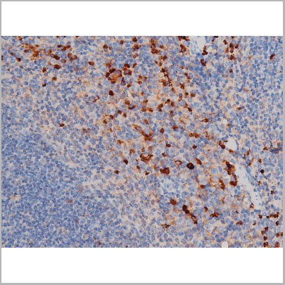
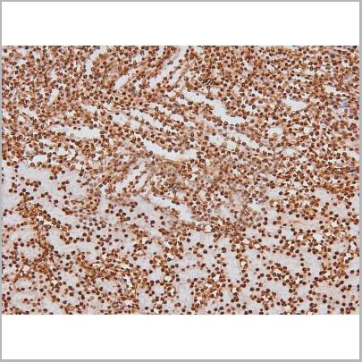
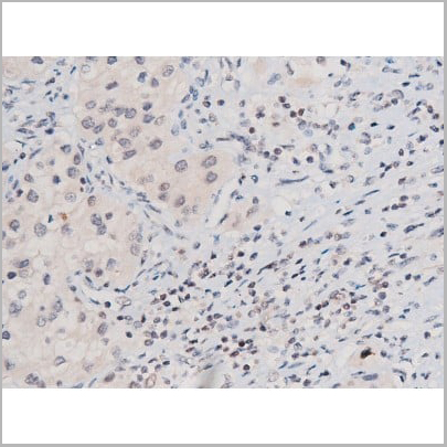
(AAA31043 at 1/200 staining Rat kidney tissue sections by IHC-P. The tissue was formaldehyde fixed and a heat mediated antigen retrieval step in citrate buffer was performed. The tissue was then blocked and incubated with the antibody for 1.5 hours at 22 degree C. An HRP conjugated goat anti-rabbit antibody was used as the secondary.)
IHC (Immunohistochemistry)
(AAA31043 at 1/200 staining Rat kidney tissue sections by IHC-P. The tissue was formaldehyde fixed and a heat mediated antigen retrieval step in citrate buffer was performed. The tissue was then blocked and incubated with the antibody for 1.5 hours at 22 degree C. An HRP conjugated goat anti-rabbit antibody was used as the secondary.)
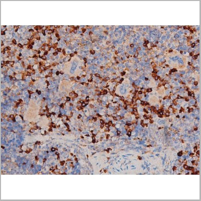
STAT1, Polyclonal Antibody (Cat# AAA31043)
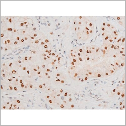

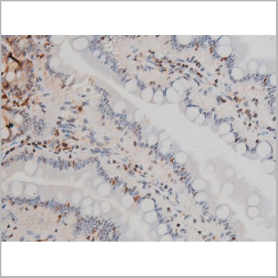
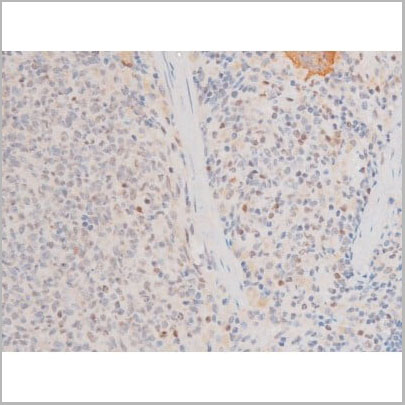
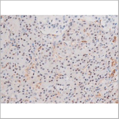
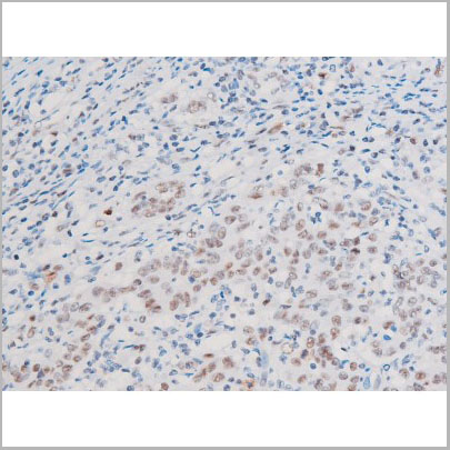
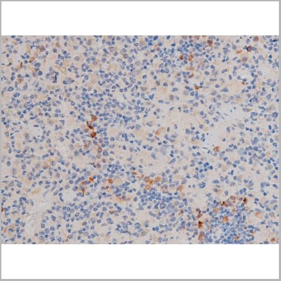
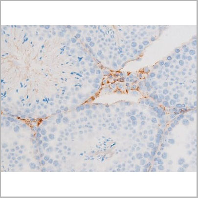
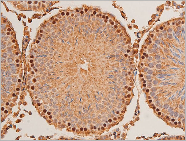
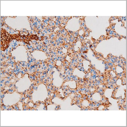
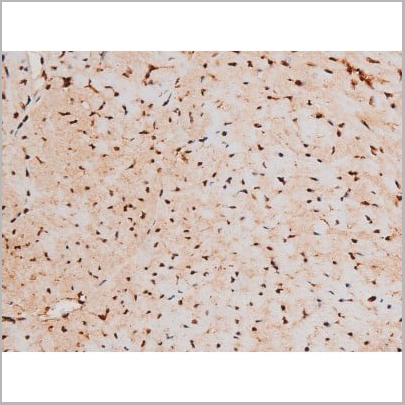
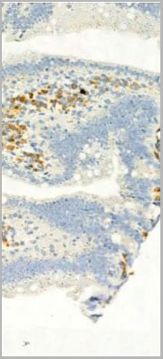
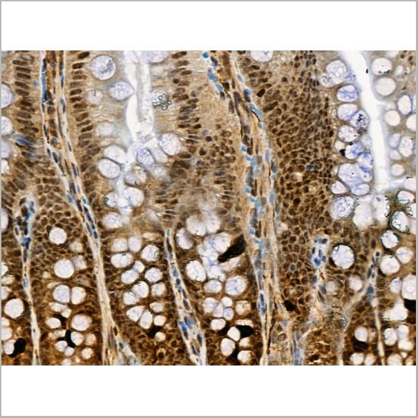
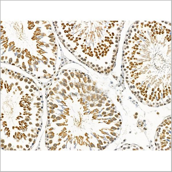
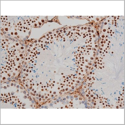
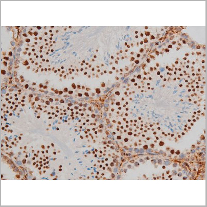
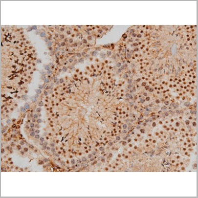
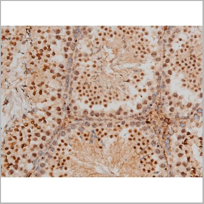
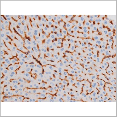
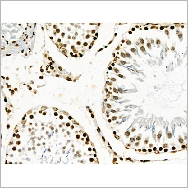
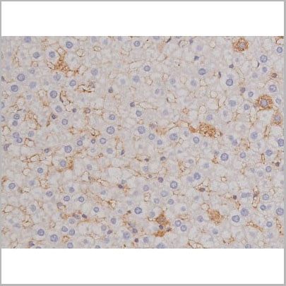
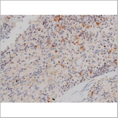
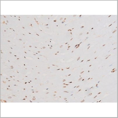
 IHC (Immunohistochemistry)
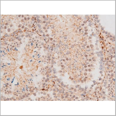
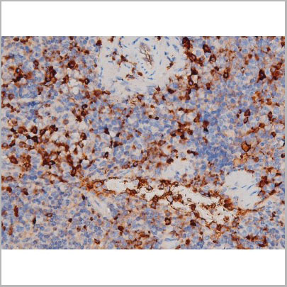
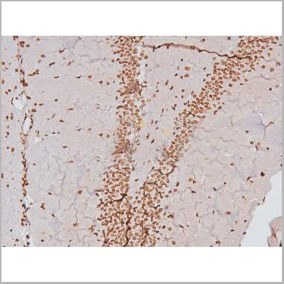
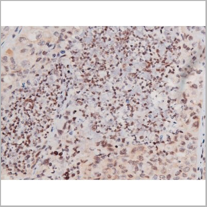
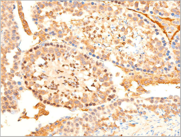
(AAA31054 at 1/200 staining Mouse testis tissue sections by IHC-P. The tissue was formaldehyde fixed and a heat mediated antigen retrieval step in citrate buffer was performed. The tissue was then blocked and incubated with the antibody for 1.5 hours at 22 degree C. An HRP conjugated goat anti-rabbit antibody was used as the secondary.)
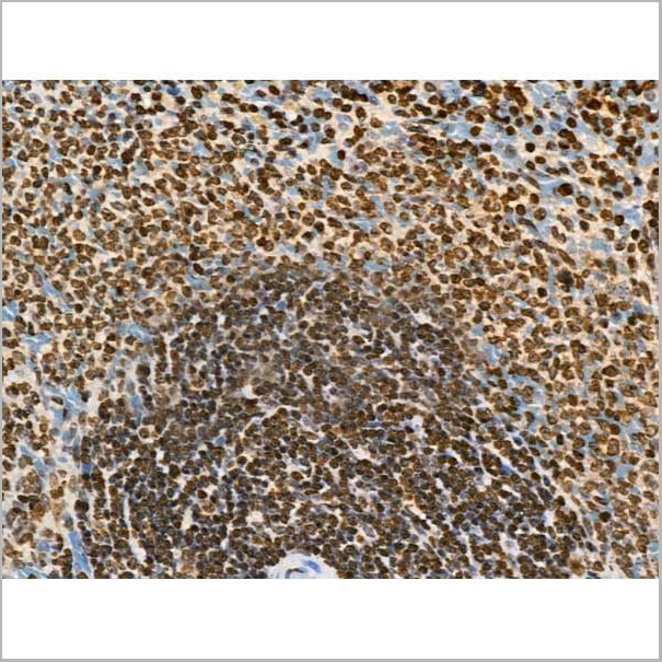
IHC (Immunohistochemistry)
(AAA31054 at 1/200 staining Mouse testis tissue sections by IHC-P. The tissue was formaldehyde fixed and a heat mediated antigen retrieval step in citrate buffer was performed. The tissue was then blocked and incubated with the antibody for 1.5 hours at 22 degree C. An HRP conjugated goat anti-rabbit antibody was used as the secondary.)
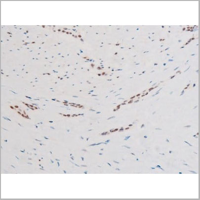
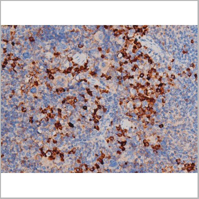
JNK1/2/3, Polyclonal Antibody (Cat# AAA31054)
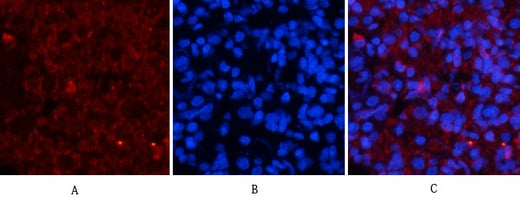
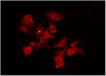
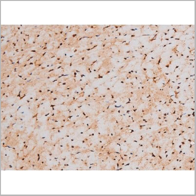
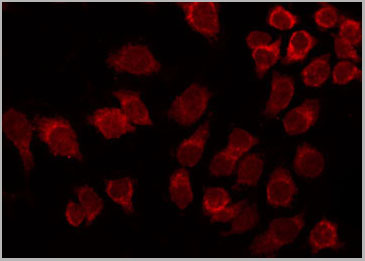
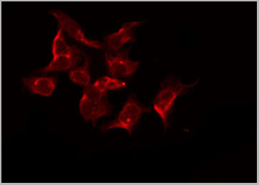
 IF (Immunoflouorescence)
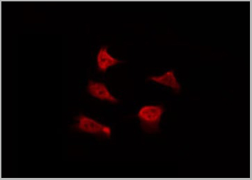
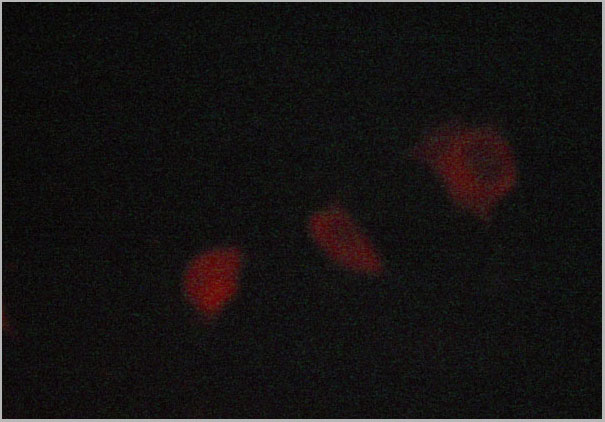
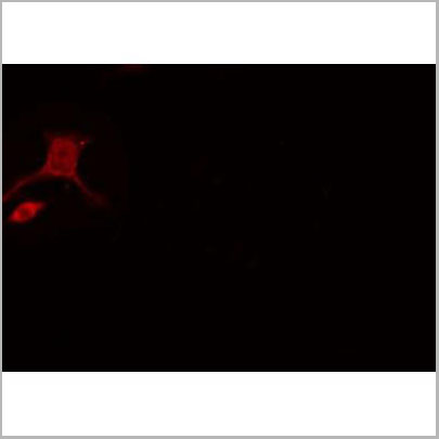
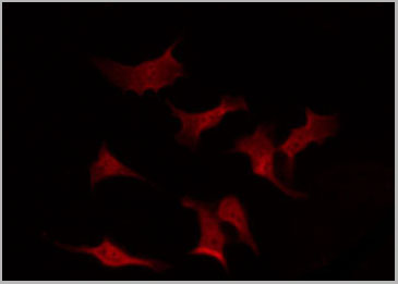
(AAA31056 staining NIH-3T3 by IF/ICC. The sample were fixed with PFA and permeabilized in 0.1% Triton X-100, then blocked in 10% serum for 45 minutes at 25 degree C. The primary antibody was diluted at 1/200 and incubated with the sample for 1 hour at 37 degree C. An Alexa Fluor 594 conjugated goat anti-rabbit IgG (H+L) Ab, diluted at 1/600, was used as the secondary antibody.)
IF (Immunoflouorescence)
(AAA31056 staining NIH-3T3 by IF/ICC. The sample were fixed with PFA and permeabilized in 0.1% Triton X-100, then blocked in 10% serum for 45 minutes at 25 degree C. The primary antibody was diluted at 1/200 and incubated with the sample for 1 hour at 37 degree C. An Alexa Fluor 594 conjugated goat anti-rabbit IgG (H+L) Ab, diluted at 1/600, was used as the secondary antibody.)
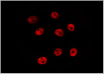
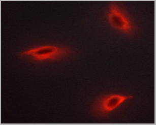
LIMK2, Polyclonal Antibody (Cat# AAA31056)
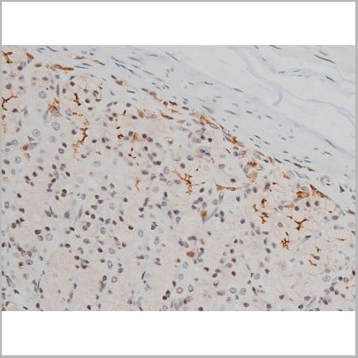
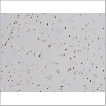
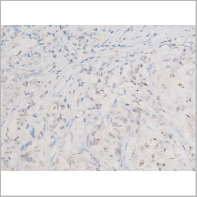
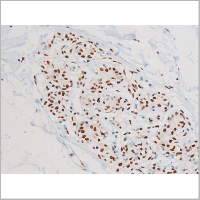
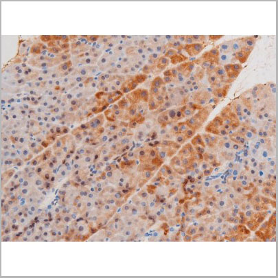
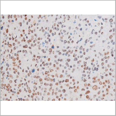
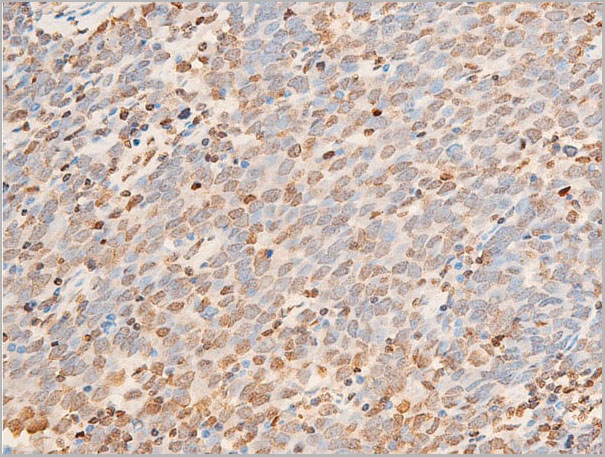
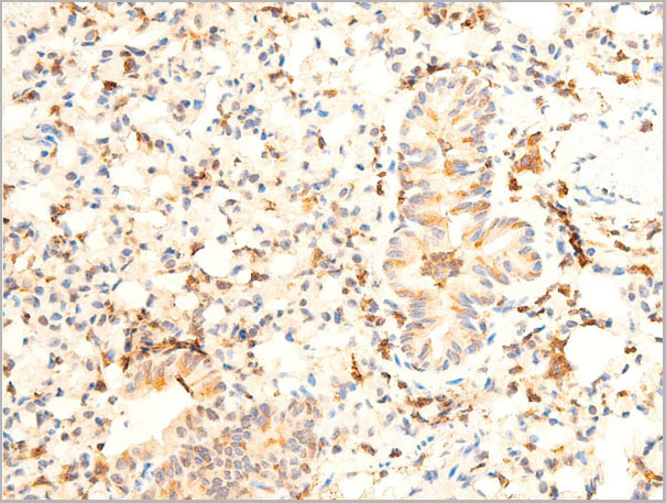
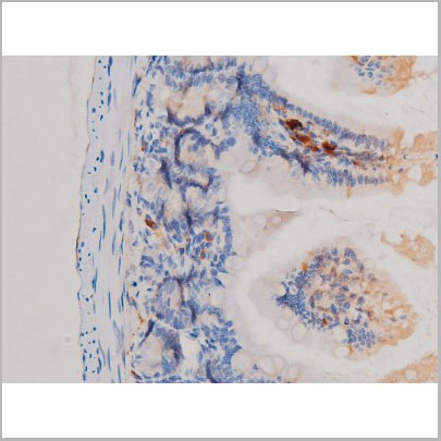
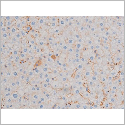
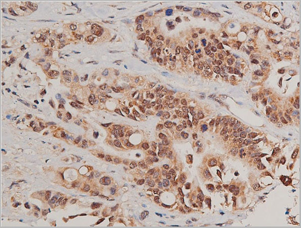
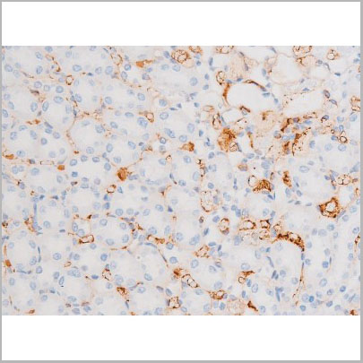
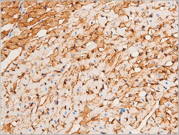
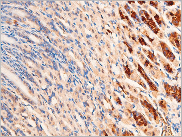
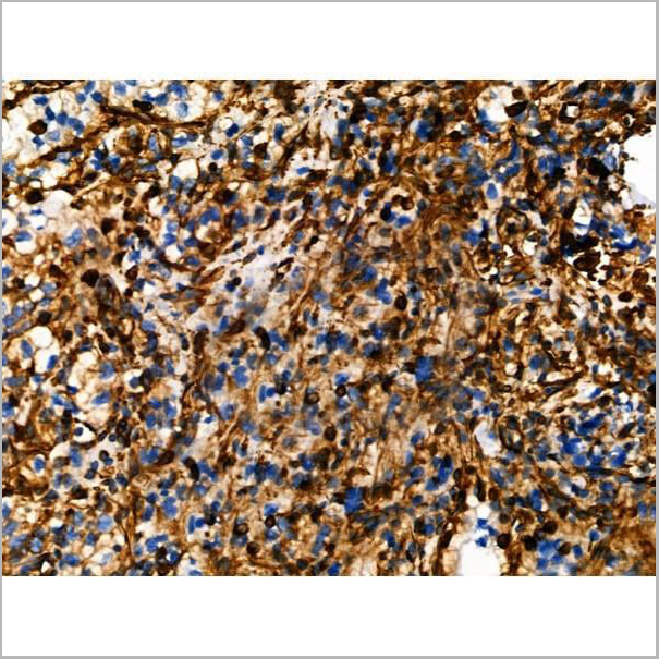
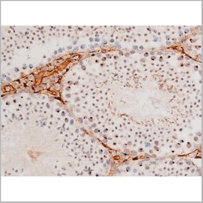
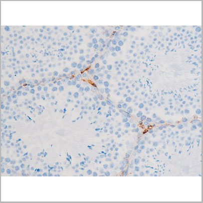
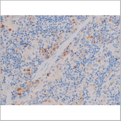
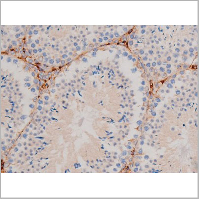
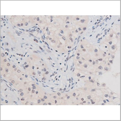
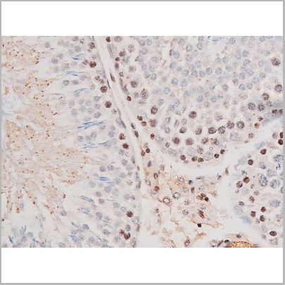
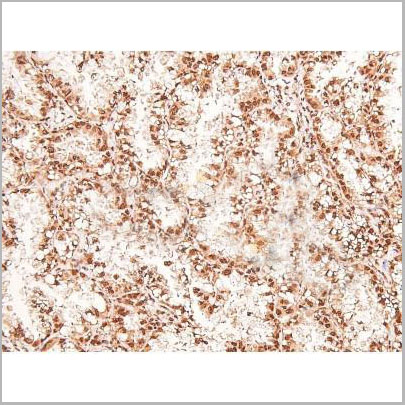
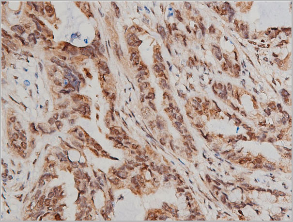
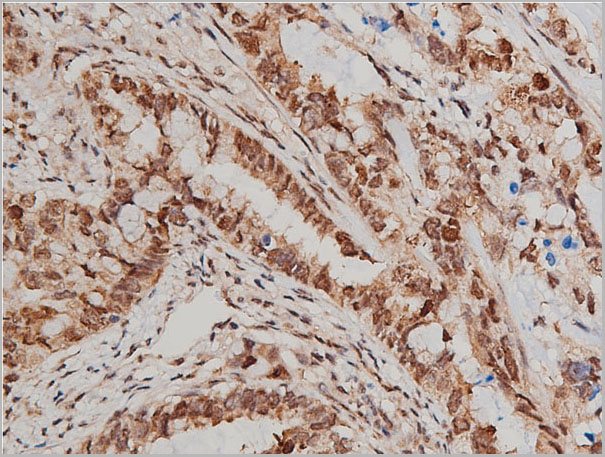
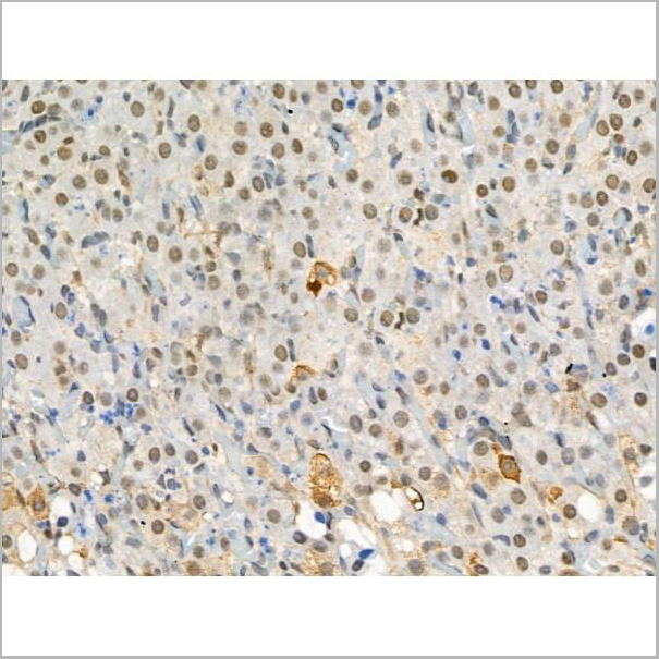
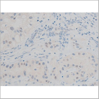
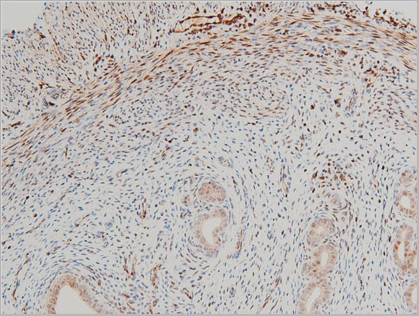
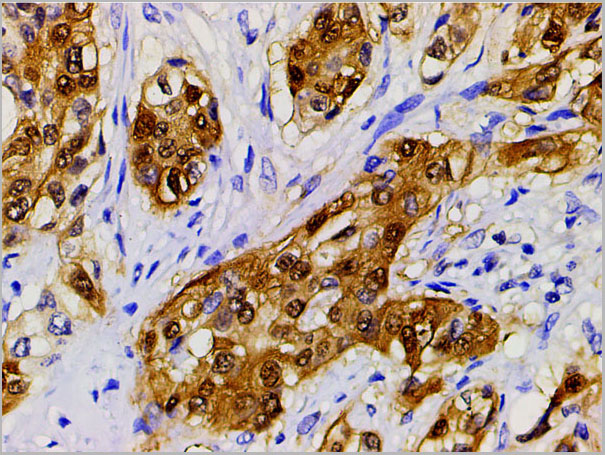
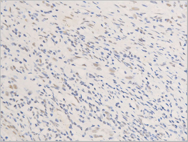
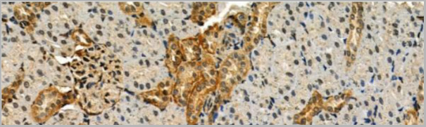
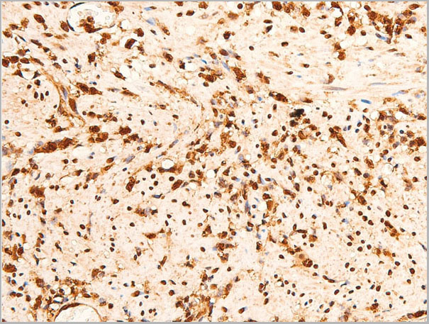
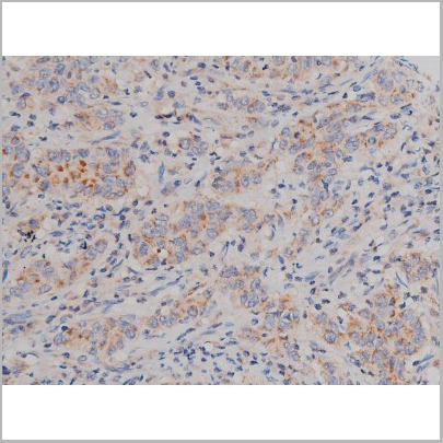
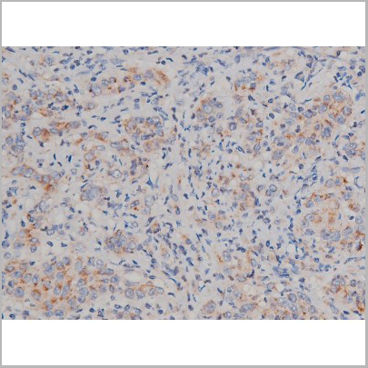
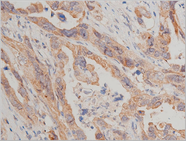
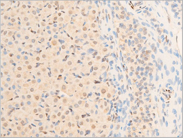
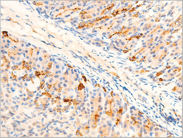
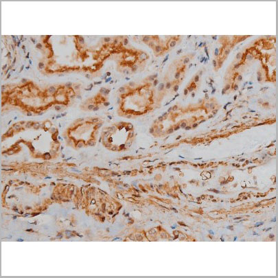
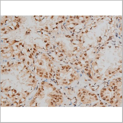
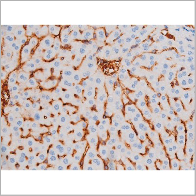
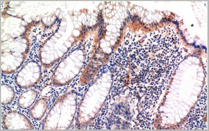
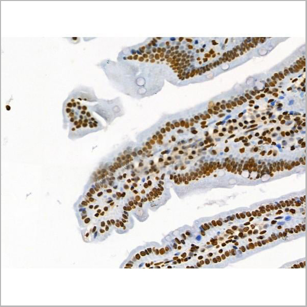
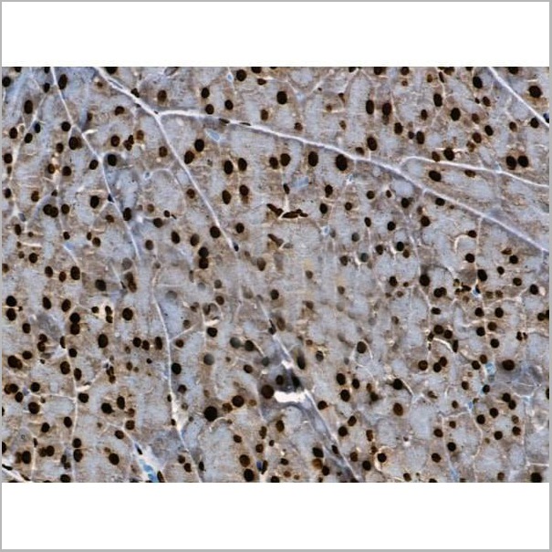
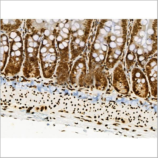
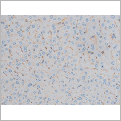
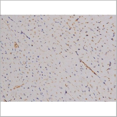
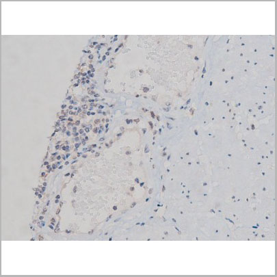
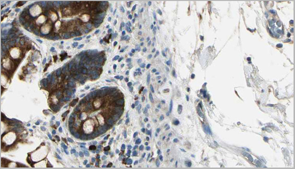
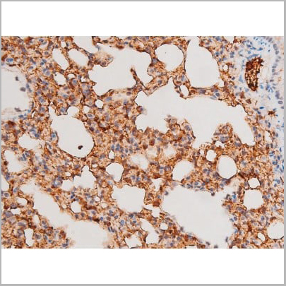
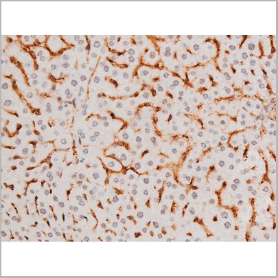
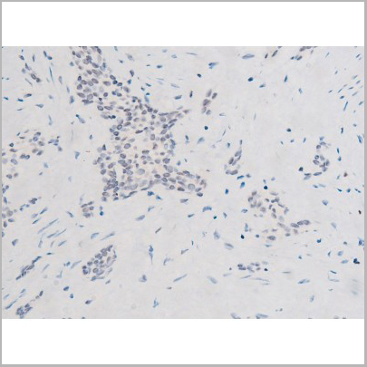
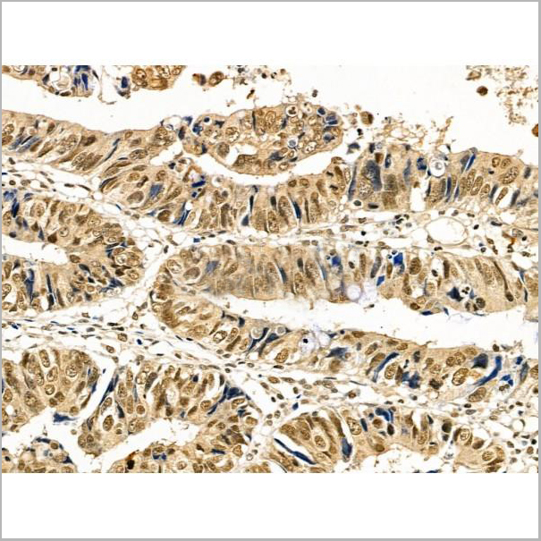 IHC (Immunohistochemistry)
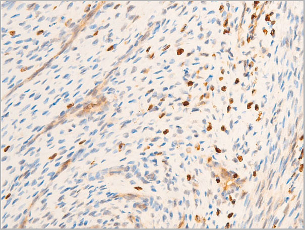
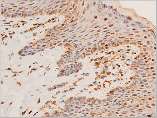
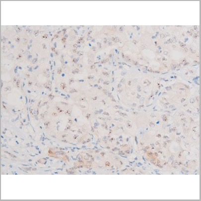
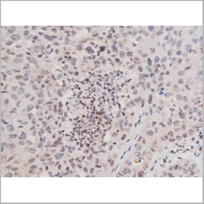
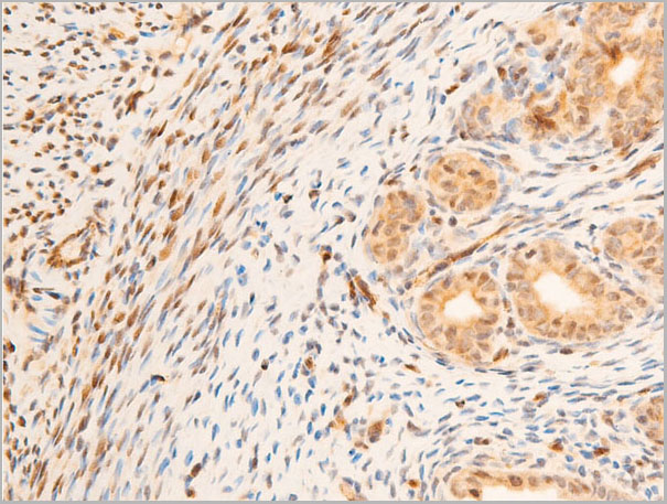
(At 1/100 staining Human colorectal cancer by IHC-P. The sample was formaldehyde fixed and a heat mediated antigen retrieval step in citrate buffer was performed. The sample was then blocked and incubated with the primary antibody at 4 degree C overnight. An HRP conjugated anti-Rabbit antibody was used as the secondary antibody.)
IHC (Immunohistochemistry)
(At 1/100 staining Human colorectal cancer by IHC-P. The sample was formaldehyde fixed and a heat mediated antigen retrieval step in citrate buffer was performed. The sample was then blocked and incubated with the primary antibody at 4 degree C overnight. An HRP conjugated anti-Rabbit antibody was used as the secondary antibody.)
Cyclin E1, Polyclonal Antibody (Cat# AAA31362)
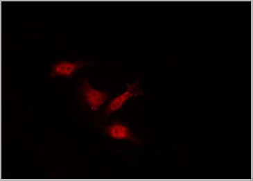
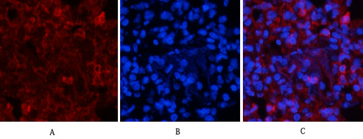
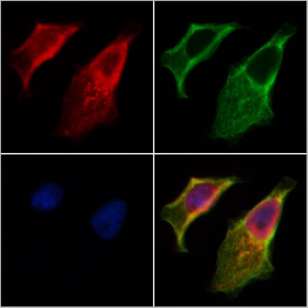
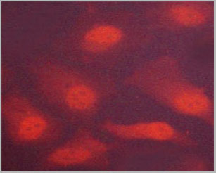
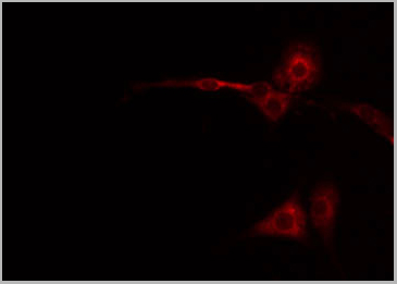
 IF (Immunofluorescence)
(AAA30937 staining A-431 cells by IF/ICC. The sample were fixed with PFA and permeabilized in 0.1% Triton X-100, then blocked in 10% serum for 45 minutes at 25 degree C. The primary antibody was diluted at 1/200 and incubated with the sample for 1 hour at 37 degree C. An Alexa Fluor 594 conjugated goat anti-rabbit IgG (H+L) antibody, diluted at 1/600, was used as secondary antibody.)
IF (Immunofluorescence)
(AAA30937 staining A-431 cells by IF/ICC. The sample were fixed with PFA and permeabilized in 0.1% Triton X-100, then blocked in 10% serum for 45 minutes at 25 degree C. The primary antibody was diluted at 1/200 and incubated with the sample for 1 hour at 37 degree C. An Alexa Fluor 594 conjugated goat anti-rabbit IgG (H+L) antibody, diluted at 1/600, was used as secondary antibody.)
SYK, Polyclonal Antibody (Cat# AAA30937)

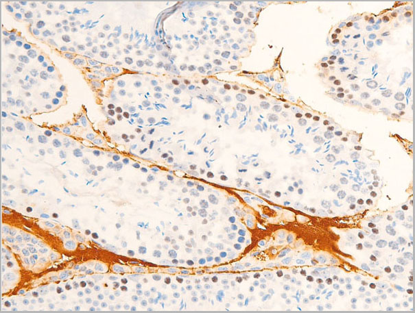
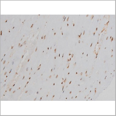
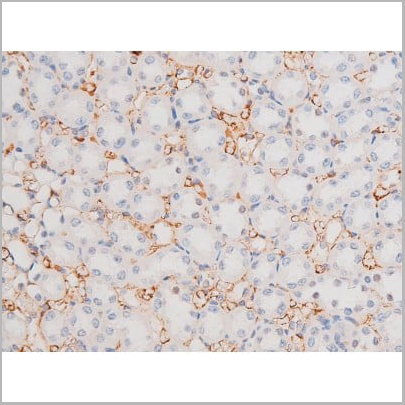
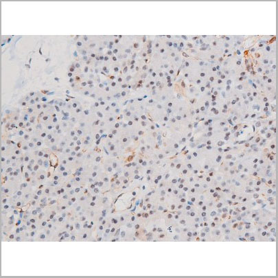
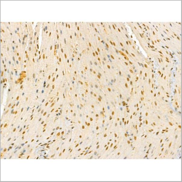
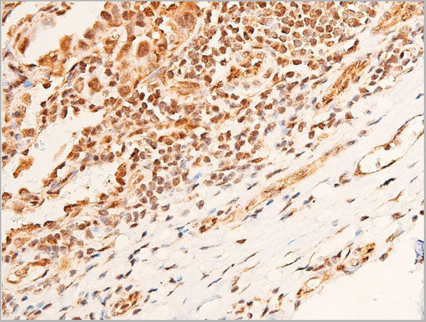
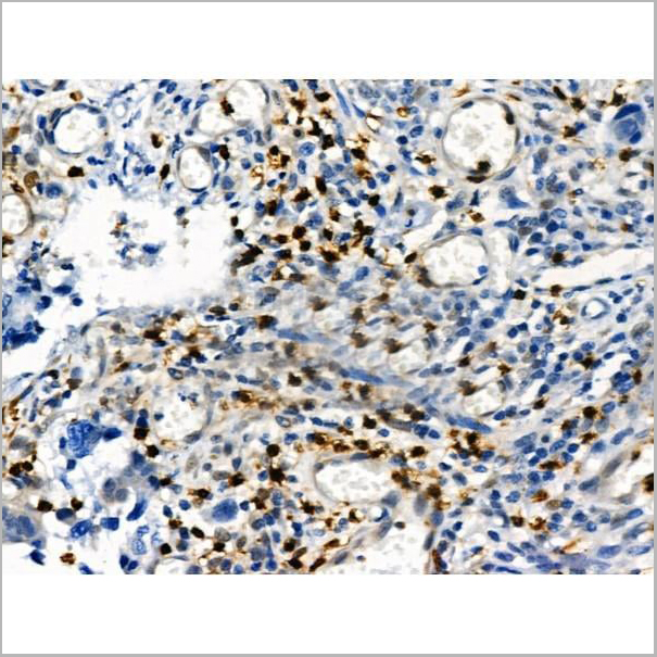
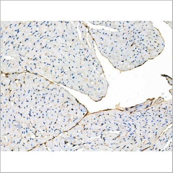
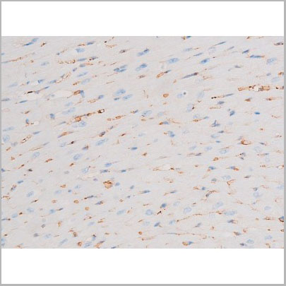
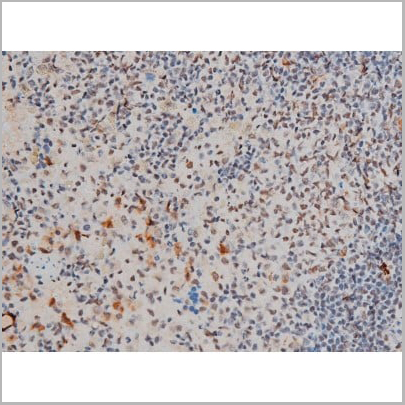
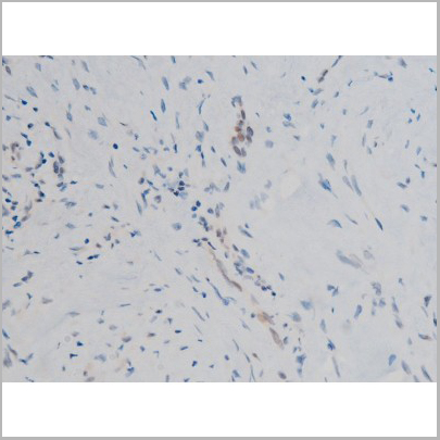
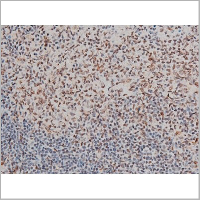
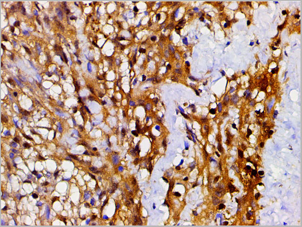
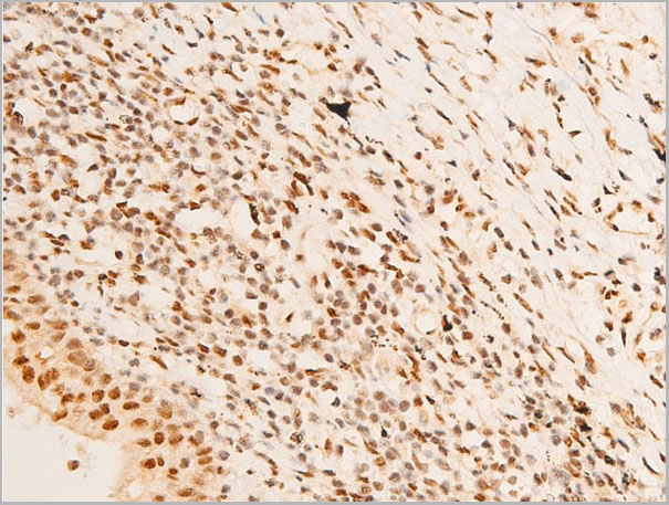
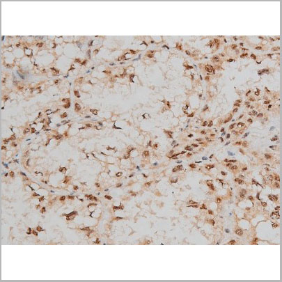
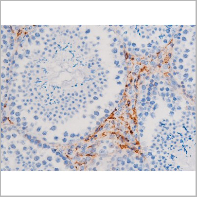
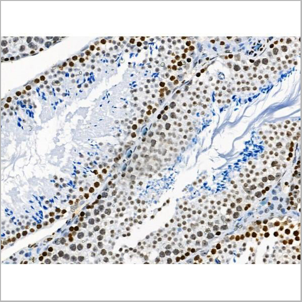
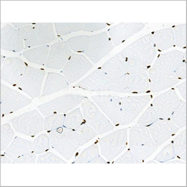
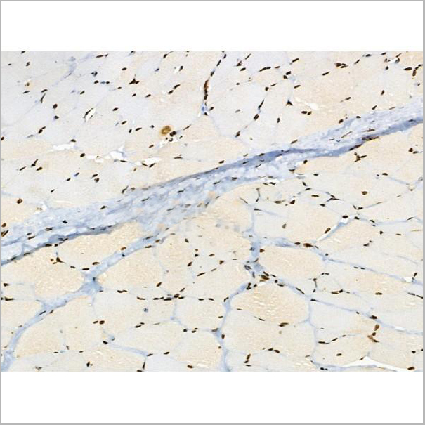
 IHC (Immunohistochemistry)
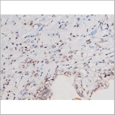
(AAA30938 at 1/100 staining human muscle tumor tissue sections by IHC-P. The tissue was formaldehyde fixed and a heat mediated antigen retrieval step in citrate buffer was performed. The tissue was then blocked and incubated with the antibody for 1.5 hours at 22 degree C. An HRP conjugated goat anti-rabbit antibody was used as the secondary.)
IHC (Immunohistochemistry)
(AAA30938 at 1/100 staining human muscle tumor tissue sections by IHC-P. The tissue was formaldehyde fixed and a heat mediated antigen retrieval step in citrate buffer was performed. The tissue was then blocked and incubated with the antibody for 1.5 hours at 22 degree C. An HRP conjugated goat anti-rabbit antibody was used as the secondary.)
JAK1, Polyclonal Antibody (Cat# AAA30938)
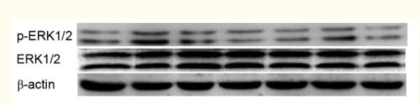 Application Data
(Xu, Yini, et al. "Inhibitory effects of oxymatrine on TGF??1?induced proliferation and abnormal differentiation in rat cardiac fibroblasts via the p38MAPK and ERK1/2 signaling pathways." Molecular medicine reports 16.4 (2017): 5354-5362.)
Application Data
(Xu, Yini, et al. "Inhibitory effects of oxymatrine on TGF??1?induced proliferation and abnormal differentiation in rat cardiac fibroblasts via the p38MAPK and ERK1/2 signaling pathways." Molecular medicine reports 16.4 (2017): 5354-5362.)
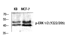
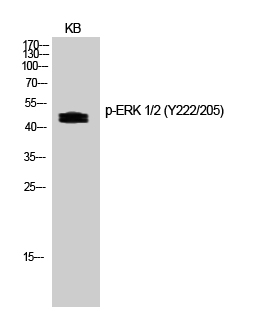
ERK 1/2, Polyclonal Antibody (Cat# AAA30747)
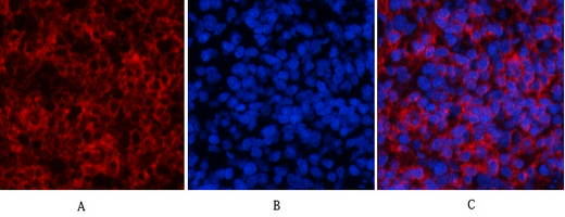
 IF (Immunofluorescence)
(AAA31029 staining HeLa by IF/ICC. The sample were fixed with PFA and permeabilized in 0.1% Triton X-100, then blocked in 10% serum for 45 minutes at 25 degree C. The primary antibody was diluted at 1/200 and incubated with the sample for 1 hour at 37 degree C. An Alexa Fluor 594 conjugated goat anti-rabbit IgG (H+L) Ab, diluted at 1/600, was used as the secondary antibody.)
IF (Immunofluorescence)
(AAA31029 staining HeLa by IF/ICC. The sample were fixed with PFA and permeabilized in 0.1% Triton X-100, then blocked in 10% serum for 45 minutes at 25 degree C. The primary antibody was diluted at 1/200 and incubated with the sample for 1 hour at 37 degree C. An Alexa Fluor 594 conjugated goat anti-rabbit IgG (H+L) Ab, diluted at 1/600, was used as the secondary antibody.)
CDC25A, Polyclonal Antibody (Cat# AAA31029)
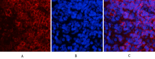
 IF (Immunofluorescence)
(AAA31032 staining HeLa by IF/ICC. The sample were fixed with PFA and permeabilized in 0.1% Triton X-100, then blocked in 10% serum for 45 minutes at 25 degree C. The primary antibody was diluted at 1/200 and incubated with the sample for 1 hour at 37 degree C. An Alexa Fluor 594 conjugated goat anti-rabbit IgG (H+L) Ab, diluted at 1/600, was used as the secondary antibody.)
IF (Immunofluorescence)
(AAA31032 staining HeLa by IF/ICC. The sample were fixed with PFA and permeabilized in 0.1% Triton X-100, then blocked in 10% serum for 45 minutes at 25 degree C. The primary antibody was diluted at 1/200 and incubated with the sample for 1 hour at 37 degree C. An Alexa Fluor 594 conjugated goat anti-rabbit IgG (H+L) Ab, diluted at 1/600, was used as the secondary antibody.)
CDC25A, Polyclonal Antibody (Cat# AAA31032)
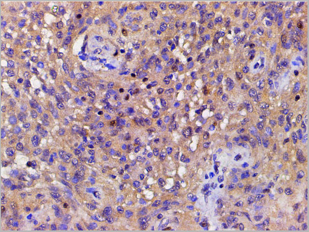
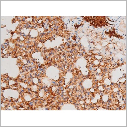
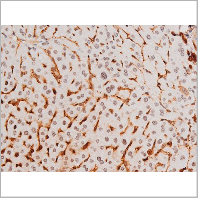
 IHC (Immunohistchemistry)
(AAA31049 at 1/50 staining human colon cancer tissue sections by IHC-P. The tissue was formaldehyde fixed and a heat mediated antigen retrieval step in citrate buffer was performed. The tissue was then blocked and incubated with the antibody for 1.5 hours at 22 degree C. An HRP conjugated goat anti-rabbit antibody was used as the secondary.)
IHC (Immunohistchemistry)
(AAA31049 at 1/50 staining human colon cancer tissue sections by IHC-P. The tissue was formaldehyde fixed and a heat mediated antigen retrieval step in citrate buffer was performed. The tissue was then blocked and incubated with the antibody for 1.5 hours at 22 degree C. An HRP conjugated goat anti-rabbit antibody was used as the secondary.)
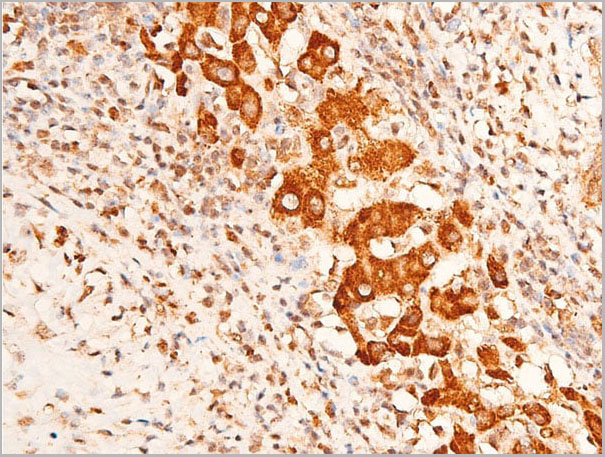
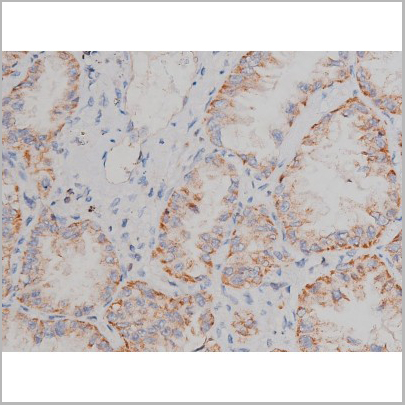
Caspase 3, Polyclonal Antibody (Cat# AAA31049)
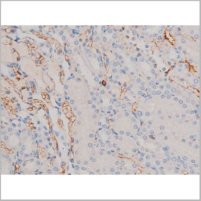
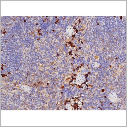
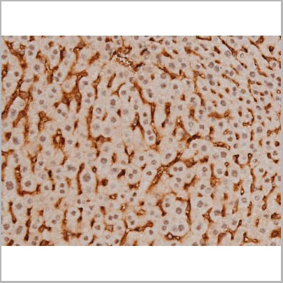
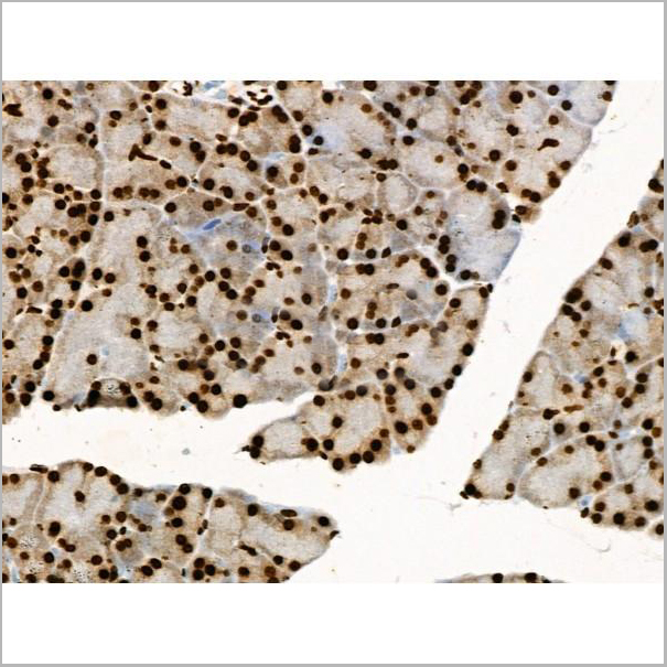
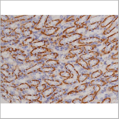
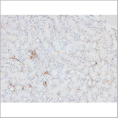
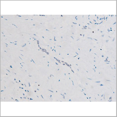
 IHC (Immunohistchemistry)
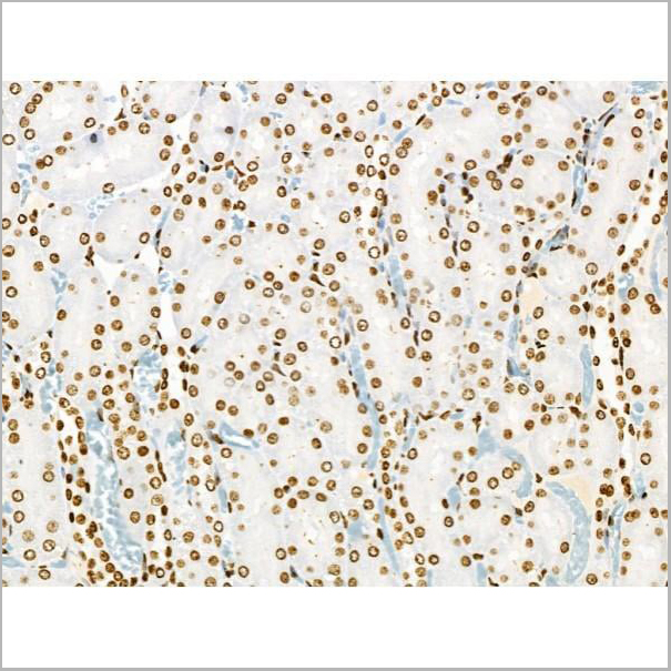
(AAA31065 at 1/200 staining Rat kidney tissue sections by IHC-P. The tissue was formaldehyde fixed and a heat mediated antigen retrieval step in citrate buffer was performed. The tissue was then blocked and incubated with the antibody for 1.5 hours at 22 degree C. An HRP conjugated goat anti-rabbit antibody was used as the secondary.)
IHC (Immunohistchemistry)
(AAA31065 at 1/200 staining Rat kidney tissue sections by IHC-P. The tissue was formaldehyde fixed and a heat mediated antigen retrieval step in citrate buffer was performed. The tissue was then blocked and incubated with the antibody for 1.5 hours at 22 degree C. An HRP conjugated goat anti-rabbit antibody was used as the secondary.)
Dynamin-1, Polyclonal Antibody (Cat# AAA31065)
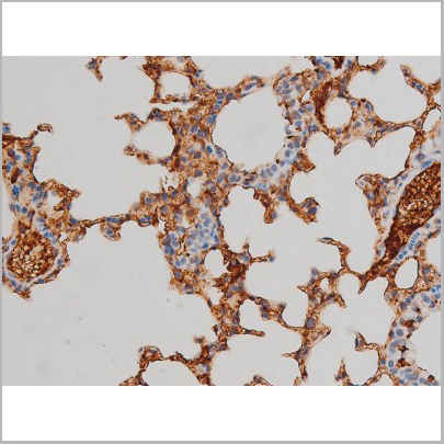
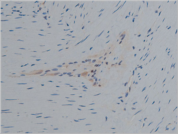
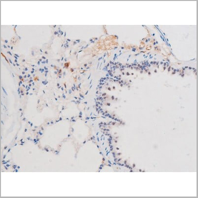
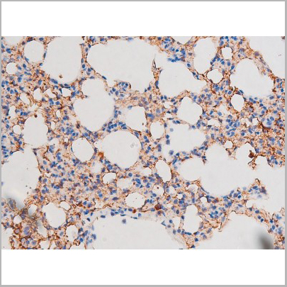
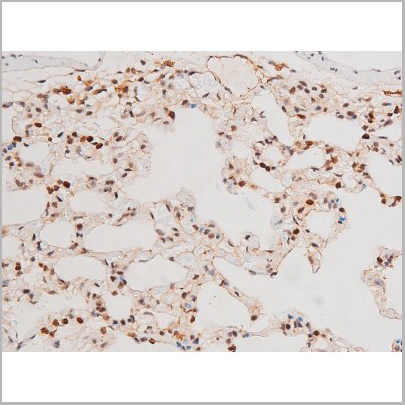
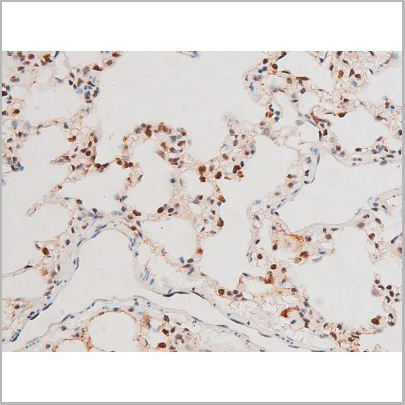
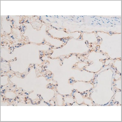
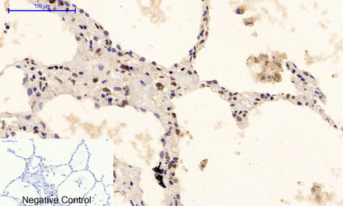
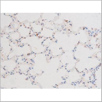
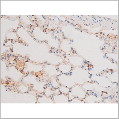
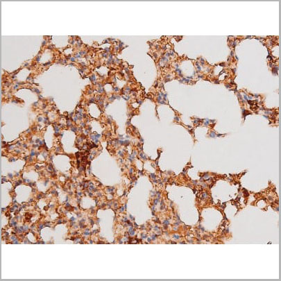
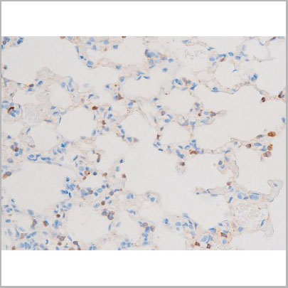
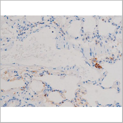
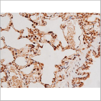
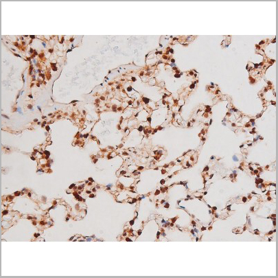
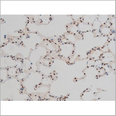
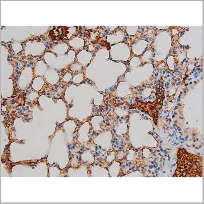
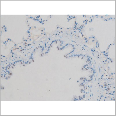
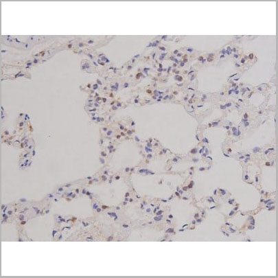
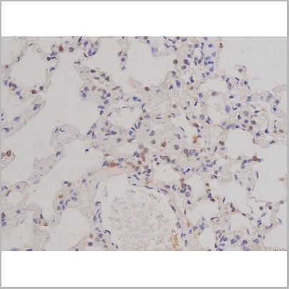
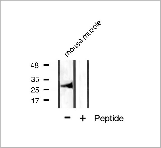
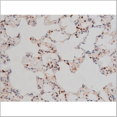
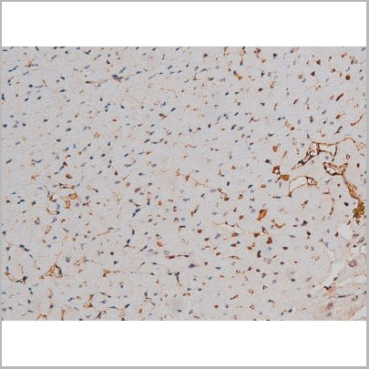
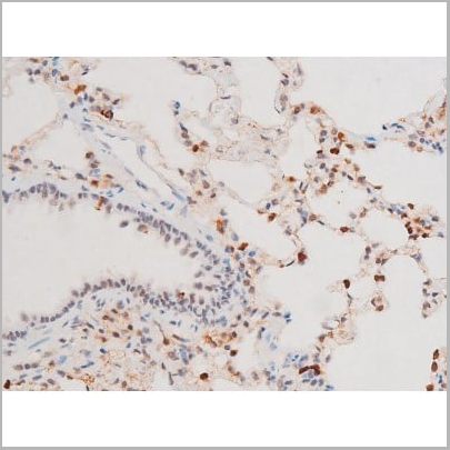
 IHC (Immunohistochemistry)
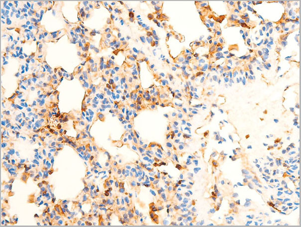
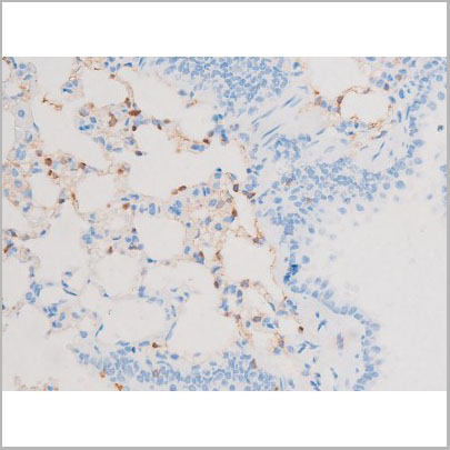
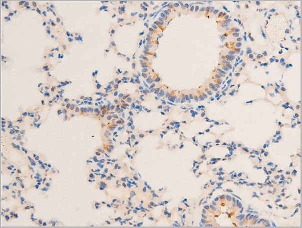
(AAA30945 at 1/100 staining rat lung tissue sections by IHC-P. The tissue was formaldehyde fixed and a heat mediated antigen retrieval step in citrate buffer was performed. The tissue was then blocked and incubated with the antibody for 1.5 hours at 22 degree C. An HRP conjugated goat anti-rabbit antibody was used as the secondary.)
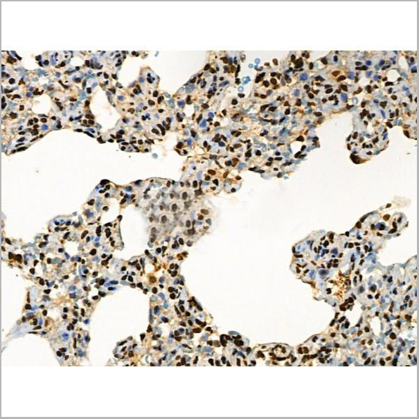
IHC (Immunohistochemistry)
(AAA30945 at 1/100 staining rat lung tissue sections by IHC-P. The tissue was formaldehyde fixed and a heat mediated antigen retrieval step in citrate buffer was performed. The tissue was then blocked and incubated with the antibody for 1.5 hours at 22 degree C. An HRP conjugated goat anti-rabbit antibody was used as the secondary.)
Chk1, Polyclonal Antibody (Cat# AAA30945)
 Application Data
(At 25 degree C. The primary antibody was diluted at 1/200 and incubated with the sample for 1 hour at 37 degree C. An Alexa Fluor 594 conjugated goat anti-rabbit IgG (H+L) Ab, diluted at 1/600, was used as the secondary antibody.)
Application Data
(At 25 degree C. The primary antibody was diluted at 1/200 and incubated with the sample for 1 hour at 37 degree C. An Alexa Fluor 594 conjugated goat anti-rabbit IgG (H+L) Ab, diluted at 1/600, was used as the secondary antibody.)
Vimentin, Polyclonal Antibody (Cat# AAA31419)
 Application Data
(At 25 degree C. The primary antibody was diluted at 1/200 and incubated with the sample for 1 hour at 37 degree C. An Alexa Fluor 594 conjugated goat anti-rabbit IgG (H+L) antibody(Red), diluted at 1/600, was used as secondary antibody.)
Application Data
(At 25 degree C. The primary antibody was diluted at 1/200 and incubated with the sample for 1 hour at 37 degree C. An Alexa Fluor 594 conjugated goat anti-rabbit IgG (H+L) antibody(Red), diluted at 1/600, was used as secondary antibody.)
CSK, Polyclonal Antibody (Cat# AAA31403)


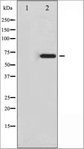
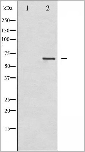
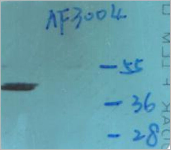
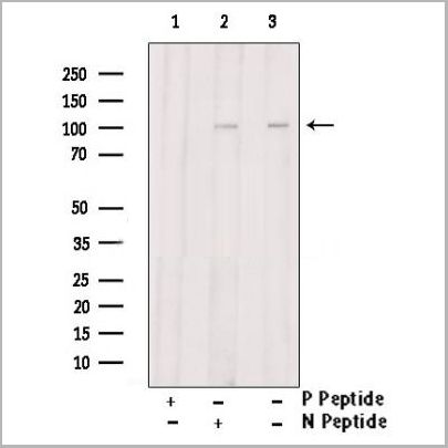
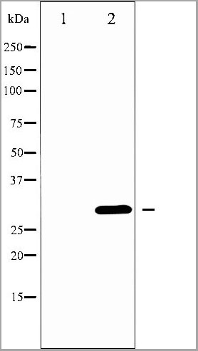
 WB (Western Blot)
(Western blot analysis of AFX phosphorylation expression in serum treated 293 whole cell lysates, The lane on the left is treated with the antigen-specific peptide.)
WB (Western Blot)
(Western blot analysis of AFX phosphorylation expression in serum treated 293 whole cell lysates, The lane on the left is treated with the antigen-specific peptide.)
AFX, Polyclonal Antibody (Cat# AAA31062)
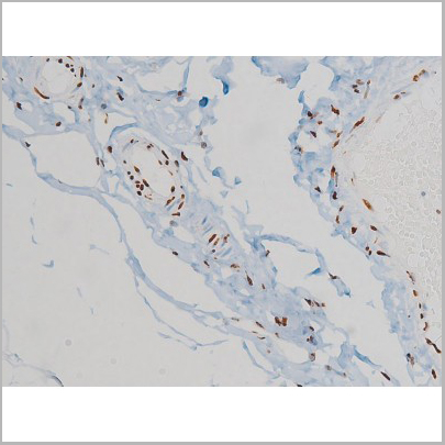
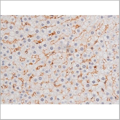
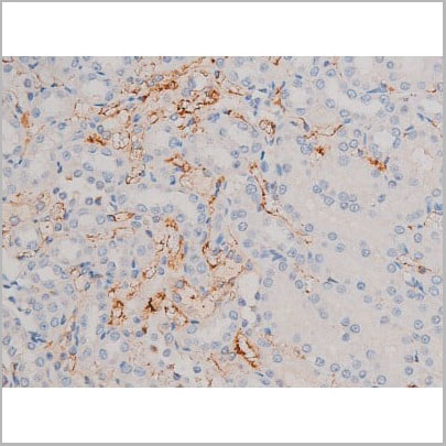
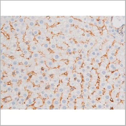
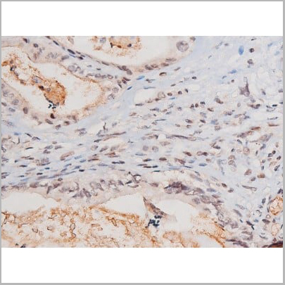
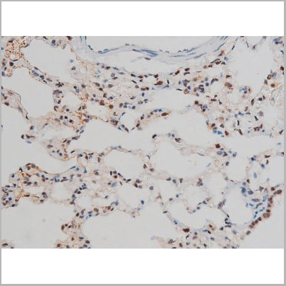
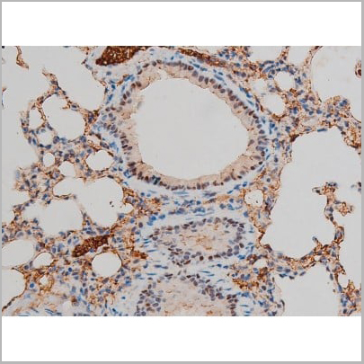
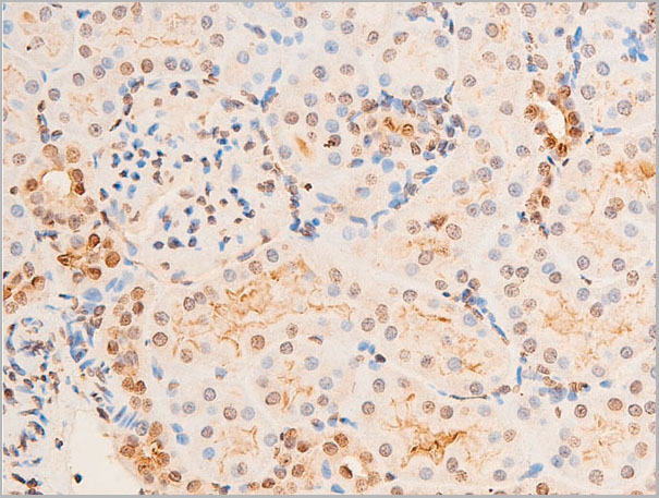
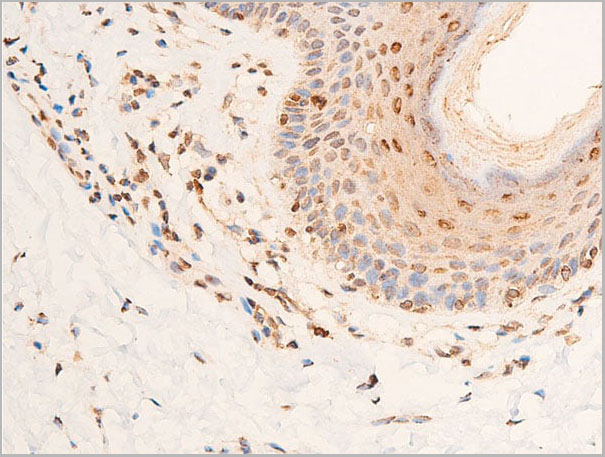
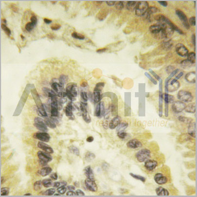
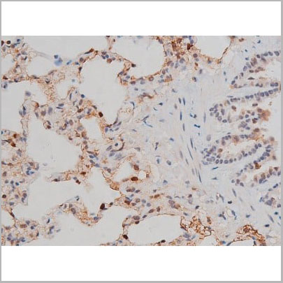
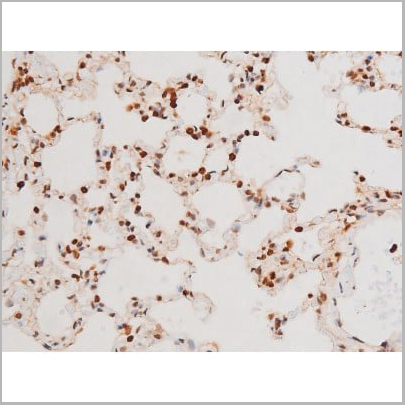
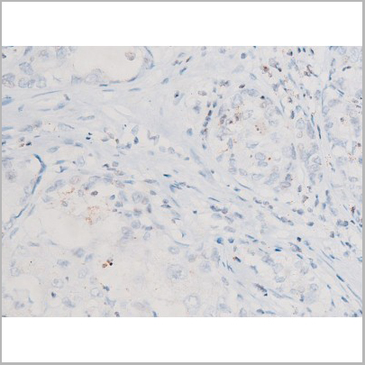
 IHC (Immunohistchemistry)
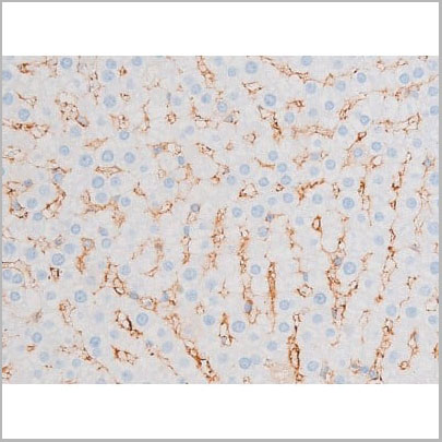
(AAA31064 at 1/200 staining Rat lung tissue sections by IHC-P. The tissue was formaldehyde fixed and a heat mediated antigen retrieval step in citrate buffer was performed. The tissue was then blocked and incubated with the antibody for 1.5 hours at 22 degree C. An HRP conjugated goat anti-rabbit antibody was used as the secondary.)
IHC (Immunohistchemistry)
(AAA31064 at 1/200 staining Rat lung tissue sections by IHC-P. The tissue was formaldehyde fixed and a heat mediated antigen retrieval step in citrate buffer was performed. The tissue was then blocked and incubated with the antibody for 1.5 hours at 22 degree C. An HRP conjugated goat anti-rabbit antibody was used as the secondary.)
PKC theta, Polyclonal Antibody (Cat# AAA31064)
 IF (Immunofluorescence)
(AAA31031 staining HeLa by IF/ICC. The sample were fixed with PFA and permeabilized in 0.1% Triton X-100, then blocked in 10% serum for 45 minutes at 25 degree C. The primary antibody was diluted at 1/200 and incubated with the sample for 1 hour at 37 degree C. An Alexa Fluor 594 conjugated goat anti-rabbit IgG (H+L) Ab, diluted at 1/600, was used as the secondary antibody.)
IF (Immunofluorescence)
(AAA31031 staining HeLa by IF/ICC. The sample were fixed with PFA and permeabilized in 0.1% Triton X-100, then blocked in 10% serum for 45 minutes at 25 degree C. The primary antibody was diluted at 1/200 and incubated with the sample for 1 hour at 37 degree C. An Alexa Fluor 594 conjugated goat anti-rabbit IgG (H+L) Ab, diluted at 1/600, was used as the secondary antibody.)
CDC25A, Polyclonal Antibody (Cat# AAA31031)
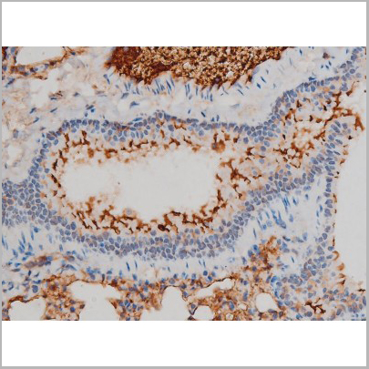
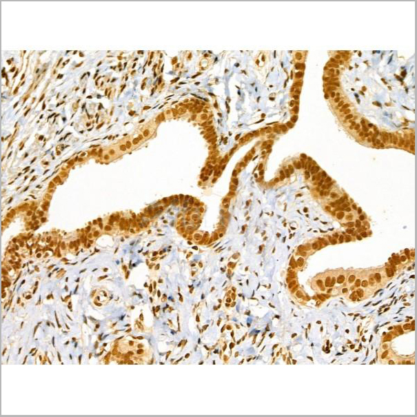
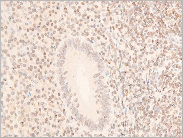
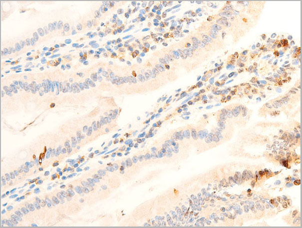
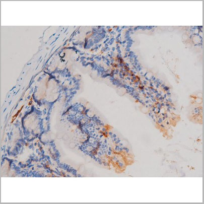
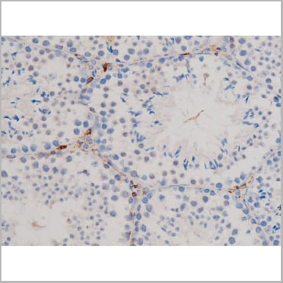
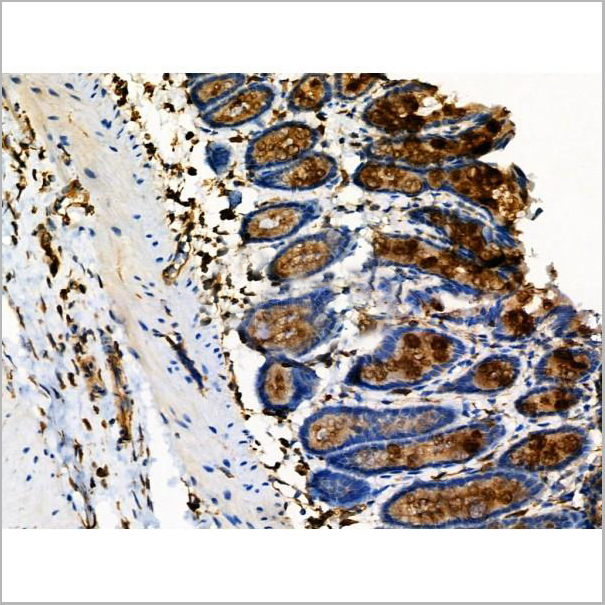
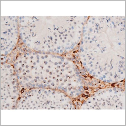
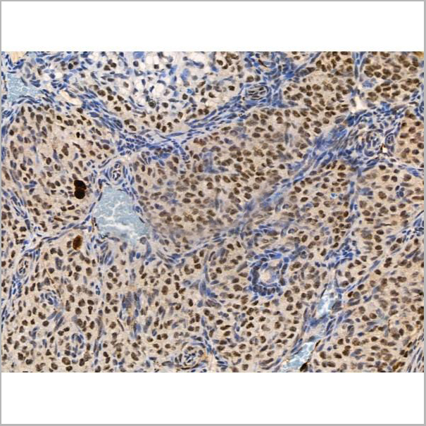
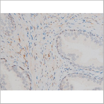
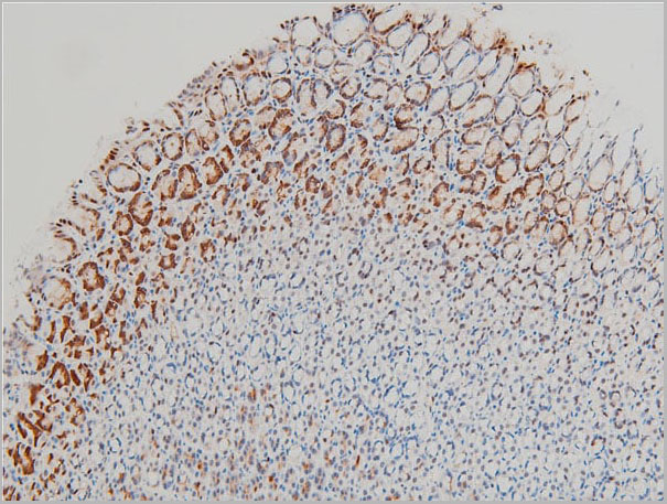
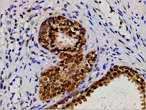
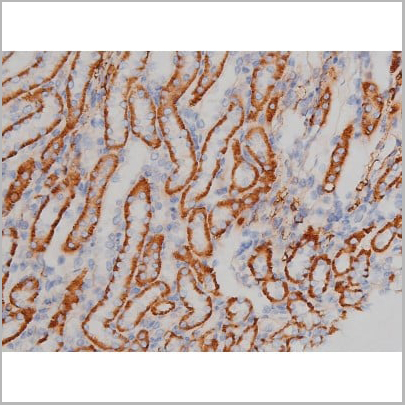
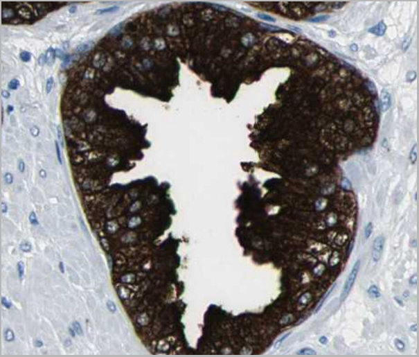
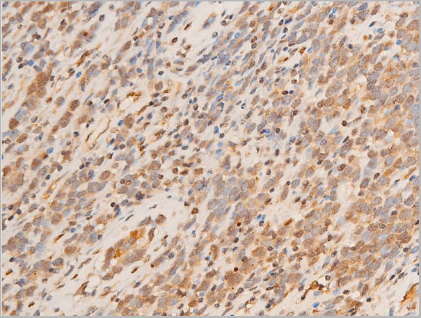
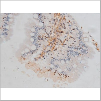
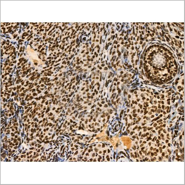
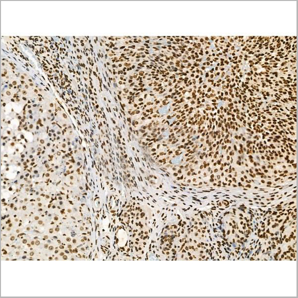
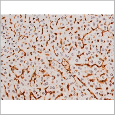
 IHC-P (Immunohistochemistry Paraffin)
(AAA31041 at 1/100 staining Mouse colon tissue by IHC-P. The sample was formaldehyde fixed and a heat mediated antigen retrieval step in citrate buffer was performed. The sample was then blocked and incubated with the primary antibody at 4°C overnight. An HRP conjugated anti-Rabbit antibody was use das the secondary antibody.)
IHC-P (Immunohistochemistry Paraffin)
(AAA31041 at 1/100 staining Mouse colon tissue by IHC-P. The sample was formaldehyde fixed and a heat mediated antigen retrieval step in citrate buffer was performed. The sample was then blocked and incubated with the primary antibody at 4°C overnight. An HRP conjugated anti-Rabbit antibody was use das the secondary antibody.)
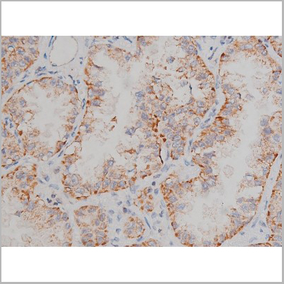
STAT3, Polyclonal Antibody (Cat# AAA31041)
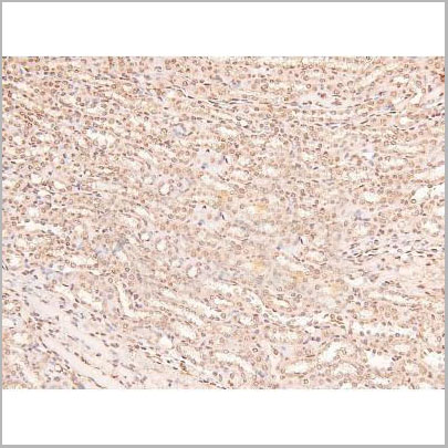
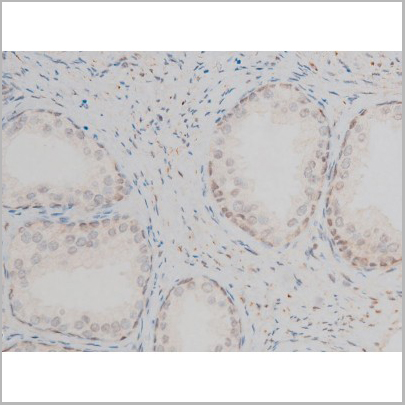
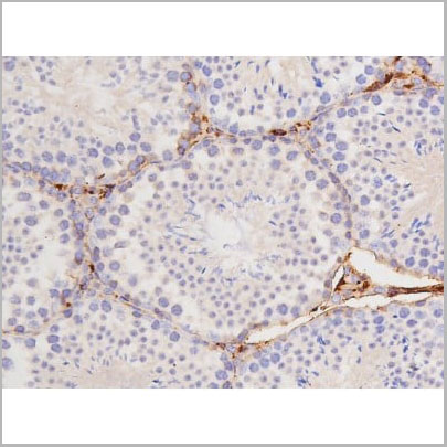
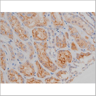
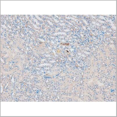
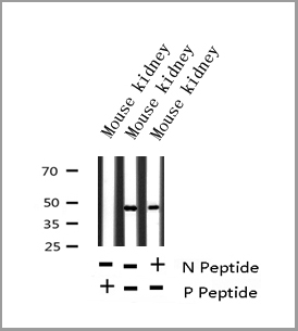
 IHC (Immunohistchemistry)
(At 1/200 staining Mouse kidney tissue sections by IHC-P. The tissue was formaldehyde fixed and a heat mediated antigen retrieval step in citrate buffer was performed. The tissue was then blocked and incubated with the antibody for 1.5 hours at 22 degree C. An HRP conjugated goat anti-rabbit antibody was used as the secondary antibody.)
IHC (Immunohistchemistry)
(At 1/200 staining Mouse kidney tissue sections by IHC-P. The tissue was formaldehyde fixed and a heat mediated antigen retrieval step in citrate buffer was performed. The tissue was then blocked and incubated with the antibody for 1.5 hours at 22 degree C. An HRP conjugated goat anti-rabbit antibody was used as the secondary antibody.)
eIF2B epsilon, Polyclonal Antibody (Cat# AAA31414)
Predicted Reactivity: Pig (80%), Bovine (80%), Horse (80%), Rabbit (80%), Dog (80%), Chicken (80%)
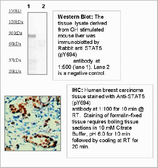
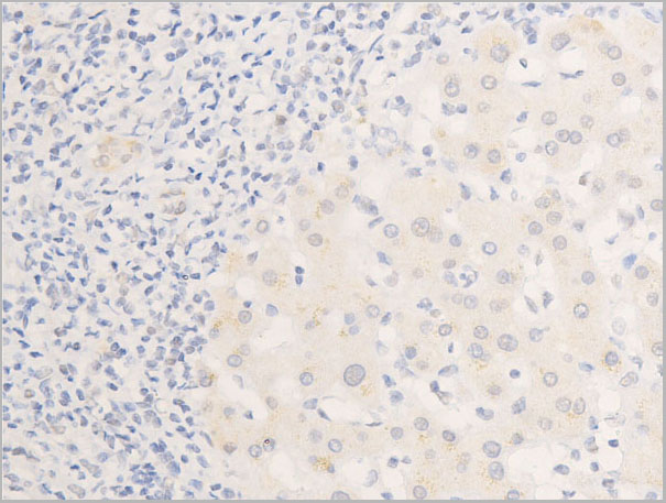
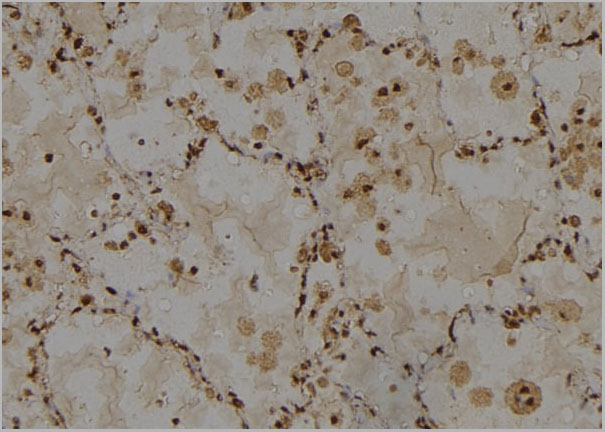
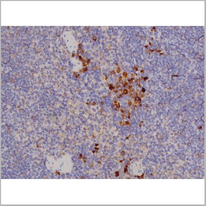
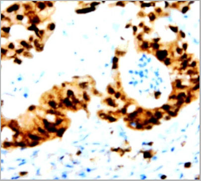 IHC (Immunohistochemistry)
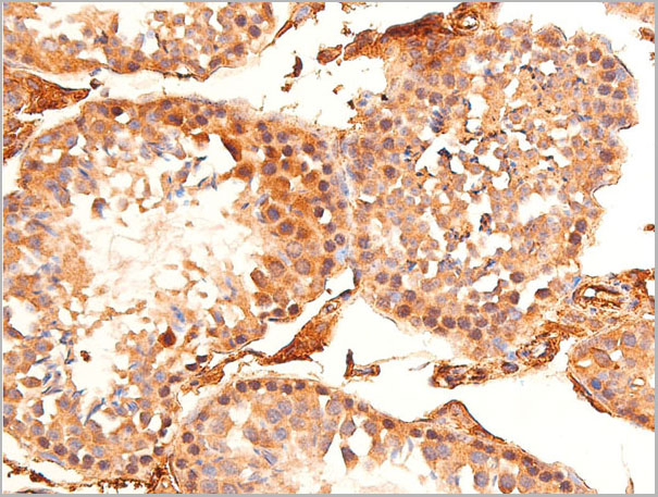
(Immunohistochemistry: Human breast carcinoma tissue (FFPE) stained with Anti-STAT5 (pY694) (Cat# AAA14089) antibody at 1:100 for 10 min @ RT. Staining of formalin-fixed tissue requires boiling tissue sections in 10 mM Citrate Buffer, pH 6.0 for 10 min followed by cooling at RT for 20 min.)
IHC (Immunohistochemistry)
(Immunohistochemistry: Human breast carcinoma tissue (FFPE) stained with Anti-STAT5 (pY694) (Cat# AAA14089) antibody at 1:100 for 10 min @ RT. Staining of formalin-fixed tissue requires boiling tissue sections in 10 mM Citrate Buffer, pH 6.0 for 10 min followed by cooling at RT for 20 min.)
STAT5 (pY694), Antibody (Cat# AAA14089)

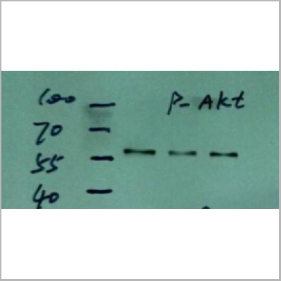
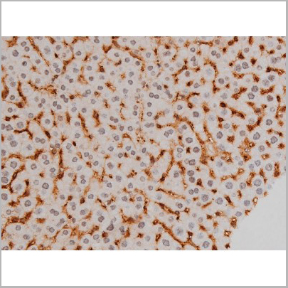
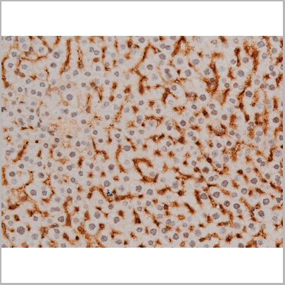
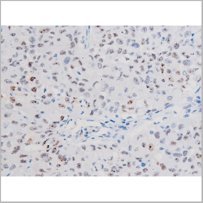
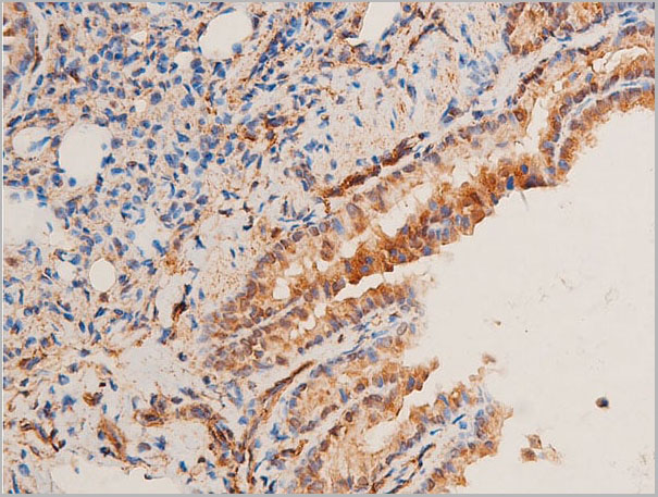
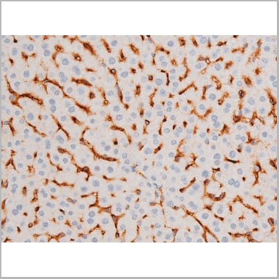
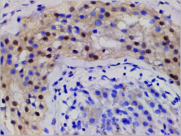
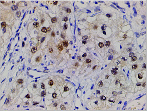
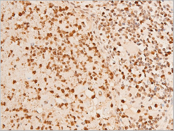
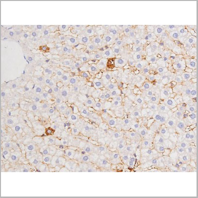
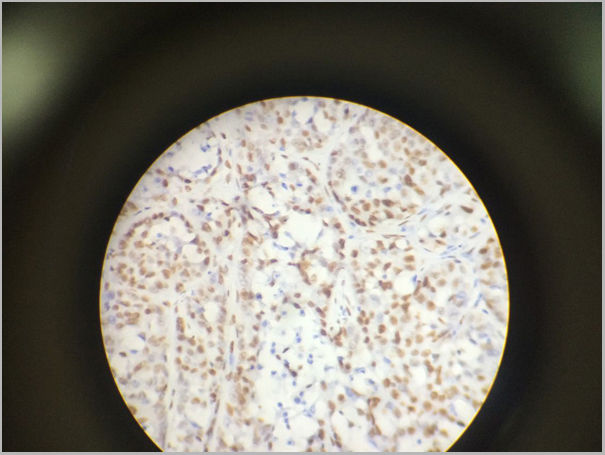
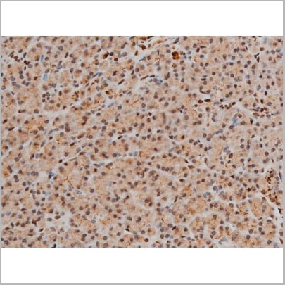
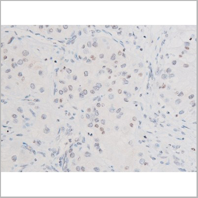
 IHC (Immunohistochemistry)
(AAA30920 at 1/200 staining human lung cancer tissue sections by IHC-P. The tissue was formaldehyde fixed and a heat mediated antigen retrieval step in citrate buffer was performed. The tissue was then blocked and incubated with the antibody for 1.5 hours at 22 degree C. An HRP conjugated goat anti-rabbit antibody was used as the secondary.)
IHC (Immunohistochemistry)
(AAA30920 at 1/200 staining human lung cancer tissue sections by IHC-P. The tissue was formaldehyde fixed and a heat mediated antigen retrieval step in citrate buffer was performed. The tissue was then blocked and incubated with the antibody for 1.5 hours at 22 degree C. An HRP conjugated goat anti-rabbit antibody was used as the secondary.)
Akt, Polyclonal Antibody (Cat# AAA30920)
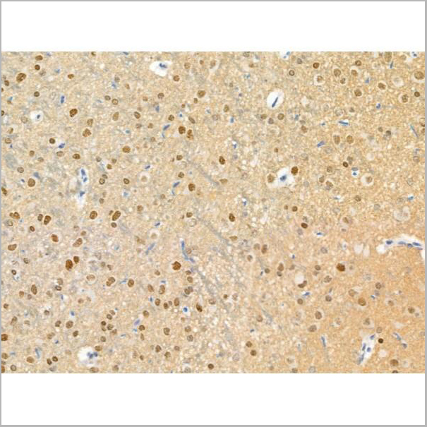
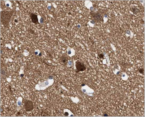
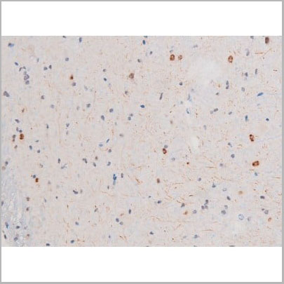
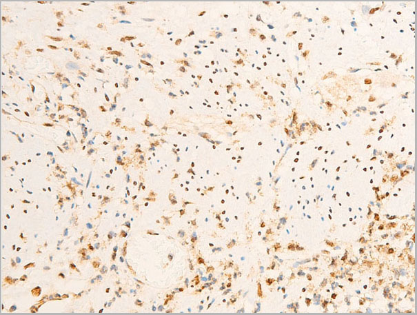
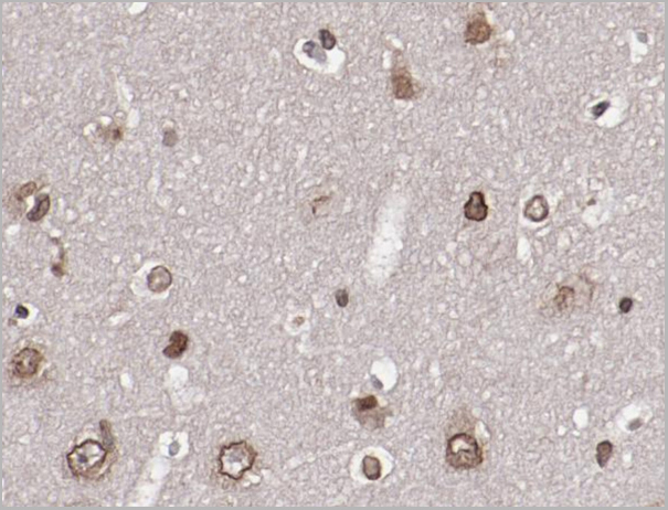
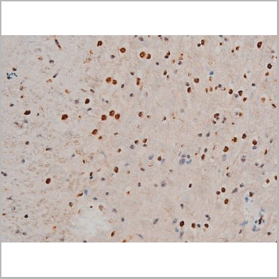
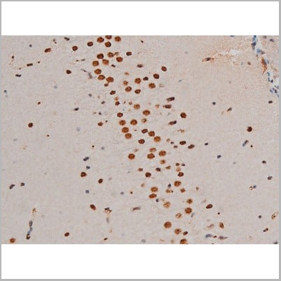
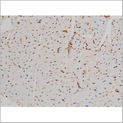
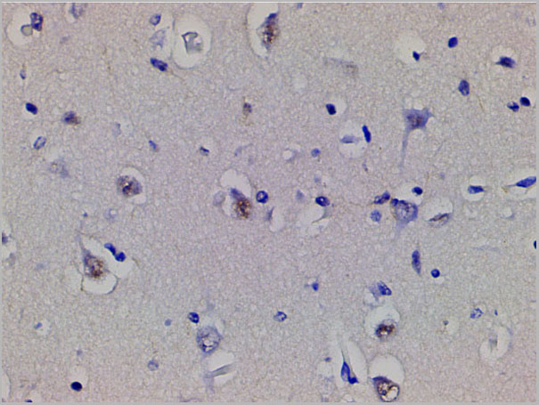
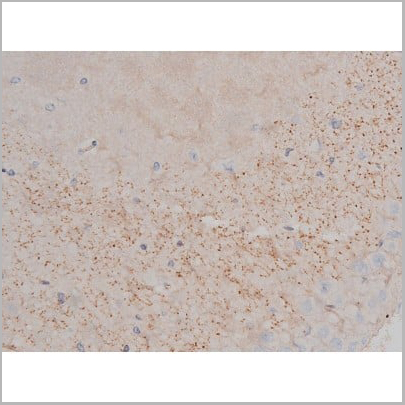
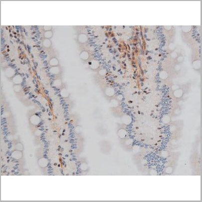
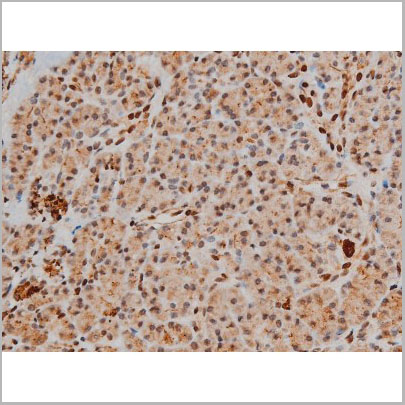
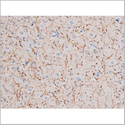
 IHC (Immunohistochemistry)
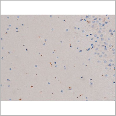
(At 1/100 staining Mouse brain tissue by IHC-P. The sample was formaldehyde fixed and a heat mediated antigen retrieval step in citrate buffer was performed. The sample was then blocked and incubated with the primary antibody at 4 degree C overnight. An HRP conjugated anti-Rabbit antibody was used as the secondary antibody.)
IHC (Immunohistochemistry)
(At 1/100 staining Mouse brain tissue by IHC-P. The sample was formaldehyde fixed and a heat mediated antigen retrieval step in citrate buffer was performed. The sample was then blocked and incubated with the primary antibody at 4 degree C overnight. An HRP conjugated anti-Rabbit antibody was used as the secondary antibody.)
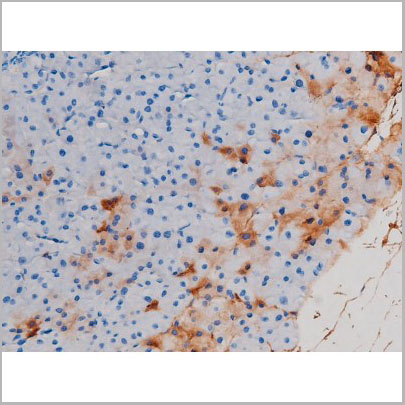
ATF6, Polyclonal Antibody (Cat# AAA31376)
Predicted Reactivity: Pig (100%), Horse (100%), Rabbit (100%), Dog (100%)
 IF (Immunofluorescence)
(AAA31012 staining Hela by IF/ICC. The sample were fixed with PFA and permeabilized in 0.1% Triton X-100, then blocked in 10% serum for 45 minutes at 25 degree C. The primary antibody was diluted at 1/200 and incubated with the sample for 1 hour at 37 degree C. An Alexa Fluor 594 conjugated goat anti-rabbit IgG (H+L) Ab, diluted at 1/600, was used as the secondary antibody.)
IF (Immunofluorescence)
(AAA31012 staining Hela by IF/ICC. The sample were fixed with PFA and permeabilized in 0.1% Triton X-100, then blocked in 10% serum for 45 minutes at 25 degree C. The primary antibody was diluted at 1/200 and incubated with the sample for 1 hour at 37 degree C. An Alexa Fluor 594 conjugated goat anti-rabbit IgG (H+L) Ab, diluted at 1/600, was used as the secondary antibody.)
ATF2, Polyclonal Antibody (Cat# AAA31012)
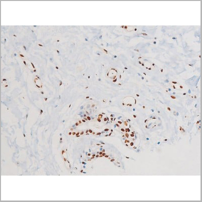
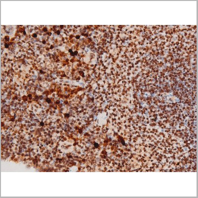
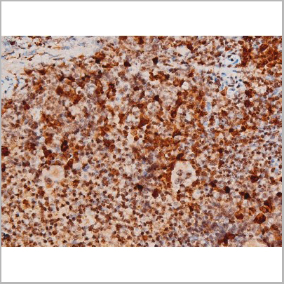
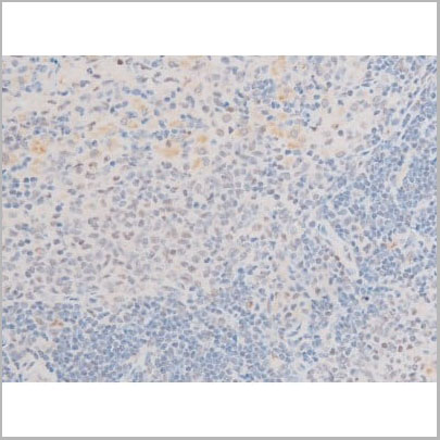
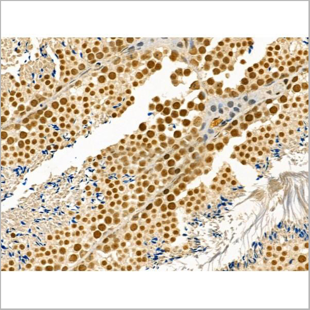
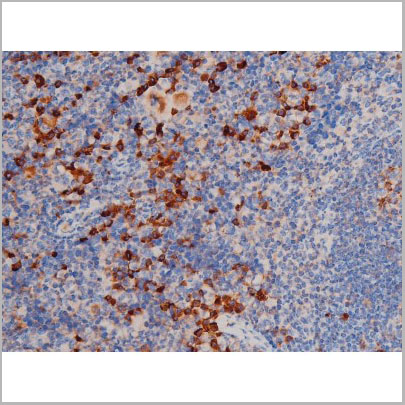
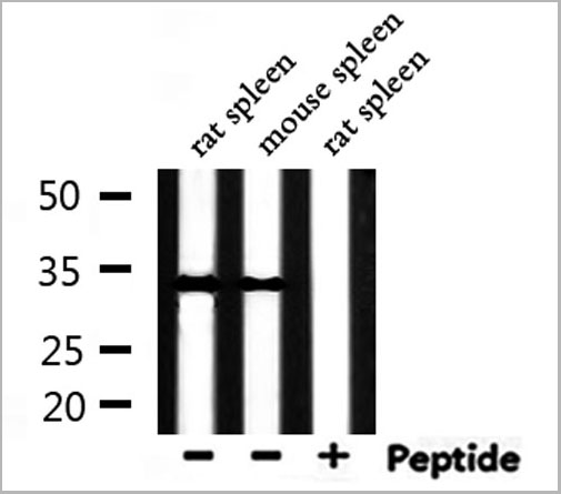
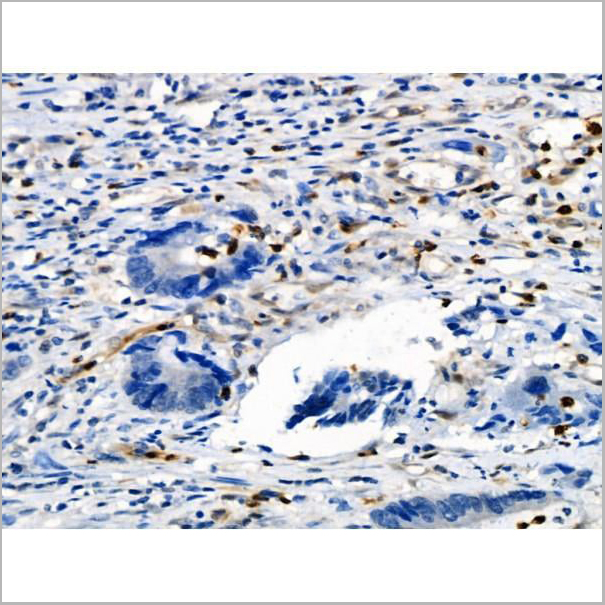
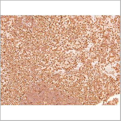
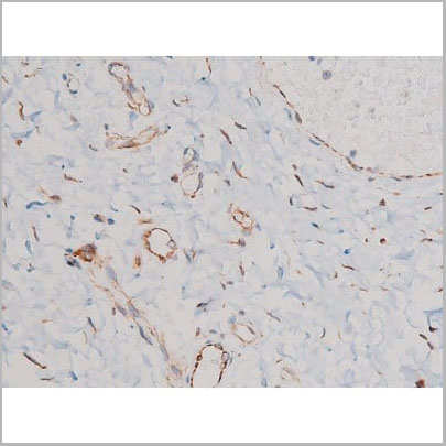
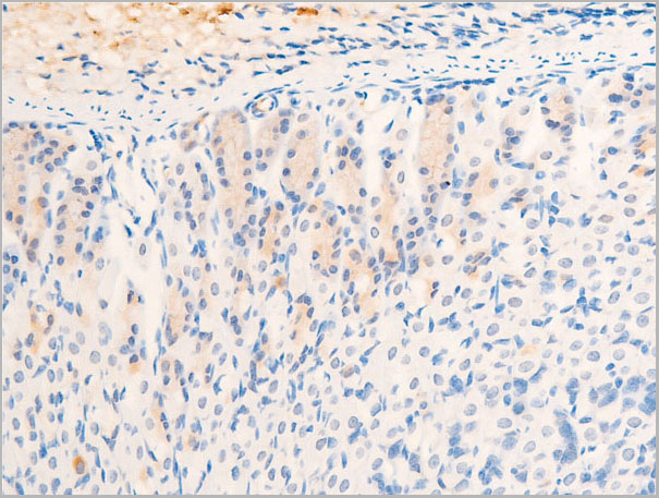
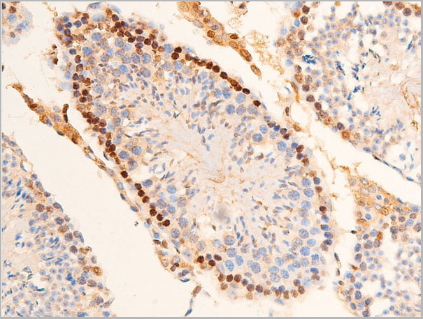
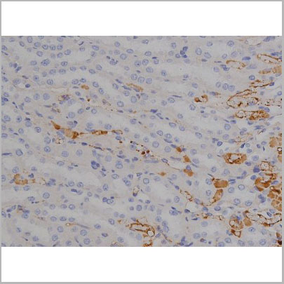
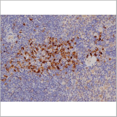
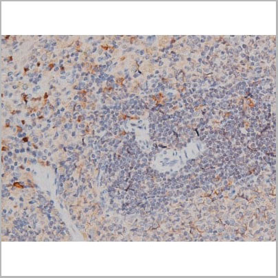
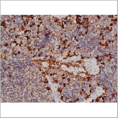
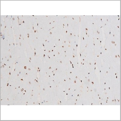
 IHC (Immunohistochemistry)
(AAA31013 at 1/200 staining Rat spleen tissue sections by IHC-P. The tissue was formaldehyde fixed and a heat mediated antigen retrieval step in citrate buffer was performed. The tissue was then blocked and incubated with the antibody for 1.5 hours at 22 degree C. An HRP conjugated goat anti-rabbit antibody was used as the secondary.)
IHC (Immunohistochemistry)
(AAA31013 at 1/200 staining Rat spleen tissue sections by IHC-P. The tissue was formaldehyde fixed and a heat mediated antigen retrieval step in citrate buffer was performed. The tissue was then blocked and incubated with the antibody for 1.5 hours at 22 degree C. An HRP conjugated goat anti-rabbit antibody was used as the secondary.)
GATA1, Polyclonal Antibody (Cat# AAA31013)


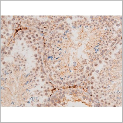
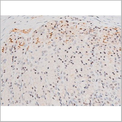
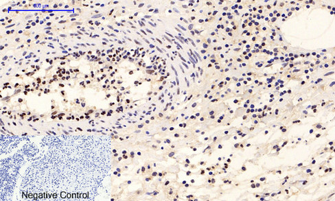
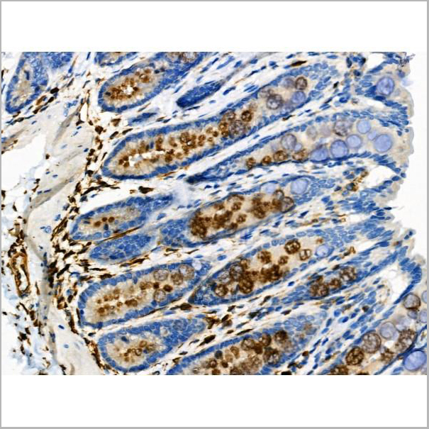
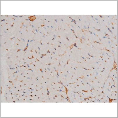
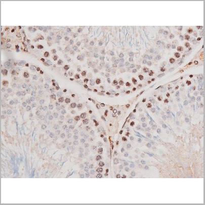
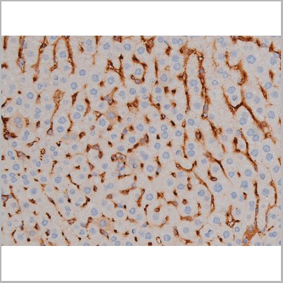
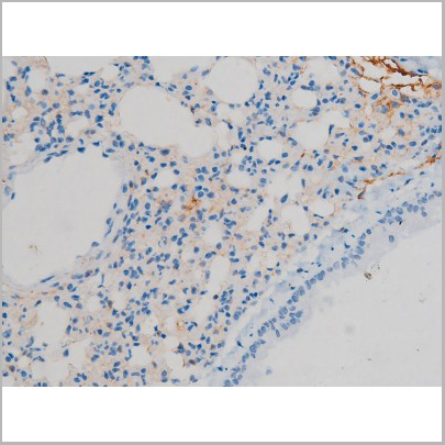
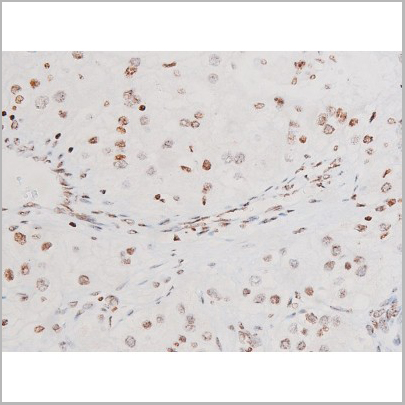
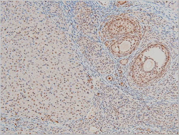 IHC (Immunohistochemistry)
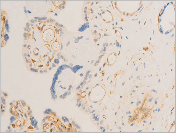
(AAA31025 at 1/100 staining rat ovarian tissue sections by IHC-P. The tissue was formaldehyde fixed and a heat mediated antigen retrieval step in citrate buffer was performed. The tissue was then blocked and incubated with the antibody for 1.5 hours at 22 degree C. An HRP conjugated goat anti-rabbit antibody was used as the secondary.)
IHC (Immunohistochemistry)
(AAA31025 at 1/100 staining rat ovarian tissue sections by IHC-P. The tissue was formaldehyde fixed and a heat mediated antigen retrieval step in citrate buffer was performed. The tissue was then blocked and incubated with the antibody for 1.5 hours at 22 degree C. An HRP conjugated goat anti-rabbit antibody was used as the secondary.)
Cofilin, Polyclonal Antibody (Cat# AAA31025)
 IF (Immunofluorescence)
(AAA31030 staining HeLa by IF/ICC. The sample were fixed with PFA and permeabilized in 0.1% Triton X-100, then blocked in 10% serum for 45 minutes at 25 degree C. The primary antibody was diluted at 1/200 and incubated with the sample for 1 hour at 37 degree C. An Alexa Fluor 594 conjugated goat anti-rabbit IgG (H+L) Ab, diluted at 1/600, was used as the secondary antibody.)
IF (Immunofluorescence)
(AAA31030 staining HeLa by IF/ICC. The sample were fixed with PFA and permeabilized in 0.1% Triton X-100, then blocked in 10% serum for 45 minutes at 25 degree C. The primary antibody was diluted at 1/200 and incubated with the sample for 1 hour at 37 degree C. An Alexa Fluor 594 conjugated goat anti-rabbit IgG (H+L) Ab, diluted at 1/600, was used as the secondary antibody.)
CDC25A, Polyclonal Antibody (Cat# AAA31030)

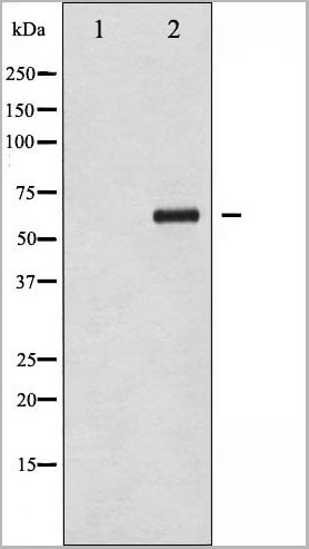
 WB (Western Blot)
(Western blot analysis of EGFR phosphorylation expression in TSA treated HeLa whole cell lysates, The lane on the left is treated with the antigen-specific peptide.)
WB (Western Blot)
(Western blot analysis of EGFR phosphorylation expression in TSA treated HeLa whole cell lysates, The lane on the left is treated with the antigen-specific peptide.)
EGFR, Polyclonal Antibody (Cat# AAA30963)
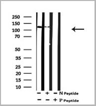
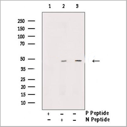
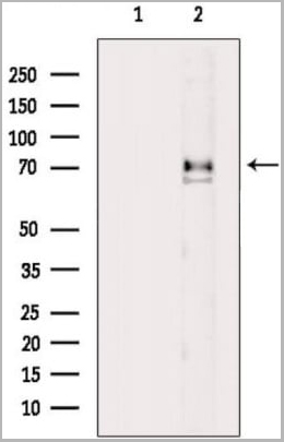
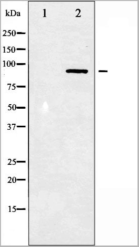
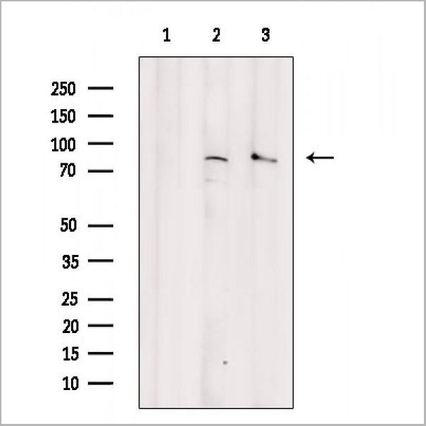
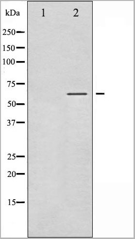
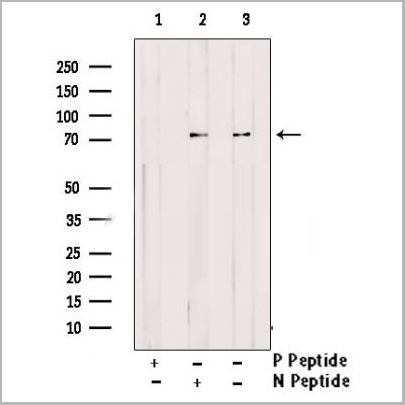
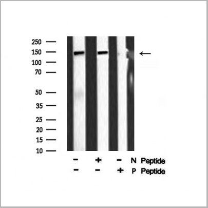
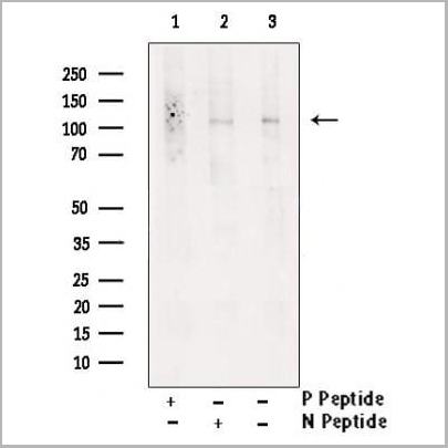 WB (Western Blot)
(Western blot analysis of extracts from K562 , using Phospho-IRE1 (Ser724) Antibody. Lane1 was treated with phospho-blocking peptide, Lane2 was treated with non-phospho-blocking peptide.)
WB (Western Blot)
(Western blot analysis of extracts from K562 , using Phospho-IRE1 (Ser724) Antibody. Lane1 was treated with phospho-blocking peptide, Lane2 was treated with non-phospho-blocking peptide.)
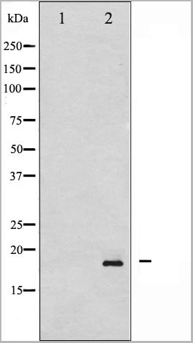
IRE1, Polyclonal Antibody (Cat# AAA31151)
 WB (Western Blot)
(Western blot analysis of Chk2 phosphorylation expression in UV treated COS7 whole cell lysates, The lane on the left is treated with the antigen-specific peptide.)
WB (Western Blot)
(Western blot analysis of Chk2 phosphorylation expression in UV treated COS7 whole cell lysates, The lane on the left is treated with the antigen-specific peptide.)
Chk2, Polyclonal Antibody (Cat# AAA30958)

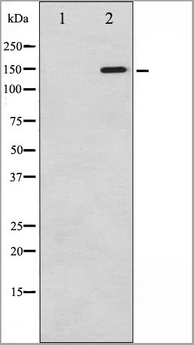
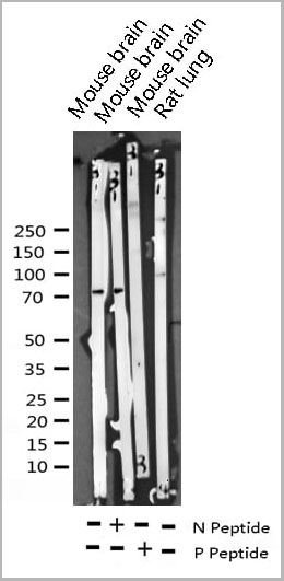
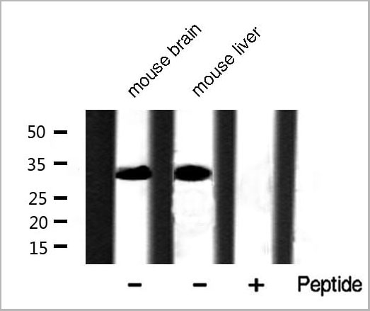
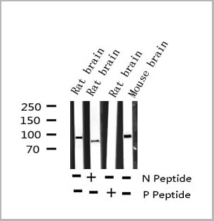
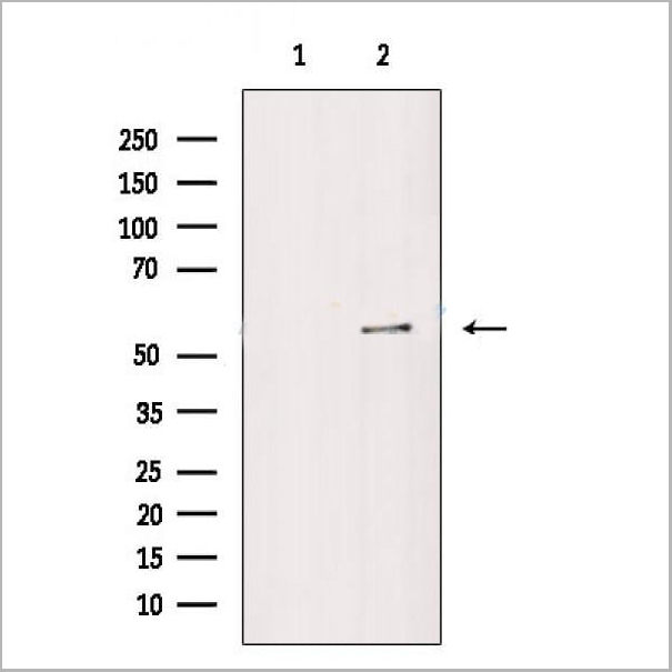
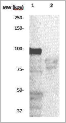
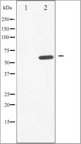
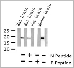
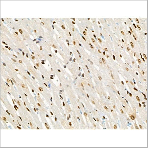
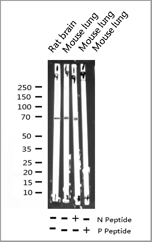
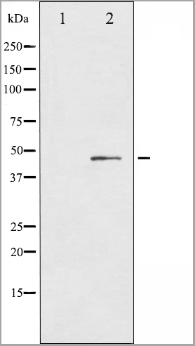
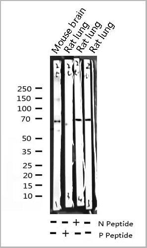
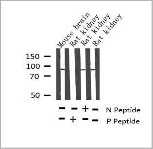 WB (Western Blot)
(Western blot analysis of Phospho-Tau (Thr212) Antibody expression in mouse brain and rat kidney tissues lysates.)
WB (Western Blot)
(Western blot analysis of Phospho-Tau (Thr212) Antibody expression in mouse brain and rat kidney tissues lysates.)
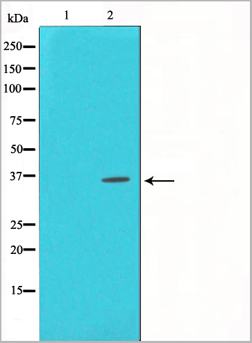
Tau, Polyclonal Antibody (Cat# AAA31001)
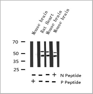
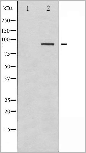
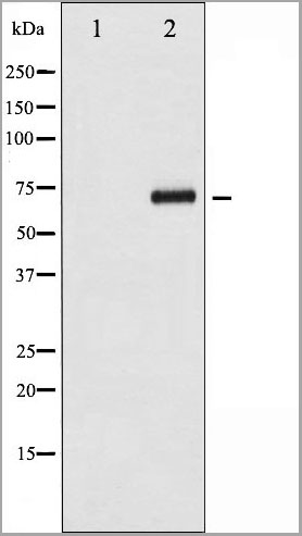
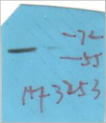
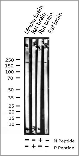
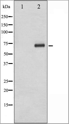
 WB (Western Blot)
(Western blot analysis of Synuclein phosphorylation expression in Mouse brain tissue lysates, The lane on the left is treated with the antigen-specific peptide.)
WB (Western Blot)
(Western blot analysis of Synuclein phosphorylation expression in Mouse brain tissue lysates, The lane on the left is treated with the antigen-specific peptide.)
Synuclein, Polyclonal Antibody (Cat# AAA31040)
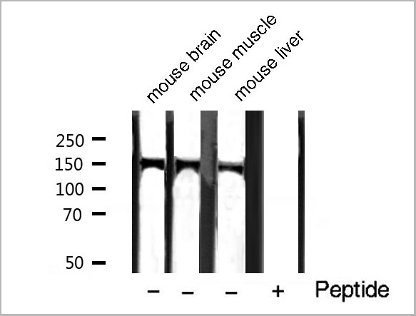
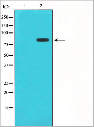
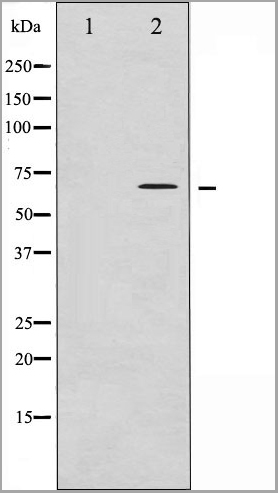
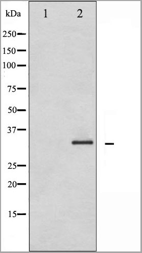
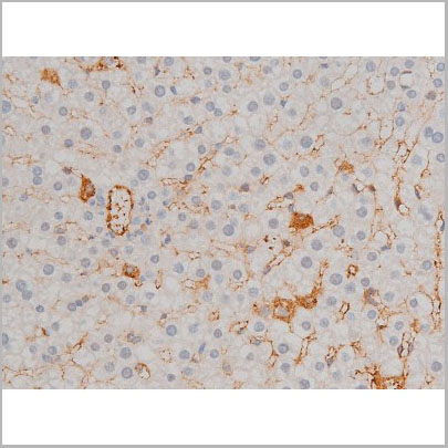
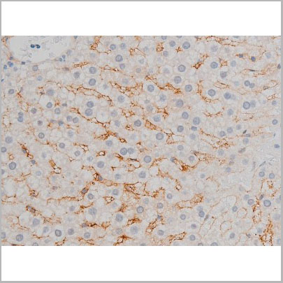
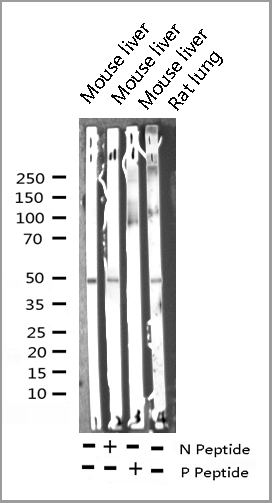
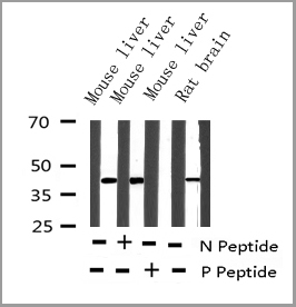
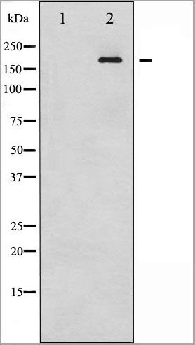
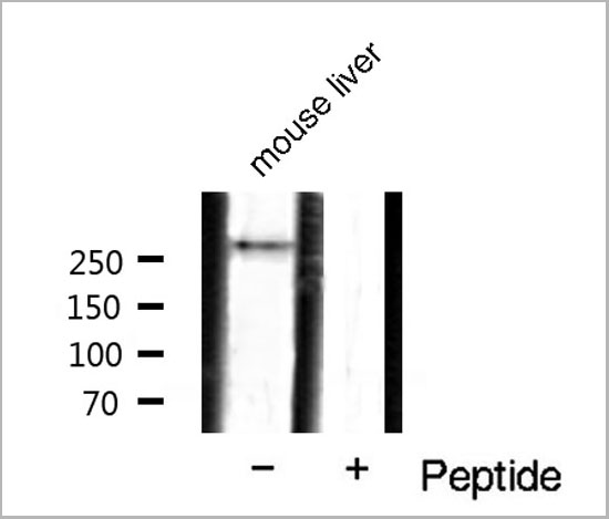 WB (Western Blot)
(Western blot analysis of mTOR phosphorylation expression in mouse liver tissue lysates, The lane on the right is treated with the antigen-specific peptide.)
WB (Western Blot)
(Western blot analysis of mTOR phosphorylation expression in mouse liver tissue lysates, The lane on the right is treated with the antigen-specific peptide.)
mTOR, Polyclonal Antibody (Cat# AAA31048)
 WB (Western Blot)
(Western blot analysis of SGK phosphorylation expression in Insulin treated HeLa whole cell lysates, The lane on the left is treated with the antigen-specific peptide.)
WB (Western Blot)
(Western blot analysis of SGK phosphorylation expression in Insulin treated HeLa whole cell lysates, The lane on the left is treated with the antigen-specific peptide.)
SGK, Polyclonal Antibody (Cat# AAA30942)
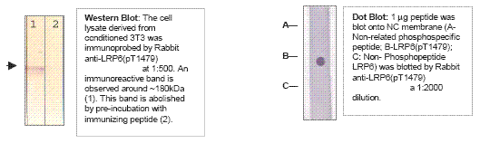 Application Data
LRP6 (phospho), Antibody (Cat# AAA14084)
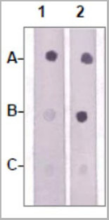 DB (Dot Blot)
(Dot Blot: 1 ug peptide was blot onto NC membraneA: JAK2 (pY1008, pY1009)B: JAK2 (Non phosphospecific)C: Non-related Phosphopeptide) were blotted at a 1:2000 dilution by:1: Rabbit anti-JAK2(pY1008/pY1009) (AAA14087)2: Rabbit anti-JAK2 (Nonphospho specific).)
DB (Dot Blot)
(Dot Blot: 1 ug peptide was blot onto NC membraneA: JAK2 (pY1008, pY1009)B: JAK2 (Non phosphospecific)C: Non-related Phosphopeptide) were blotted at a 1:2000 dilution by:1: Rabbit anti-JAK2(pY1008/pY1009) (AAA14087)2: Rabbit anti-JAK2 (Nonphospho specific).)
JAK2 (pY1007/Y1008), Antibody (Cat# AAA14087)
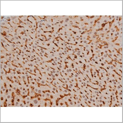

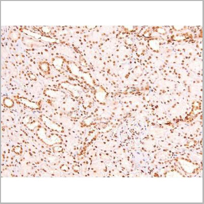
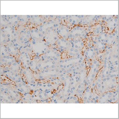
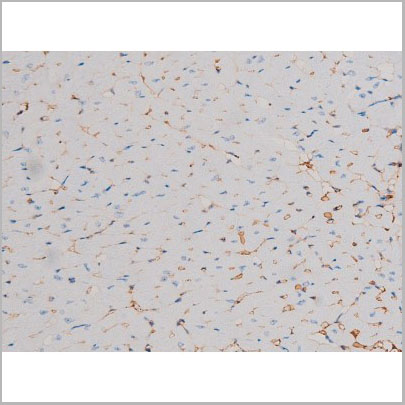
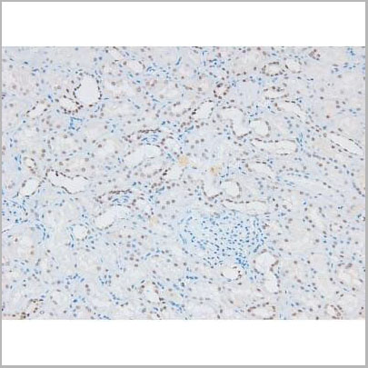
 IHC (Immunohistochemistry)
(AAA31016 at 1/200 staining Rat kidney tissue sections by IHC-P. The tissue was formaldehyde fixed and a heat mediated antigen retrieval step in citrate buffer was performed. The tissue was then blocked and incubated with the antibody for 1.5 hours at 22 degree C. An HRP conjugated goat anti-rabbit antibody was used as the secondary.)
IHC (Immunohistochemistry)
(AAA31016 at 1/200 staining Rat kidney tissue sections by IHC-P. The tissue was formaldehyde fixed and a heat mediated antigen retrieval step in citrate buffer was performed. The tissue was then blocked and incubated with the antibody for 1.5 hours at 22 degree C. An HRP conjugated goat anti-rabbit antibody was used as the secondary.)
CREB, Polyclonal Antibody (Cat# AAA31016)
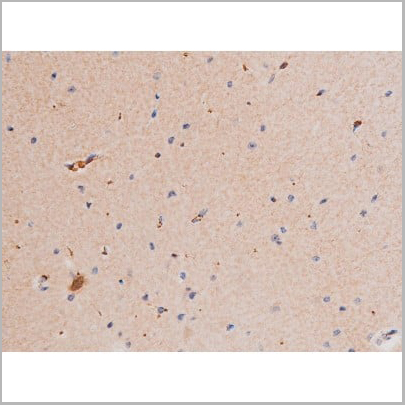
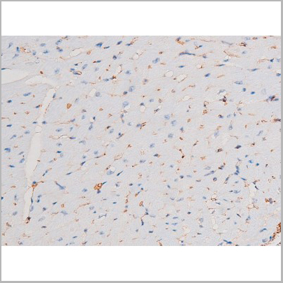
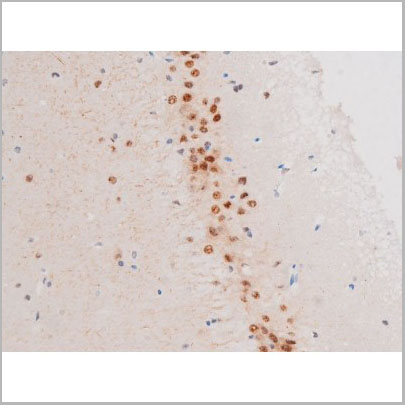
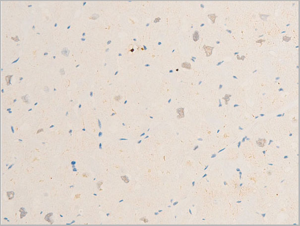
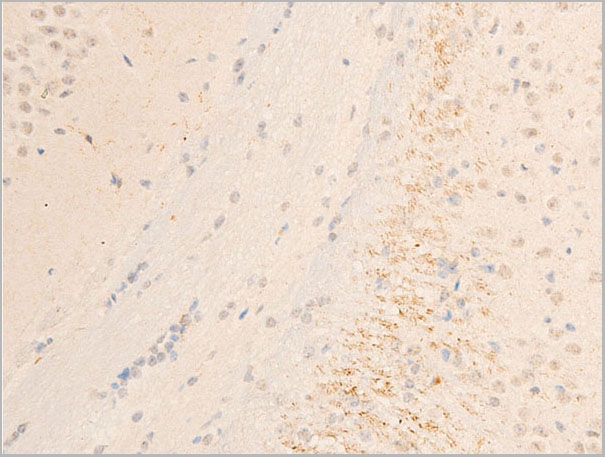
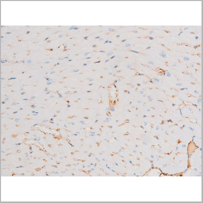
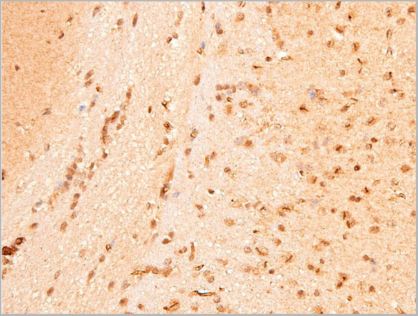
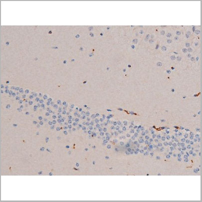
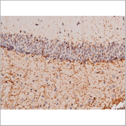
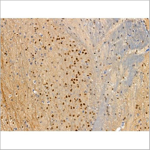
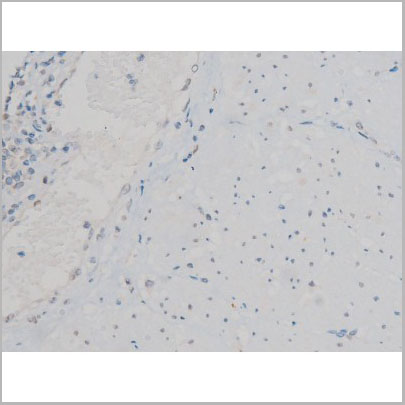
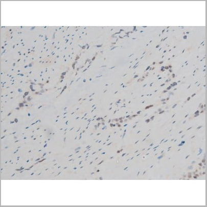
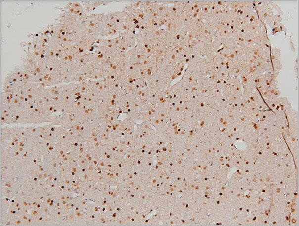
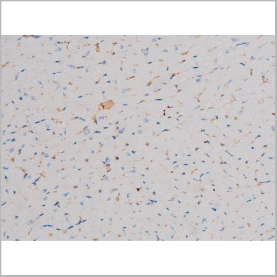
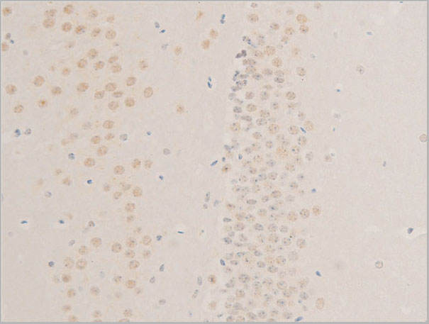
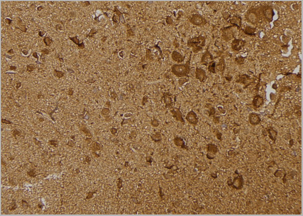
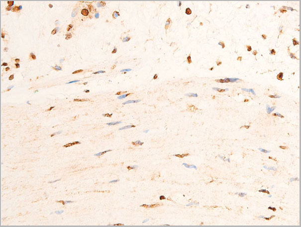
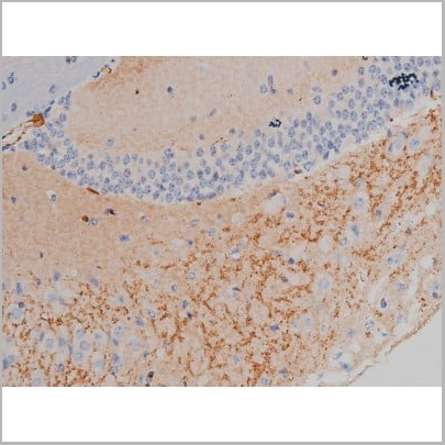
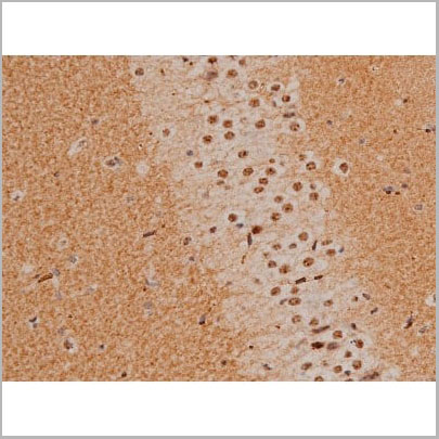
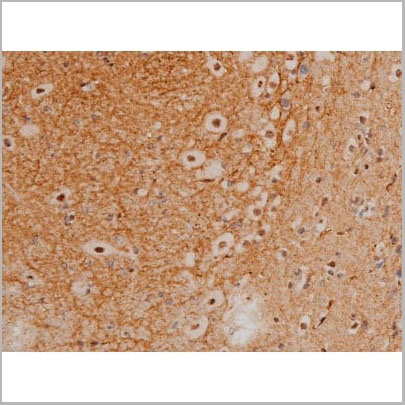
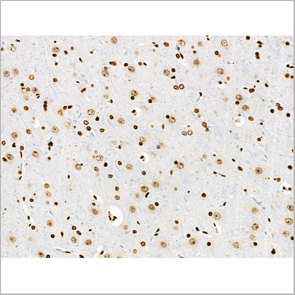
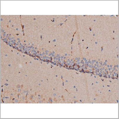
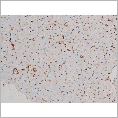
 IHC (Immunohistochemistry)
(AAA31036 at 1/200 staining Rat brain tissue sections by IHC-P. The tissue was formaldehyde fixed and a heat mediated antigen retrieval step in citrate buffer was performed. The tissue was then blocked and incubated with the antibody for 1.5 hours at 22 degree C. An HRP conjugated goat anti-rabbit antibody was used as the secondary.)
IHC (Immunohistochemistry)
(AAA31036 at 1/200 staining Rat brain tissue sections by IHC-P. The tissue was formaldehyde fixed and a heat mediated antigen retrieval step in citrate buffer was performed. The tissue was then blocked and incubated with the antibody for 1.5 hours at 22 degree C. An HRP conjugated goat anti-rabbit antibody was used as the secondary.)
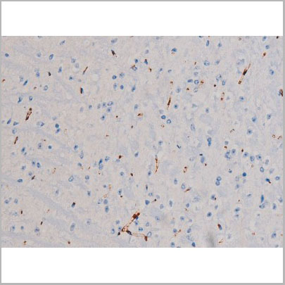
IRS-1, Polyclonal Antibody (Cat# AAA31036)
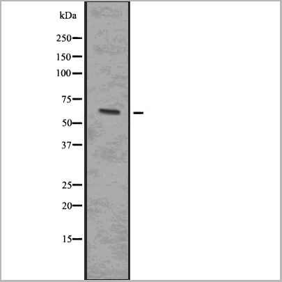
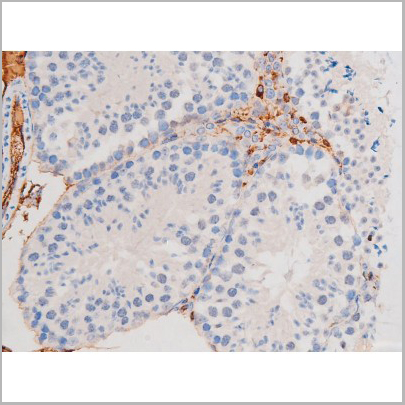
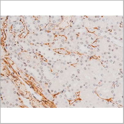
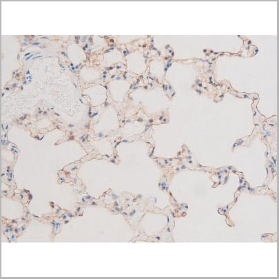
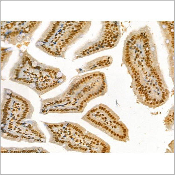
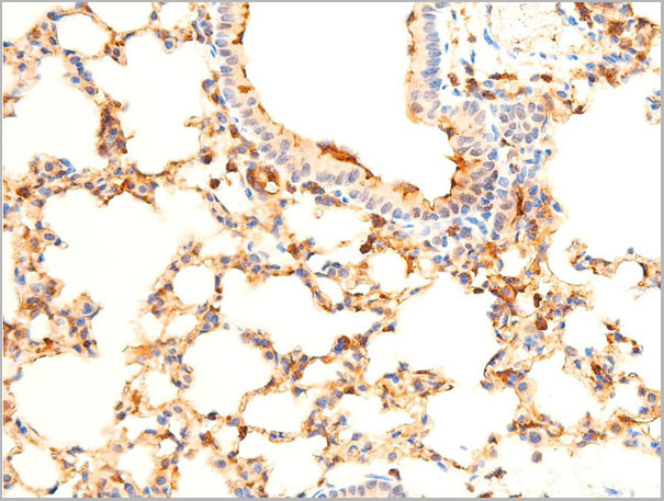
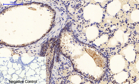
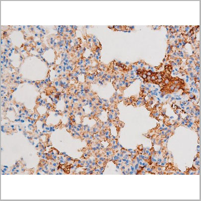
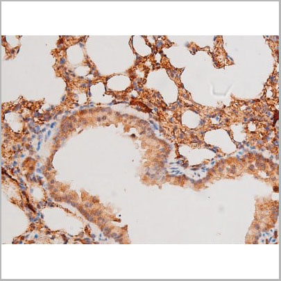
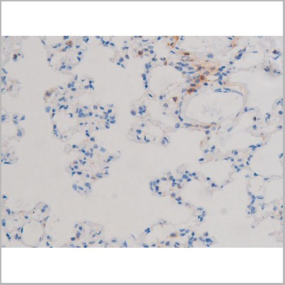
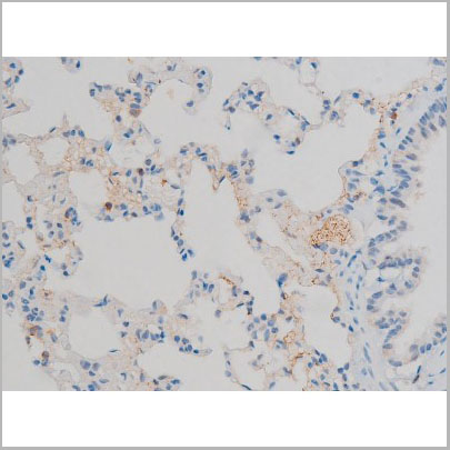
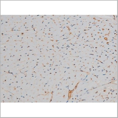
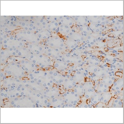
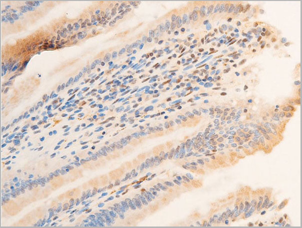
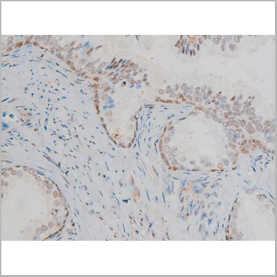
 IHC (Immunohistochemistry)
(AAA31133 at 1/100 staining Mouse lung tissue by IHC-P. The sample was formaldehyde fixed and a heat mediated antigen retrieval step in citrate buffer was performed. The sample was then blocked and incubated with the antibody for 1.5 hours at 22°C. An HRP conjugated goat anti-rabbit antibody was used as the secondary.)
IHC (Immunohistochemistry)
(AAA31133 at 1/100 staining Mouse lung tissue by IHC-P. The sample was formaldehyde fixed and a heat mediated antigen retrieval step in citrate buffer was performed. The sample was then blocked and incubated with the antibody for 1.5 hours at 22°C. An HRP conjugated goat anti-rabbit antibody was used as the secondary.)
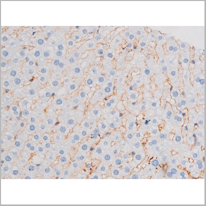
IRAK4, Polyclonal Antibody (Cat# AAA31133)
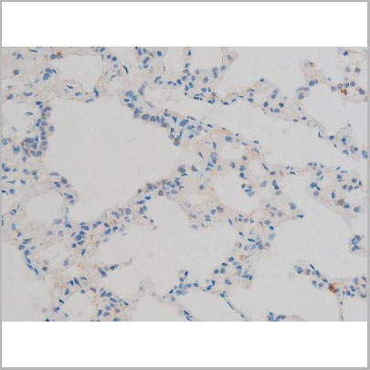
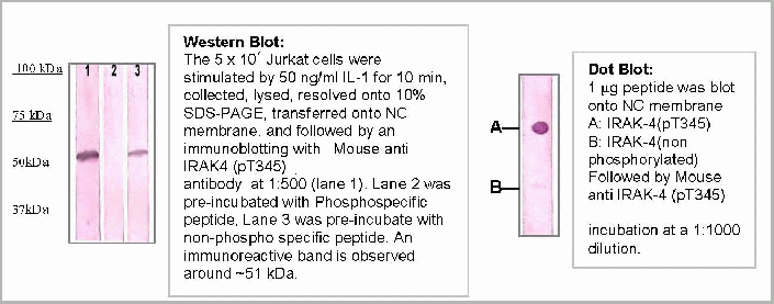 Application Data
IRAK4 (pT345), Monoclonal Antibody (Cat# AAA14096)
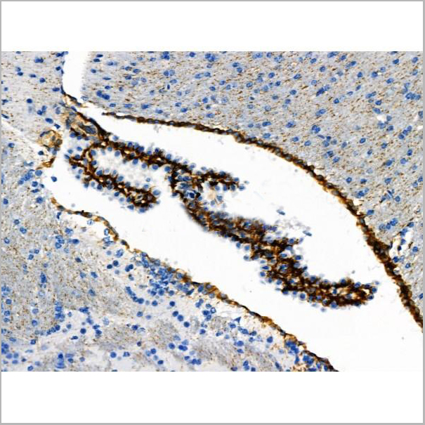
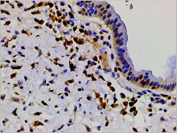
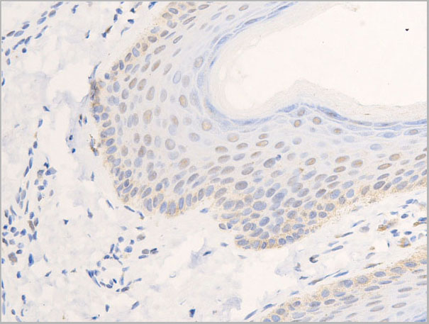
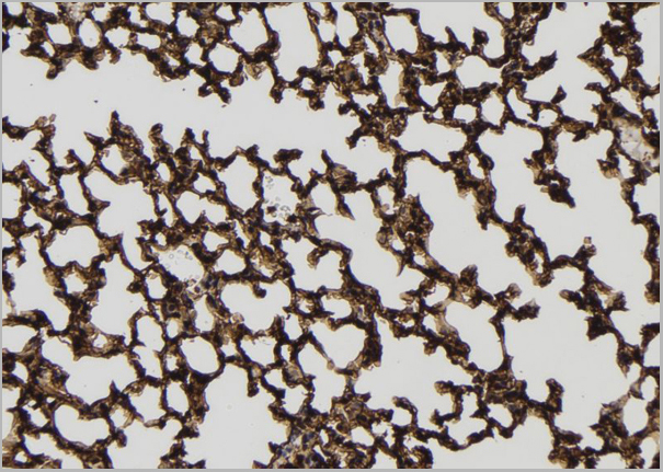
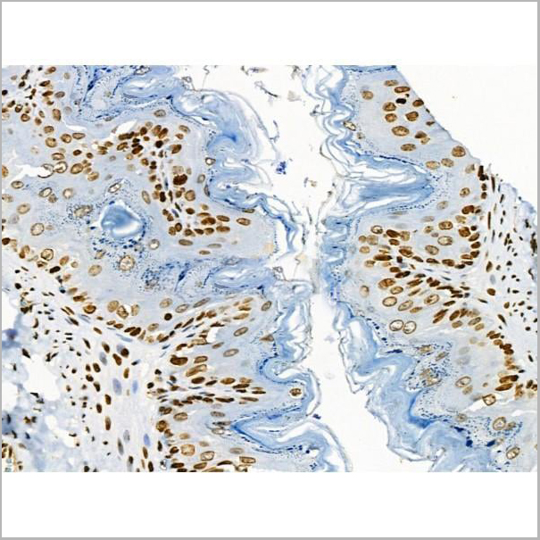
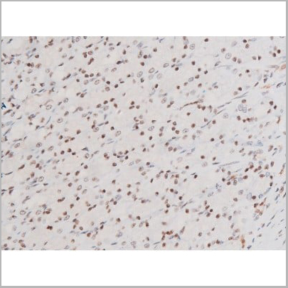
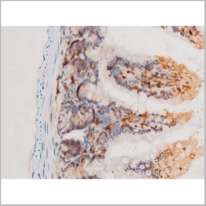
 IHC (Immunohistochemistry)
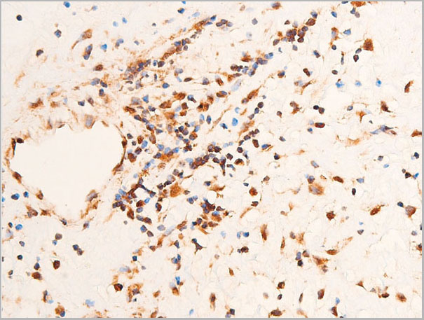
(Immunohistochemistry of paraffin-embedded Human skin tissue with Phospho-MLKL (Ser358) Monoclonal Antibody at dilution of 1:200)
IHC (Immunohistochemistry)
(Immunohistochemistry of paraffin-embedded Human skin tissue with Phospho-MLKL (Ser358) Monoclonal Antibody at dilution of 1:200)
MLKL, Monoclonal Antibody (Cat# AAA22113)

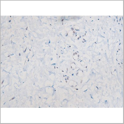
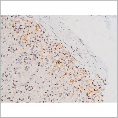
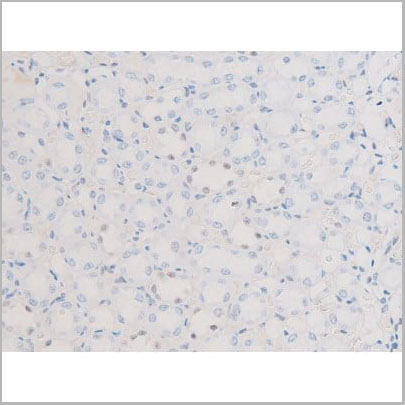
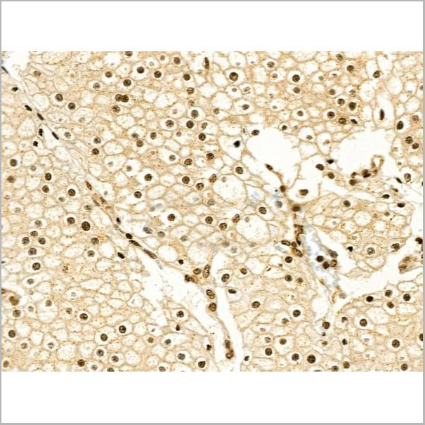
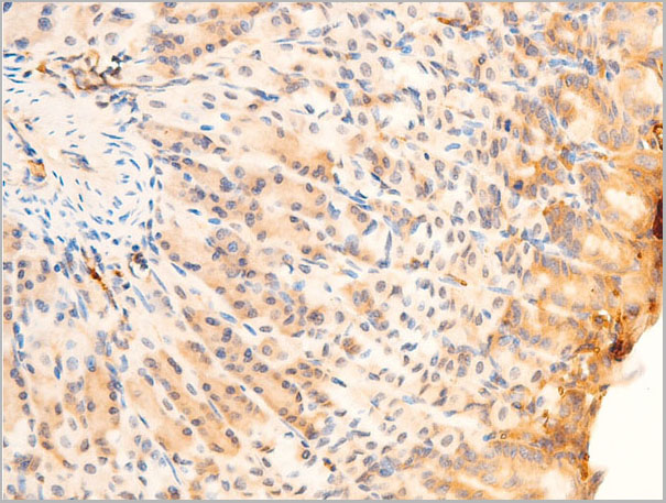
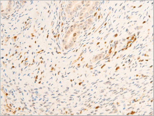
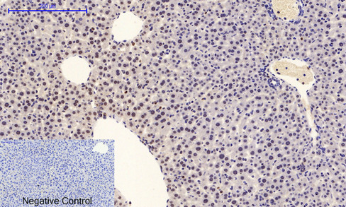
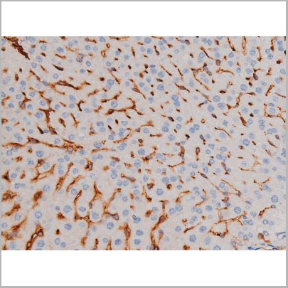
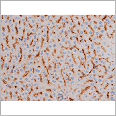
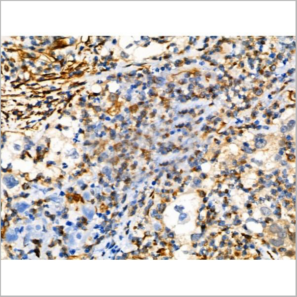
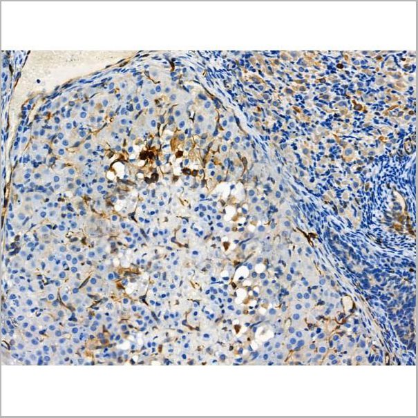
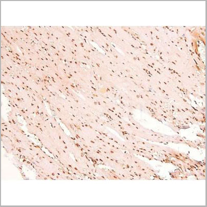
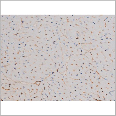
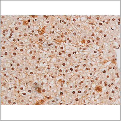
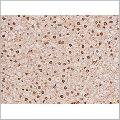
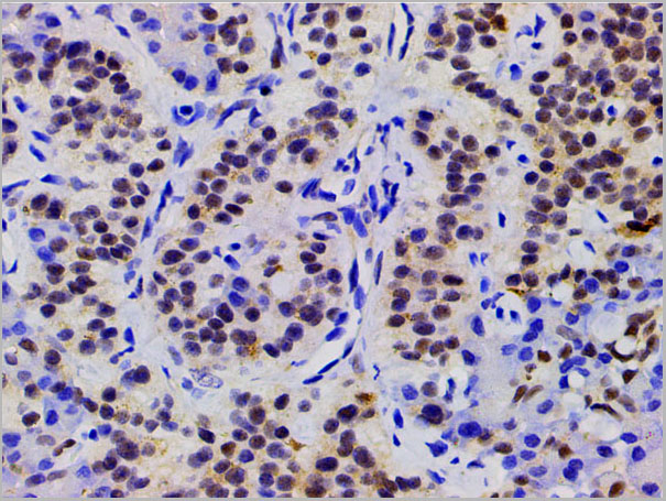
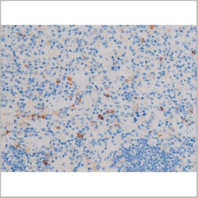
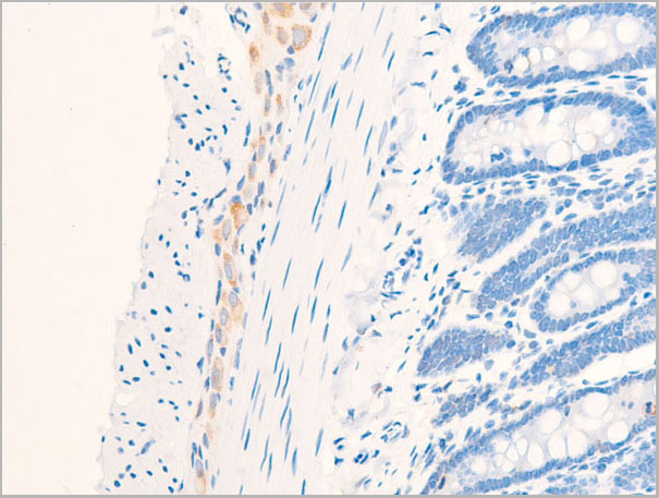
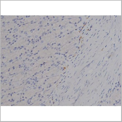
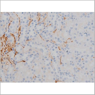
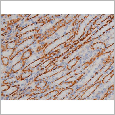
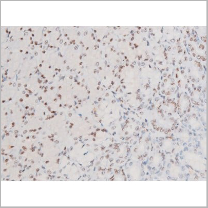
 IHC (Immunohistochemistry)
(At 1/100 staining Mouse stomach tissue by IHC-P. The sample was formaldehyde fixed and a heat mediated antigen retrieval step in citrate buffer was performed. The sample was then blocked and incubated with the primary antibody at 4 degree C overnight. An HRP conjugated anti-Rabbit antibody was used as the secondary antibody.)
IHC (Immunohistochemistry)
(At 1/100 staining Mouse stomach tissue by IHC-P. The sample was formaldehyde fixed and a heat mediated antigen retrieval step in citrate buffer was performed. The sample was then blocked and incubated with the primary antibody at 4 degree C overnight. An HRP conjugated anti-Rabbit antibody was used as the secondary antibody.)
BRK, Polyclonal Antibody (Cat# AAA31371)
Predicted Reactivity: Horse (89%)
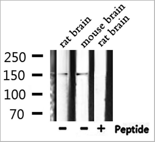
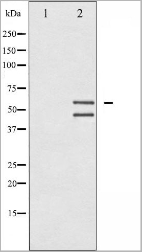
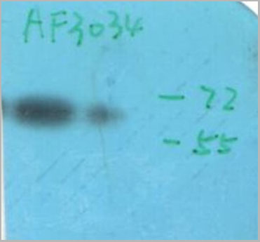
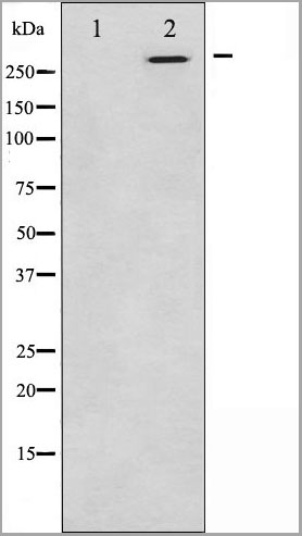
 WB (Western Blot)
(Western blot analysis of Phospho-Connexin 43 (Ser367) expression in various lysates)
WB (Western Blot)
(Western blot analysis of Phospho-Connexin 43 (Ser367) expression in various lysates)
Connexin 43, Polyclonal Antibody (Cat# AAA31018)
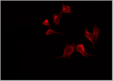
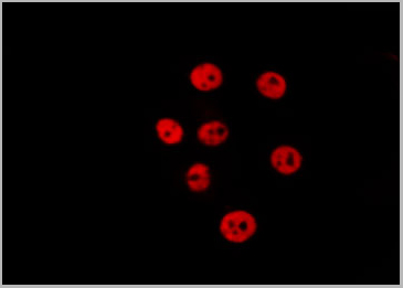
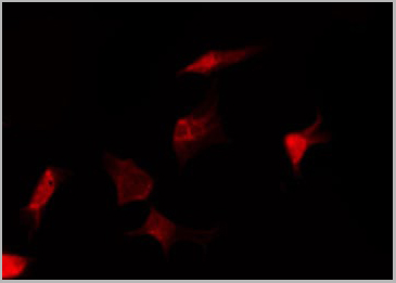
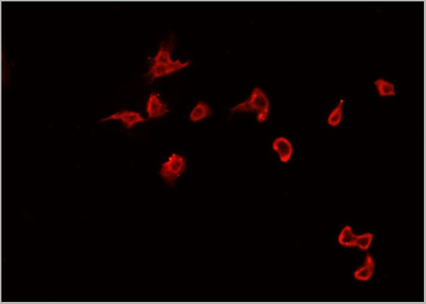
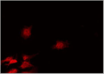
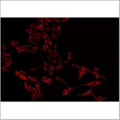
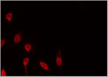
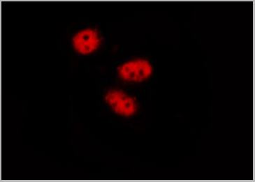
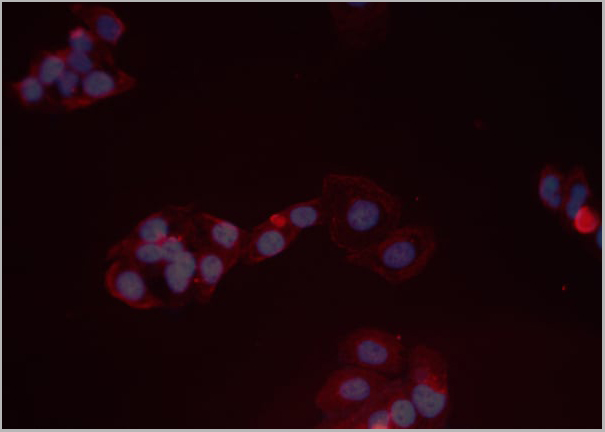
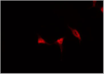
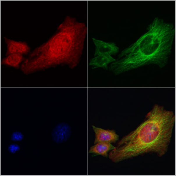
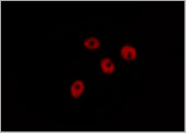
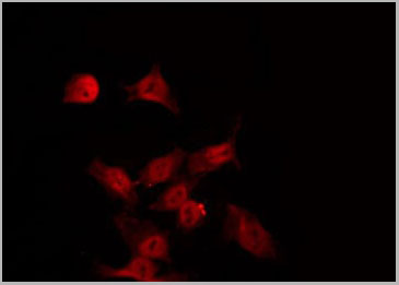
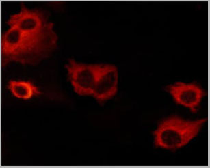 IF (Immunofluorescence)
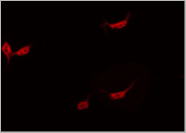
(AAA31068 staining NIH/3T3 cells by ICC/IF. Cells were fixed with PFA and permeabilized in 0.1% saponin prior to blocking in 10% serum for 45 minutes at 37 degree C. The primary antibody was diluted 1/400 and incubated with the sample for 1 hour at 37 degree C. A Alexa Fluor 594 conjugated goat polyclonal to rabbit IgG (H+L), diluted 1/600 was used as secondary antibody.)
IF (Immunofluorescence)
(AAA31068 staining NIH/3T3 cells by ICC/IF. Cells were fixed with PFA and permeabilized in 0.1% saponin prior to blocking in 10% serum for 45 minutes at 37 degree C. The primary antibody was diluted 1/400 and incubated with the sample for 1 hour at 37 degree C. A Alexa Fluor 594 conjugated goat polyclonal to rabbit IgG (H+L), diluted 1/600 was used as secondary antibody.)
BCL-XL, Polyclonal Antibody (Cat# AAA31068)
 Application Data
(At 25 degree C. The primary antibody was diluted at 1/200 and incubated with the sample for 1 hour at 37 degree C. An Alexa Fluor 594 conjugated goat anti-rabbit IgG (H+L) Ab, diluted at 1/600, was used as the secondary antibody.)
Application Data
(At 25 degree C. The primary antibody was diluted at 1/200 and incubated with the sample for 1 hour at 37 degree C. An Alexa Fluor 594 conjugated goat anti-rabbit IgG (H+L) Ab, diluted at 1/600, was used as the secondary antibody.)
eNOS, Polyclonal Antibody (Cat# AAA31394)
Predicted Reactivity: Pig (100%), Bovine (100%), Rabbit (100%), Dog (100%)
RAD50, Polyclonal Antibody (Cat# AAA31432)
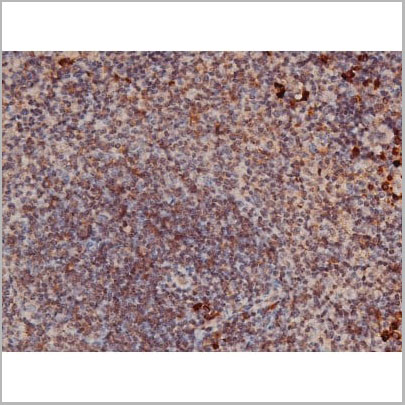
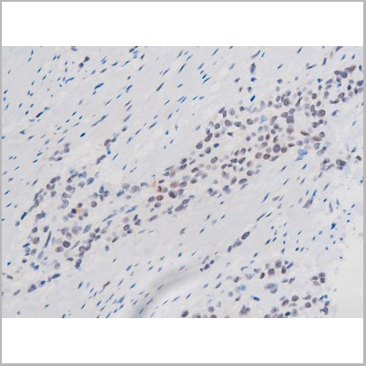
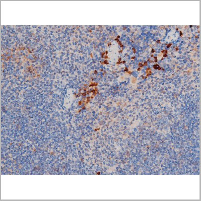
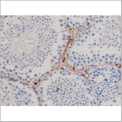
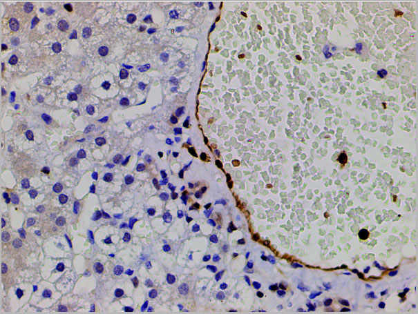
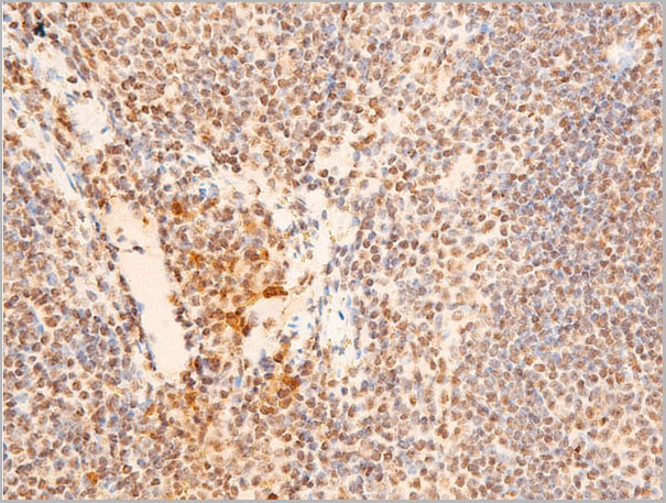
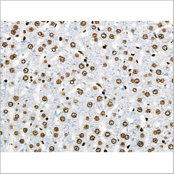
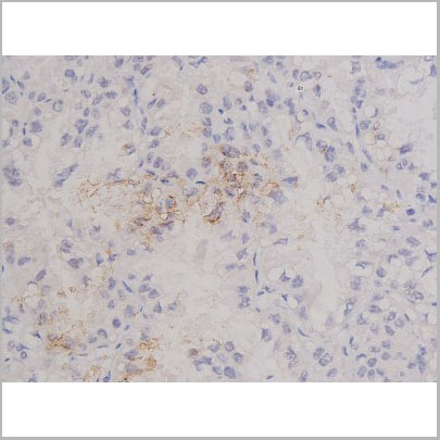
 IHC (Immunohistochemistry)
(AAA31023 at 1/200 staining Rat spleen tissue sections by IHC-P. The tissue was formaldehyde fixed and a heat mediated antigen retrieval step in citrate buffer was performed. The tissue was then blocked and incubated with the antibody for 1.5 hours at 22 degree C. An HRP conjugated goat anti-rabbit antibody was used as the secondary.)
IHC (Immunohistochemistry)
(AAA31023 at 1/200 staining Rat spleen tissue sections by IHC-P. The tissue was formaldehyde fixed and a heat mediated antigen retrieval step in citrate buffer was performed. The tissue was then blocked and incubated with the antibody for 1.5 hours at 22 degree C. An HRP conjugated goat anti-rabbit antibody was used as the secondary.)
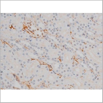
p70 S6 Kinase, Polyclonal Antibody (Cat# AAA31023)

 WB (Western Blot)
(Western blot analysis of Phospho-IL-8R beta/CDw128 beta (Ser347) expression in Mouse kidney lysate)
WB (Western Blot)
(Western blot analysis of Phospho-IL-8R beta/CDw128 beta (Ser347) expression in Mouse kidney lysate)
IL-8R beta/CDw128 beta, Polyclonal Antibody (Cat# AAA31026)
 WB (Western Blot)
(Western blot analysis of Phospho-SYK (Tyr525) expression in various lysates)
WB (Western Blot)
(Western blot analysis of Phospho-SYK (Tyr525) expression in various lysates)
SYK, Polyclonal Antibody (Cat# AAA31052)
What Are Phospho Antibodies?
Protein phosphorylation is a process where a phosphate group is added to certain amino acid residues of a protein – usually serine (S), threonine (T), or tyrosine (Y) - by enzymes called kinases. This process is integral in controlling cellular signaling, cellular growth, and other biological functions.
Our catalog includes a wide range of phospho-specific antibodies that can accurately detect this important marker. They perform strongly in widely-used laboratory applications such as Western blot, flow cytometry, immunohistochemistry, and immunofluorescence microscopy. We value your trust in us and are committed to providing top-quality products and services. All of our antibodies are guaranteed to work for the applications and species indicated on our website & associated product pages.
What Are The Key Applications of Phospho Antibodies?
1. Western Blotting
One of the first steps a researcher can take in utilizing these phospho-specific antibodies, is to check if the antibody works using a technique referred to as “Western blot”. For those unfamiliar, Western Blot aids in showing whether the protein that the antibody recognizes is appearing at the correct/expected size. These phospho-specific antibodies should also be able to detect changes in the target protein’s phosphorylation (on/off state) when cells are stimulated in certain ways.
2. Staining of Fixed Cells (Immunocytochemistry)
Another routine use of these phospho-specific antibodies, is to test if the antibody is able to demonstrate similar performance when used on fixed cells (intact cells that have been preserved) as it did in the Western blot tests. It is an important aspect in many cases to confirm that the antibody works in actual intact cell samples. Ideally, the method used for cellular fixation should be the same as what is used in pathology labs (like using 10% formalin). To check if the antibody works well in tissue sections (FFPE), researchers will often test it on fixed cells that are processed similar to tissue samples.
3. Specificity Tests Using Peptides
In order to make sure that the antibody is only binding to the right target:
- Laboratory technicians will mix the antibody with phospho-peptides (short segments of the protein containing the phosphate group modification).
- If the antibody signal disappears, it is confirmation that it is binding to the correct phosphorylated location.
- A more robust test is to use both the phosphorylated and non-phosphorylated (dephosphorylated) versions of the protein. The antibody should react only with the phosphorylated one.
- Another method sometimes utilized is to treat the sample with an enzyme, such as alkaline phosphatase, that specifically removes phosphate groups. If the antibody signal disappears after this, it also confirms specificity.
4. Genetic Confirmation
As a final step, scientists can genetically manipulate the nucleotide sequence and alter the target protein by removing the exact site where phosphorylation happens. If the antibody no longer appears to detect the modified protein, it is strong evidence supporting the antibody being specific for that phosphorylated site.
Why Buy Phospho Antibodies Through Us?
- The production laboratory adheres to strict and consistent protocols prior to releasing any of these phospho-specific antibodies:
- Standard methods and proper controls in all tests to ensure high quality.
- These antibodies are tested and validated in different cell types and species.
- High quality control criterion to ensure each batch is consistent, so you will obtain reliable results every time.
FAQ
1. What Are Phospho-Specific Antibodies?
Phospho-specific antibodies are made to detect proteins only when they have a phosphate group linked to a specific amino acid residue. This empowers scientists understand if a protein is "turned on" or active, based on its phosphorylation state.
2. How to Detect Phosphorylated Proteins in a Western Blot?
To find out if a protein is phosphorylated using Western blot:
- Use a phospho-specific antibody that binds only to the phosphorylated form of the protein.
- You can also use a “regular” antibody for the same amino acid sequence of the protein that the phospho-specific antibody is binding to (but in this case, this antibody will not bind if there is a phosphate group present) in order to compare how much of it is phosphorylated versus how much is non-phosphorylated (or “total” protein, if the “normal” antibody’s epitopes are non-phospho-site-specific).
3. How to Choose the Best Antibody?
Here are some simple tips to help you pick the right antibody:
- Know your target
- Match your sample characteristics
- Confirm the intended use is appropriate
- Check “host” and “type”
- Check the “quality” of the presented data/images
- Appraise whether the available validation meets your needs