Filters
▼Clonality
▼Type
▼Reactivity
▼Gene Name
▼Isotype
▼Host
▼Application
▼Clone
▼Phospho Antibodies
Phospho-specific antibodies’ typical purpose is to enable researchers to detect changes in proteins. They will exclusively bind to the amino acid sequence on a protein that has been phosphorylated (which is both a physical & chemical change) and do not bind to the same amino acid sequence on said protein if it lacks said phosphorylation. This aids in being able to clearly see and understand the data produced from this particular protein modification.
Viewing 4700-4750 of 5298 product results
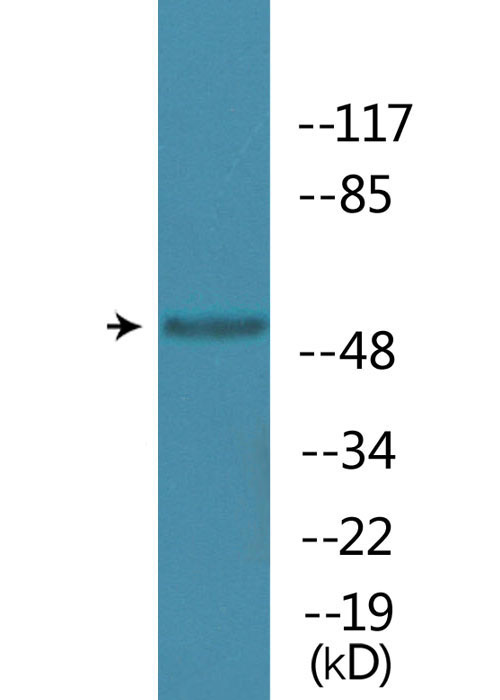
 WB (Western Blot)
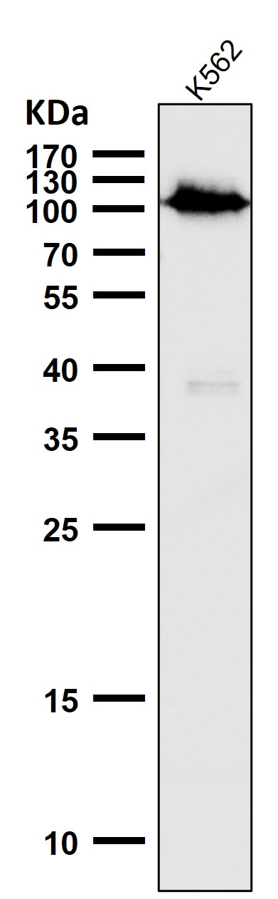
(Western blot analysis of Phospho-Rb (S780) expression in K562 cell lysate.)
WB (Western Blot)
(Western blot analysis of Phospho-Rb (S780) expression in K562 cell lysate.)
Rb, Monoclonal Antibody (Cat# AAA128103)
 WB (Western Blot)
(Western blot analysis of PC3 Cell Lysate using Phospho-Akt Ser473 Mouse mAb diluted at 1:1000.)
WB (Western Blot)
(Western blot analysis of PC3 Cell Lysate using Phospho-Akt Ser473 Mouse mAb diluted at 1:1000.)
Akt, Monoclonal Antibody (Cat# AAA320540)
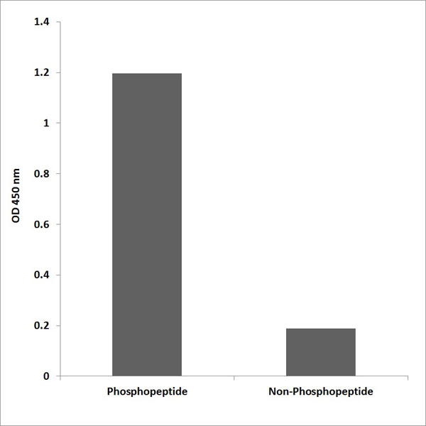
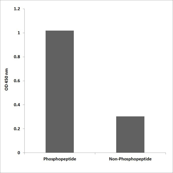
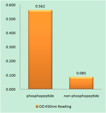
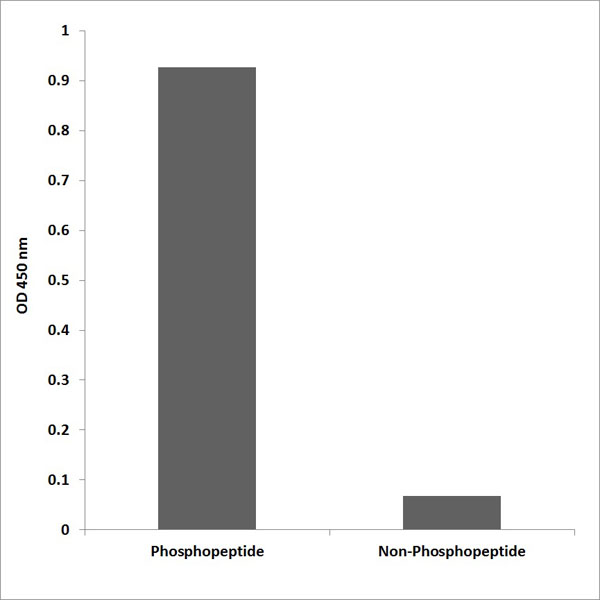
 ELISA
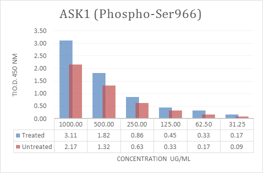
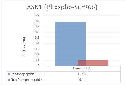
(Enzyme-Linked Immunosorbent Assay (ELISA) for immunogen phosphor-peptide (left) and non-phospho peptide (right), using Anti-ASK1 (Phospho-Ser966) Antibody.)
ELISA
(Enzyme-Linked Immunosorbent Assay (ELISA) for immunogen phosphor-peptide (left) and non-phospho peptide (right), using Anti-ASK1 (Phospho-Ser966) Antibody.)
ASK1, ELISA Kit (Cat# AAA318619)
 ELISA
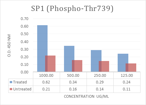
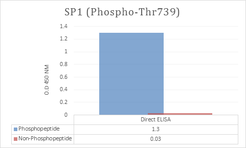
(Enzyme-Linked Immunosorbent Assay (ELISA) for immunogen phosphor-peptide (left) and non-phospho peptide (right), using Anti-SP1 (Phospho-Thr739) Antibody.)
ELISA
(Enzyme-Linked Immunosorbent Assay (ELISA) for immunogen phosphor-peptide (left) and non-phospho peptide (right), using Anti-SP1 (Phospho-Thr739) Antibody.)
SP1, ELISA Kit (Cat# AAA318629)
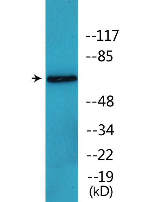 WB (Western Blot)
(Western blot analysis of lysates from COS7 cells treated with PMA 125ng/ml 30', using MUNC-18a (Phospho-Ser313) Antibody.)
WB (Western Blot)
(Western blot analysis of lysates from COS7 cells treated with PMA 125ng/ml 30', using MUNC-18a (Phospho-Ser313) Antibody.)
MUNC-18a, Polyclonal Antibody (Cat# AAA318360)
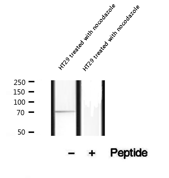 WB (Western Blot)
(Western blot analysis of extracts of HT29 cells treated with nocodazole, using Phospho-Myt1 (Ser83) Antibody.)
WB (Western Blot)
(Western blot analysis of extracts of HT29 cells treated with nocodazole, using Phospho-Myt1 (Ser83) Antibody.)
Myt1, Polyclonal Antibody (Cat# AAA321353)
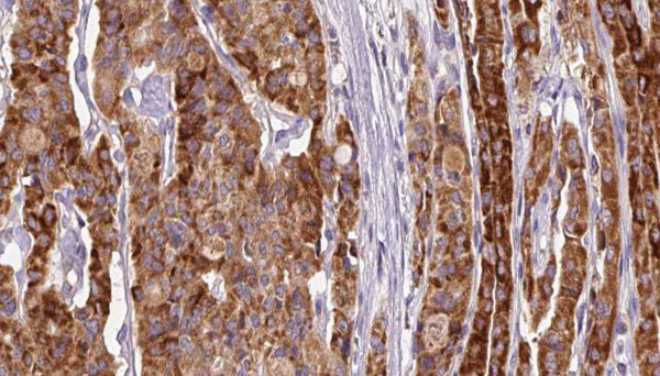
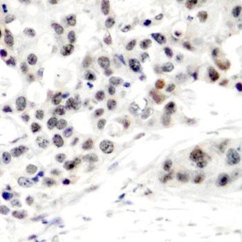
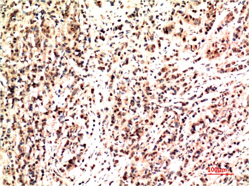
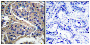
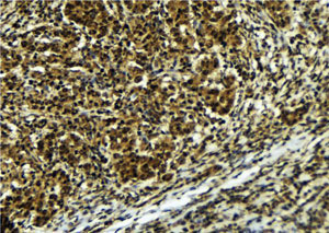 IHC (Immunohiostchemistry)
(AAA327182 at 1/100 staining Human breast cancer tissue by IHC-P. The sample was formaldehyde fixed and a heat mediated antigen retrieval step in citrate buffer was performed. The sample was then blocked and incubated with the antibody for 1.5 hours at 22 degree C. An HRP conjugated goat anti-rabbit antibody was used as the secondary.)
IHC (Immunohiostchemistry)
(AAA327182 at 1/100 staining Human breast cancer tissue by IHC-P. The sample was formaldehyde fixed and a heat mediated antigen retrieval step in citrate buffer was performed. The sample was then blocked and incubated with the antibody for 1.5 hours at 22 degree C. An HRP conjugated goat anti-rabbit antibody was used as the secondary.)
ATR, Polyclonal Antibody (Cat# AAA327182)
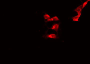
 IF (Immunofluorescence)
(AAA327189 staining A549 cells(4h of LPS treatment) by IF/ICC. The samples were fixed with PFA and permeabilized in 0.1% Triton X-100,then blocked in 10% serum for 45 minutes at 25°C. Samples were then incubated with primary Ab(AAA327189) and mouse anti-beta tubulin Ab for 1 hour at 37°C. An AlexaFluor594 conjugated goat anti-rabbit IgG(H+L) Ab(Red) and an AlexaFluor488 conjugated goat anti-mouse IgG(H+L) Ab(Green) were used as the secondary Ab. The nuclear counter stain is DAPI (blue).)
IF (Immunofluorescence)
(AAA327189 staining A549 cells(4h of LPS treatment) by IF/ICC. The samples were fixed with PFA and permeabilized in 0.1% Triton X-100,then blocked in 10% serum for 45 minutes at 25°C. Samples were then incubated with primary Ab(AAA327189) and mouse anti-beta tubulin Ab for 1 hour at 37°C. An AlexaFluor594 conjugated goat anti-rabbit IgG(H+L) Ab(Red) and an AlexaFluor488 conjugated goat anti-mouse IgG(H+L) Ab(Green) were used as the secondary Ab. The nuclear counter stain is DAPI (blue).)
nrf2, Polyclonal Antibody (Cat# AAA327189)
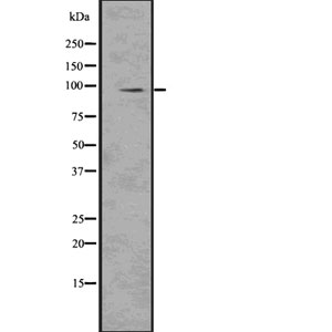
 WB (Western Blot)
(Western blot analysis of Phospho-Doublecortin (Ser334) using K562 whole cell lysates)
WB (Western Blot)
(Western blot analysis of Phospho-Doublecortin (Ser334) using K562 whole cell lysates)
Doublecortin, Polyclonal Antibody (Cat# AAA327235)
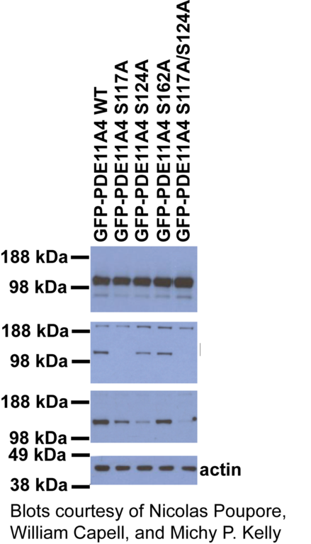
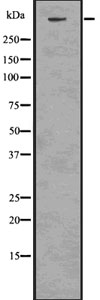
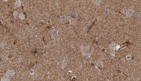
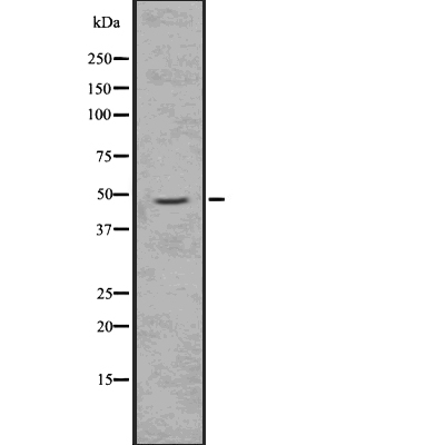
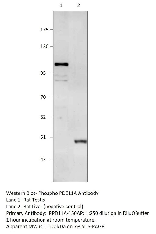 WB (Western Blot)
Phospho-PDE11A, Polyclonal Antibody (Cat# AAA76027)
 WB (Western Blot)
(Western blot analysis of lysates from K562 cells treated with Na3VO4 0.3nM 40', using MAPKAPK5 (Phospho-Thr182) Antibody.)
WB (Western Blot)
(Western blot analysis of lysates from K562 cells treated with Na3VO4 0.3nM 40', using MAPKAPK5 (Phospho-Thr182) Antibody.)
MAPKAPK5, Polyclonal Antibody (Cat# AAA318353)
 IF (Immunofluorescence)
(AAA327267 staining NIH-3T3 cells by IF/ICC. The sample were fixed with PFA and permeabilized in 0.1% Triton X-100, then blocked in 10% serum for 45 minutes at 25 degree C. The primary antibody was diluted at 1/200 and incubated with the sample for 1 hour at 37 degree C. An Alexa Fluor 594 conjugated goat anti-rabbit IgG (H+L) antibody, diluted at 1/600, was used as secondary antibody.)
IF (Immunofluorescence)
(AAA327267 staining NIH-3T3 cells by IF/ICC. The sample were fixed with PFA and permeabilized in 0.1% Triton X-100, then blocked in 10% serum for 45 minutes at 25 degree C. The primary antibody was diluted at 1/200 and incubated with the sample for 1 hour at 37 degree C. An Alexa Fluor 594 conjugated goat anti-rabbit IgG (H+L) antibody, diluted at 1/600, was used as secondary antibody.)
MAPKAP Kinase 2, Polyclonal Antibody (Cat# AAA327267)
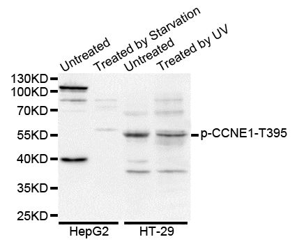
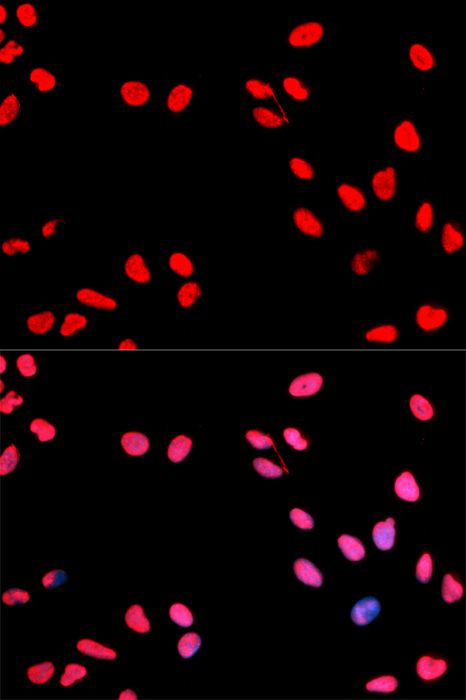 IF (Immunofluorescence)
(Immunofluorescence analysis of MCF-7 cells using Phospho-CCNE1-T395 antibody. Blue: DAPI for nuclear staining.)
IF (Immunofluorescence)
(Immunofluorescence analysis of MCF-7 cells using Phospho-CCNE1-T395 antibody. Blue: DAPI for nuclear staining.)
CCNE1-T395, Antibody (Cat# AAA36683)
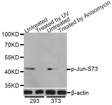
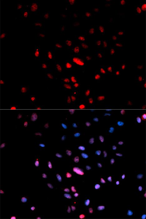 IF (Immunofluorescence)
(Immunofluorescence analysis of U2OS cells using Phospho-Jun-S73 antibody. Blue: DAPI for nuclear staining.)
IF (Immunofluorescence)
(Immunofluorescence analysis of U2OS cells using Phospho-Jun-S73 antibody. Blue: DAPI for nuclear staining.)
c-Jun-Ser73, Antibody (Cat# AAA36766)
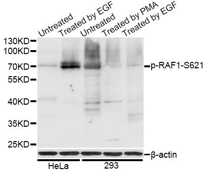
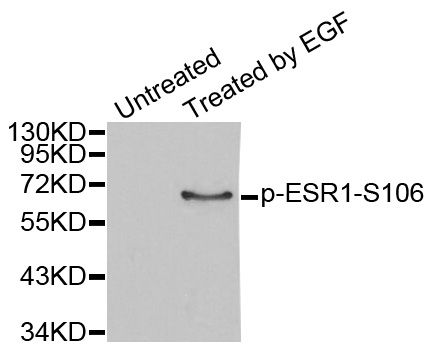
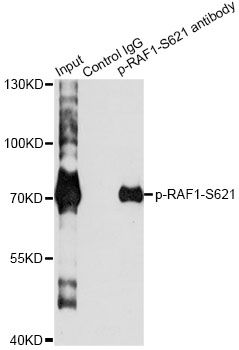 IP (Immunoprecipitation)
(Immunoprecipitation analysis of 200ug extracts of HeLa cells treated by EGF using 2.5ug Phospho-RAF1-S621 antibody. Western blot was performed from the immunoprecipitate using Phospho-RAF1-S621 antibody at a dilition of 1:1000.)
IP (Immunoprecipitation)
(Immunoprecipitation analysis of 200ug extracts of HeLa cells treated by EGF using 2.5ug Phospho-RAF1-S621 antibody. Western blot was performed from the immunoprecipitate using Phospho-RAF1-S621 antibody at a dilition of 1:1000.)
RAF1-S621, Antibody (Cat# AAA36781)
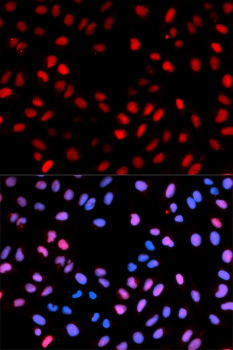
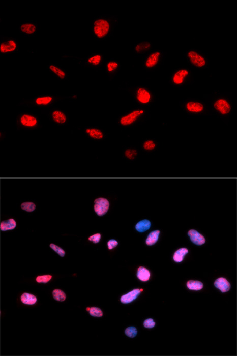 IF (Immunofluorescence)
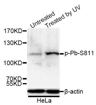
(Immunofluorescence analysis of U2OS cells using Phospho-Rb-S811 antibody. Blue: DAPI for nuclear staining.)
IF (Immunofluorescence)
(Immunofluorescence analysis of U2OS cells using Phospho-Rb-S811 antibody. Blue: DAPI for nuclear staining.)
RB-S811, Antibody (Cat# AAA36784)
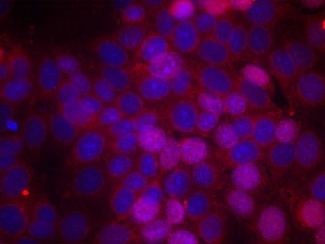 IF (Immunofluorescence)
(Immunofluorescence staining of methanol-fixed MCF-7 cells using Phospho-ESR1-S106 antibody.)
IF (Immunofluorescence)
(Immunofluorescence staining of methanol-fixed MCF-7 cells using Phospho-ESR1-S106 antibody.)
ESR1-S106, Antibody (Cat# AAA37366)
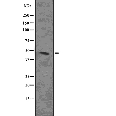
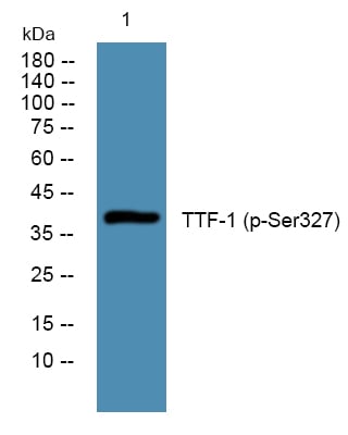 WB (Western Blot)
(Western blot analysis of lysates from HCT116 cells, primary antibody was diluted at 1:1000, 4 degree C over night)
WB (Western Blot)
(Western blot analysis of lysates from HCT116 cells, primary antibody was diluted at 1:1000, 4 degree C over night)
TTF-1, ELISA Kit (Cat# AAA319128)
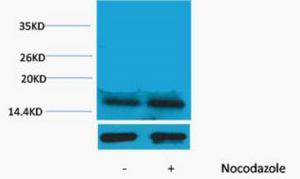 WB (Western Blot)
(Western blot analysis of extracts from Hela cells, untreated (-) or treated, 1:2000. Secondary antibody was diluted at 1:20000 cells nucleus extracted by Minute TM Cytoplasmic and Nuclear Fractionation kit (SC-003,Inventbiotech,MN,USA).)
WB (Western Blot)
(Western blot analysis of extracts from Hela cells, untreated (-) or treated, 1:2000. Secondary antibody was diluted at 1:20000 cells nucleus extracted by Minute TM Cytoplasmic and Nuclear Fractionation kit (SC-003,Inventbiotech,MN,USA).)
Histone H3, ELISA Kit (Cat# AAA319073)
 WB (Western Blot)
(Western blot analysis of K562 using p-p70 S6 kinase alpha (S427) antibody.)
WB (Western Blot)
(Western blot analysis of K562 using p-p70 S6 kinase alpha (S427) antibody.)
p70 S6 kinase alpha, ELISA Kit (Cat# AAA319186)

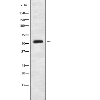
 WB (Western Blot)
(Western blot analysis of lysates from RAW264.7 cells treated with G-CSF 25ng/ml 15', using IL-3R beta (Phospho-Tyr593) Antibody. The lane on the right is blocked with the phospho peptide.)
WB (Western Blot)
(Western blot analysis of lysates from RAW264.7 cells treated with G-CSF 25ng/ml 15', using IL-3R beta (Phospho-Tyr593) Antibody. The lane on the right is blocked with the phospho peptide.)
IL-3R beta, Polyclonal Antibody (Cat# AAA316152)
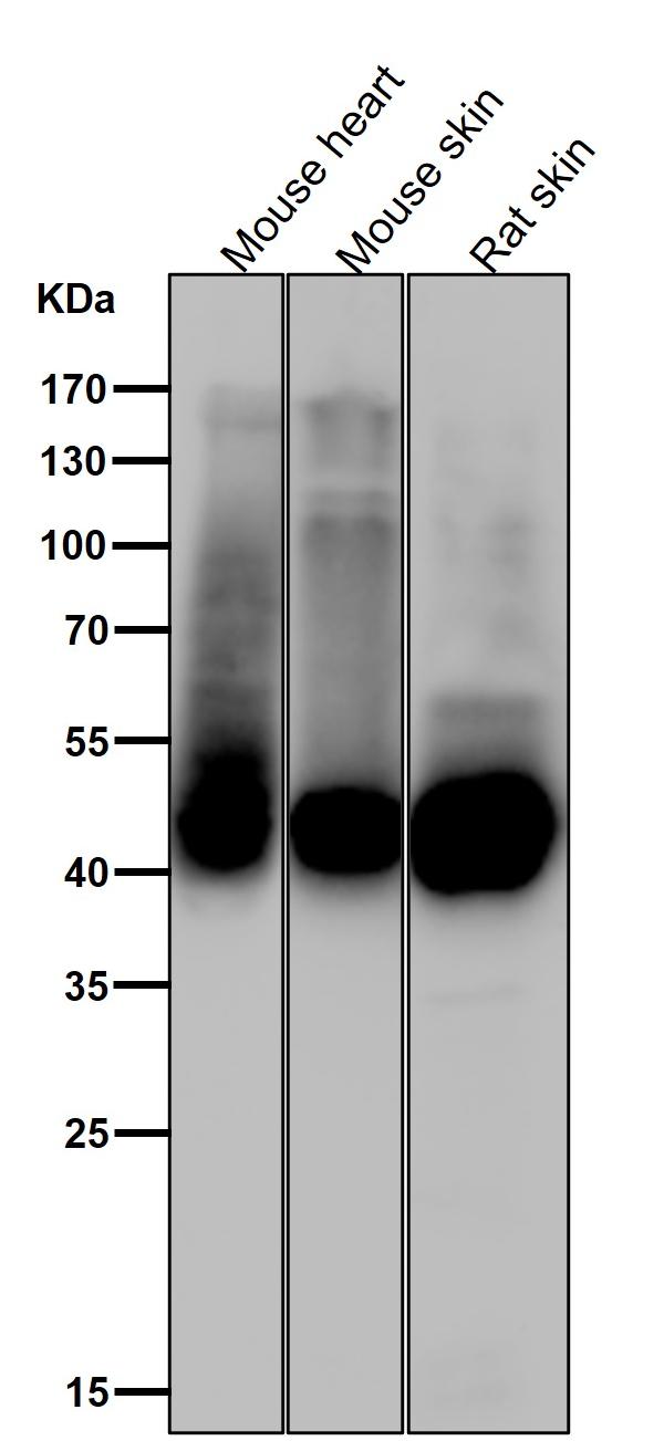
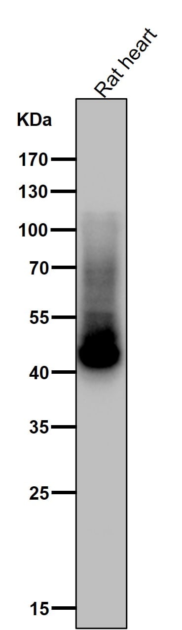
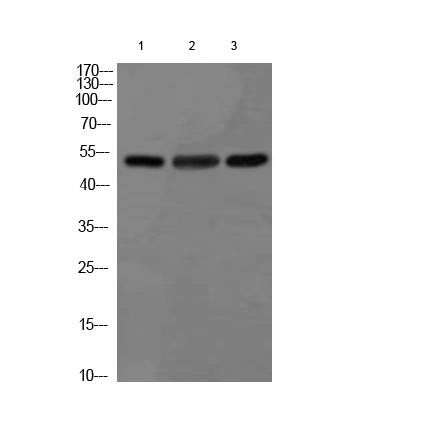 WB (Western Blot)
(Western blot analysis of 1) Hela Cell Lysate, 2) Mouse Heart Tissue Lysate, 3)Rat Heart Tissue Lysate using GSK 3? Mouse mAb diluted at 1:1000.)
WB (Western Blot)
(Western blot analysis of 1) Hela Cell Lysate, 2) Mouse Heart Tissue Lysate, 3)Rat Heart Tissue Lysate using GSK 3? Mouse mAb diluted at 1:1000.)
GSK3beta, Monoclonal Antibody (Cat# AAA320543)
CamKII, phosphorylated, Monoclonal Antibody (Cat# AAA47742)
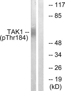
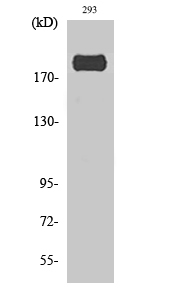
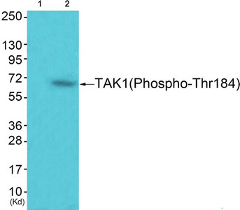 WB (Western Blot)
(Western blot analysis of extracts from 293 cells (Lane 2), using TAK1 (Phospho-Thr184) Antibody. The lane on the left is treated with synthesized peptide.)
WB (Western Blot)
(Western blot analysis of extracts from 293 cells (Lane 2), using TAK1 (Phospho-Thr184) Antibody. The lane on the left is treated with synthesized peptide.)
MAP3K7, Polyclonal Antibody (Cat# AAA243251)
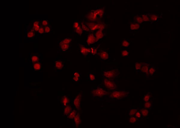 IF (Immunofluorescence)
(AAA327232 staining HeLa by IF/ICC. The sample were fixed with PFA and permeabilized in 0.1% Triton X-100, then blocked in 10% serum for 45 minutes at 25 degree C. The primary antibody was diluted at 1/200 and incubated with the sample for 1 hour at 37 degree C. An Alexa Fluor 594 conjugated goat anti-rabbit IgG (H+L) Ab, diluted at 1/600, was used as the secondary antibody.)
IF (Immunofluorescence)
(AAA327232 staining HeLa by IF/ICC. The sample were fixed with PFA and permeabilized in 0.1% Triton X-100, then blocked in 10% serum for 45 minutes at 25 degree C. The primary antibody was diluted at 1/200 and incubated with the sample for 1 hour at 37 degree C. An Alexa Fluor 594 conjugated goat anti-rabbit IgG (H+L) Ab, diluted at 1/600, was used as the secondary antibody.)
PKC epsilon, Polyclonal Antibody (Cat# AAA327232)

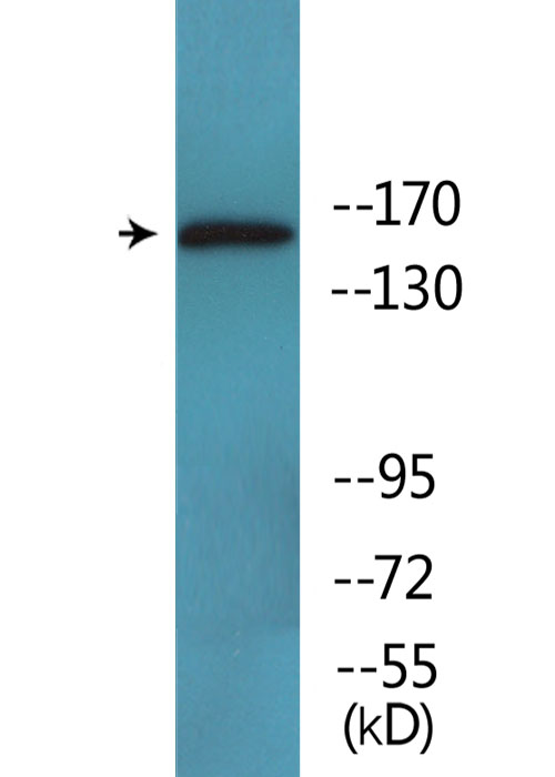
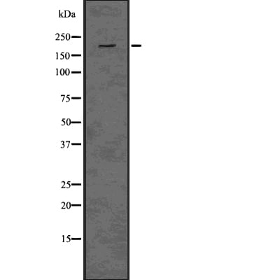 WB (Western Blot)
(Western blot analysis of Phospho-HER3/ErbB3 (Tyr1289) using HeLa whole cell lysates)
WB (Western Blot)
(Western blot analysis of Phospho-HER3/ErbB3 (Tyr1289) using HeLa whole cell lysates)
HER3/ErbB3, Polyclonal Antibody (Cat# AAA327251)
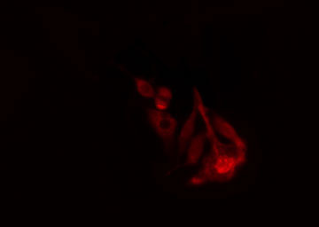
 IF (Immunofluorescence)
(AAA327321 staining NIH-3T3 cells by IF/ICC. The sample were fixed with PFA and permeabilized in 0.1% Triton X-100, then blocked in 10% serum for 45 minutes at 25 degree C. The primary antibody was diluted at 1/200 and incubated with the sample for 1 hour at 37 degree C. An Alexa Fluor 594 conjugated goat anti-rabbit IgG (H+L) antibody, diluted at 1/600, was used as secondary antibody.)
IF (Immunofluorescence)
(AAA327321 staining NIH-3T3 cells by IF/ICC. The sample were fixed with PFA and permeabilized in 0.1% Triton X-100, then blocked in 10% serum for 45 minutes at 25 degree C. The primary antibody was diluted at 1/200 and incubated with the sample for 1 hour at 37 degree C. An Alexa Fluor 594 conjugated goat anti-rabbit IgG (H+L) antibody, diluted at 1/600, was used as secondary antibody.)
MAP2K7, Polyclonal Antibody (Cat# AAA327321)
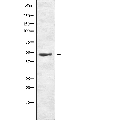
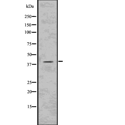
 WB (Western Blot)
(Western blot analysis of PKM2(phospho-Ser37) using HeLa whole lysates.)
WB (Western Blot)
(Western blot analysis of PKM2(phospho-Ser37) using HeLa whole lysates.)
PKM2, Polyclonal Antibody (Cat# AAA327323)
Phospho-Smad2, Monoclonal Recombinant Antibody (Cat# AAA120236)


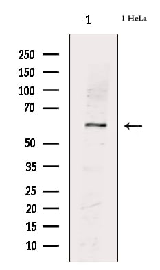
 WB (Western Blot)
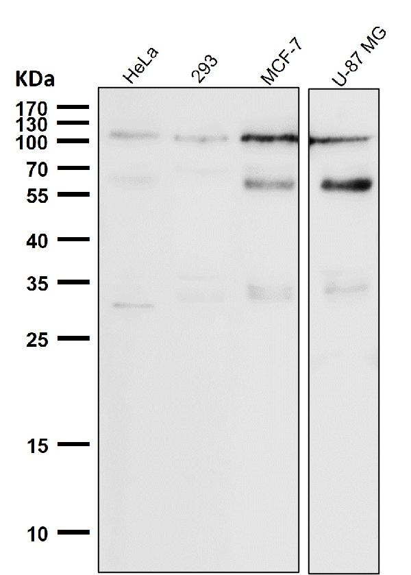
(Western Blot analysis of various cells using Phospho-NF?B-p105/p50 (S337) Polyclonal Antibody diluted at 1:500)
WB (Western Blot)
(Western Blot analysis of various cells using Phospho-NF?B-p105/p50 (S337) Polyclonal Antibody diluted at 1:500)
NFkappaB-p105/p50, Polyclonal Antibody (Cat# AAA318642)
 WB (Western Blot)
(Western Blot analysis of various cells using Phospho-IRS-1 (S307) Polyclonal Antibody diluted at 1:1000)
WB (Western Blot)
(Western Blot analysis of various cells using Phospho-IRS-1 (S307) Polyclonal Antibody diluted at 1:1000)
IRS-1, Polyclonal Antibody (Cat# AAA318646)
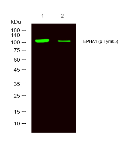
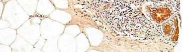
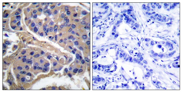
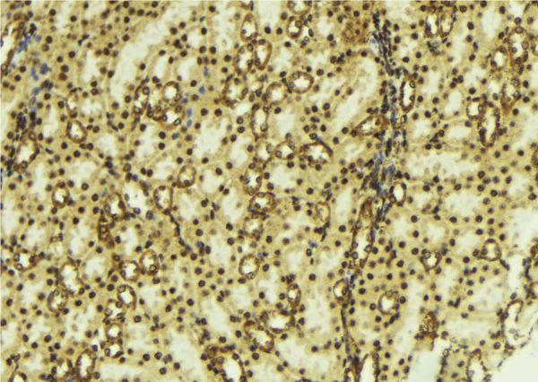
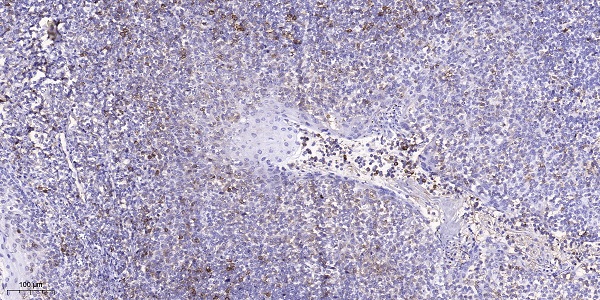 IHC (Immunohiostchemistry)
(Immunohistochemical analysis of paraffin-embedded human tonsil. 1, Antibody was diluted at 1:200(4 degree overnight). 2, Tris-EDTA,pH9.0 was used for antigen retrieval. 3,Secondary antibody was diluted at 1:200(room temperature, 45min).)
IHC (Immunohiostchemistry)
(Immunohistochemical analysis of paraffin-embedded human tonsil. 1, Antibody was diluted at 1:200(4 degree overnight). 2, Tris-EDTA,pH9.0 was used for antigen retrieval. 3,Secondary antibody was diluted at 1:200(room temperature, 45min).)
EPHA1, Polyclonal Antibody (Cat# AAA318657)
 WB (Western Blot)
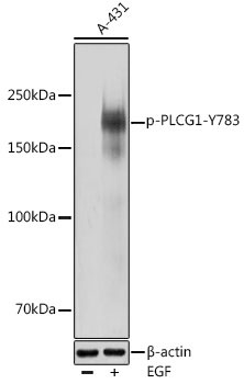
(Western blot analysis of extracts of A-431 cells, using Phospho-PLCG1-Y783 antibody (AAA37406) at 1:1000 dilution.A-431 cells were treated by EGF (25 ug/ml) at 37°C for 30 minutes after serum-starvation overnight.Secondary antibody: HRP Goat Anti-Rabbit IgG (H+L) at 1:10000 dilution.Lysates/proteins: 25ug per lane.Blocking buffer: 3% nonfat dry milk in TBST.Detection: ECL Basic Kit .Exposure time: 30s.)
WB (Western Blot)
(Western blot analysis of extracts of A-431 cells, using Phospho-PLCG1-Y783 antibody (AAA37406) at 1:1000 dilution.A-431 cells were treated by EGF (25 ug/ml) at 37°C for 30 minutes after serum-starvation overnight.Secondary antibody: HRP Goat Anti-Rabbit IgG (H+L) at 1:10000 dilution.Lysates/proteins: 25ug per lane.Blocking buffer: 3% nonfat dry milk in TBST.Detection: ECL Basic Kit .Exposure time: 30s.)
PLCG1-Y783, Antibody (Cat# AAA37406)
 IF (Immunofluorescence)
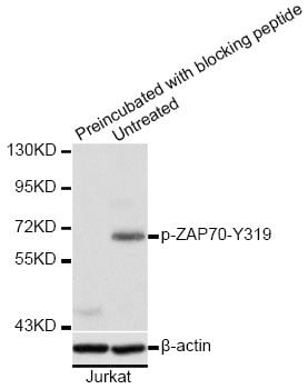
(Immunofluorescence staining of methanol-fixed HeLa cells using Phospho-ZAP70-Y319 antibody.)
IF (Immunofluorescence)
(Immunofluorescence staining of methanol-fixed HeLa cells using Phospho-ZAP70-Y319 antibody.)
ZAP70-Y319, Antibody (Cat# AAA37421)
 IF (Immunofluorescence)
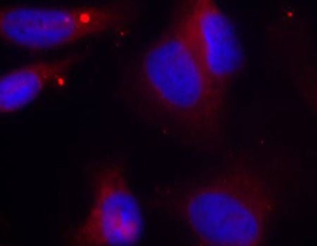
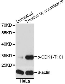
(Immunofluorescence analysis of MCF-7 cells using Phospho-CDK1-T161 antibody. Blue: DAPI for nuclear staining.)
IF (Immunofluorescence)
(Immunofluorescence analysis of MCF-7 cells using Phospho-CDK1-T161 antibody. Blue: DAPI for nuclear staining.)
CDK1-T161, Antibody (Cat# AAA37422)
 Application Data
b-Dystroglycan (Tyr-892), Monoclonal Antibody (Cat# AAA71498)
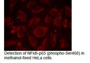
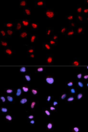
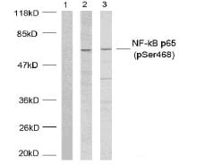 Application Data
NFkB-p65, Polyclonal Antibody (Cat# AAA47797)
 IF (Immunofluorescence)
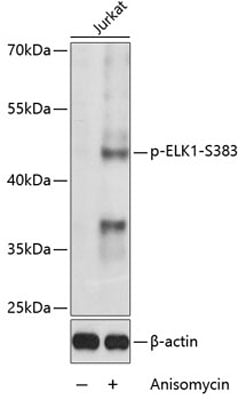
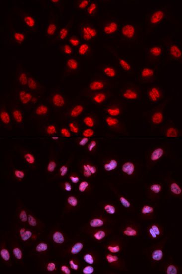
(Immunofluorescence analysis of MCF-7 cells using Phospho-ELK1-S383 antibody. Blue: DAPI for nuclear staining.)
IF (Immunofluorescence)
(Immunofluorescence analysis of MCF-7 cells using Phospho-ELK1-S383 antibody. Blue: DAPI for nuclear staining.)
ELK1-S383, Antibody (Cat# AAA36768)
FGF Receptor 1 - Y654, Antibody (Cat# AAA36686)
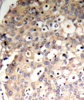
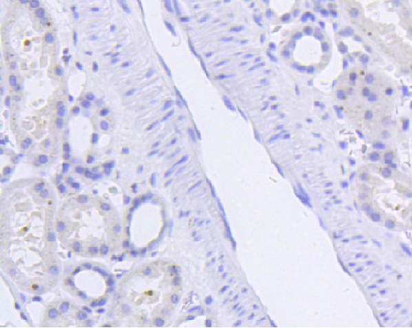
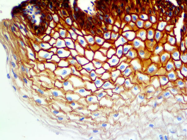 IHC (Immunohiostchemistry)
(EGFR Phospho Antibody Immunohistochemistry on an FFPE Cervix Tissue)
IHC (Immunohiostchemistry)
(EGFR Phospho Antibody Immunohistochemistry on an FFPE Cervix Tissue)
EGFR Phospho, Monoclonal Antibody (Cat# AAA59345)
 IF (Immunofluorescence)
(Immunofluorescence staining of methanol-fixed HeLa cells using Phospho-PTPN6-Y536 antibody.)
IF (Immunofluorescence)
(Immunofluorescence staining of methanol-fixed HeLa cells using Phospho-PTPN6-Y536 antibody.)
PTPN6-Y536, Antibody (Cat# AAA37394)
phospho-extracellular signal-regulated kinase, pERK, ELISA Kit (Cat# AAA41781)
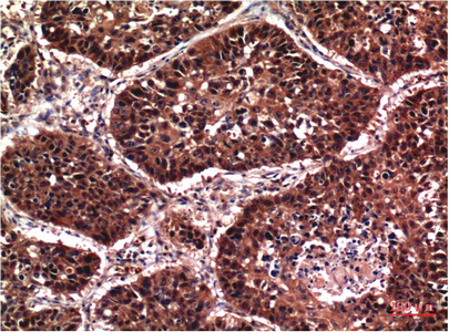
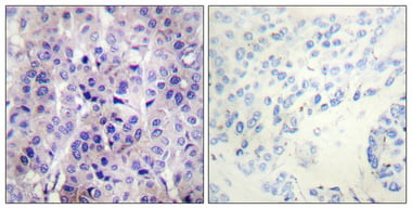
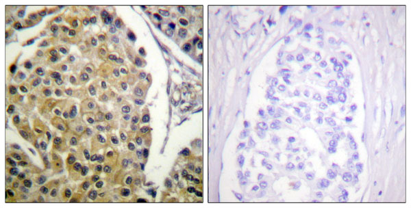
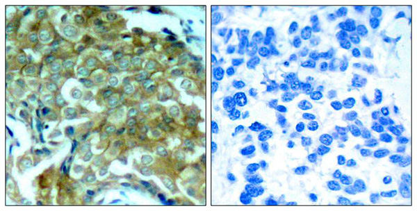 IHC (Immunohiostchemistry)
(Immunohistochemical analysis of paraffin-embedded human breast carcinoma tissue using EGFR(Phospho-Tyr1172) Antibody (left) or the same antibody preincubated with blocking peptide(right).)
IHC (Immunohiostchemistry)
(Immunohistochemical analysis of paraffin-embedded human breast carcinoma tissue using EGFR(Phospho-Tyr1172) Antibody (left) or the same antibody preincubated with blocking peptide(right).)
EGFR, Polyclonal Antibody (Cat# AAA302796)
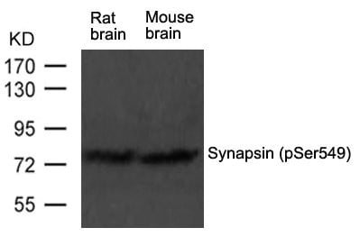
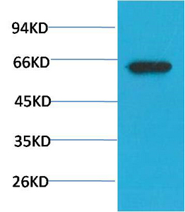
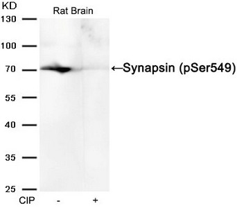 WB (Western Blot)
(Western blot analysis of extracts from Rat brain tissue or calf intestinal phosphatase (CIP), using Synapsin (phospho-Ser549) Antibody.)
WB (Western Blot)
(Western blot analysis of extracts from Rat brain tissue or calf intestinal phosphatase (CIP), using Synapsin (phospho-Ser549) Antibody.)
Syn1, Polyclonal Antibody (Cat# AAA243166)
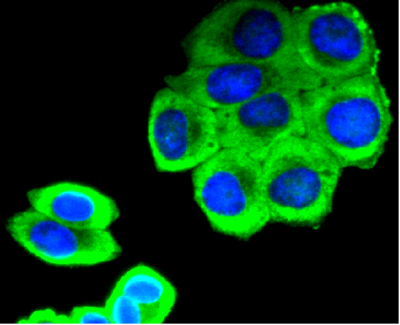
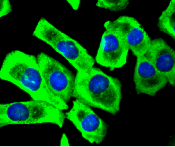
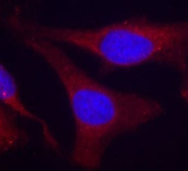
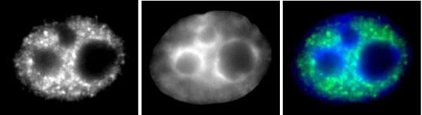
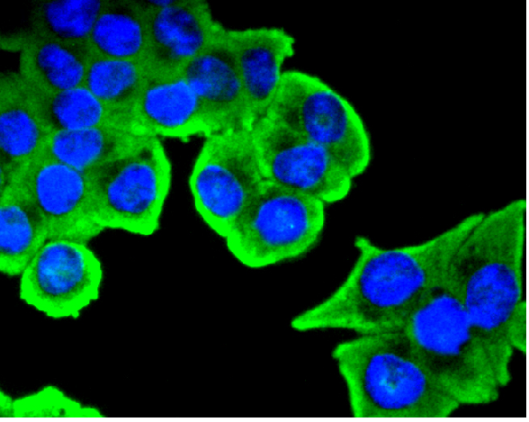 ICC (Immunocytochemistry)
(ICC staining SIRT1 (phospho S47) in SW480 cells (green). The nuclear counter stain is DAPI (blue). Cells were fixed in paraformaldehyde, permeabilised with 0.25% Triton X100/PBS.)
ICC (Immunocytochemistry)
(ICC staining SIRT1 (phospho S47) in SW480 cells (green). The nuclear counter stain is DAPI (blue). Cells were fixed in paraformaldehyde, permeabilised with 0.25% Triton X100/PBS.)
SIRT1, Monoclonal Antibody (Cat# AAA311022)
 WB (Western Blot)
(Western blot analysis of lysates from HepG2 cells, COLO205 cells and HUVEC cells, using PKC delta (Phospho-Tyr313) Antibody. The lane on the right is blocked with the phospho peptide.)
WB (Western Blot)
(Western blot analysis of lysates from HepG2 cells, COLO205 cells and HUVEC cells, using PKC delta (Phospho-Tyr313) Antibody. The lane on the right is blocked with the phospho peptide.)
PKC delta, Polyclonal Antibody (Cat# AAA316277)
 WB (Western Blot)
(Western blot analysis of lysates from Jurkat cells, using MAP3K1 (Phospho-Thr1402) Antibody.)
WB (Western Blot)
(Western blot analysis of lysates from Jurkat cells, using MAP3K1 (Phospho-Thr1402) Antibody.)
MAP3K1, Polyclonal Antibody (Cat# AAA318350)
 WB (Western Blot)
(Western blot analysis of Phospho-PKA C (Thr197) using COLO205 whole cell lysates)
WB (Western Blot)
(Western blot analysis of Phospho-PKA C (Thr197) using COLO205 whole cell lysates)
PKA C, Polyclonal Antibody (Cat# AAA327245)
 WB (Western Blot)
(All lanes use the Antibody at 1:3K dilution for 1 hour at room temperature.)
WB (Western Blot)
(All lanes use the Antibody at 1:3K dilution for 1 hour at room temperature.)
GLUT4, Monoclonal Antibody (Cat# AAA128119)
 WB (Western Blot)
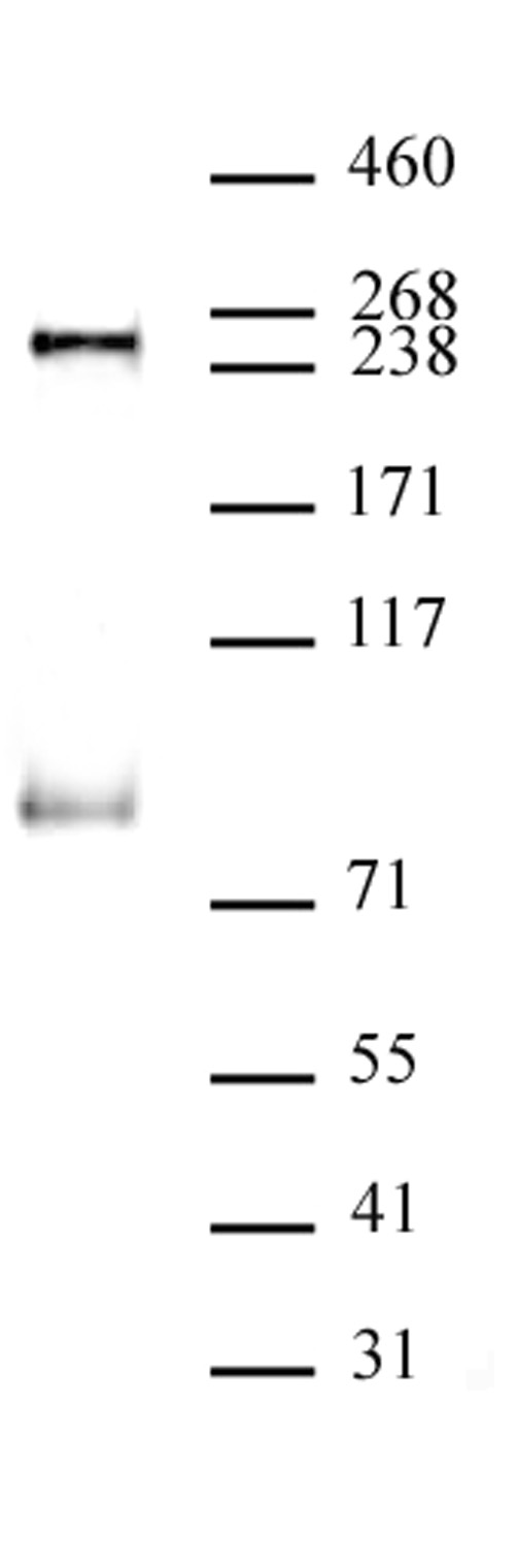
(RNA pol II phospho Ser5 antibody (mAb) (Clone 1H4B6) tested by Western blot. HeLa nuclear extract (40 ug per lane) probed with RNA pol II phospho Ser5 antibody (mAb) at a 1 ug/ml dilution.)
WB (Western Blot)
(RNA pol II phospho Ser5 antibody (mAb) (Clone 1H4B6) tested by Western blot. HeLa nuclear extract (40 ug per lane) probed with RNA pol II phospho Ser5 antibody (mAb) at a 1 ug/ml dilution.)
RNA pol II CTD phospho Ser5, Monoclonal Antibody (Cat# AAA60078)
What Are Phospho Antibodies?
Protein phosphorylation is a process where a phosphate group is added to certain amino acid residues of a protein – usually serine (S), threonine (T), or tyrosine (Y) - by enzymes called kinases. This process is integral in controlling cellular signaling, cellular growth, and other biological functions.
Our catalog includes a wide range of phospho-specific antibodies that can accurately detect this important marker. They perform strongly in widely-used laboratory applications such as Western blot, flow cytometry, immunohistochemistry, and immunofluorescence microscopy. We value your trust in us and are committed to providing top-quality products and services. All of our antibodies are guaranteed to work for the applications and species indicated on our website & associated product pages.
What Are The Key Applications of Phospho Antibodies?
1. Western Blotting
One of the first steps a researcher can take in utilizing these phospho-specific antibodies, is to check if the antibody works using a technique referred to as “Western blot”. For those unfamiliar, Western Blot aids in showing whether the protein that the antibody recognizes is appearing at the correct/expected size. These phospho-specific antibodies should also be able to detect changes in the target protein’s phosphorylation (on/off state) when cells are stimulated in certain ways.
2. Staining of Fixed Cells (Immunocytochemistry)
Another routine use of these phospho-specific antibodies, is to test if the antibody is able to demonstrate similar performance when used on fixed cells (intact cells that have been preserved) as it did in the Western blot tests. It is an important aspect in many cases to confirm that the antibody works in actual intact cell samples. Ideally, the method used for cellular fixation should be the same as what is used in pathology labs (like using 10% formalin). To check if the antibody works well in tissue sections (FFPE), researchers will often test it on fixed cells that are processed similar to tissue samples.
3. Specificity Tests Using Peptides
In order to make sure that the antibody is only binding to the right target:
- Laboratory technicians will mix the antibody with phospho-peptides (short segments of the protein containing the phosphate group modification).
- If the antibody signal disappears, it is confirmation that it is binding to the correct phosphorylated location.
- A more robust test is to use both the phosphorylated and non-phosphorylated (dephosphorylated) versions of the protein. The antibody should react only with the phosphorylated one.
- Another method sometimes utilized is to treat the sample with an enzyme, such as alkaline phosphatase, that specifically removes phosphate groups. If the antibody signal disappears after this, it also confirms specificity.
4. Genetic Confirmation
As a final step, scientists can genetically manipulate the nucleotide sequence and alter the target protein by removing the exact site where phosphorylation happens. If the antibody no longer appears to detect the modified protein, it is strong evidence supporting the antibody being specific for that phosphorylated site.
Why Buy Phospho Antibodies Through Us?
- The production laboratory adheres to strict and consistent protocols prior to releasing any of these phospho-specific antibodies:
- Standard methods and proper controls in all tests to ensure high quality.
- These antibodies are tested and validated in different cell types and species.
- High quality control criterion to ensure each batch is consistent, so you will obtain reliable results every time.
FAQ
1. What Are Phospho-Specific Antibodies?
Phospho-specific antibodies are made to detect proteins only when they have a phosphate group linked to a specific amino acid residue. This empowers scientists understand if a protein is "turned on" or active, based on its phosphorylation state.
2. How to Detect Phosphorylated Proteins in a Western Blot?
To find out if a protein is phosphorylated using Western blot:
- Use a phospho-specific antibody that binds only to the phosphorylated form of the protein.
- You can also use a “regular” antibody for the same amino acid sequence of the protein that the phospho-specific antibody is binding to (but in this case, this antibody will not bind if there is a phosphate group present) in order to compare how much of it is phosphorylated versus how much is non-phosphorylated (or “total” protein, if the “normal” antibody’s epitopes are non-phospho-site-specific).
3. How to Choose the Best Antibody?
Here are some simple tips to help you pick the right antibody:
- Know your target
- Match your sample characteristics
- Confirm the intended use is appropriate
- Check “host” and “type”
- Check the “quality” of the presented data/images
- Appraise whether the available validation meets your needs