Filters
▼Clonality
▼Type
▼Reactivity
▼Gene Name
▼Isotype
▼Host
▼Application
▼Clone
▼Phospho Antibodies
Phospho-specific antibodies’ typical purpose is to enable researchers to detect changes in proteins. They will exclusively bind to the amino acid sequence on a protein that has been phosphorylated (which is both a physical & chemical change) and do not bind to the same amino acid sequence on said protein if it lacks said phosphorylation. This aids in being able to clearly see and understand the data produced from this particular protein modification.
Viewing 4550-4600 of 5298 product results
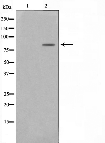
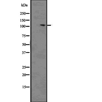
 WB (Western Blot)
(Western blot analysis of lysates from HCT116 cells, primary antibody was diluted at 1:1000, 4 degree over night)
WB (Western Blot)
(Western blot analysis of lysates from HCT116 cells, primary antibody was diluted at 1:1000, 4 degree over night)
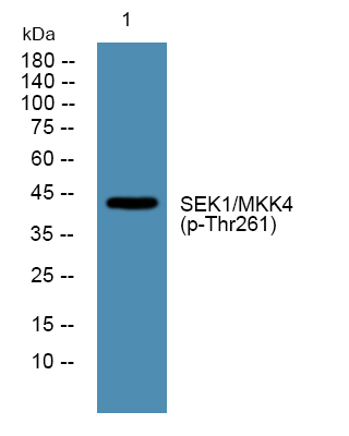
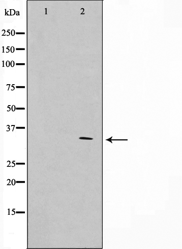
SEK1/MKK4, Polyclonal Antibody (Cat# AAA319467)
 Application Data
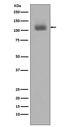
(Diluted at 1:10000)
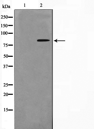
Application Data
(Diluted at 1:10000)
Kinesin-like Protein KIF1C, Polyclonal Antibody (Cat# AAA319470)

 IF (Immunofluorescence)
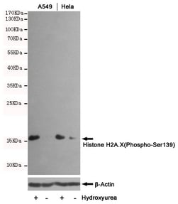
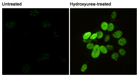
(Immunofluorescent analysis of Phosphorylation of H2A.X at Serine 139 in A549(upper.untreated or Hydroxyurea-treated) and Hela(lower.untreated or Hydroxyurea-treated) using Phospho-Histone H2A.X (Ser139) mouse mAb (1:400).)
IF (Immunofluorescence)
(Immunofluorescent analysis of Phosphorylation of H2A.X at Serine 139 in A549(upper.untreated or Hydroxyurea-treated) and Hela(lower.untreated or Hydroxyurea-treated) using Phospho-Histone H2A.X (Ser139) mouse mAb (1:400).)
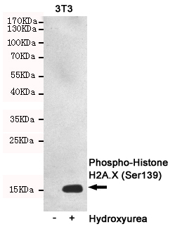
Histone H2A.X, Monoclonal Antibody (Cat# AAA320396)

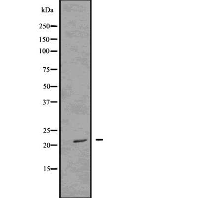
 WB (Western Blot)
(Western blot analysis on HuvEc cell lysate using Phospho-APC(Ser2054) Antibody. The lane on the left is treated with the antigen-specific peptide.)
WB (Western Blot)
(Western blot analysis on HuvEc cell lysate using Phospho-APC(Ser2054) Antibody. The lane on the left is treated with the antigen-specific peptide.)
APC, Polyclonal Antibody (Cat# AAA321246)

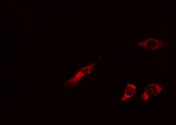
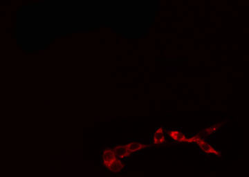
 IF (Immunofluorescence)
(AAA321250 staining HuvEc by IF/ICC. The sample were fixed with PFA and permeabilized in 0.1% Triton X-100, then blocked in 10% serum for 45 minutes at 25 degree C. The primary antibody was diluted at 1/200 and incubated with the sample for 1 hour at 37 degree C. An Alexa Fluor 594 conjugated goat anti-rabbit IgG (H+L) Ab, diluted at 1/600, was used as the secondary antibody.)
IF (Immunofluorescence)
(AAA321250 staining HuvEc by IF/ICC. The sample were fixed with PFA and permeabilized in 0.1% Triton X-100, then blocked in 10% serum for 45 minutes at 25 degree C. The primary antibody was diluted at 1/200 and incubated with the sample for 1 hour at 37 degree C. An Alexa Fluor 594 conjugated goat anti-rabbit IgG (H+L) Ab, diluted at 1/600, was used as the secondary antibody.)
Nibrin, Polyclonal Antibody (Cat# AAA321250)
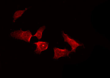
 IF (Immunofluorescence)
(AAA321257 staining Hela cells by IF/ICC. The sample were fixed with PFA and permeabilized in 0.1% Triton X-100, then blocked in 10% serum for 45 minutes at 25 degree C. The primary antibody was diluted at 1/200 and incubated with the sample for 1 hour at 37 degree C. An Alexa Fluor 594 conjugated goat anti-rabbit IgG (H+L) antibody, diluted at 1/600, was used as secondary antibody.)
IF (Immunofluorescence)
(AAA321257 staining Hela cells by IF/ICC. The sample were fixed with PFA and permeabilized in 0.1% Triton X-100, then blocked in 10% serum for 45 minutes at 25 degree C. The primary antibody was diluted at 1/200 and incubated with the sample for 1 hour at 37 degree C. An Alexa Fluor 594 conjugated goat anti-rabbit IgG (H+L) antibody, diluted at 1/600, was used as secondary antibody.)
BTK, Polyclonal Antibody (Cat# AAA321257)
 WB (Western Blot)
(Western blot analysis on HeLa cell lysate using Phospho-CDC25A(Ser75) Antibody. The lane on the left is treated with the antigen-specific peptide.)
WB (Western Blot)
(Western blot analysis on HeLa cell lysate using Phospho-CDC25A(Ser75) Antibody. The lane on the left is treated with the antigen-specific peptide.)
CDC25A, Polyclonal Antibody (Cat# AAA321275)
 IF (Immunofluorescence)
(AAA321284 staining HepG2 by IF/ICC. The sample were fixed with PFA and permeabilized in 0.1% Triton X-100, then blocked in 10% serum for 45 minutes at 25 degree C. The primary antibody was diluted at 1/200 and incubated with the sample for 1 hour at 37 degree C. An Alexa Fluor 594 conjugated goat anti-rabbit IgG (H+L) Ab, diluted at 1/600, was used as the secondary antibody.)
IF (Immunofluorescence)
(AAA321284 staining HepG2 by IF/ICC. The sample were fixed with PFA and permeabilized in 0.1% Triton X-100, then blocked in 10% serum for 45 minutes at 25 degree C. The primary antibody was diluted at 1/200 and incubated with the sample for 1 hour at 37 degree C. An Alexa Fluor 594 conjugated goat anti-rabbit IgG (H+L) Ab, diluted at 1/600, was used as the secondary antibody.)
AKT1/2/3, Polyclonal Antibody (Cat# AAA321284)
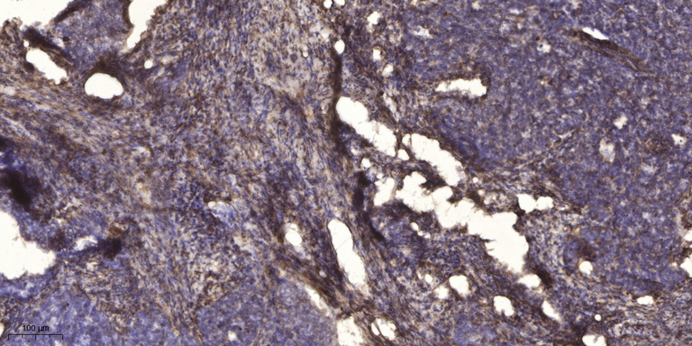
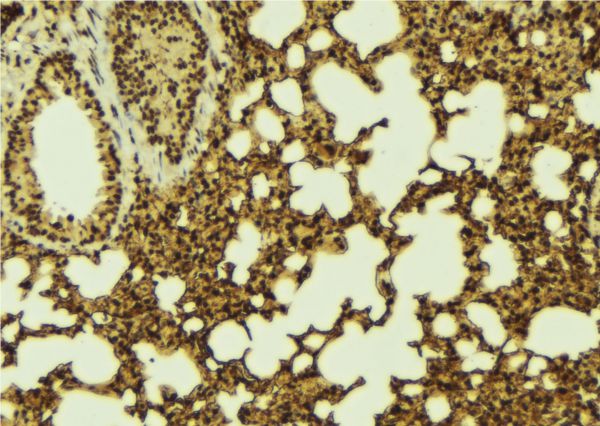
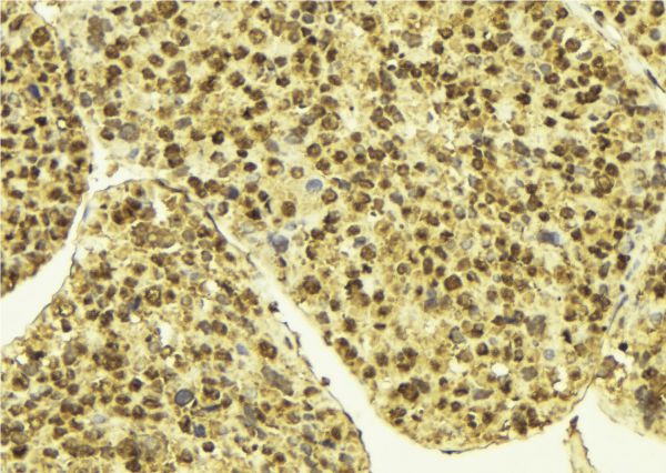
 IHC (Immunohiostchemistry)
(AAA321287 at 1/100 staining Mouse lung tissue by IHC-P. The sample was formaldehyde fixed and a heat mediated antigen retrieval step in citrate buffer was performed. The sample was then blocked and incubated with the antibody for 1.5 hours at 22 degree C. An HRP conjugated goat anti-rabbit antibody was used as the secondary.)
IHC (Immunohiostchemistry)
(AAA321287 at 1/100 staining Mouse lung tissue by IHC-P. The sample was formaldehyde fixed and a heat mediated antigen retrieval step in citrate buffer was performed. The sample was then blocked and incubated with the antibody for 1.5 hours at 22 degree C. An HRP conjugated goat anti-rabbit antibody was used as the secondary.)
FOS, Polyclonal Antibody (Cat# AAA321287)
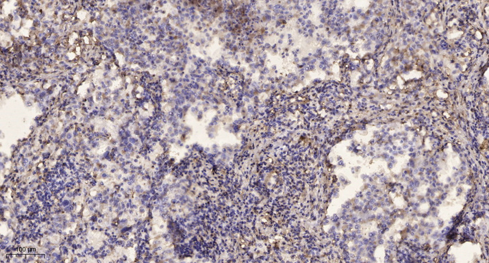
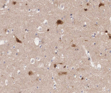
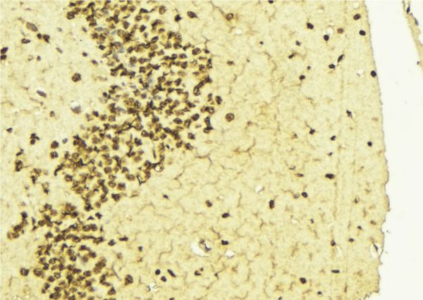
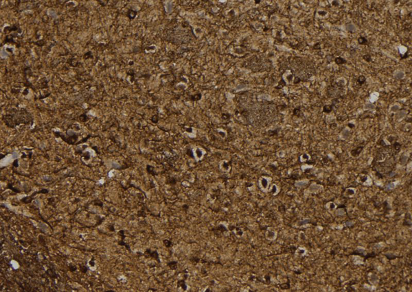 IHC (Immunohiostchemistry)
(AAA321294 at 1/100 staining Rat brain tissue by IHC-P. The sample was formaldehyde fixed and a heat mediated antigen retrieval step in citrate buffer was performed. The sample was then blocked and incubated with the antibody for 1.5 hours at 22 degree C. An HRP conjugated goat anti-rabbit antibody was used as the secondary.)
IHC (Immunohiostchemistry)
(AAA321294 at 1/100 staining Rat brain tissue by IHC-P. The sample was formaldehyde fixed and a heat mediated antigen retrieval step in citrate buffer was performed. The sample was then blocked and incubated with the antibody for 1.5 hours at 22 degree C. An HRP conjugated goat anti-rabbit antibody was used as the secondary.)
BTK, Polyclonal Antibody (Cat# AAA321294)
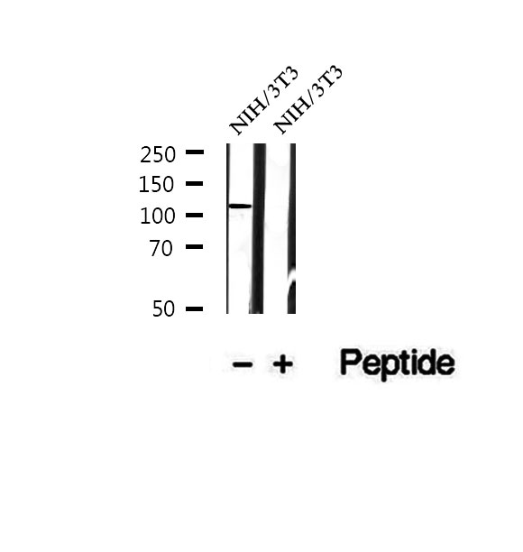 WB (Western Blot)
(Western blot analysis of extracts of NIH/3T3 cells, using Phospho-PKD2(Ser812) Antibody.)
WB (Western Blot)
(Western blot analysis of extracts of NIH/3T3 cells, using Phospho-PKD2(Ser812) Antibody.)
PKD2, Polyclonal Antibody (Cat# AAA321297)
 WB (Western Blot)
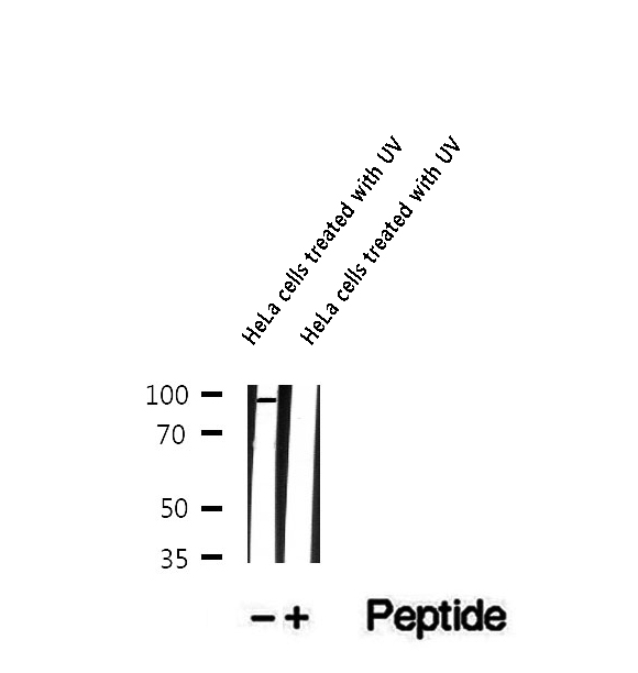
(Western blot analysis of extracts of HeLa cells treated with UV, using Phospho-BAP1 (Ser592) Antibody.)
WB (Western Blot)
(Western blot analysis of extracts of HeLa cells treated with UV, using Phospho-BAP1 (Ser592) Antibody.)
BAP1, Polyclonal Antibody (Cat# AAA321305)
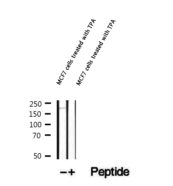 WB (Western Blot)
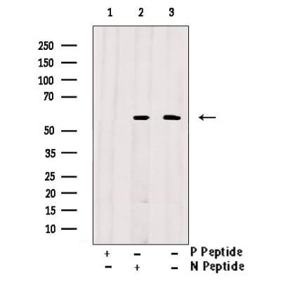
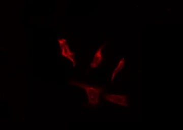
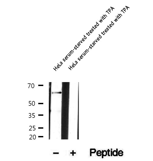
(Western blot analysis of extracts of MCF7 cells treated with TPA, using Phospho-BCL9L (Ser915) Antibody.)
WB (Western Blot)
(Western blot analysis of extracts of MCF7 cells treated with TPA, using Phospho-BCL9L (Ser915) Antibody.)
BCL9L, Polyclonal Antibody (Cat# AAA321306)
 WB (Western Blot)
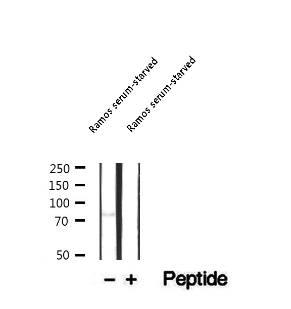
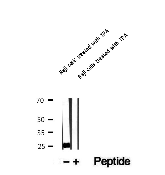
(Western blot analysis of extracts of Raji cells treated with TPA, using Phospho-Bim (Ser69) Antibody.)
WB (Western Blot)
(Western blot analysis of extracts of Raji cells treated with TPA, using Phospho-Bim (Ser69) Antibody.)
Bim, Polyclonal Antibody (Cat# AAA321308)
 WB (Western Blot)
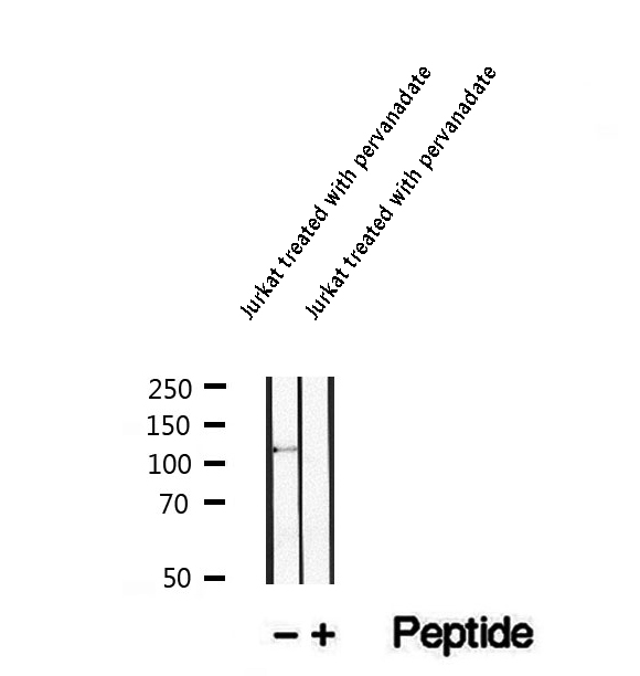
(Western blot analysis of extracts of Jurkat treated with pervanadate, using Phospho-c-Cbl (Tyr700) Antibody.)
WB (Western Blot)
(Western blot analysis of extracts of Jurkat treated with pervanadate, using Phospho-c-Cbl (Tyr700) Antibody.)
c-Cbl, Polyclonal Antibody (Cat# AAA321312)
 WB (Western Blot)
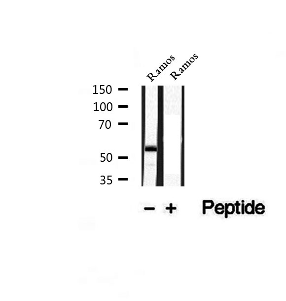
(Western blot analysis of extracts of Ramos cells, using Phospho-CD79A (Tyr182) Antibody.)
WB (Western Blot)
(Western blot analysis of extracts of Ramos cells, using Phospho-CD79A (Tyr182) Antibody.)
CD79A, Polyclonal Antibody (Cat# AAA321313)
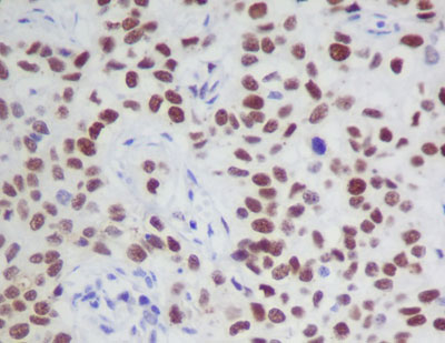
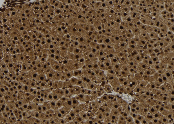 IHC (Immunohiostchemistry)
(AAA321314 at 1/100 staining Rat liver tissue by IHC-P. The sample was formaldehyde fixed and a heat mediated antigen retrieval step in citrate buffer was performed. The sample was then blocked and incubated with the antibody for 1.5 hours at 22 degree C. An HRP conjugated goat anti-rabbit antibody was used as the secondary.)
IHC (Immunohiostchemistry)
(AAA321314 at 1/100 staining Rat liver tissue by IHC-P. The sample was formaldehyde fixed and a heat mediated antigen retrieval step in citrate buffer was performed. The sample was then blocked and incubated with the antibody for 1.5 hours at 22 degree C. An HRP conjugated goat anti-rabbit antibody was used as the secondary.)
CENP-A, Polyclonal Antibody (Cat# AAA321314)
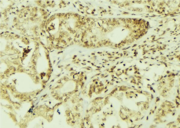
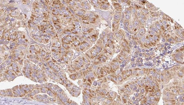
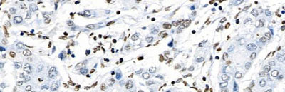
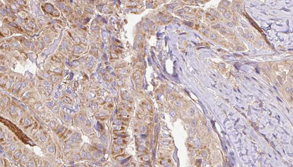
 IHC (Immunohiostchemistry)
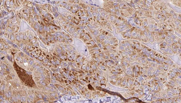
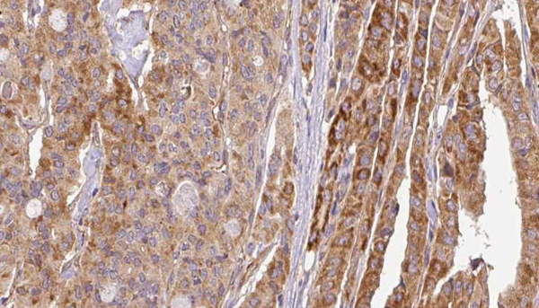
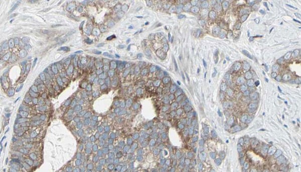
(AAA320723 at 1/100 staining Human breast cancer tissue by IHC-P. The sample was formaldehyde fixed and a heat mediated antigen retrieval step in citrate buffer was performed. The sample was then blocked and incubated with the antibody for 1.5 hours at 22 degree C. An HRP conjugated goat anti-rabbit antibody was used as the secondary.)
IHC (Immunohiostchemistry)
(AAA320723 at 1/100 staining Human breast cancer tissue by IHC-P. The sample was formaldehyde fixed and a heat mediated antigen retrieval step in citrate buffer was performed. The sample was then blocked and incubated with the antibody for 1.5 hours at 22 degree C. An HRP conjugated goat anti-rabbit antibody was used as the secondary.)
VEGFR2, Polyclonal Antibody (Cat# AAA320723)
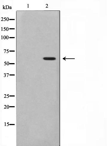
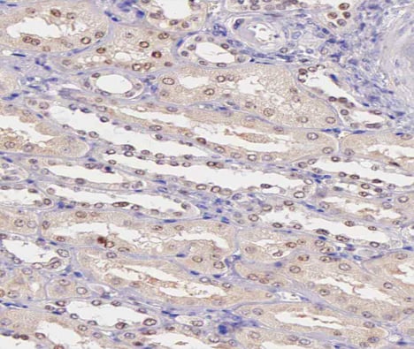
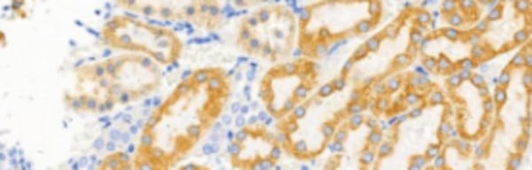 IHC (Immunohistochemistry)
(AAA320729 at 1/100 staining Rat kidney tissue by IHC-P. The sample was formaldehyde fixed and a heat mediated antigen retrieval step in citrate buffer was performed. The sample was then blocked and incubated with the primary Ab at 4 degree C overnight. An HRP conjugated anti-Rabbit Ab was used as the secondary Ab.)
IHC (Immunohistochemistry)
(AAA320729 at 1/100 staining Rat kidney tissue by IHC-P. The sample was formaldehyde fixed and a heat mediated antigen retrieval step in citrate buffer was performed. The sample was then blocked and incubated with the primary Ab at 4 degree C overnight. An HRP conjugated anti-Rabbit Ab was used as the secondary Ab.)
EPHA2/3/4, Polyclonal Antibody (Cat# AAA320729)
 IF (Immunofluorescence)
(AAA320735 staining MCF-7 cells by IF/ICC. The sample were fixed with PFA and permeabilized in 0.1% Triton X-100, then blocked in 10% serum for 45 minutes at 25 degree C. The primary antibody was diluted at 1/200 and incubated with the sample for 1 hour at 37 degree C. An Alexa Fluor 594 conjugated goat anti-rabbit IgG (H+L) antibody, diluted at 1/600, was used as secondary antibody.)
IF (Immunofluorescence)
(AAA320735 staining MCF-7 cells by IF/ICC. The sample were fixed with PFA and permeabilized in 0.1% Triton X-100, then blocked in 10% serum for 45 minutes at 25 degree C. The primary antibody was diluted at 1/200 and incubated with the sample for 1 hour at 37 degree C. An Alexa Fluor 594 conjugated goat anti-rabbit IgG (H+L) antibody, diluted at 1/600, was used as secondary antibody.)
CD227/MUC1, Antibody (Cat# AAA320735)
 IHC (Immunohiostchemistry)
(AAA320738 at 1/200 staining human Cerebellum tissue sections by IHC-P. The tissue was formaldehyde fixed and a heat mediated antigen retrieval step in citrate buffer was performed. The tissue was then blocked and incubated with the antibody for 1.5 hours at 22 degree C. An HRP conjugated goat anti-rabbit antibody was used as the secondary.)
IHC (Immunohiostchemistry)
(AAA320738 at 1/200 staining human Cerebellum tissue sections by IHC-P. The tissue was formaldehyde fixed and a heat mediated antigen retrieval step in citrate buffer was performed. The tissue was then blocked and incubated with the antibody for 1.5 hours at 22 degree C. An HRP conjugated goat anti-rabbit antibody was used as the secondary.)
PAK3, Polyclonal Antibody (Cat# AAA320738)
 IHC (Immunohistochemisry)
(AAA320744 at 1/200 staining human kidney tissue sections by IHC-P. The tissue was formaldehyde fixed and a heat mediated antigen retrieval step in citrate buffer was performed. The tissue was then blocked and incubated with the antibody for 1.5 hours at 22 degree C. An HRP conjugated goat anti-rabbit antibody was used as the secondary.)
IHC (Immunohistochemisry)
(AAA320744 at 1/200 staining human kidney tissue sections by IHC-P. The tissue was formaldehyde fixed and a heat mediated antigen retrieval step in citrate buffer was performed. The tissue was then blocked and incubated with the antibody for 1.5 hours at 22 degree C. An HRP conjugated goat anti-rabbit antibody was used as the secondary.)
MAPKAPK5, Polyclonal Antibody (Cat# AAA320744)
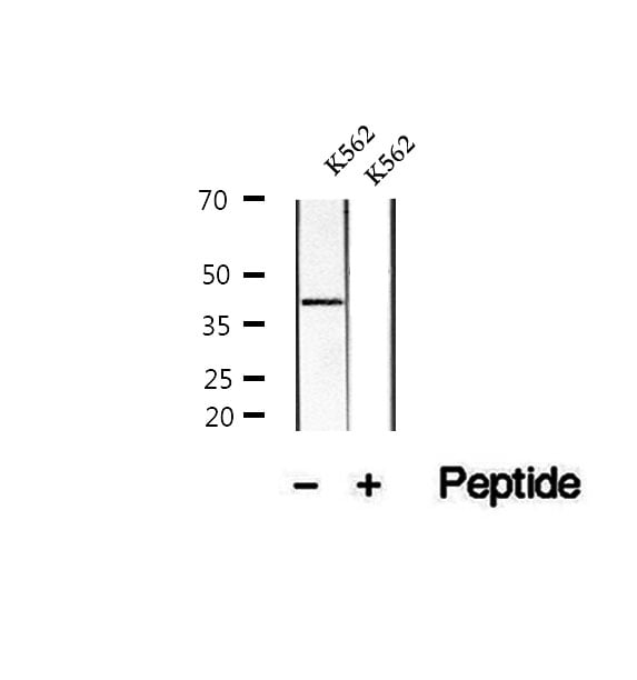 WB (Western Blot)
(Western blot analysis of extracts of K562 cells, using Phospho-CrkL (Tyr207) Antibody.)
WB (Western Blot)
(Western blot analysis of extracts of K562 cells, using Phospho-CrkL (Tyr207) Antibody.)
CrkL, Polyclonal Antibody (Cat# AAA321316)
 IHC (Immunohiostchemistry)
(AAA321318 at 1/100 staining Human gastric tissue by IHC-P. The sample was formaldehyde fixed and a heat mediated antigen retrieval step in citrate buffer was performed. The sample was then blocked and incubated with the antibody for 1.5 hours at 22 degree C. An HRP conjugated goat anti-rabbit antibody was used as the secondary.)
IHC (Immunohiostchemistry)
(AAA321318 at 1/100 staining Human gastric tissue by IHC-P. The sample was formaldehyde fixed and a heat mediated antigen retrieval step in citrate buffer was performed. The sample was then blocked and incubated with the antibody for 1.5 hours at 22 degree C. An HRP conjugated goat anti-rabbit antibody was used as the secondary.)
Cyclin E1, Polyclonal Antibody (Cat# AAA321318)
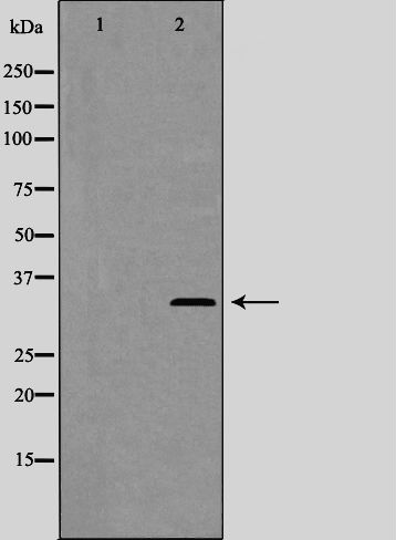
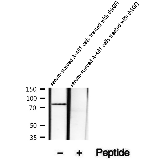 WB (Western Blot)
(Western blot analysis of extracts of serum-starved A-431 cells treated with (hEGF), using Phospho-DR6 (Ser562) Antibody.)
WB (Western Blot)
(Western blot analysis of extracts of serum-starved A-431 cells treated with (hEGF), using Phospho-DR6 (Ser562) Antibody.)
DR6, Polyclonal Antibody (Cat# AAA321323)
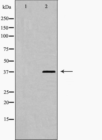
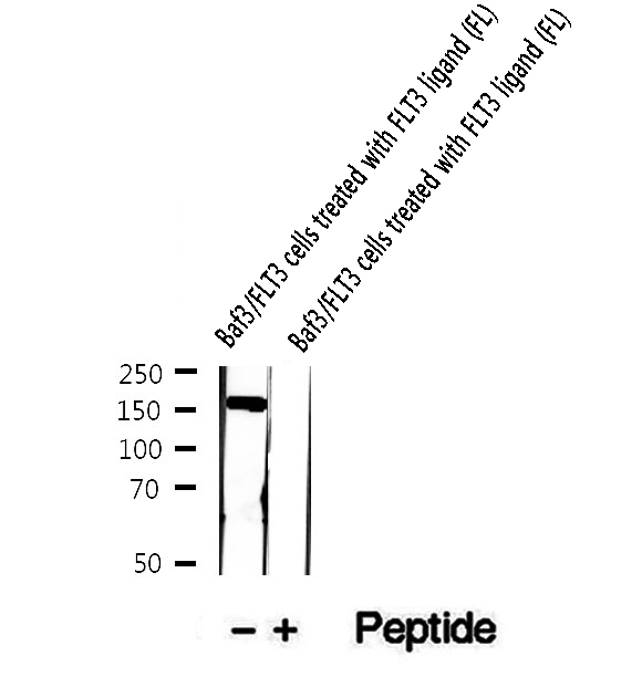 WB (Western Blot)
(Western blot analysis of extracts of Baf3/FLT3 cells treated with FLT3 ligand (FL), using Phospho-FLT3 (Tyr969) Antibody.)
WB (Western Blot)
(Western blot analysis of extracts of Baf3/FLT3 cells treated with FLT3 ligand (FL), using Phospho-FLT3 (Tyr969) Antibody.)
FLT3, Polyclonal Antibody (Cat# AAA321326)
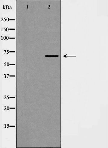
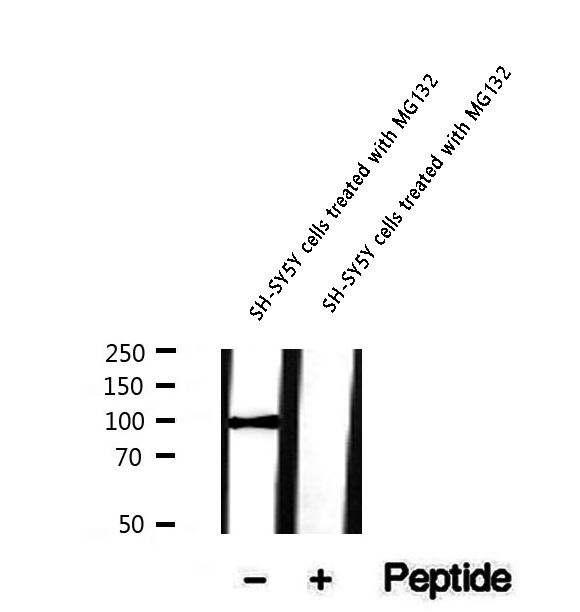 WB (Western Blot)
(Western blot analysis of extracts of SH-SY5Y cells treated with MG132, using Phospho-FoxO3a (Ser413) Antibody.)
WB (Western Blot)
(Western blot analysis of extracts of SH-SY5Y cells treated with MG132, using Phospho-FoxO3a (Ser413) Antibody.)
FoxO3a, Polyclonal Antibody (Cat# AAA321327)
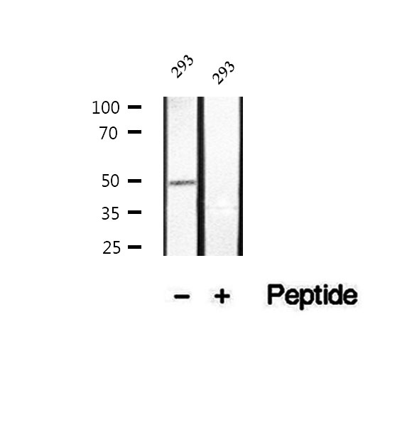
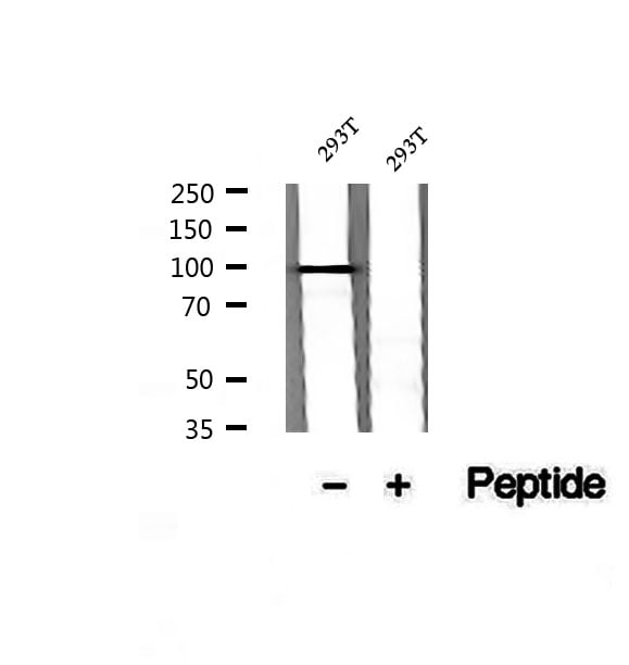 WB (Western Blot)
(Western blot analysis of extracts of 293T cells, using Phospho-FoxO3a (Ser7) Antibody.)
WB (Western Blot)
(Western blot analysis of extracts of 293T cells, using Phospho-FoxO3a (Ser7) Antibody.)
FoxO3a, Polyclonal Antibody (Cat# AAA321328)
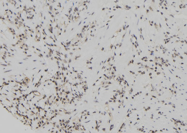
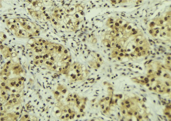
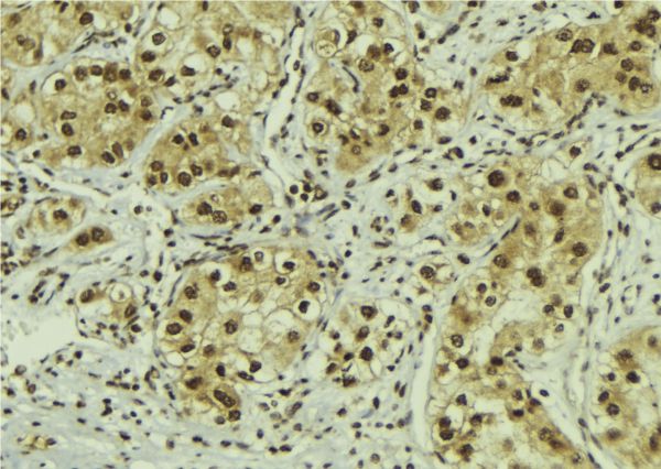
 IHC (Immunohistochemisry)
(AAA322067 at 1/100 staining Human prostate tissue by IHC-P. The sample was formaldehyde fixed and a heat mediated antigen retrieval step in citrate buffer was performed. The sample was then blocked and incubated with the antibody for 1.5 hours at 22 degree C. An HRP conjugated goat anti-rabbit antibody was used as the secondary.)
IHC (Immunohistochemisry)
(AAA322067 at 1/100 staining Human prostate tissue by IHC-P. The sample was formaldehyde fixed and a heat mediated antigen retrieval step in citrate buffer was performed. The sample was then blocked and incubated with the antibody for 1.5 hours at 22 degree C. An HRP conjugated goat anti-rabbit antibody was used as the secondary.)
eIF4B, Polyclonal Antibody (Cat# AAA322067)
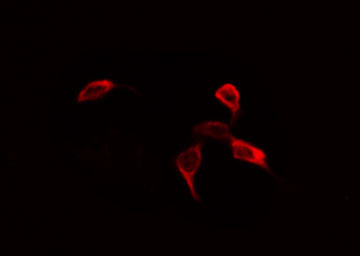
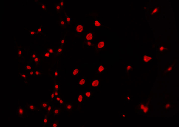
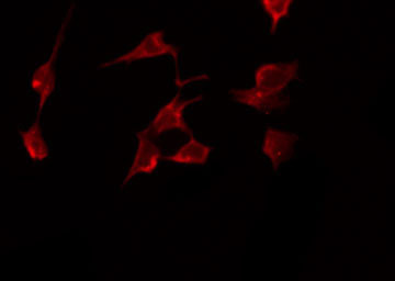
 IF (Immunofluorescence)
(AAA322069 staining 293 by IF/ICC. The sample were fixed with PFA and permeabilized in 0.1% Triton X-100, then blocked in 10% serum for 45 minutes at 25 degree C. The primary antibody was diluted at 1/200 and incubated with the sample for 1 hour at 37 degree C. An Alexa Fluor 594 conjugated goat anti-rabbit IgG (H+L) Ab, diluted at 1/600, was used as the secondary antibody.)
IF (Immunofluorescence)
(AAA322069 staining 293 by IF/ICC. The sample were fixed with PFA and permeabilized in 0.1% Triton X-100, then blocked in 10% serum for 45 minutes at 25 degree C. The primary antibody was diluted at 1/200 and incubated with the sample for 1 hour at 37 degree C. An Alexa Fluor 594 conjugated goat anti-rabbit IgG (H+L) Ab, diluted at 1/600, was used as the secondary antibody.)
IKK-gamma, Polyclonal Antibody (Cat# AAA322069)
 IF (Immunofluorescence)
(AAA322072 staining NIH-3T3 by IF/ICC. The sample were fixed with PFA and permeabilized in 0.1% Triton X-100, then blocked in 10% serum for 45 minutes at 25 degree C. The primary antibody was diluted at 1/200 and incubated with the sample for 1 hour at 37 degree C. An Alexa Fluor 594 conjugated goat anti-rabbit IgG (H+L) Ab, diluted at 1/600, was used as the secondary antibody.)
IF (Immunofluorescence)
(AAA322072 staining NIH-3T3 by IF/ICC. The sample were fixed with PFA and permeabilized in 0.1% Triton X-100, then blocked in 10% serum for 45 minutes at 25 degree C. The primary antibody was diluted at 1/200 and incubated with the sample for 1 hour at 37 degree C. An Alexa Fluor 594 conjugated goat anti-rabbit IgG (H+L) Ab, diluted at 1/600, was used as the secondary antibody.)
Cyclin C, Polyclonal Antibody (Cat# AAA322072)

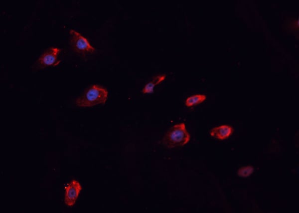
 IF (Immunofluorescence)
(AAA322075 staining HuvEc by IF/ICC. The sample were fixed with PFA and permeabilized in 0.1% Triton X-100, then blocked in 10% serum for 45 minutes at 25 degree C. The primary antibody was diluted at 1/200 and incubated with the sample for 1 hour at 37 degree C. An Alexa Fluor 594 conjugated goat anti-rabbit IgG (H+L) Ab, diluted at 1/600, was used as the secondary antibody.)
IF (Immunofluorescence)
(AAA322075 staining HuvEc by IF/ICC. The sample were fixed with PFA and permeabilized in 0.1% Triton X-100, then blocked in 10% serum for 45 minutes at 25 degree C. The primary antibody was diluted at 1/200 and incubated with the sample for 1 hour at 37 degree C. An Alexa Fluor 594 conjugated goat anti-rabbit IgG (H+L) Ab, diluted at 1/600, was used as the secondary antibody.)
hnRPD, Polyclonal Antibody (Cat# AAA322075)
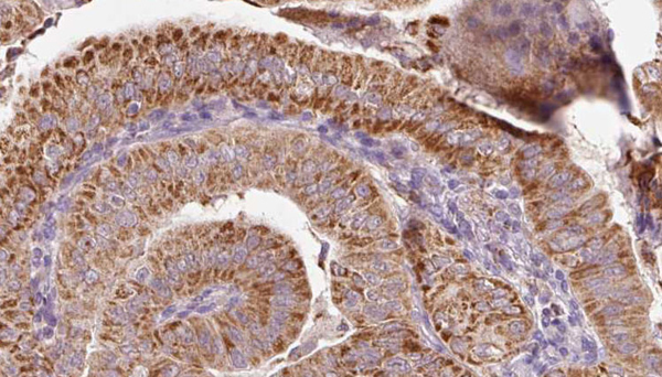
 IHC (Immunohistochemisry)
(AAA322076 at 1/100 staining Human uterus tissue by IHC-P. The sample was formaldehyde fixed and a heat mediated antigen retrieval step in citrate buffer was performed. The sample was then blocked and incubated with the antibody for 1.5 hours at 22 degree C. An HRP conjugated goat anti-rabbit antibody was used as the secondary.)
IHC (Immunohistochemisry)
(AAA322076 at 1/100 staining Human uterus tissue by IHC-P. The sample was formaldehyde fixed and a heat mediated antigen retrieval step in citrate buffer was performed. The sample was then blocked and incubated with the antibody for 1.5 hours at 22 degree C. An HRP conjugated goat anti-rabbit antibody was used as the secondary.)
ANXA2, Polyclonal Antibody (Cat# AAA322076)
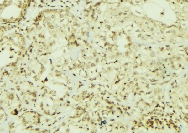
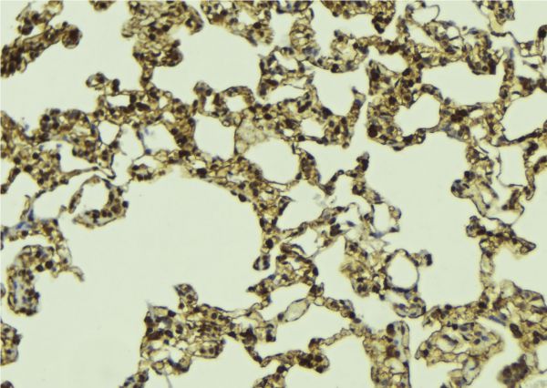 IHC (Immunohistochemisry)
(AAA322077 at 1/100 staining Mouse lung tissue by IHC-P. The sample was formaldehyde fixed and a heat mediated antigen retrieval step in citrate buffer was performed. The sample was then blocked and incubated with the antibody for 1.5 hours at 22 degree C. An HRP conjugated goat anti-rabbit antibody was used as the secondary.)
IHC (Immunohistochemisry)
(AAA322077 at 1/100 staining Mouse lung tissue by IHC-P. The sample was formaldehyde fixed and a heat mediated antigen retrieval step in citrate buffer was performed. The sample was then blocked and incubated with the antibody for 1.5 hours at 22 degree C. An HRP conjugated goat anti-rabbit antibody was used as the secondary.)
Daxx, Polyclonal Antibody (Cat# AAA322077)
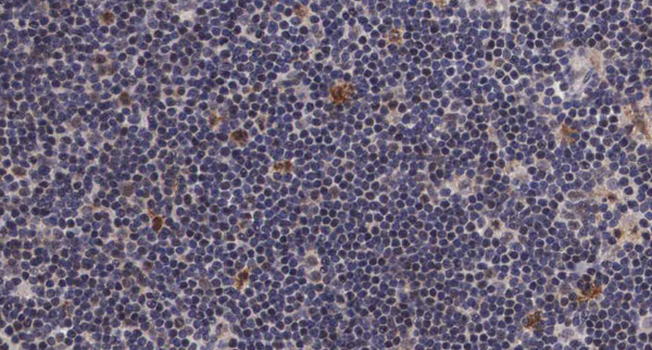
 IHC (Immunohistochemisry)
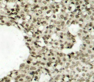
(Immunohistochemical analysis of paraffin-embedded human cervix cancer, using Phospho-JAK2 (Y1007 + Y1008) Antibody.)
IHC (Immunohistochemisry)
(Immunohistochemical analysis of paraffin-embedded human cervix cancer, using Phospho-JAK2 (Y1007 + Y1008) Antibody.)
JAK2, Monoclonal Antibody (Cat# AAA125166)
 IF (Immunofluorescence)
(AAA323700 staining SK-OV3 by IF/ICC. The sample were fixed with PFA and permeabilized in 0.1% Triton X-100, then blocked in 10% serum for 45 minutes at 25 degree C. The primary antibody was diluted at 1/200 and incubated with the sample for 1 hour at 37 degree C. An Alexa Fluor 594 conjugated goat anti-rabbit IgG (H+L) Ab, diluted at 1/600, was used as the secondary antibody.)
IF (Immunofluorescence)
(AAA323700 staining SK-OV3 by IF/ICC. The sample were fixed with PFA and permeabilized in 0.1% Triton X-100, then blocked in 10% serum for 45 minutes at 25 degree C. The primary antibody was diluted at 1/200 and incubated with the sample for 1 hour at 37 degree C. An Alexa Fluor 594 conjugated goat anti-rabbit IgG (H+L) Ab, diluted at 1/600, was used as the secondary antibody.)
EGFR, Polyclonal Antibody (Cat# AAA323700)
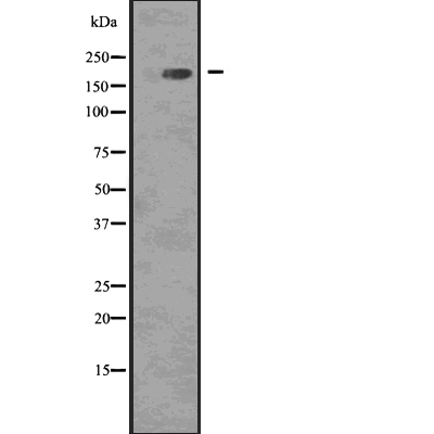
 WB (Western Blot)
(Western blot analysis of Phospho-53BP1 (Ser25/29) using 293 whole cell lysates)
WB (Western Blot)
(Western blot analysis of Phospho-53BP1 (Ser25/29) using 293 whole cell lysates)
53BP1, Polyclonal Antibody (Cat# AAA323704)
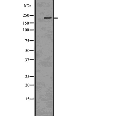 WB (Western Blot)
(Western blot analysis of Phospho-53BP1 (Thr543) using COLO205 whole cell lysates)
WB (Western Blot)
(Western blot analysis of Phospho-53BP1 (Thr543) using COLO205 whole cell lysates)
53BP1, Polyclonal Antibody (Cat# AAA323705)
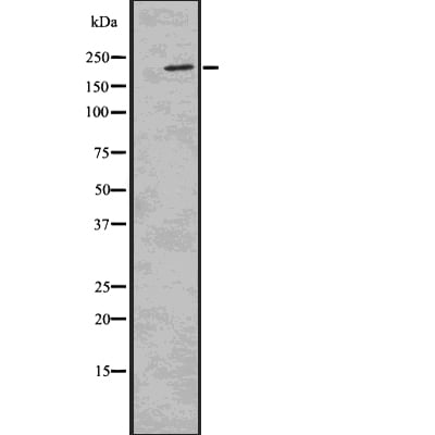
 WB (Western Blot)
(Western blot analysis of Phospho-53BP1 (Ser1778) using HUVEC whole cell lysates)
WB (Western Blot)
(Western blot analysis of Phospho-53BP1 (Ser1778) using HUVEC whole cell lysates)
53BP1, Polyclonal Antibody (Cat# AAA323706)

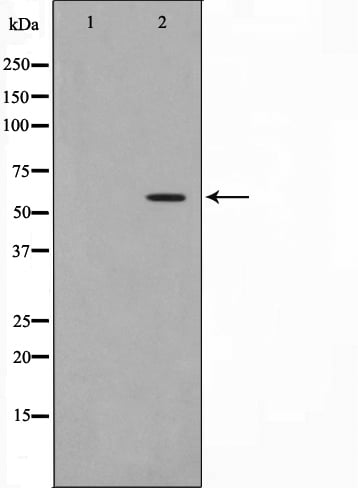
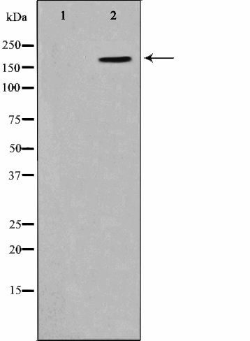
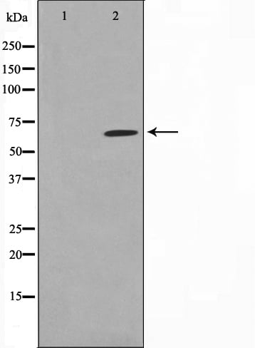
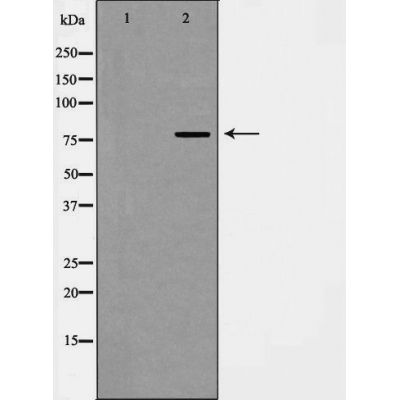
 WB (Western Blot)
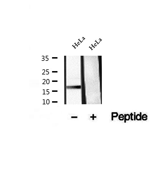
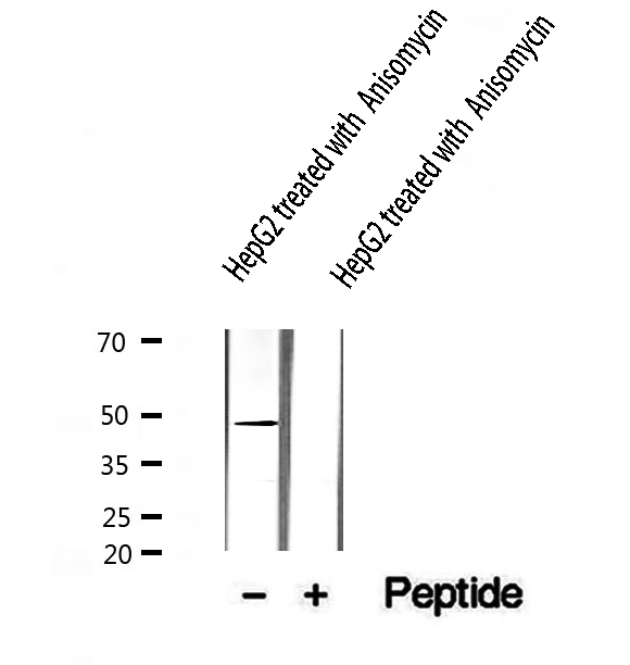
(Western blot analysis of extracts from 293 whole cell lysates ,using Phospho-DRP1 (Ser637) Antibody. Lane1 was treatedwith phospho-blocking peptide, Lane2 was treated with non-phospho-blocking peptide)
WB (Western Blot)
(Western blot analysis of extracts from 293 whole cell lysates ,using Phospho-DRP1 (Ser637) Antibody. Lane1 was treatedwith phospho-blocking peptide, Lane2 was treated with non-phospho-blocking peptide)
DRP1, Polyclonal Antibody (Cat# AAA323707)
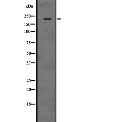
 WB (Western Blot)
(Western blot analysis of Phospho-FADD (Ser194) using COLO205 whole cell lysates)
WB (Western Blot)
(Western blot analysis of Phospho-FADD (Ser194) using COLO205 whole cell lysates)
FADD, Polyclonal Antibody (Cat# AAA323712)
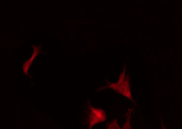
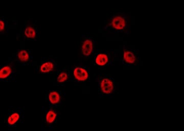
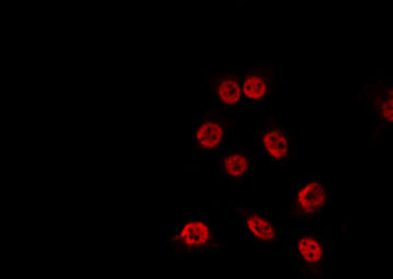
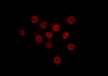
 IF (Immunofluorescence)
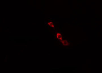
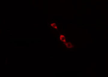
(AAA323714 staining HepG2 by IF/ICC. The sample were fixed with PFA and permeabilized in 0.1% Triton X-100, then blocked in 10% serum for 45 minutes at 25 degree C. The primary antibody was diluted at 1/200 and incubated with the sample for 1 hour at 37 degree C. An Alexa Fluor 594 conjugated goat anti-rabbit IgG (H+L) Ab, diluted at 1/600, was used as the secondary antibody.)
IF (Immunofluorescence)
(AAA323714 staining HepG2 by IF/ICC. The sample were fixed with PFA and permeabilized in 0.1% Triton X-100, then blocked in 10% serum for 45 minutes at 25 degree C. The primary antibody was diluted at 1/200 and incubated with the sample for 1 hour at 37 degree C. An Alexa Fluor 594 conjugated goat anti-rabbit IgG (H+L) Ab, diluted at 1/600, was used as the secondary antibody.)
Ack1, Polyclonal Antibody (Cat# AAA323714)
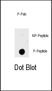 DB (Dot Blot)
(Dot blot analysis of anti-PAK1-pT423 Phospho-specific Pab on nitrocellulose membrane. 50ng of Phospho-peptide or Non Phospho-peptide per dot were adsorbed. Antibody working concentrations are 0.5ug per ml.)
DB (Dot Blot)
(Dot blot analysis of anti-PAK1-pT423 Phospho-specific Pab on nitrocellulose membrane. 50ng of Phospho-peptide or Non Phospho-peptide per dot were adsorbed. Antibody working concentrations are 0.5ug per ml.)
Phospho-PAK1 (T423), Polyclonal Antibody (Cat# AAA288458)
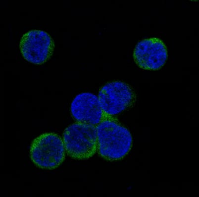
 IF (Immunofluorescence)
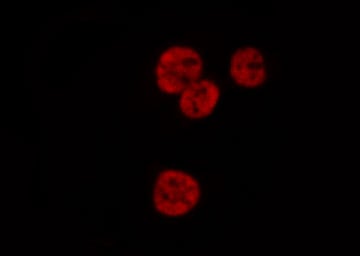
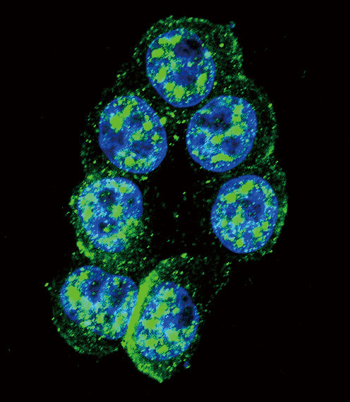
(Immunofluorescence analysis of U2OS cells using Phospho-MAX-S11 antibody. Blue: DAPI for nuclear staining.)
IF (Immunofluorescence)
(Immunofluorescence analysis of U2OS cells using Phospho-MAX-S11 antibody. Blue: DAPI for nuclear staining.)
MAX-S11, Antibody (Cat# AAA36631)
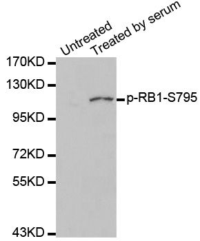
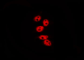
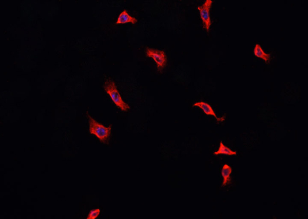
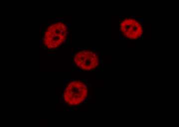
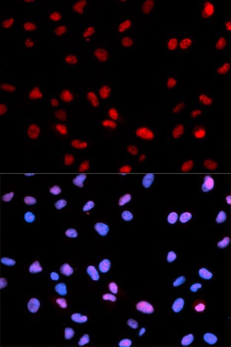 IF (Immunofluorescence)
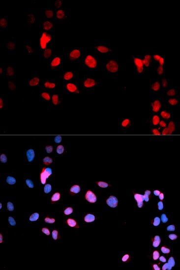
(Immunofluorescence analysis of U2OS cells using Phospho-RB-S795 antibody. Blue: DAPI for nuclear staining.)
IF (Immunofluorescence)
(Immunofluorescence analysis of U2OS cells using Phospho-RB-S795 antibody. Blue: DAPI for nuclear staining.)
RB-S795, Antibody (Cat# AAA36667)
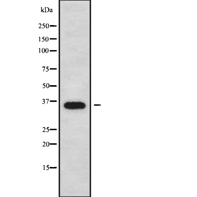
 WB (Western Blot)
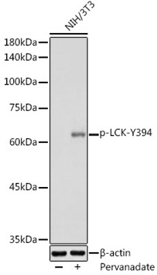
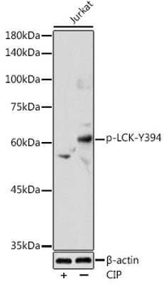
(Western blot analysis of extracts of Jurkat cells, using Phospho-LCK-Y394 antibody (AAA37349) at 1:1000 dilution. Jurkat cells were treated by CIP(20uL/400ul) at 37°C for 1 hour. Secondary antibody: HRP Goat Anti-Rabbit IgG (H+L) at 1:10000 dilution. Lysates/proteins: 25ug per lane. Blocking buffer: 3% nonfat dry milk in TBST. Detection: ECL Enhanced Kit (RM00021). Exposure time: 180s.)
WB (Western Blot)
(Western blot analysis of extracts of Jurkat cells, using Phospho-LCK-Y394 antibody (AAA37349) at 1:1000 dilution. Jurkat cells were treated by CIP(20uL/400ul) at 37°C for 1 hour. Secondary antibody: HRP Goat Anti-Rabbit IgG (H+L) at 1:10000 dilution. Lysates/proteins: 25ug per lane. Blocking buffer: 3% nonfat dry milk in TBST. Detection: ECL Enhanced Kit (RM00021). Exposure time: 180s.)
LCK-Y394, Antibody (Cat# AAA37349)
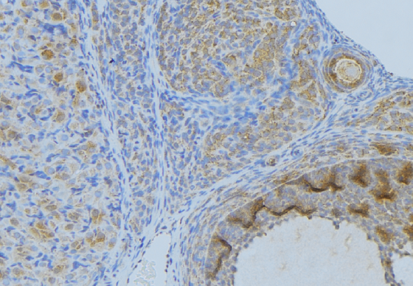
 IF (Immunofluorescence)
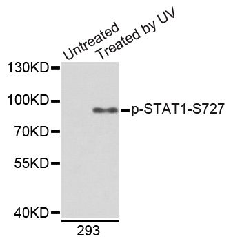
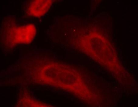
(Immunofluorescence staining of methanol-fixed HeLa cells using Phospho-STAT1-S727 antibody.)
IF (Immunofluorescence)
(Immunofluorescence staining of methanol-fixed HeLa cells using Phospho-STAT1-S727 antibody.)
STAT1-S727, Antibody (Cat# AAA37363)
 WB (Western Blot)
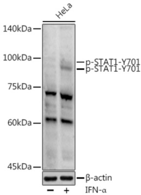
(Western blot analysis of extracts of HeLa cells, using Phospho-STAT1-Y701 antibody (AAA37377) at 1:1000 dilution.HeLa cells were treated by IFN-α (100 ng/ml) at 37°C for 30 minutes after serum-starvation overnight. Secondary antibody: HRP Goat Anti-Rabbit IgG (H+L) at 1:10000 dilution. Lysates/proteins: 25ug per lane. Blocking buffer: 3% nonfat dry milk in TBST. Detection: ECL Enhanced Kit. Exposure time: 180s.)
WB (Western Blot)
(Western blot analysis of extracts of HeLa cells, using Phospho-STAT1-Y701 antibody (AAA37377) at 1:1000 dilution.HeLa cells were treated by IFN-α (100 ng/ml) at 37°C for 30 minutes after serum-starvation overnight. Secondary antibody: HRP Goat Anti-Rabbit IgG (H+L) at 1:10000 dilution. Lysates/proteins: 25ug per lane. Blocking buffer: 3% nonfat dry milk in TBST. Detection: ECL Enhanced Kit. Exposure time: 180s.)
STAT1-Y701, Antibody (Cat# AAA37377)
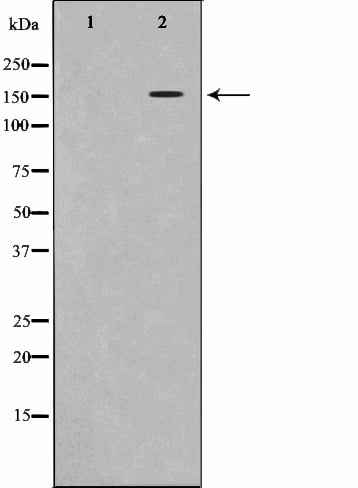
 WB (Western Blot)
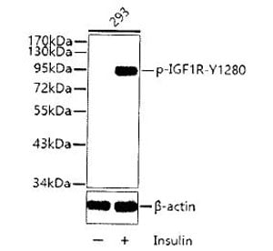
(Western blot analysis of extract from 293 cells, using Phospho-IGF1R-Y1280 antibody.Secondary antibody: HRP Goat Anti-Rabbit IgG (H+L) at 1:10000 dilution.Lysates/proteins: 25ug per lane.Blocking buffer: 3% BSA.)
WB (Western Blot)
(Western blot analysis of extract from 293 cells, using Phospho-IGF1R-Y1280 antibody.Secondary antibody: HRP Goat Anti-Rabbit IgG (H+L) at 1:10000 dilution.Lysates/proteins: 25ug per lane.Blocking buffer: 3% BSA.)
IGF1R-Y1280, Antibody (Cat# AAA37387)
 WB (Western Blot)
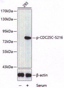
(Western blot analysis of extracts of HT-29 cells, using Phospho-cdc25CS216 antibody.Secondary antibody: HRP Goat Anti-Rabbit IgG (H+L) at 1:10000 dilution.Lysates/proteins: 25ug per lane.Blocking buffer: 3% BSA.)
WB (Western Blot)
(Western blot analysis of extracts of HT-29 cells, using Phospho-cdc25CS216 antibody.Secondary antibody: HRP Goat Anti-Rabbit IgG (H+L) at 1:10000 dilution.Lysates/proteins: 25ug per lane.Blocking buffer: 3% BSA.)
CDC25C-S216, Antibody (Cat# AAA37389)
What Are Phospho Antibodies?
Protein phosphorylation is a process where a phosphate group is added to certain amino acid residues of a protein – usually serine (S), threonine (T), or tyrosine (Y) - by enzymes called kinases. This process is integral in controlling cellular signaling, cellular growth, and other biological functions.
Our catalog includes a wide range of phospho-specific antibodies that can accurately detect this important marker. They perform strongly in widely-used laboratory applications such as Western blot, flow cytometry, immunohistochemistry, and immunofluorescence microscopy. We value your trust in us and are committed to providing top-quality products and services. All of our antibodies are guaranteed to work for the applications and species indicated on our website & associated product pages.
What Are The Key Applications of Phospho Antibodies?
1. Western Blotting
One of the first steps a researcher can take in utilizing these phospho-specific antibodies, is to check if the antibody works using a technique referred to as “Western blot”. For those unfamiliar, Western Blot aids in showing whether the protein that the antibody recognizes is appearing at the correct/expected size. These phospho-specific antibodies should also be able to detect changes in the target protein’s phosphorylation (on/off state) when cells are stimulated in certain ways.
2. Staining of Fixed Cells (Immunocytochemistry)
Another routine use of these phospho-specific antibodies, is to test if the antibody is able to demonstrate similar performance when used on fixed cells (intact cells that have been preserved) as it did in the Western blot tests. It is an important aspect in many cases to confirm that the antibody works in actual intact cell samples. Ideally, the method used for cellular fixation should be the same as what is used in pathology labs (like using 10% formalin). To check if the antibody works well in tissue sections (FFPE), researchers will often test it on fixed cells that are processed similar to tissue samples.
3. Specificity Tests Using Peptides
In order to make sure that the antibody is only binding to the right target:
- Laboratory technicians will mix the antibody with phospho-peptides (short segments of the protein containing the phosphate group modification).
- If the antibody signal disappears, it is confirmation that it is binding to the correct phosphorylated location.
- A more robust test is to use both the phosphorylated and non-phosphorylated (dephosphorylated) versions of the protein. The antibody should react only with the phosphorylated one.
- Another method sometimes utilized is to treat the sample with an enzyme, such as alkaline phosphatase, that specifically removes phosphate groups. If the antibody signal disappears after this, it also confirms specificity.
4. Genetic Confirmation
As a final step, scientists can genetically manipulate the nucleotide sequence and alter the target protein by removing the exact site where phosphorylation happens. If the antibody no longer appears to detect the modified protein, it is strong evidence supporting the antibody being specific for that phosphorylated site.
Why Buy Phospho Antibodies Through Us?
- The production laboratory adheres to strict and consistent protocols prior to releasing any of these phospho-specific antibodies:
- Standard methods and proper controls in all tests to ensure high quality.
- These antibodies are tested and validated in different cell types and species.
- High quality control criterion to ensure each batch is consistent, so you will obtain reliable results every time.
FAQ
1. What Are Phospho-Specific Antibodies?
Phospho-specific antibodies are made to detect proteins only when they have a phosphate group linked to a specific amino acid residue. This empowers scientists understand if a protein is "turned on" or active, based on its phosphorylation state.
2. How to Detect Phosphorylated Proteins in a Western Blot?
To find out if a protein is phosphorylated using Western blot:
- Use a phospho-specific antibody that binds only to the phosphorylated form of the protein.
- You can also use a “regular” antibody for the same amino acid sequence of the protein that the phospho-specific antibody is binding to (but in this case, this antibody will not bind if there is a phosphate group present) in order to compare how much of it is phosphorylated versus how much is non-phosphorylated (or “total” protein, if the “normal” antibody’s epitopes are non-phospho-site-specific).
3. How to Choose the Best Antibody?
Here are some simple tips to help you pick the right antibody:
- Know your target
- Match your sample characteristics
- Confirm the intended use is appropriate
- Check “host” and “type”
- Check the “quality” of the presented data/images
- Appraise whether the available validation meets your needs