Phospho-specific antibodies’ typical purpose is to enable researchers to detect changes in proteins. They will exclusively bind to the amino acid sequence on a protein that has been phosphorylated (which is both a physical & chemical change) and do not bind to the same amino acid sequence on said protein if it lacks said phosphorylation. This aids in being able to clearly see and understand the data produced from this particular protein modification.
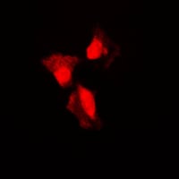
IF (Immunofluorescence) (Immunofluorescent analysis of MKK1 (pT292) staining in HeLa cells. Formalin-fixed cells were permeabilized with 0.1% Triton X-100 in TBS for 5-10 minutes and blocked with 3% BSA-PBS for 30 minutes at room temperature. Cells were probed with the primary antibody in 3% BSA-PBS and incubated overnight at 4 °C in a humidified chamber. Cells were washed with PBST and incubated with a DyLight 594-conjugated secondary antibody (red) in PBS at room temperature in the dark.)
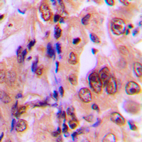
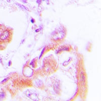
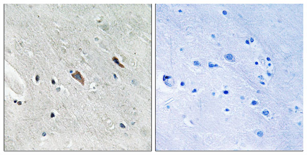
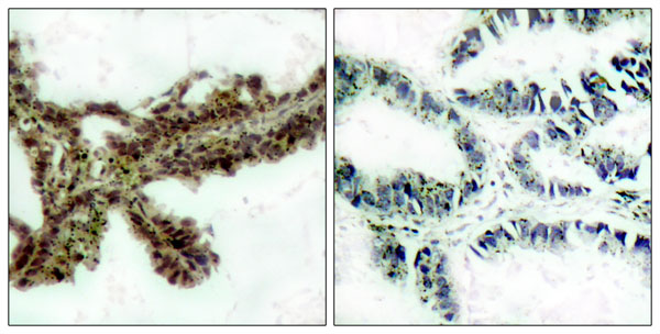
IHC (Immunohiostchemistry) (Immunohistochemical analysis of MKK1 (pT292) staining in human breast cancer formalin fixed paraffin embedded tissue section. The section was pre-treated using heat mediated antigen retrieval with sodium citrate buffer (pH 6.0). The section was then incubated with the antibody at room temperature and detected using an HRP conjugated compact polymer system. DAB was used as the chromogen. The section was then counterstained with haematoxylin and mounted with DPX.)
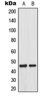
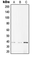
WB (Western Blot) (Western blot analysis of MKK1 (pT292) expression in A431 EGF-treated (A), HeLa EGF-treated (B) whole cell lysates.)
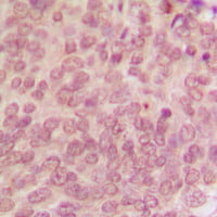
IHC (Immunohiostchemistry) (Immunohistochemical analysis of AMPK beta 1 (pS182) staining in human lung cancer formalin fixed paraffin embedded tissue section. The section was pre-treated using heat mediated antigen retrieval with sodium citrate buffer (pH 6.0). The section was then incubated with the antibody at room temperature and detected using an HRP conjugated compact polymer system. DAB was used as the chromogen. The section was then counterstained with haematoxylin and mounted with DPX.)
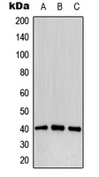
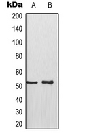
WB (Western Blot) (Western blot analysis of AMPK beta 1 (pS182) expression in A431 (A), NIH3T3 (B), PC12 (C) whole cell lysates.)
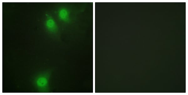
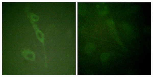
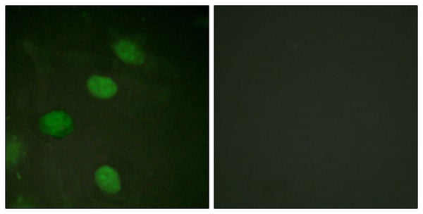
IF (Immunofluorescence) (Immunofluorescent analysis of p53 (pS315) staining in A431 cells. Formalin-fixed cells were permeabilized with 0.1% Triton X-100 in TBS for 5-10 minutes and blocked with 3% BSA-PBS for 30 minutes at room temperature. Cells were probed with the primary antibody in 3% BSA-PBS and incubated overnight at 4 °C in a humidified chamber. Cells were washed with PBST and incubated with a DyLight 594-conjugated secondary antibody (red) in PBS at room temperature in the dark.)
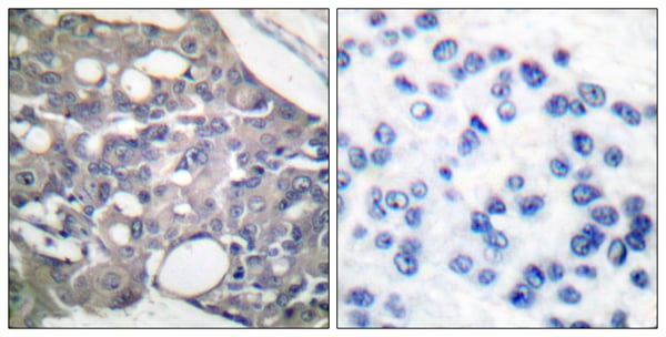
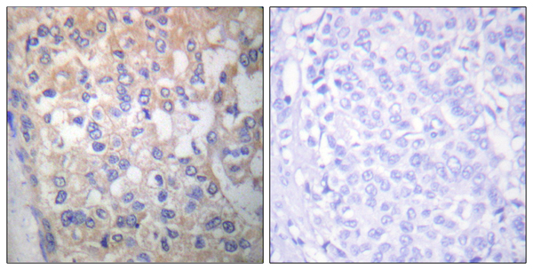
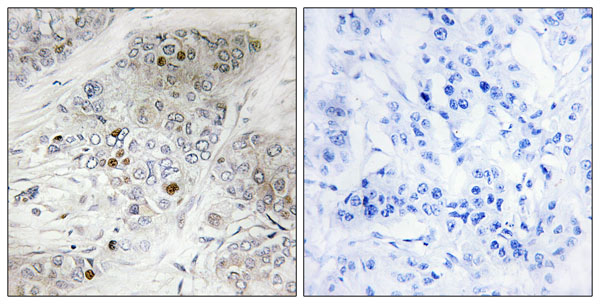
IHC (Immunohiostchemistry) (Immunohistochemical analysis of p53 (pS315) staining in human breast cancer formalin fixed paraffin embedded tissue section. The section was pre-treated using heat mediated antigen retrieval with sodium citrate buffer (pH 6.0). The section was then incubated with the antibody at room temperature and detected using an HRP conjugated compact polymer system. DAB was used as the chromogen. The section was then counterstained with haematoxylin and mounted with DPX.)
WB (Western Blot) (Western blot analysis of p53 (pS315) expression in HeLa (A), A431 (B) whole cell lysates.)
Immunoprecipitation, Immunocytochemistry, Immunofluorescence, Immunohistochemistry, Western Blot
Purity
The antibody was purified by immunogen affinity chromatography.
Pricing
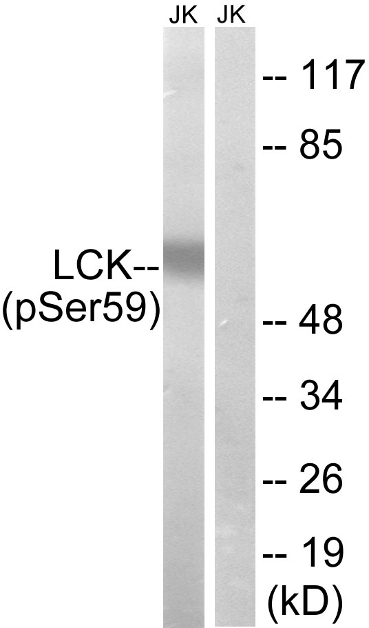
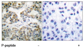
Application Data (Lane 1: The extracts from Jurkat cells treated with IFN (2500U/ML, 30mins) using LCK (Phospho-Ser59) antibody antibody Lane 2: The extracts from Jurkat cells treated with IFN (2500U/ML, 30mins) plus phosphopeptide using LCK (Phospho-Ser59) antibody antibody)
Affinity-purified from rabbit antiserum by affinity-chromatography using epitope-specific phosphopeptide. The antibody against non-phosphopeptide was removed by chromatography using non-phosphopeptide corresponding to the phosphorylation site.
IHC (Immunohiostchemistry) (Immunohistochemistry analysis of paraffin-embedded human brain tissue using Gab2 (Phospho-Tyr643) antibody. Western blot analysis of extracts from Jurkat cells, treated with IFN (2500U/ML, 30mins), using Gab2 (Phospho-Tyr643) antibody.)
Affinity-purified from rabbit antiserum by affinity-chromatography using epitope-specific phosphopeptide. The antibody againstnon-phosphopeptide was removed by chromatography using non-phosphopeptide corresponding to the phosphorylation site.
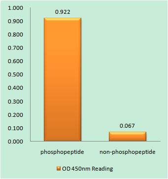
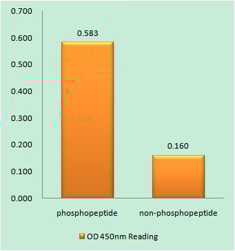
IHC (Immunohiostchemistry) (Immunohistochemistry analysis of paraffin-embedded human breast carcinoma tissue using p53 (Phospho-Thr387) antibody. p53 (Phospho-Thr387) antibody reacts with epitope-specific phosphopeptide and corresponding non-phosphopeptide. The absorbance readings at 450 nM are shown in the ELISA figure.)
Affinity-purified from rabbit antiserum by affinity-chromatography using epitope-specific phosphopeptide. The antibody against non-phosphopeptide was removed by chromatography using non-phosphopeptide corresponding to the phosphorylation site.
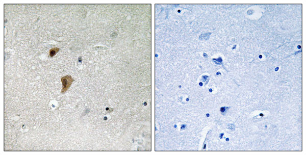
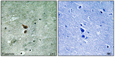
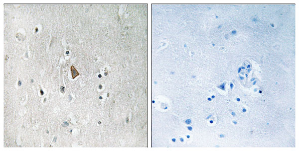
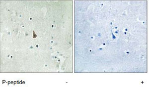
Application Data (P-peptide-+Immunohistochemistry analysis of paraffin-embedded human brain using PAK3 (Phospho-Ser154) Antibody.Western blot analysis of extracts from rat heart, using PAK3 (Phospho-Ser154) Antibody.)
IHC (Immunohiostchemistry) (Immunohistochemistry analysis of paraffin-embedded human brain tissue using HSF1 (Phospho-Thr142) antibody. HSF1 (Phospho-Thr142) antibody reacts with epitope-specific phosphopeptide and corresponding non-phosphopeptide. The absorbance readings at 450 nM are shown in the ELISA figure.)
Affinity-purified from rabbit antiserum by affinity-chromatography using epitope-specific phosphopeptide. The antibody againstnon-phosphopeptide was removed by chromatography using non-phosphopeptide corresponding to the phosphorylation site.
Pricing
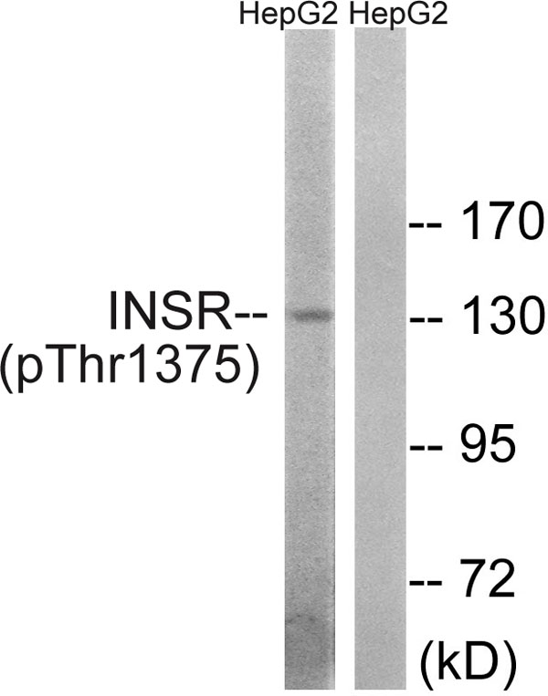
WB (Western Blot) (Western blot analysis of extracts from HepG2 cells, using INSR (Phospho-Thr1375) antibody.)
Affinity-purified fromrabbit antiserum by affinity-chromatography using epitope-specific phosphopeptide. The antibody against non-phosphopeptide was removed by chromatography using non-phosphopeptide corresponding to the phosphorylation site.
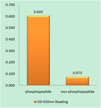
IHC (Immunohiostchemistry) (Immunohistochemistry analysis of paraffin-embedded human breast carcinoma tissue using AKT1/2/3 (Phospho-Tyr315/316/312) antibody. AKT1/2/3 (Phospho-Tyr315/316/312) antibody reacts with epitope-specific phosphopeptide and corresponding non-phosphopeptide. The absorbance readings at 450 nM are shown in the ELISA figure.)
Affinity-purified from rabbit antiserum by affinity-chromatography using epitope-specific phosphopeptide. The antibody against non-phosphopeptide was removed by chromatography using non-phosphopeptide corresponding to the phosphorylation site.
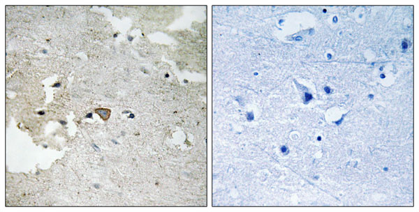
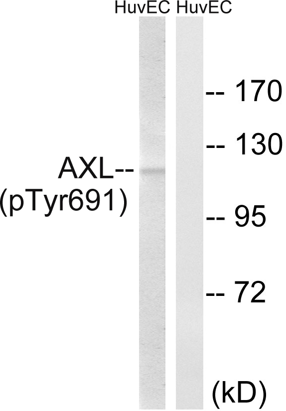
IHC (Immunohiostchemistry) (Immunohistochemistry analysis of paraffin-embedded human brain tissue using AXL (Phospho-Tyr691) antibody. Western blot analysis of extracts from HUVEC cells, treated with EGF (200ng/ml, 15mins), using AXL (Phospho-Tyr691) antibody.)
Affinity-purified from rabbit antiserum by affinity-chromatography using epitope-specific phosphopeptide. The antibody against non-phosphopeptide was removed by chromatography using non-phosphopeptide corresponding to the phosphorylation site.
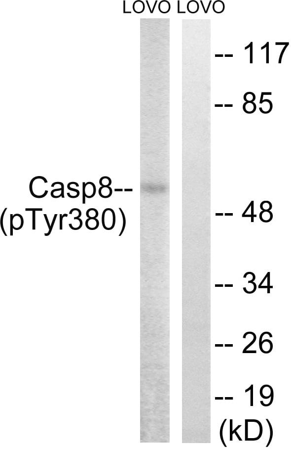
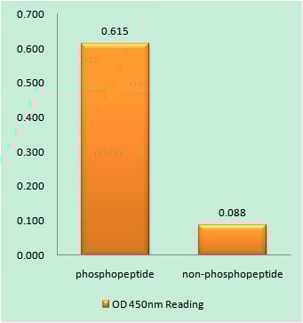
WB (Western Blot) (Western blot analysis of extracts from LOVO cells, treated with UV (15mins), using Caspase 8 (Phospho-Tyr380) antibody. Caspase 8 (Phospho-Tyr380) antibody reacts with epitope-specific phosphopeptide and corresponding non-phosphopeptide. The absorbance readings at 450 nM are shown in the ELISA figure.)
Affinity-purified from rabbit antiserum by affinity-chromatography using epitope-specific phosphopeptide. The antibody against non-phosphopeptide was removed by chromatography using non-phosphopeptide corresponding to the phosphorylation site.
WB (Western Blot) (Western blot analysis using Akt(Phospho-Thr308) Antibody: Line1: The extracts from 293 cells untreated; Line2: The extracts from 293 cells treated with insulin.)
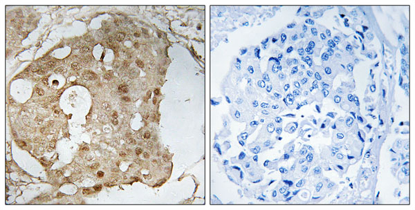
IHC (Immunohistochemistry) (Immunohistochemical analysis of paraffin-embedded lung carcinoma, using Akt(Phospho-Thr308) Antibody. Left: Untreated; Right: Treated with synthesized phosphopeptide.)
Affinity-purified from rabbit antiserum by affinity-chromatography using epitope-specific phosphopeptide. The antibody against non-phosphopeptide was removed by chromatogramphy using non-phosphopeptide corresponding to the phosphorylation site.
IHC (Immunohiostchemistry) (Immunohistochemistry analysis of paraffin-embedded human breast carcinoma tissue using p53 (Phospho-Ser366) antibody. Western blot analysis of extracts from HeLa cells, treated with Adriamycin (0.5ug/ml, 24hours), using p53 (Phospho-Ser366) antibody.)
Affinity-purified fromrabbit antiserum by affinity-chromatography using epitope-specific phosphopeptide. The antibody against non-phosphopeptide was removed by chromatography using non-phosphopeptide corresponding to the phosphorylation site.
Human (Identities=100%, Positives=100%) Mouse (Identities=100%, Positives=100%)
Applications
Immunohistochemistry, Western Blot
Purity
Affinity-purified from rabbit antiserum by affinity-chromatography using epitope-specific phosphopeptide. The antibody against non-phosphopeptide was removed by chromatography using non-phosphopeptide corresponding to the phosphorylation site.
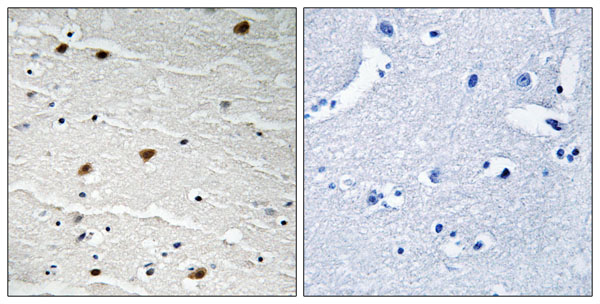
IHC (Immunohiostchemistry) (Immunohistochemistry analysis of paraffin-embedded human brain tissue using IGF1R (Phospho-Tyr1346) antibody. IGF1R (Phospho-Tyr1346) antibody reacts with epitope-specific phosphopeptide and corresponding non-phosphopeptide. The absorbance readings at 450 nM are shown in the ELISA figure.)
Affinity-purified from rabbit antiserum by affinity-chromatography using epitope-specific phosphopeptide. The antibody against non-phosphopeptide was removed by chromatography using non-phosphopeptide corresponding to the phosphorylation site.
Affinity-purified from rabbit antiserum by affinity-chromatography using epitope-specific phosphopeptide. The antibody against non-phosphopeptide was removed by chromatography using non-phosphopeptide corresponding to the phosphorylation site.
Pricing
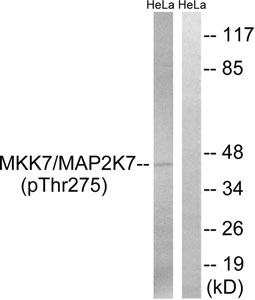
WB (Western Blot) (Western blot analysis of extracts from HeLa cells, treated with calyculinA (50ng/ml, 30mins), using MAP2K7 (Phospho-Thr275) antibody.)
Affinity-purified fromrabbit antiserum by affinity-chromatography using epitope-specific phosphopeptide. The antibody against non-phosphopeptide was removed by chromatography using non-phosphopeptide corresponding to the phosphorylation site.
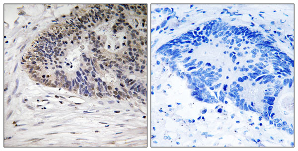
IHC (Immunohiostchemistry) (Immunohistochemistry analysis of paraffin-embedded human colon carcinoma tissue, using ATRIP (Phospho-Ser224) antibody. Western blot analysis of extracts from 292 cells, treated with UV (15mins), using ATRIP (Phospho-Ser224) antibody.)
Affinity-purified from rabbit antiserum by affinity-chromatography using epitope-specific phosphopeptide. The antibody against non-phosphopeptide was removed by chromatography using non-phosphopeptide corresponding to the phosphorylation site.
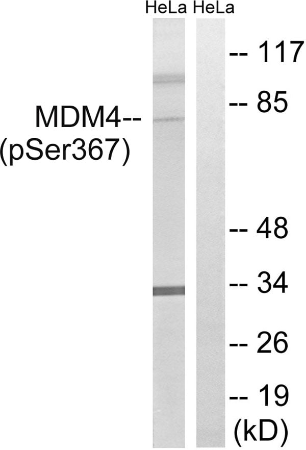
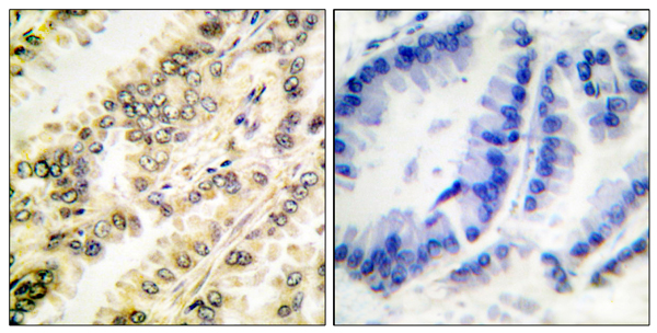
IHC (Immunohiostchemistry) (Immunohistochemistry analysis of paraffin-embedded human colon carcinoma tissue using MDM4 (Phospho-Ser367) antibody. Western blot analysis of extracts from HeLa cells, treated with calyculinA (50ng/ml, 30mins), using MDM4 (Phospho-Ser367) antibody.)
Affinity-purified from rabbit antiserum by affinity-chromatography using epitope-specific phosphopeptide. The antibody againstnon-phosphopeptide was removed by chromatography using non-phosphopeptide corresponding to the phosphorylation site.
WB (Western Blot) (Western blot analysis of extracts from HeLa cells treated with Insulin (0.01U/ml, 15mins), using SGK (phospho-Ser422) antibody (#A0087, Line 1 and 2).)
IHC (Immunohistochemistry) (P-peptide-+ Immunohistochemical analysis of paraffin-embedded human breast carcinoma tissue using SGK (phospho-Ser422) antibody (#A0087).)
Affinity-purified from rabbit antiserum by affinity-chromatography using epitope-specific phosphopeptide. The antibody against non-phosphopeptide was removed by chromatography using non-phosphopeptide corresponding to the phosphorylation site.
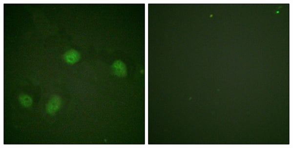
IF (Immunofluorescence) (P-peptide-+ Immunofluorescence analysis of HeLa cells, using Raf1 (Phospho-Ser621) antibody.)
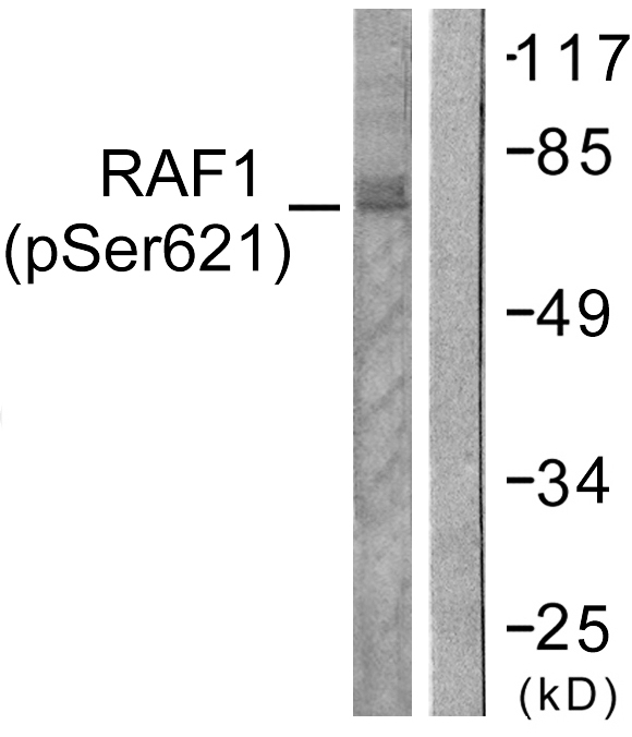
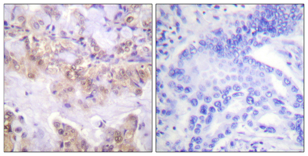
IHC (Immunohiostchemistry) (Immunohistochemistry analysis of paraffin-embedded human lung carcinoma tissue using Raf1 (Phospho-Ser621) antibody. Western blot analysis of extracts from HeLa cells, treated with UV (5mins), using Raf1 (Phospho-Ser621) antibody.)
Immunofluorescence, Immunohistochemistry, Western Blot
Purity
Affinity-purified from rabbit antiserum by affinity-chromatography using epitope-specific phosphopeptide. The antibody against non-phosphopeptide was removed by chromatography using non-phosphopeptide corresponding to the phosphorylation site.
Affinity-purified from rabbit antiserum by affinity-chromatography using epitope-specific phosphopeptide. The antibody against non-phosphopeptide was removed by chromatography using non-phosphopeptide corresponding to the phosphorylation site.
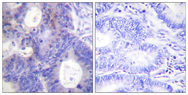
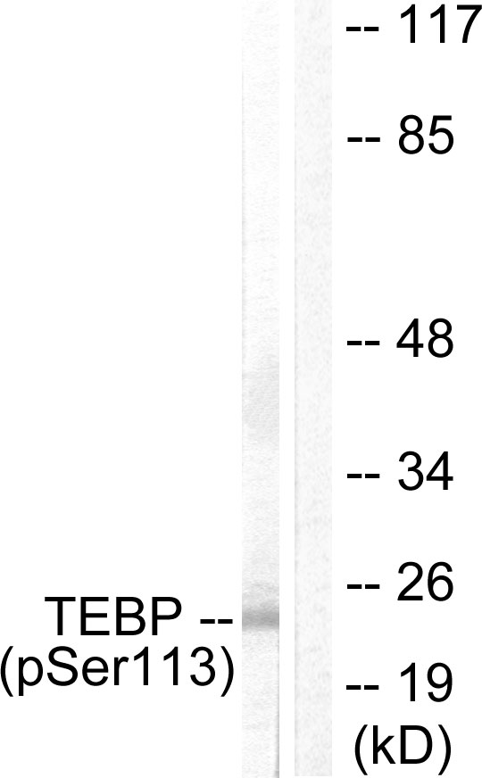
IHC (Immunohiostchemistry) (Immunohistochemistry analysis of paraffin-embedded human colon carcinoma tissue using TEBP (Phospho-Ser113) antibody. Western blot analysis of extracts from NIH/3T3 cells, treated with EGF (200ng/ml, 30mins), using TEBP (Phospho-Ser113) antibody.)
Affinity-purified from rabbit antiserum by affinity-chromatography using epitope-specific phosphopeptide. The antibody against non-phosphopeptide was removed by chromatography using non-phosphopeptide corresponding to the phosphorylation site.
IHC (Immunohiostchemistry) (Immunohistochemistry analysis of paraffin-embedded human breast carcinoma tissue using PKC delta (Phospho-Thr505) antibody. Western blot analysis of extracts from NIH/3T3cells, treated with UV (15mins), using PKC delta (Phospho-Thr505) antibody.)
Affinity-purified from rabbit antiserum by affinity-chromatography using epitope-specific phosphopeptide. The antibody againstnon-phosphopeptide was removed by chromatography using non-phosphopeptide corresponding to the phosphorylation site.
IHC (Immunohiostchemistry) (Immunohistochemistry analysis of paraffin-embedded human thymus gland tissue using ZAP-70 (Phospho-Tyr493) antibody. Western blot analysis of extracts from Jurkat cells, using ZAP-70 (Phospho-Tyr493) antibody.)
Affinity-purified from rabbit antiserum by affinity-chromatography using epitope-specific phosphopeptide. The antibody against non-phosphopeptide was removed by chromatography using non-phosphopeptide corresponding to the phosphorylation site.
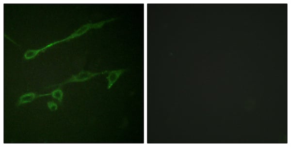
IF (Immunofluorescence) (P-peptide-+ Immunofluorescence analysis of NIH/3T3 cells, using VAV2 (Phospho-Tyr142) antibody.)
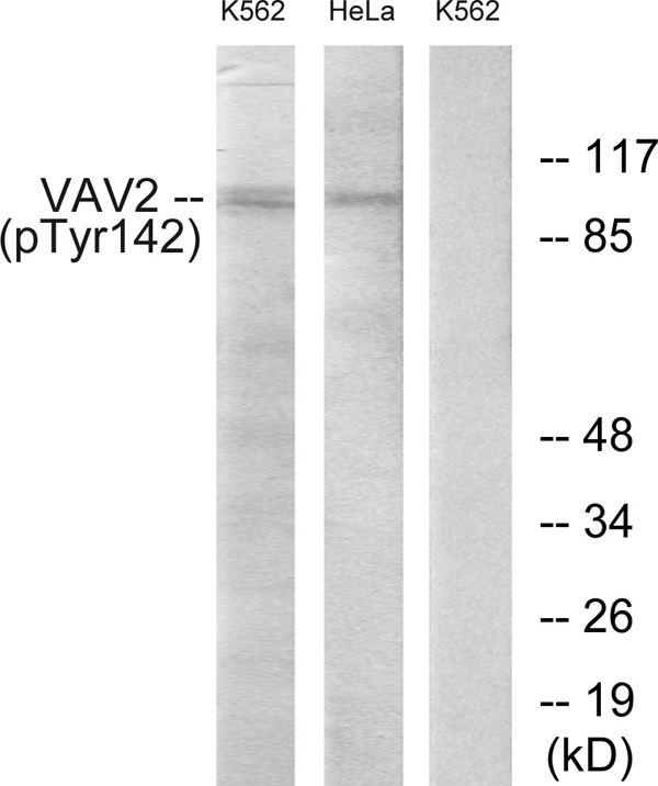
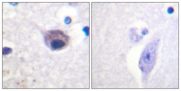
IHC (Immunohiostchemistry) (Immunohistochemistry analysis of paraffin-embedded human brain tissue using VAV2 (Phospho-Tyr142) antibody. Western blot analysis of extracts from K562 cells and HeLa cells, treated with TNF (200ng/ml, 30mins), using VAV2 (Phospho-Tyr142) antibody.)
Immunofluorescence, Immunohistochemistry, Western Blot
Purity
Affinity-purified from rabbit antiserum by affinity-chromatography using epitope-specific phosphopeptide. The antibody against non-phosphopeptide was removed by chromatography using non-phosphopeptide corresponding to the phosphorylation site.
Application Data (P-peptide-+ Immunofluorescence analysis of HuvEc cells, using HER3/ErbB3 (Phospho-Tyr1222) antibody.)
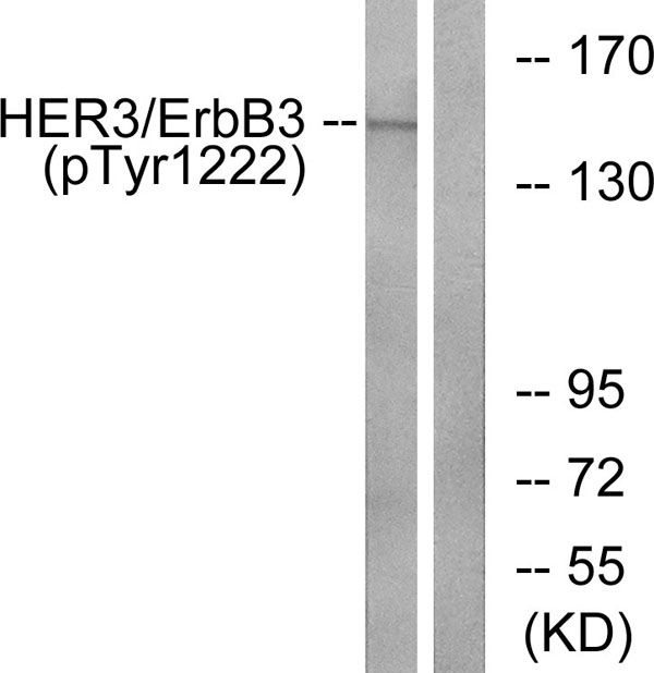
WB (Western Blot) (Western blot analysis of extracts from HUVEC cells, treated with EGF (200ng/ml, 30mins), using HER3/ErbB3 (Phospho-Tyr1222) antibody.)
Immunofluorescence, Immunohistochemistry, Western Blot
Purity
Affinity-purified fromrabbit antiserum by affinity-chromatography using epitope-specific phosphopeptide. The antibody against non-phosphopeptide was removed by chromatography using non-phosphopeptide corresponding to the phosphorylation site.
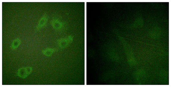
IF (Immunofluorescence) (P-peptide-+ Immunofluorescence analysis of HepG2 cells, using GAB1 (Phospho-Tyr659) antibody.)
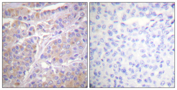
IHC (Immunohiostchemistry) (Immunohistochemistry analysis of paraffin-embedded human breast carcinoma tissue using GAB1 (Phospho-Tyr659) antibody. Western blot analysis of extracts from NIH/3T3 cells, treated with Insulin (0.01U/ml, 15mins), using GAB1 (Phospho-Tyr659) antibody.)
Immunofluorescence, Immunohistochemistry, Western Blot
Purity
Affinity-purified from rabbit antiserum by affinity-chromatography using epitope-specific phosphopeptide. The antibody againstnon-phosphopeptide was removed by chromatography using non-phosphopeptide corresponding to the phosphorylation site.
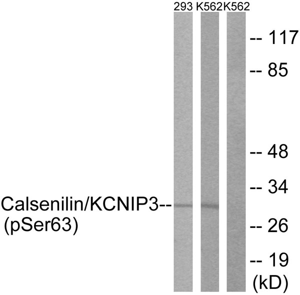
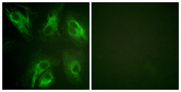
IF (Immunofluorescence) (P-peptide-+ Immunofluorescence analysis of HeLa cells, using Calsenilin/KCNIP3 (Phospho-Ser63) antibody.)
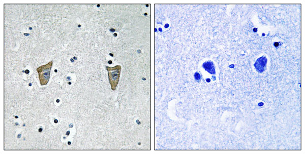
IHC (Immunohiostchemistry) (Immunohistochemistry analysis of paraffin-embedded human brain tissue using Calsenilin/KCNIP3 (Phospho-Ser63) antibody. Western blot analysis of extracts from 293 cells treated with PMA (125ng/ml, 30mins) and Jurkat cells treated with forskolin (40nM, 30mins), using Calsenilin/KCNIP3 (Phospho-Ser63) antibody.)
Immunofluorescence, Immunohistochemistry, Western Blot
Purity
Affinity-purified fromrabbit antiserum by affinity-chromatography using epitope-specific phosphopeptide. The antibody against non-phosphopeptide was removed by chromatography using non-phosphopeptide corresponding to the phosphorylation site.
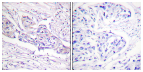
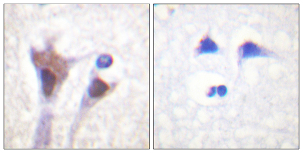
IHC (Immunohiostchemistry) (Immunohistochemistry analysis of paraffin-embedded human breast carcinoma tissue using FOXO1A (Phospho-Ser329) antibody. Western blot analysis of extracts from HeLa cells, treated with Serum (20%, 15mins), using FOXO1A (Phospho-Ser329) antibody.)
Affinity-purified from rabbit antiserum by affinity-chromatography using epitope-specific phosphopeptide. The antibody against non-phosphopeptide was removed by chromatography using non-phosphopeptide corresponding to the phosphorylation site.
IHC (Immunohiostchemistry) (Immunohistochemistry analysis of paraffin-embedded human brain tissue using 14-3-3 zeta/delta (Phospho-Thr232) antibody. Western blot analysis of extracts from Jurkat cells, treated with UV (15mins), using 14-3-3 zeta/delta (Phospho-Thr232) antibody.)
Affinity-purified from rabbit antiserum by affinity-chromatography using epitope-specific phosphopeptide. The antibody against non-phosphopeptide was removed by chromatography using non-phosphopeptide corresponding to the phosphorylation site.
WB (Western Blot) (Western blot analysis of the extracts from NIH/3T3 cells untreated or treated with forskolin (40muM, 30min), using VASP (Ab-238) antibody (Line 1 and 2) and VASP (Phospho-Ser238) antibody (Line 3 and 4).)
IHC (Immunohistochemistry) (P-peptide-+ Immunohistochemical analysis of paraffin-embedded human tonsil tissue using VASP (Phospho-Ser238) antibody.)
Affinity-purified from rabbit antiserum by affinity-chromatography using epitope-specific phosphopeptide. The antibody against non-phosphopeptide was removed by chromatogramphy using non-phosphopeptide corresponding to the phosphorylation site.
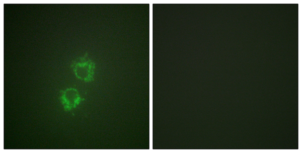
IF (Immunofluorescence) (P-peptide-+ Immunofluorescence analysis of HeLa cells, using 14-3-3 theta/tau (Phospho-Ser232) antibody.)
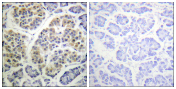
IHC (Immunohiostchemistry) (Immunohistochemistry analysis of paraffin-embedded human pancreas tissue using 14-3-3 theta/tau (Phospho-Ser232) antibody. Western blot analysis of extracts from HeLa cells, using 14-3-3 theta/tau (Phospho-Ser232) antibody.)
Immunofluorescence, Immunohistochemistry, Western Blot
Purity
Affinity-purified fromrabbit antiserum by affinity-chromatography using epitope-specific phosphopeptide. The antibody against non-phosphopeptide was removed by chromatography using non-phosphopeptide corresponding to the phosphorylation site.
WB (Western Blot) (Western blot analysis of extracts from RAW264.7 cells treated with EGF (200ng/ml, 5mins), using SHP-1 (phospho-Tyr536) antibody.)
IHC (Immunohistochemistry) (P-peptide-+ Immunohistochemical analysis of paraffin-embedded human breast carcinoma tissue using SHP-1 (phospho-Tyr536) antibody.)
Affinity-purified from rabbit antiserum by affinity-chromatography using epitope-specific phosphopeptide. The antibody against non-phosphopeptide was removed by chromatography using non-phosphopeptide corresponding to the phosphorylation site.
WB (Western Blot) (P-peptide-+ Western blot analysis of extracts from HepG2 cells, using NEK9 (Phospho-Thr210) antibody. Immunofluorescence analysis of HeLa cells, using NEK9 (Phospho-Thr210) antibody.)
Affinity-purified from rabbit antiserum by affinity-chromatography using epitope-specific phosphopeptide. The antibody against non-phosphopeptide was removed by chromatography using non-phosphopeptide corresponding to the phosphorylation site.
WB (Western Blot) (Western blot analysis of the extracts from MCF7 cells using LIMK2 (phospho-Thr505) antibody.)
IHC (Immunohistochemistry) (P-Peptide-+ Immunohistochemical analysis of paraffin-embedded human breast carcinoma tissue using LIMK2 (phospho-Thr505) antibody.)
Affinity-purified from rabbit antiserum by affinity-chromatography using epitope-specific phosphopeptide. The antibody against non-phosphopeptide was removed by chromatography using non-phosphopeptide corresponding to the phosphorylation site.
IHC (Immunohiostchemistry) (Immunohistochemistry analysis of paraffin-embedded human brain tissue using hnRNP C1/2 (Phospho-Ser260) antibody. Western blot analysis of extracts from 293 cells, treated with H2O2 (100uM, 15mins), using hnRNP C1/2 (Phospho-Ser260) antibody.)
Affinity-purified from rabbit antiserum by affinity-chromatography using epitope-specific phosphopeptide. The antibody against non-phosphopeptide was removed by chromatography using non-phosphopeptide corresponding to the phosphorylation site.
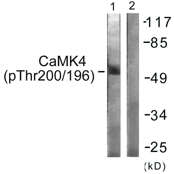
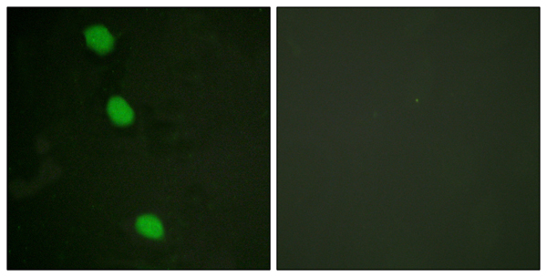
IF (Immunofluorescence) (P-peptide-+ Immunofluorescence analysis of HeLa cells, using CaMK4 (Phospho-Thr196/200) antibody.)
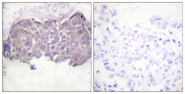
IHC (Immunohiostchemistry) (Immunohistochemistry analysis of paraffin-embedded human brain tissue using CaMK4 (Phospho-Thr196/200) antibody. Western blot analysis of extracts from K562 cells, treated with H2O2 (100uM, 30mins), using CaMK4 (Phospho-Thr196/200) antibody.)
Immunofluorescence, Immunohistochemistry, Western Blot
Purity
Affinity-purified fromrabbit antiserum by affinity-chromatography using epitope-specific phosphopeptide. The antibody against non-phosphopeptide was removed by chromatography using non-phosphopeptide corresponding to the phosphorylation site.
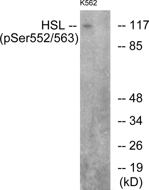
IHC (Immunohiostchemistry) (Immunohistochemistry analysis of paraffin-embedded human breast carcinoma tissue using HSL (Phospho-Ser552) antibody. Western blot analysis of extracts from K562 cells, using using HSL (Phospho-Ser552) antibody.)
Affinity-purified from rabbit antiserum by affinity-chromatography using epitope-specific phosphopeptide. The antibody against non-phosphopeptide was removed by chromatography using non-phosphopeptide corresponding to the phosphorylation site.
IHC (Immunohiostchemistry) (Immunohistochemistry analysis of paraffin-embedded human tonsil tissue using PLCG2 (Phospho-Tyr1217) antibody. Western blot analysis of extracts from Jurkat cells, treated with UV (15mins), using PLCG2 (Phospho-Tyr1217) antibody.)
Immunofluorescence, Immunohistochemistry, Western Blot
Purity
Affinity-purified from rabbit antiserum by affinity-chromatography using epitope-specific phosphopeptide. The antibody against non-phosphopeptide was removed by chromatography using non-phosphopeptide corresponding to the phosphorylation site.
WB (Western Blot) (Western blot analysis of extracts from HeLa cells treated with PMA (125ng/ml, 15mins), using Smad2 (Phospho-Ser467) antibody (#A0030).)
IHC (Immunohistochemistry) (P-peptide-+ Immunohistochemical analysis of paraffin-embedded human breast carcinoma tissue using Smad2 (Phospho-Ser467) antibody (#A0030).)
Affinity-purified from rabbit antiserum by affinity-chromatography using epitope-specific phosphopeptide. The antibody against non-phosphopeptide was removed by chromatography using non-phosphopeptide corresponding to the phosphorylation site.
WB (Western Blot) (Western blot analysis of extracts from 293 cells, treated with H2O2 (100uM, 15mins), using Fyn (Phospho-Tyr530) antibody (#A0430).)
IHC (Immunohistochemistry) (P-peptide-+ Immunohistochemical analysis of paraffin-embedded human breast carcinoma tissue using Fyn (Phospho-Tyr530) antibody (#A0430).)
Affinity-purified from rabbit antiserum by affinity-chromatography using epitope-specific phosphopeptide. The antibody against non-phosphopeptide was removed by chromatography using non-phosphopeptide corresponding to the phosphorylation site.
WB (Western Blot) (P-peptide-+ Western blot analysis of extracts from HeLa cells, using IL-7R/CD127 (Phospho-Tyr449) antibody. Immunofluorescence analysis of HUVEC cells, using IL-7R/CD127 (Phospho-Tyr449) antibody.)
Affinity-purified fromrabbit antiserum by affinity-chromatography using epitope-specific phosphopeptide. The antibody against non-phosphopeptide was removed by chromatography using non-phosphopeptide corresponding to the phosphorylation site.
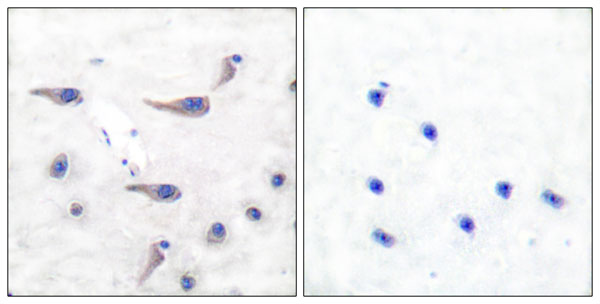
IHC (Immunohiostchemistry) (P-peptide-+ Immunohistochemistry analysis of paraffin-embedded human testis tissue using c-Jun (Phospho-Thr231) antibody. Immunofluorescence analysis of HeLa cells, using c-Jun (Phospho-Thr231) antibody.)
Affinity-purified fromrabbit antiserum by affinity-chromatography using epitope-specific phosphopeptide. The antibody against non-phosphopeptide was removed by chromatography using non-phosphopeptide corresponding to the phosphorylation site.
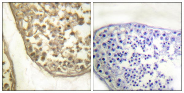
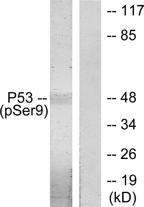
IHC (Immunohiostchemistry) (Immunohistochemistry analysis of paraffin-embedded human breast carcinoma tissue using p53 (Phospho-Ser9) antibody. Western blot analysis of extracts from LOVO cells, using p53 (Phospho-Ser9) antibody.)
Affinity-purified from rabbit antiserum by affinity-chromatography using epitope-specific phosphopeptide. The antibody against non-phosphopeptide was removed by chromatography using non-phosphopeptide corresponding to the phosphorylation site.
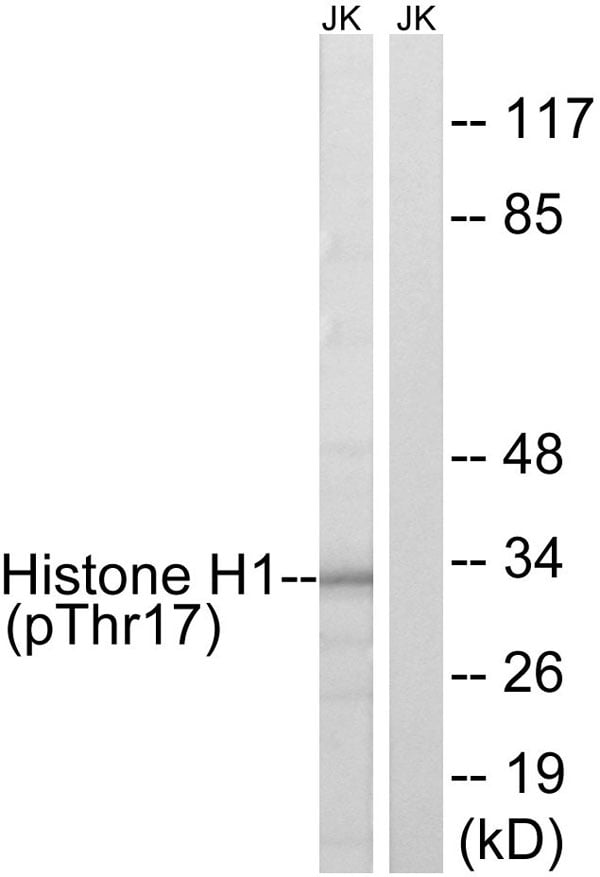
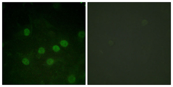
IF (Immunofluorescence) (serum +- Immunofluorescence analysis of HuvEc cells, treated with serum (20%, 30mins), using Histone H1 (Phospho-Thr17) antibody.)
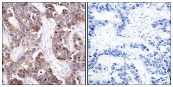
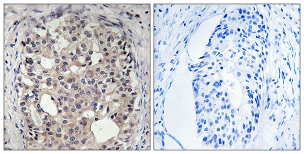
IHC (Immunohiostchemistry) (Immunohistochemistry analysis of paraffin-embedded human colon carcinoma tissue using Histone H1 (Phospho-Thr17) antibody. Western blot analysis of extracts from Jurkat cells, treated with UV (15mins), using Histone H1 (Phospho-Thr17) antibody.)
Immunofluorescence, Immunohistochemistry, Western Blot
Purity
Affinity-purified fromrabbit antiserum by affinity-chromatography using epitope-specific phosphopeptide. The antibody against non-phosphopeptide was removed by chromatography using non-phosphopeptide corresponding to the phosphorylation site.
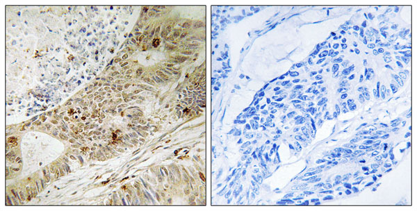
IHC (Immunohiostchemistry) (Immunohistochemistry analysis of paraffin-embedded human breast carcinoma tissue using CDC25A (Phospho-Ser178) antibody. Western blot analysis of extracts from A549 cells, treated with UV (15mins), using CDC25A (Phospho-Ser178) antibody.)
Affinity-purified from rabbit antiserum by affinity-chromatography using epitope-specific phosphopeptide. The antibody against non-phosphopeptide was removed by chromatography using non-phosphopeptide corresponding to the phosphorylation site.
WB (Western Blot) (Western blot analysis of extracts from HeLa cells treated with PMA (125ng/ml, 30mins), using GluR1 (phospho-Ser863) antibody (Line 1 and 2).)
IHC (Immunohistochemistry) (P-peptide-+ Immunohistochemical analysis of paraffin-embedded human brain tissue using GluR1 (phospho-Ser863) antibody.)
Affinity-purified from rabbit antiserum by affinity-chromatography using epitope-specific phosphopeptide. The antibody against non-phosphopeptide was removed by chromatography using non-phosphopeptide corresponding to the phosphorylation site.
Pricing
What Are Phospho Antibodies?
Protein phosphorylation is a process where a phosphate group is added to certain amino acid residues of a protein – usually serine (S), threonine (T), or tyrosine (Y) - by enzymes called kinases. This process is integral in controlling cellular signaling, cellular growth, and other biological functions.
Our catalog includes a wide range of phospho-specific antibodies that can accurately detect this important marker. They perform strongly in widely-used laboratory applications such as Western blot, flow cytometry, immunohistochemistry, and immunofluorescence microscopy. We value your trust in us and are committed to providing top-quality products and services. All of our antibodies are guaranteed to work for the applications and species indicated on our website & associated product pages.
What Are The Key Applications of Phospho Antibodies?
1. Western Blotting
One of the first steps a researcher can take in utilizing these phospho-specific antibodies, is to check if the antibody works using a technique referred to as “Western blot”. For those unfamiliar, Western Blot aids in showing whether the protein that the antibody recognizes is appearing at the correct/expected size. These phospho-specific antibodies should also be able to detect changes in the target protein’s phosphorylation (on/off state) when cells are stimulated in certain ways.
2. Staining of Fixed Cells (Immunocytochemistry)
Another routine use of these phospho-specific antibodies, is to test if the antibody is able to demonstrate similar performance when used on fixed cells (intact cells that have been preserved) as it did in the Western blot tests. It is an important aspect in many cases to confirm that the antibody works in actual intact cell samples. Ideally, the method used for cellular fixation should be the same as what is used in pathology labs (like using 10% formalin). To check if the antibody works well in tissue sections (FFPE), researchers will often test it on fixed cells that are processed similar to tissue samples.
3. Specificity Tests Using Peptides
In order to make sure that the antibody is only binding to the right target:
Laboratory technicians will mix the antibody with phospho-peptides (short segments of the protein containing the phosphate group modification).
If the antibody signal disappears, it is confirmation that it is binding to the correct phosphorylated location.
A more robust test is to use both the phosphorylated and non-phosphorylated (dephosphorylated) versions of the protein. The antibody should react only with the phosphorylated one.
Another method sometimes utilized is to treat the sample with an enzyme, such as alkaline phosphatase, that specifically removes phosphate groups. If the antibody signal disappears after this, it also confirms specificity.
4. Genetic Confirmation
As a final step, scientists can genetically manipulate the nucleotide sequence and alter the target protein by removing the exact site where phosphorylation happens. If the antibody no longer appears to detect the modified protein, it is strong evidence supporting the antibody being specific for that phosphorylated site.
Why Buy Phospho Antibodies Through Us?
The production laboratory adheres to strict and consistent protocols prior to releasing any of these phospho-specific antibodies:
Standard methods and proper controls in all tests to ensure high quality.
These antibodies are tested and validated in different cell types and species.
High quality control criterion to ensure each batch is consistent, so you will obtain reliable results every time.
FAQ
1. What Are Phospho-Specific Antibodies?
Phospho-specific antibodies are made to detect proteins only when they have a phosphate group linked to a specific amino acid residue. This empowers scientists understand if a protein is "turned on" or active, based on its phosphorylation state.
2. How to Detect Phosphorylated Proteins in a Western Blot?
To find out if a protein is phosphorylated using Western blot:
Use a phospho-specific antibody that binds only to the phosphorylated form of the protein.
You can also use a “regular” antibody for the same amino acid sequence of the protein that the phospho-specific antibody is binding to (but in this case, this antibody will not bind if there is a phosphate group present) in order to compare how much of it is phosphorylated versus how much is non-phosphorylated (or “total” protein, if the “normal” antibody’s epitopes are non-phospho-site-specific).
3. How to Choose the Best Antibody?
Here are some simple tips to help you pick the right antibody:
Know your target
Match your sample characteristics
Confirm the intended use is appropriate
Check “host” and “type”
Check the “quality” of the presented data/images
Appraise whether the available validation meets your needs
Submit a Question
Please complete the form below and a representative will contact you as soon as possible.
Request more Information
Please complete the form below and a representative will contact you as soon as possible.
Request a Manual
Please complete the form below and a representative will contact you as soon as possible.
Request a Quote
Please complete the form below and a representative will contact you as soon as possible.
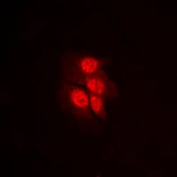 IF (Immunofluorescence)
(Immunofluorescent analysis of MKK1 (pT292) staining in HeLa cells. Formalin-fixed cells were permeabilized with 0.1% Triton X-100 in TBS for 5-10 minutes and blocked with 3% BSA-PBS for 30 minutes at room temperature. Cells were probed with the primary antibody in 3% BSA-PBS and incubated overnight at 4 °C in a humidified chamber. Cells were washed with PBST and incubated with a DyLight 594-conjugated secondary antibody (red) in PBS at room temperature in the dark.)
IF (Immunofluorescence)
(Immunofluorescent analysis of MKK1 (pT292) staining in HeLa cells. Formalin-fixed cells were permeabilized with 0.1% Triton X-100 in TBS for 5-10 minutes and blocked with 3% BSA-PBS for 30 minutes at room temperature. Cells were probed with the primary antibody in 3% BSA-PBS and incubated overnight at 4 °C in a humidified chamber. Cells were washed with PBST and incubated with a DyLight 594-conjugated secondary antibody (red) in PBS at room temperature in the dark.)
 IHC (Immunohiostchemistry)
(Immunohistochemical analysis of AMPK beta 1 (pS182) staining in human lung cancer formalin fixed paraffin embedded tissue section. The section was pre-treated using heat mediated antigen retrieval with sodium citrate buffer (pH 6.0). The section was then incubated with the antibody at room temperature and detected using an HRP conjugated compact polymer system. DAB was used as the chromogen. The section was then counterstained with haematoxylin and mounted with DPX.)
IHC (Immunohiostchemistry)
(Immunohistochemical analysis of AMPK beta 1 (pS182) staining in human lung cancer formalin fixed paraffin embedded tissue section. The section was pre-treated using heat mediated antigen retrieval with sodium citrate buffer (pH 6.0). The section was then incubated with the antibody at room temperature and detected using an HRP conjugated compact polymer system. DAB was used as the chromogen. The section was then counterstained with haematoxylin and mounted with DPX.)
 IF (Immunofluorescence)
(Immunofluorescent analysis of p53 (pS315) staining in A431 cells. Formalin-fixed cells were permeabilized with 0.1% Triton X-100 in TBS for 5-10 minutes and blocked with 3% BSA-PBS for 30 minutes at room temperature. Cells were probed with the primary antibody in 3% BSA-PBS and incubated overnight at 4 °C in a humidified chamber. Cells were washed with PBST and incubated with a DyLight 594-conjugated secondary antibody (red) in PBS at room temperature in the dark.)
IF (Immunofluorescence)
(Immunofluorescent analysis of p53 (pS315) staining in A431 cells. Formalin-fixed cells were permeabilized with 0.1% Triton X-100 in TBS for 5-10 minutes and blocked with 3% BSA-PBS for 30 minutes at room temperature. Cells were probed with the primary antibody in 3% BSA-PBS and incubated overnight at 4 °C in a humidified chamber. Cells were washed with PBST and incubated with a DyLight 594-conjugated secondary antibody (red) in PBS at room temperature in the dark.)
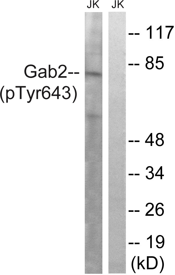 IHC (Immunohiostchemistry)
(Immunohistochemistry analysis of paraffin-embedded human brain tissue using Gab2 (Phospho-Tyr643) antibody. Western blot analysis of extracts from Jurkat cells, treated with IFN (2500U/ML, 30mins), using Gab2 (Phospho-Tyr643) antibody.)
IHC (Immunohiostchemistry)
(Immunohistochemistry analysis of paraffin-embedded human brain tissue using Gab2 (Phospho-Tyr643) antibody. Western blot analysis of extracts from Jurkat cells, treated with IFN (2500U/ML, 30mins), using Gab2 (Phospho-Tyr643) antibody.)
 IHC (Immunohiostchemistry)
(Immunohistochemistry analysis of paraffin-embedded human breast carcinoma tissue using p53 (Phospho-Thr387) antibody. p53 (Phospho-Thr387) antibody reacts with epitope-specific phosphopeptide and corresponding non-phosphopeptide. The absorbance readings at 450 nM are shown in the ELISA figure.)
IHC (Immunohiostchemistry)
(Immunohistochemistry analysis of paraffin-embedded human breast carcinoma tissue using p53 (Phospho-Thr387) antibody. p53 (Phospho-Thr387) antibody reacts with epitope-specific phosphopeptide and corresponding non-phosphopeptide. The absorbance readings at 450 nM are shown in the ELISA figure.)
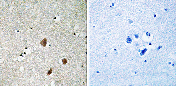
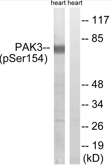 Application Data
(P-peptide-+Immunohistochemistry analysis of paraffin-embedded human brain using PAK3 (Phospho-Ser154) Antibody.Western blot analysis of extracts from rat heart, using PAK3 (Phospho-Ser154) Antibody.)
Application Data
(P-peptide-+Immunohistochemistry analysis of paraffin-embedded human brain using PAK3 (Phospho-Ser154) Antibody.Western blot analysis of extracts from rat heart, using PAK3 (Phospho-Ser154) Antibody.)
 IHC (Immunohiostchemistry)
(Immunohistochemistry analysis of paraffin-embedded human brain tissue using HSF1 (Phospho-Thr142) antibody. HSF1 (Phospho-Thr142) antibody reacts with epitope-specific phosphopeptide and corresponding non-phosphopeptide. The absorbance readings at 450 nM are shown in the ELISA figure.)
IHC (Immunohiostchemistry)
(Immunohistochemistry analysis of paraffin-embedded human brain tissue using HSF1 (Phospho-Thr142) antibody. HSF1 (Phospho-Thr142) antibody reacts with epitope-specific phosphopeptide and corresponding non-phosphopeptide. The absorbance readings at 450 nM are shown in the ELISA figure.)
 IHC (Immunohiostchemistry)
(Immunohistochemistry analysis of paraffin-embedded human breast carcinoma tissue using AKT1/2/3 (Phospho-Tyr315/316/312) antibody. AKT1/2/3 (Phospho-Tyr315/316/312) antibody reacts with epitope-specific phosphopeptide and corresponding non-phosphopeptide. The absorbance readings at 450 nM are shown in the ELISA figure.)
IHC (Immunohiostchemistry)
(Immunohistochemistry analysis of paraffin-embedded human breast carcinoma tissue using AKT1/2/3 (Phospho-Tyr315/316/312) antibody. AKT1/2/3 (Phospho-Tyr315/316/312) antibody reacts with epitope-specific phosphopeptide and corresponding non-phosphopeptide. The absorbance readings at 450 nM are shown in the ELISA figure.)
 IHC (Immunohiostchemistry)
(Immunohistochemistry analysis of paraffin-embedded human brain tissue using AXL (Phospho-Tyr691) antibody. Western blot analysis of extracts from HUVEC cells, treated with EGF (200ng/ml, 15mins), using AXL (Phospho-Tyr691) antibody.)
IHC (Immunohiostchemistry)
(Immunohistochemistry analysis of paraffin-embedded human brain tissue using AXL (Phospho-Tyr691) antibody. Western blot analysis of extracts from HUVEC cells, treated with EGF (200ng/ml, 15mins), using AXL (Phospho-Tyr691) antibody.)
 WB (Western Blot)
(Western blot analysis of extracts from LOVO cells, treated with UV (15mins), using Caspase 8 (Phospho-Tyr380) antibody. Caspase 8 (Phospho-Tyr380) antibody reacts with epitope-specific phosphopeptide and corresponding non-phosphopeptide. The absorbance readings at 450 nM are shown in the ELISA figure.)
WB (Western Blot)
(Western blot analysis of extracts from LOVO cells, treated with UV (15mins), using Caspase 8 (Phospho-Tyr380) antibody. Caspase 8 (Phospho-Tyr380) antibody reacts with epitope-specific phosphopeptide and corresponding non-phosphopeptide. The absorbance readings at 450 nM are shown in the ELISA figure.)
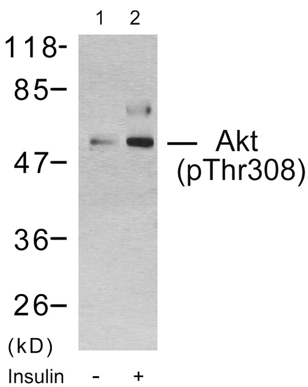 WB (Western Blot)
(Western blot analysis using Akt(Phospho-Thr308) Antibody: Line1: The extracts from 293 cells untreated; Line2: The extracts from 293 cells treated with insulin.)
WB (Western Blot)
(Western blot analysis using Akt(Phospho-Thr308) Antibody: Line1: The extracts from 293 cells untreated; Line2: The extracts from 293 cells treated with insulin.)
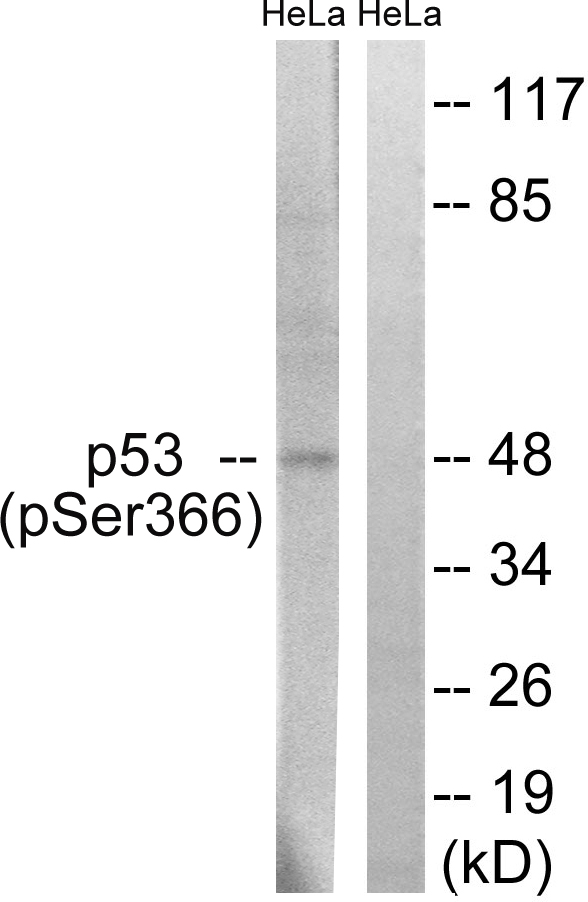 IHC (Immunohiostchemistry)
(Immunohistochemistry analysis of paraffin-embedded human breast carcinoma tissue using p53 (Phospho-Ser366) antibody. Western blot analysis of extracts from HeLa cells, treated with Adriamycin (0.5ug/ml, 24hours), using p53 (Phospho-Ser366) antibody.)
IHC (Immunohiostchemistry)
(Immunohistochemistry analysis of paraffin-embedded human breast carcinoma tissue using p53 (Phospho-Ser366) antibody. Western blot analysis of extracts from HeLa cells, treated with Adriamycin (0.5ug/ml, 24hours), using p53 (Phospho-Ser366) antibody.)
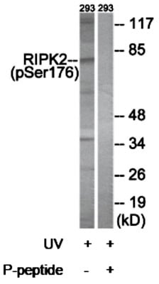 WB (Western Blot)
(Western blot analysis of extracts from 293 cells, treated with UV (15mins), using RIPK2 (Phospho-Ser176) antibody.)
WB (Western Blot)
(Western blot analysis of extracts from 293 cells, treated with UV (15mins), using RIPK2 (Phospho-Ser176) antibody.)
 IHC (Immunohiostchemistry)
(Immunohistochemistry analysis of paraffin-embedded human brain tissue using IGF1R (Phospho-Tyr1346) antibody. IGF1R (Phospho-Tyr1346) antibody reacts with epitope-specific phosphopeptide and corresponding non-phosphopeptide. The absorbance readings at 450 nM are shown in the ELISA figure.)
IHC (Immunohiostchemistry)
(Immunohistochemistry analysis of paraffin-embedded human brain tissue using IGF1R (Phospho-Tyr1346) antibody. IGF1R (Phospho-Tyr1346) antibody reacts with epitope-specific phosphopeptide and corresponding non-phosphopeptide. The absorbance readings at 450 nM are shown in the ELISA figure.)
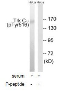 WB (Western Blot)
(Western blot analysis of extracts from HeLa cells, treated with serum (20%, 15mins), usingTrk C (Phospho-Tyr516)antibody)
WB (Western Blot)
(Western blot analysis of extracts from HeLa cells, treated with serum (20%, 15mins), usingTrk C (Phospho-Tyr516)antibody)
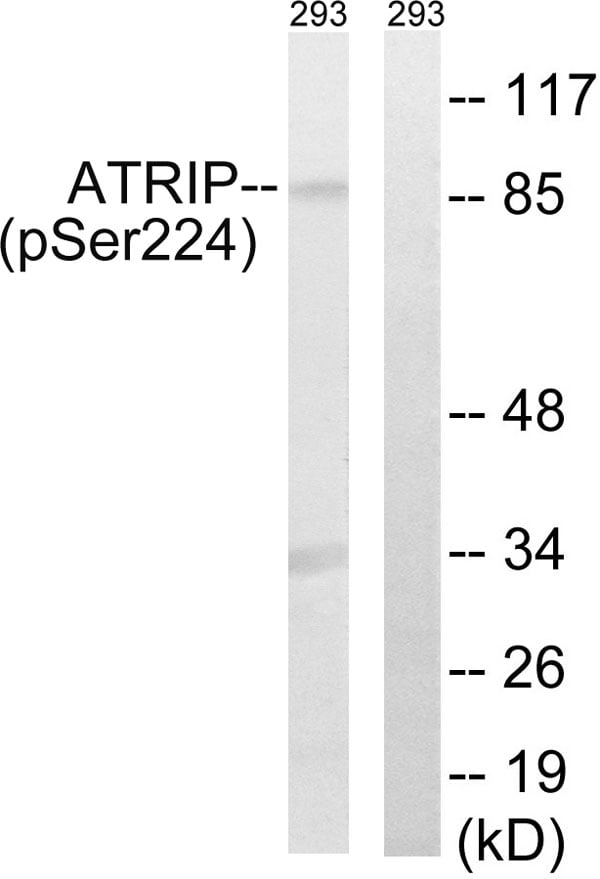 IHC (Immunohiostchemistry)
(Immunohistochemistry analysis of paraffin-embedded human colon carcinoma tissue, using ATRIP (Phospho-Ser224) antibody. Western blot analysis of extracts from 292 cells, treated with UV (15mins), using ATRIP (Phospho-Ser224) antibody.)
IHC (Immunohiostchemistry)
(Immunohistochemistry analysis of paraffin-embedded human colon carcinoma tissue, using ATRIP (Phospho-Ser224) antibody. Western blot analysis of extracts from 292 cells, treated with UV (15mins), using ATRIP (Phospho-Ser224) antibody.)
 IHC (Immunohiostchemistry)
(Immunohistochemistry analysis of paraffin-embedded human colon carcinoma tissue using MDM4 (Phospho-Ser367) antibody. Western blot analysis of extracts from HeLa cells, treated with calyculinA (50ng/ml, 30mins), using MDM4 (Phospho-Ser367) antibody.)
IHC (Immunohiostchemistry)
(Immunohistochemistry analysis of paraffin-embedded human colon carcinoma tissue using MDM4 (Phospho-Ser367) antibody. Western blot analysis of extracts from HeLa cells, treated with calyculinA (50ng/ml, 30mins), using MDM4 (Phospho-Ser367) antibody.)
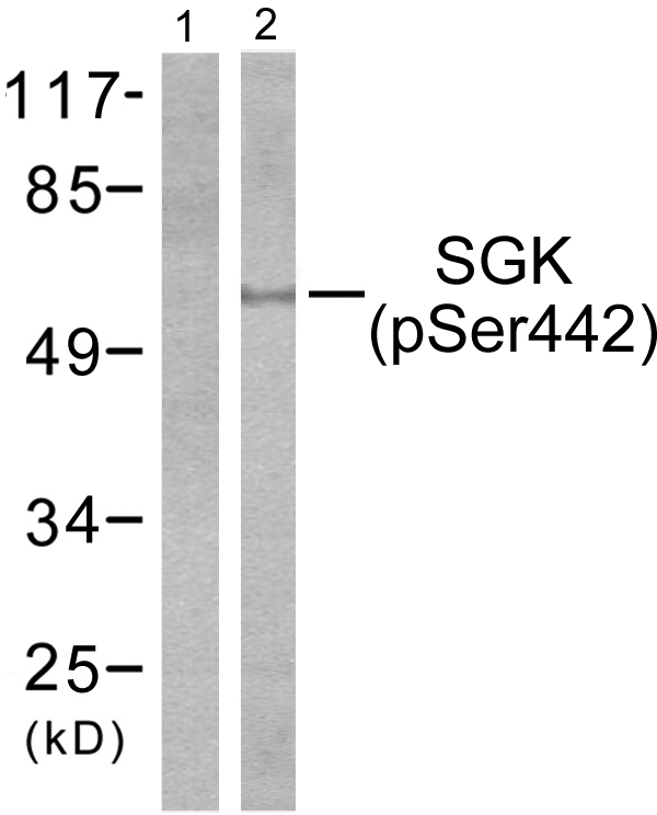 WB (Western Blot)
(Western blot analysis of extracts from HeLa cells treated with Insulin (0.01U/ml, 15mins), using SGK (phospho-Ser422) antibody (#A0087, Line 1 and 2).)
WB (Western Blot)
(Western blot analysis of extracts from HeLa cells treated with Insulin (0.01U/ml, 15mins), using SGK (phospho-Ser422) antibody (#A0087, Line 1 and 2).)
 IF (Immunofluorescence)
(P-peptide-+ Immunofluorescence analysis of HeLa cells, using Raf1 (Phospho-Ser621) antibody.)
IF (Immunofluorescence)
(P-peptide-+ Immunofluorescence analysis of HeLa cells, using Raf1 (Phospho-Ser621) antibody.)
 WB (Western Blot)
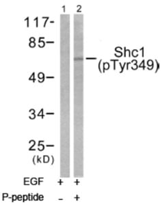
(Western blot analysis of extracts from 293 cells treated with EGF (200ng/ml, 30mins), using Shc (phospho-Tyr349) antibody.)
WB (Western Blot)
(Western blot analysis of extracts from 293 cells treated with EGF (200ng/ml, 30mins), using Shc (phospho-Tyr349) antibody.)
 IHC (Immunohiostchemistry)
(Immunohistochemistry analysis of paraffin-embedded human colon carcinoma tissue using TEBP (Phospho-Ser113) antibody. Western blot analysis of extracts from NIH/3T3 cells, treated with EGF (200ng/ml, 30mins), using TEBP (Phospho-Ser113) antibody.)
IHC (Immunohiostchemistry)
(Immunohistochemistry analysis of paraffin-embedded human colon carcinoma tissue using TEBP (Phospho-Ser113) antibody. Western blot analysis of extracts from NIH/3T3 cells, treated with EGF (200ng/ml, 30mins), using TEBP (Phospho-Ser113) antibody.)
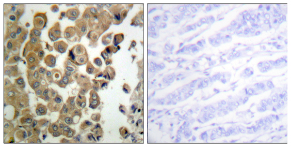
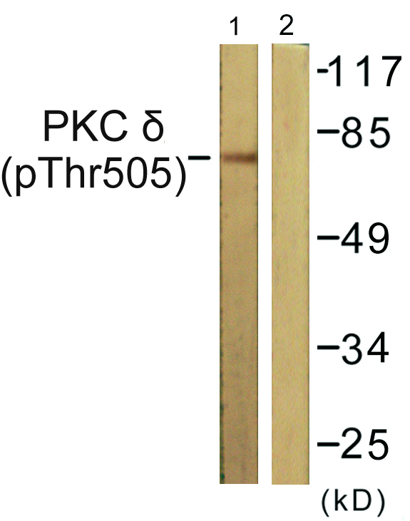 IHC (Immunohiostchemistry)
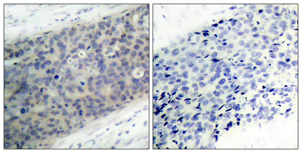
(Immunohistochemistry analysis of paraffin-embedded human breast carcinoma tissue using PKC delta (Phospho-Thr505) antibody. Western blot analysis of extracts from NIH/3T3cells, treated with UV (15mins), using PKC delta (Phospho-Thr505) antibody.)
IHC (Immunohiostchemistry)
(Immunohistochemistry analysis of paraffin-embedded human breast carcinoma tissue using PKC delta (Phospho-Thr505) antibody. Western blot analysis of extracts from NIH/3T3cells, treated with UV (15mins), using PKC delta (Phospho-Thr505) antibody.)
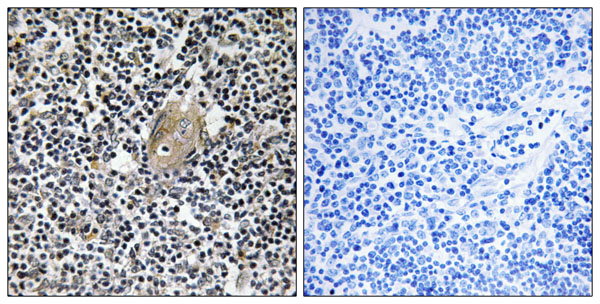
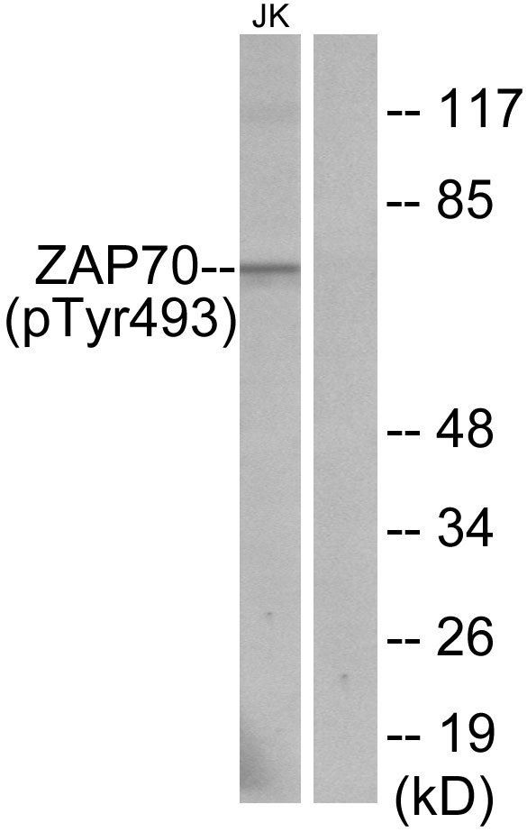 IHC (Immunohiostchemistry)
(Immunohistochemistry analysis of paraffin-embedded human thymus gland tissue using ZAP-70 (Phospho-Tyr493) antibody. Western blot analysis of extracts from Jurkat cells, using ZAP-70 (Phospho-Tyr493) antibody.)
IHC (Immunohiostchemistry)
(Immunohistochemistry analysis of paraffin-embedded human thymus gland tissue using ZAP-70 (Phospho-Tyr493) antibody. Western blot analysis of extracts from Jurkat cells, using ZAP-70 (Phospho-Tyr493) antibody.)
 IF (Immunofluorescence)
(P-peptide-+ Immunofluorescence analysis of NIH/3T3 cells, using VAV2 (Phospho-Tyr142) antibody.)
IF (Immunofluorescence)
(P-peptide-+ Immunofluorescence analysis of NIH/3T3 cells, using VAV2 (Phospho-Tyr142) antibody.)
 Application Data
(P-peptide-+ Immunofluorescence analysis of HuvEc cells, using HER3/ErbB3 (Phospho-Tyr1222) antibody.)
Application Data
(P-peptide-+ Immunofluorescence analysis of HuvEc cells, using HER3/ErbB3 (Phospho-Tyr1222) antibody.)
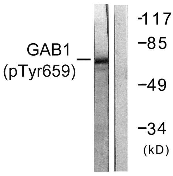
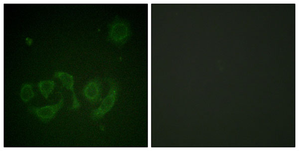 IF (Immunofluorescence)
(P-peptide-+ Immunofluorescence analysis of HepG2 cells, using GAB1 (Phospho-Tyr659) antibody.)
IF (Immunofluorescence)
(P-peptide-+ Immunofluorescence analysis of HepG2 cells, using GAB1 (Phospho-Tyr659) antibody.)
 IF (Immunofluorescence)
(P-peptide-+ Immunofluorescence analysis of HeLa cells, using Calsenilin/KCNIP3 (Phospho-Ser63) antibody.)
IF (Immunofluorescence)
(P-peptide-+ Immunofluorescence analysis of HeLa cells, using Calsenilin/KCNIP3 (Phospho-Ser63) antibody.)
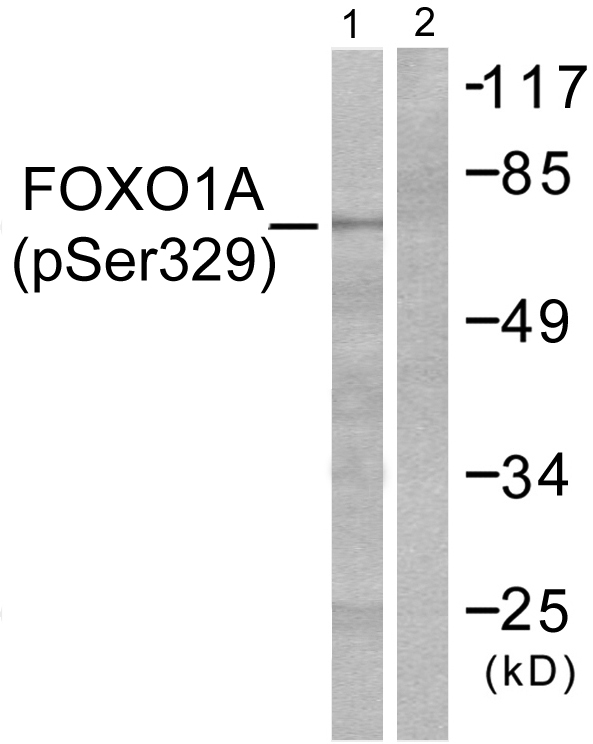 IHC (Immunohiostchemistry)
(Immunohistochemistry analysis of paraffin-embedded human breast carcinoma tissue using FOXO1A (Phospho-Ser329) antibody. Western blot analysis of extracts from HeLa cells, treated with Serum (20%, 15mins), using FOXO1A (Phospho-Ser329) antibody.)
IHC (Immunohiostchemistry)
(Immunohistochemistry analysis of paraffin-embedded human breast carcinoma tissue using FOXO1A (Phospho-Ser329) antibody. Western blot analysis of extracts from HeLa cells, treated with Serum (20%, 15mins), using FOXO1A (Phospho-Ser329) antibody.)
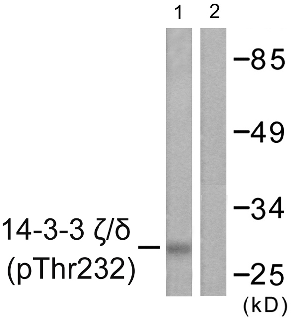 IHC (Immunohiostchemistry)
(Immunohistochemistry analysis of paraffin-embedded human brain tissue using 14-3-3 zeta/delta (Phospho-Thr232) antibody. Western blot analysis of extracts from Jurkat cells, treated with UV (15mins), using 14-3-3 zeta/delta (Phospho-Thr232) antibody.)
IHC (Immunohiostchemistry)
(Immunohistochemistry analysis of paraffin-embedded human brain tissue using 14-3-3 zeta/delta (Phospho-Thr232) antibody. Western blot analysis of extracts from Jurkat cells, treated with UV (15mins), using 14-3-3 zeta/delta (Phospho-Thr232) antibody.)
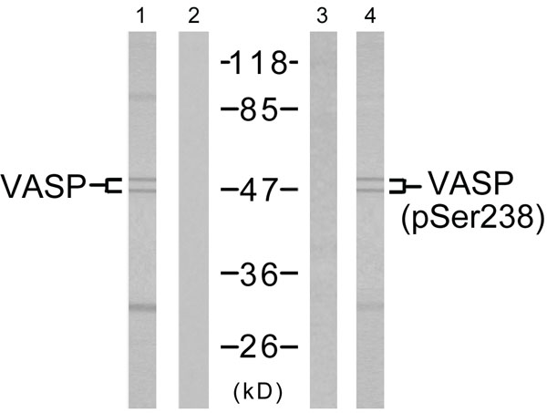 WB (Western Blot)
(Western blot analysis of the extracts from NIH/3T3 cells untreated or treated with forskolin (40muM, 30min), using VASP (Ab-238) antibody (Line 1 and 2) and VASP (Phospho-Ser238) antibody (Line 3 and 4).)
WB (Western Blot)
(Western blot analysis of the extracts from NIH/3T3 cells untreated or treated with forskolin (40muM, 30min), using VASP (Ab-238) antibody (Line 1 and 2) and VASP (Phospho-Ser238) antibody (Line 3 and 4).)
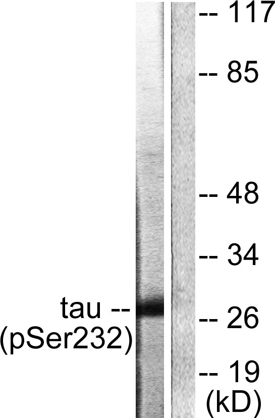
 IF (Immunofluorescence)
(P-peptide-+ Immunofluorescence analysis of HeLa cells, using 14-3-3 theta/tau (Phospho-Ser232) antibody.)
IF (Immunofluorescence)
(P-peptide-+ Immunofluorescence analysis of HeLa cells, using 14-3-3 theta/tau (Phospho-Ser232) antibody.)
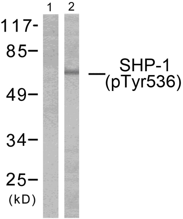 WB (Western Blot)
(Western blot analysis of extracts from RAW264.7 cells treated with EGF (200ng/ml, 5mins), using SHP-1 (phospho-Tyr536) antibody.)
WB (Western Blot)
(Western blot analysis of extracts from RAW264.7 cells treated with EGF (200ng/ml, 5mins), using SHP-1 (phospho-Tyr536) antibody.)
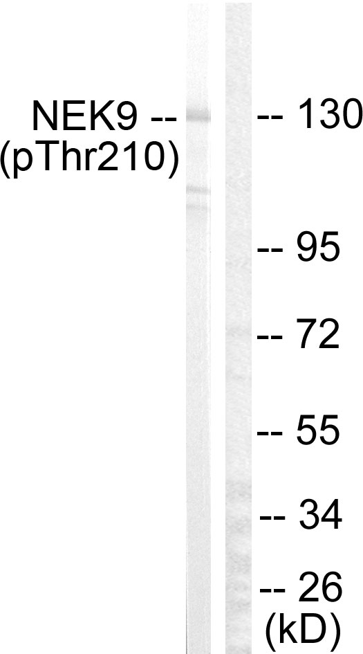
 WB (Western Blot)
(P-peptide-+ Western blot analysis of extracts from HepG2 cells, using NEK9 (Phospho-Thr210) antibody. Immunofluorescence analysis of HeLa cells, using NEK9 (Phospho-Thr210) antibody.)
WB (Western Blot)
(P-peptide-+ Western blot analysis of extracts from HepG2 cells, using NEK9 (Phospho-Thr210) antibody. Immunofluorescence analysis of HeLa cells, using NEK9 (Phospho-Thr210) antibody.)
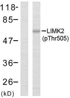 WB (Western Blot)
(Western blot analysis of the extracts from MCF7 cells using LIMK2 (phospho-Thr505) antibody.)
WB (Western Blot)
(Western blot analysis of the extracts from MCF7 cells using LIMK2 (phospho-Thr505) antibody.)
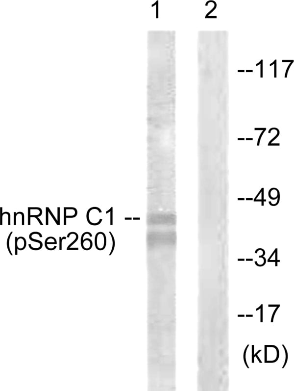 IHC (Immunohiostchemistry)
(Immunohistochemistry analysis of paraffin-embedded human brain tissue using hnRNP C1/2 (Phospho-Ser260) antibody. Western blot analysis of extracts from 293 cells, treated with H2O2 (100uM, 15mins), using hnRNP C1/2 (Phospho-Ser260) antibody.)
IHC (Immunohiostchemistry)
(Immunohistochemistry analysis of paraffin-embedded human brain tissue using hnRNP C1/2 (Phospho-Ser260) antibody. Western blot analysis of extracts from 293 cells, treated with H2O2 (100uM, 15mins), using hnRNP C1/2 (Phospho-Ser260) antibody.)

 IF (Immunofluorescence)
(P-peptide-+ Immunofluorescence analysis of HeLa cells, using CaMK4 (Phospho-Thr196/200) antibody.)
IF (Immunofluorescence)
(P-peptide-+ Immunofluorescence analysis of HeLa cells, using CaMK4 (Phospho-Thr196/200) antibody.)
 IHC (Immunohiostchemistry)
(Immunohistochemistry analysis of paraffin-embedded human breast carcinoma tissue using HSL (Phospho-Ser552) antibody. Western blot analysis of extracts from K562 cells, using using HSL (Phospho-Ser552) antibody.)
IHC (Immunohiostchemistry)
(Immunohistochemistry analysis of paraffin-embedded human breast carcinoma tissue using HSL (Phospho-Ser552) antibody. Western blot analysis of extracts from K562 cells, using using HSL (Phospho-Ser552) antibody.)
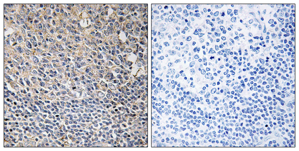
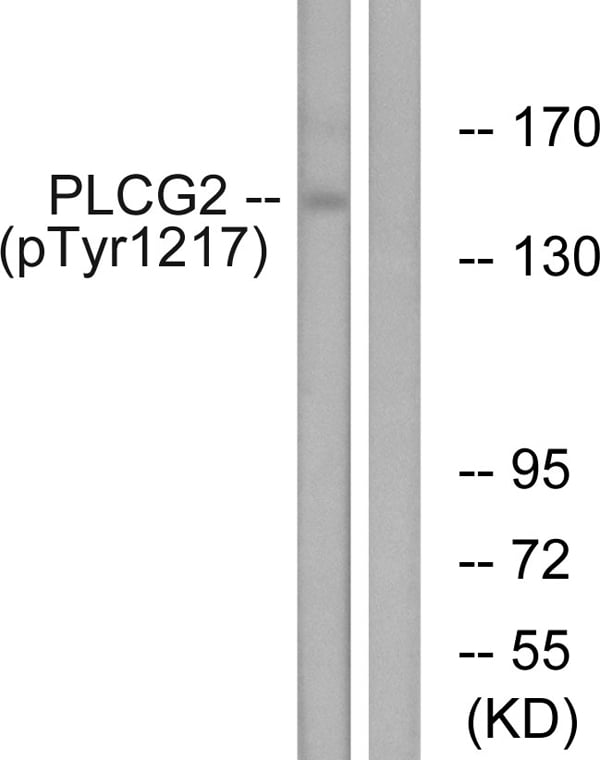 IHC (Immunohiostchemistry)
(Immunohistochemistry analysis of paraffin-embedded human tonsil tissue using PLCG2 (Phospho-Tyr1217) antibody. Western blot analysis of extracts from Jurkat cells, treated with UV (15mins), using PLCG2 (Phospho-Tyr1217) antibody.)
IHC (Immunohiostchemistry)
(Immunohistochemistry analysis of paraffin-embedded human tonsil tissue using PLCG2 (Phospho-Tyr1217) antibody. Western blot analysis of extracts from Jurkat cells, treated with UV (15mins), using PLCG2 (Phospho-Tyr1217) antibody.)
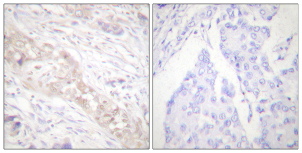
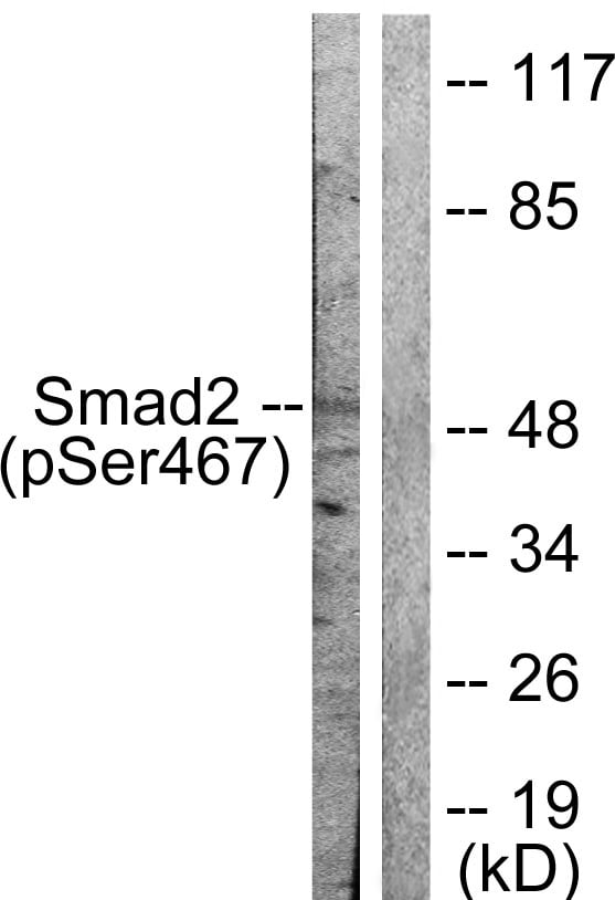 WB (Western Blot)
(Western blot analysis of extracts from HeLa cells treated with PMA (125ng/ml, 15mins), using Smad2 (Phospho-Ser467) antibody (#A0030).)
WB (Western Blot)
(Western blot analysis of extracts from HeLa cells treated with PMA (125ng/ml, 15mins), using Smad2 (Phospho-Ser467) antibody (#A0030).)
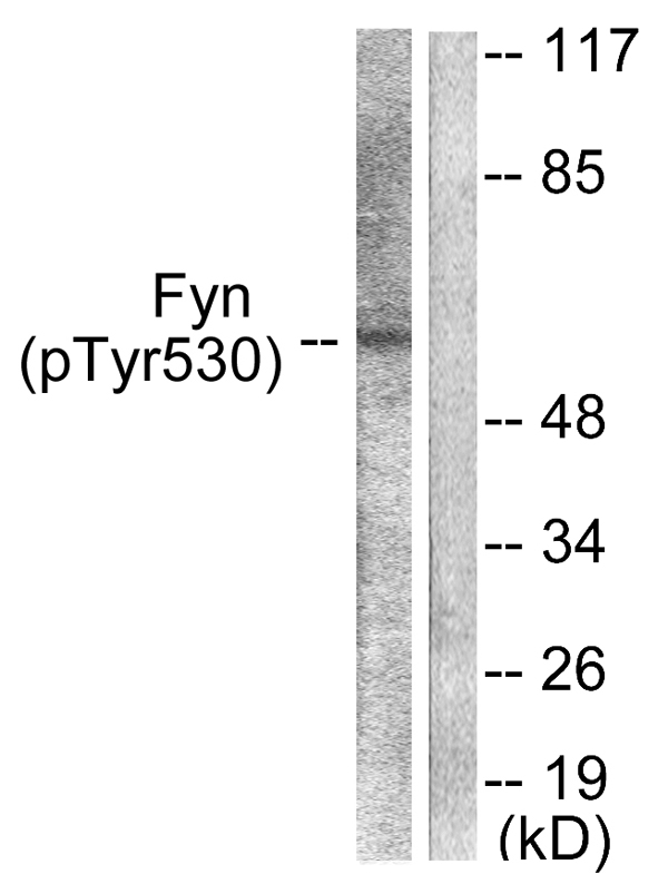 WB (Western Blot)
(Western blot analysis of extracts from 293 cells, treated with H2O2 (100uM, 15mins), using Fyn (Phospho-Tyr530) antibody (#A0430).)
WB (Western Blot)
(Western blot analysis of extracts from 293 cells, treated with H2O2 (100uM, 15mins), using Fyn (Phospho-Tyr530) antibody (#A0430).)
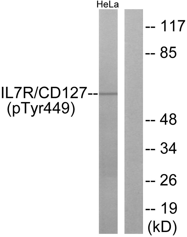
 WB (Western Blot)
(P-peptide-+ Western blot analysis of extracts from HeLa cells, using IL-7R/CD127 (Phospho-Tyr449) antibody. Immunofluorescence analysis of HUVEC cells, using IL-7R/CD127 (Phospho-Tyr449) antibody.)
WB (Western Blot)
(P-peptide-+ Western blot analysis of extracts from HeLa cells, using IL-7R/CD127 (Phospho-Tyr449) antibody. Immunofluorescence analysis of HUVEC cells, using IL-7R/CD127 (Phospho-Tyr449) antibody.)
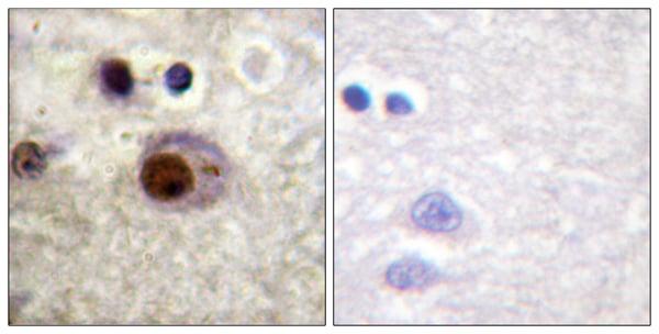
 IHC (Immunohiostchemistry)
(P-peptide-+ Immunohistochemistry analysis of paraffin-embedded human testis tissue using c-Jun (Phospho-Thr231) antibody. Immunofluorescence analysis of HeLa cells, using c-Jun (Phospho-Thr231) antibody.)
IHC (Immunohiostchemistry)
(P-peptide-+ Immunohistochemistry analysis of paraffin-embedded human testis tissue using c-Jun (Phospho-Thr231) antibody. Immunofluorescence analysis of HeLa cells, using c-Jun (Phospho-Thr231) antibody.)
 IHC (Immunohiostchemistry)
(Immunohistochemistry analysis of paraffin-embedded human breast carcinoma tissue using p53 (Phospho-Ser9) antibody. Western blot analysis of extracts from LOVO cells, using p53 (Phospho-Ser9) antibody.)
IHC (Immunohiostchemistry)
(Immunohistochemistry analysis of paraffin-embedded human breast carcinoma tissue using p53 (Phospho-Ser9) antibody. Western blot analysis of extracts from LOVO cells, using p53 (Phospho-Ser9) antibody.)
 IF (Immunofluorescence)
(serum +- Immunofluorescence analysis of HuvEc cells, treated with serum (20%, 30mins), using Histone H1 (Phospho-Thr17) antibody.)
IF (Immunofluorescence)
(serum +- Immunofluorescence analysis of HuvEc cells, treated with serum (20%, 30mins), using Histone H1 (Phospho-Thr17) antibody.)
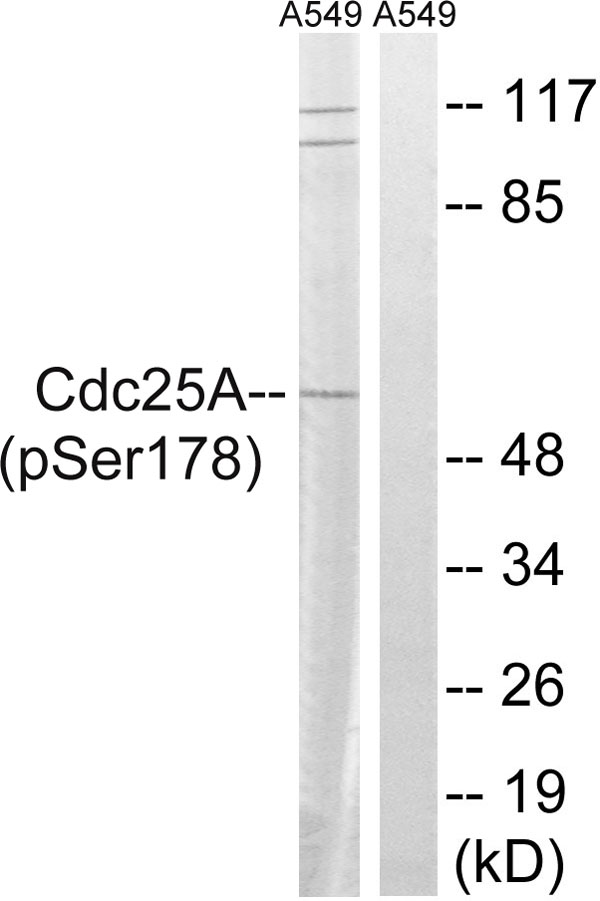 IHC (Immunohiostchemistry)
(Immunohistochemistry analysis of paraffin-embedded human breast carcinoma tissue using CDC25A (Phospho-Ser178) antibody. Western blot analysis of extracts from A549 cells, treated with UV (15mins), using CDC25A (Phospho-Ser178) antibody.)
IHC (Immunohiostchemistry)
(Immunohistochemistry analysis of paraffin-embedded human breast carcinoma tissue using CDC25A (Phospho-Ser178) antibody. Western blot analysis of extracts from A549 cells, treated with UV (15mins), using CDC25A (Phospho-Ser178) antibody.)
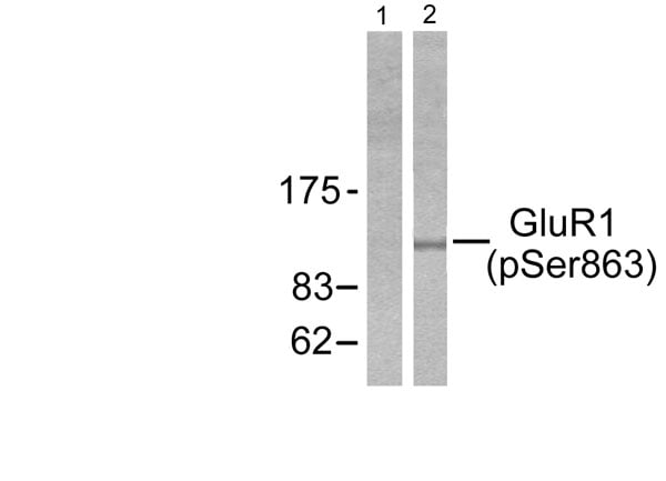 WB (Western Blot)
(Western blot analysis of extracts from HeLa cells treated with PMA (125ng/ml, 30mins), using GluR1 (phospho-Ser863) antibody (Line 1 and 2).)
WB (Western Blot)
(Western blot analysis of extracts from HeLa cells treated with PMA (125ng/ml, 30mins), using GluR1 (phospho-Ser863) antibody (Line 1 and 2).)