Filters
▼Clonality
▼Type
▼Reactivity
▼Gene Name
▼Isotype
▼Host
▼Application
▼Clone
▼Phospho Antibodies
Phospho-specific antibodies’ typical purpose is to enable researchers to detect changes in proteins. They will exclusively bind to the amino acid sequence on a protein that has been phosphorylated (which is both a physical & chemical change) and do not bind to the same amino acid sequence on said protein if it lacks said phosphorylation. This aids in being able to clearly see and understand the data produced from this particular protein modification.
Viewing 2550-2600 of 5298 product results

 DB (Dot Blot)
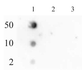
(RNA Pol II CTD phospho Ser2 antibody (pAb) tested by dot blot analysis. Dot blot analysis was used to confirm the specificity of RNA Pol II CTD phospho Ser2 pAb. Peptides corresponding to the immunogen and related peptides were spotted onto PVDF and probed with the antibody at a dilution of 1:2,000. The amount of peptide (picomoles) spotted is indicated next to each row. Lane 1: Phospho Ser2 of RNA Pol II CTD peptide. Lane 2: Unmodified RNA Pol II CTD peptide. Lane 3: Phospho Ser5 of RNA Pol II CTD peptide.)
DB (Dot Blot)
(RNA Pol II CTD phospho Ser2 antibody (pAb) tested by dot blot analysis. Dot blot analysis was used to confirm the specificity of RNA Pol II CTD phospho Ser2 pAb. Peptides corresponding to the immunogen and related peptides were spotted onto PVDF and probed with the antibody at a dilution of 1:2,000. The amount of peptide (picomoles) spotted is indicated next to each row. Lane 1: Phospho Ser2 of RNA Pol II CTD peptide. Lane 2: Unmodified RNA Pol II CTD peptide. Lane 3: Phospho Ser5 of RNA Pol II CTD peptide.)
RNA pol II CTD phospho Ser2, Polyclonal Antibody (Cat# AAA59880)
 ICC (Immunocytochemistry)
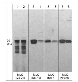
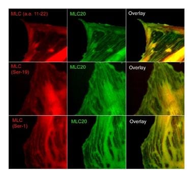
(Immunocytochemical labeling of phosphorylated MLC in paraformaldehyde fixed A7r5 cells. The cells were dual-labeled with anti-MLC (MM3441; middle) and anti-MLC (MP4201; top left), anti-MLC (Ser-19) (MP4221; middle left) and anti-MLC (Ser-1) (MP3461; bottom left). Goat anti-Mouse DyLight 488 and Goat anti-Rabbit DyLight 594 were used for detection of primary antibodies. The overlay of staining patterns are shown to the right.)
ICC (Immunocytochemistry)
(Immunocytochemical labeling of phosphorylated MLC in paraformaldehyde fixed A7r5 cells. The cells were dual-labeled with anti-MLC (MM3441; middle) and anti-MLC (MP4201; top left), anti-MLC (Ser-19) (MP4221; middle left) and anti-MLC (Ser-1) (MP3461; bottom left). Goat anti-Mouse DyLight 488 and Goat anti-Rabbit DyLight 594 were used for detection of primary antibodies. The overlay of staining patterns are shown to the right.)
Myosin, Polyclonal Antibody (Cat# AAA71660)
 ICC (Immunocytochemistry)
(Immunocytochemical labeling of myosin IIA heavy chain phosphorylation relative to F-actin in chick fibroblasts. The cells were labeled with rabbit polyclonal Myosin IIA Heavy Chain (Ser-1943) antibody (MP3831), then detected using appropriate secondary antibody (Bottom, Red). This labeling is compared to F-actin staining (Bottom, Green) and to secondary only (Top). (Image provided by Dr. Gianluca Gallo at Drexel University).)
ICC (Immunocytochemistry)
(Immunocytochemical labeling of myosin IIA heavy chain phosphorylation relative to F-actin in chick fibroblasts. The cells were labeled with rabbit polyclonal Myosin IIA Heavy Chain (Ser-1943) antibody (MP3831), then detected using appropriate secondary antibody (Bottom, Red). This labeling is compared to F-actin staining (Bottom, Green) and to secondary only (Top). (Image provided by Dr. Gianluca Gallo at Drexel University).)
Myosin IIA Heavy Chain, Polyclonal Antibody (Cat# AAA71662)
 ICC (Immunocytochemistry)
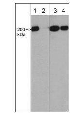
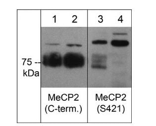
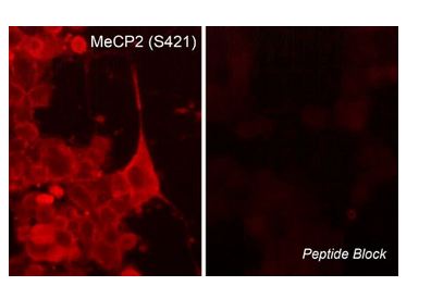
(Immunocytochemical labeling of MeCP2 phosphorylation in rat PC12 cells differentiated with NGF. The cells were probed with MeCP2 (Ser-421) rabbit polyclonal antibody (MP4611) in the absence (left) or presence (right) of blocking peptide (MX4615). The antibody was detected using appropriate secondary antibody conjugated to DyLight 594.)
ICC (Immunocytochemistry)
(Immunocytochemical labeling of MeCP2 phosphorylation in rat PC12 cells differentiated with NGF. The cells were probed with MeCP2 (Ser-421) rabbit polyclonal antibody (MP4611) in the absence (left) or presence (right) of blocking peptide (MX4615). The antibody was detected using appropriate secondary antibody conjugated to DyLight 594.)
MeCP2, Polyclonal Antibody (Cat# AAA71670)
 ICC (Immunocytochemistry)
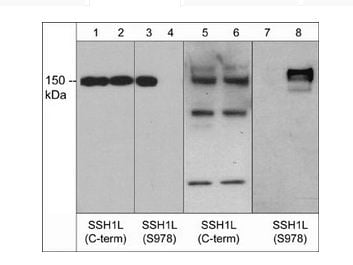
(Immunocytochemical labeling of Slingshot-1L in rat PC12 cells differentiated with NGF. The cells were labeled with rabbit polyclonal anti-SSH1L (C-term.) and anti-SSH1L (Ser-978) antibodies, then detected using appropriate secondary antibody conjugated to Cy3 (Right panel). Phase image of corresponding PC12 cells (Left panel).)
ICC (Immunocytochemistry)
(Immunocytochemical labeling of Slingshot-1L in rat PC12 cells differentiated with NGF. The cells were labeled with rabbit polyclonal anti-SSH1L (C-term.) and anti-SSH1L (Ser-978) antibodies, then detected using appropriate secondary antibody conjugated to Cy3 (Right panel). Phase image of corresponding PC12 cells (Left panel).)
Slingshot-1L, Polyclonal Antibody (Cat# AAA71713)
 ICC (Immunocytochemistry)
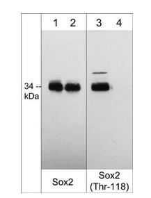
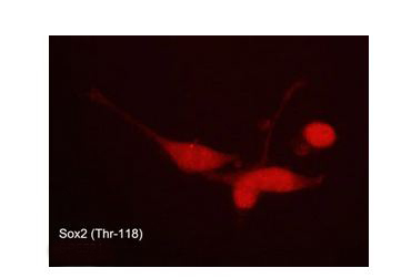
(Immunocytochemical labeling of phosphorylated Sox2 in aldehyde fixed and NP-40 permeabilized human NCI-H446 lung carcinoma cells. The cells were labeled with rabbit polyclonal anti-Sox2 (Thr-118) phospho-specific (SP5521). The antibody was detected using goat anti-rabbit DyLight 594.)
ICC (Immunocytochemistry)
(Immunocytochemical labeling of phosphorylated Sox2 in aldehyde fixed and NP-40 permeabilized human NCI-H446 lung carcinoma cells. The cells were labeled with rabbit polyclonal anti-Sox2 (Thr-118) phospho-specific (SP5521). The antibody was detected using goat anti-rabbit DyLight 594.)
Sox2, Polyclonal Antibody (Cat# AAA71715)
 Application Data
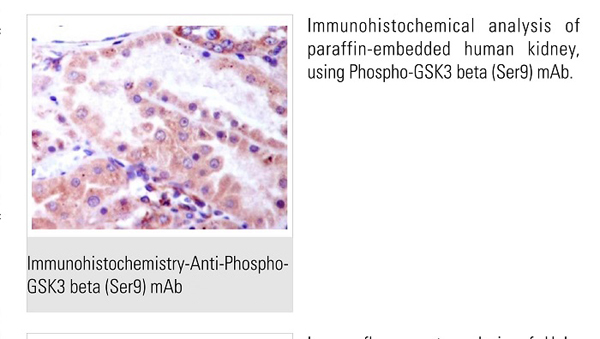
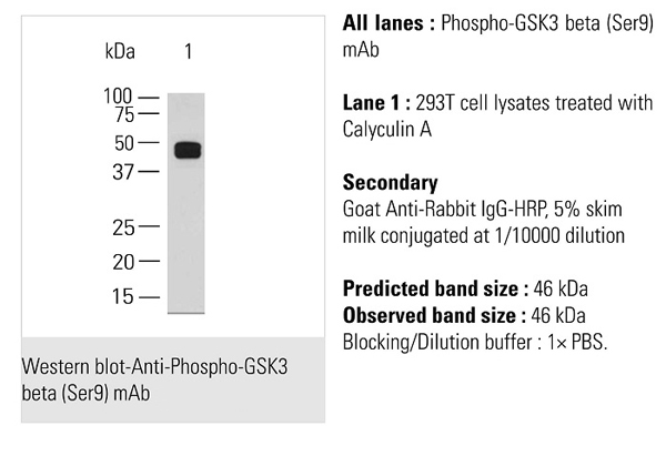
GSK3b (pS9), Antibody (Cat# AAA71401)
 Application Data
STAT1 (pY701), Antibody (Cat# AAA71403)
 Application Data
Rho Kinase/ROCKII (pT249), Antibody (Cat# AAA71408)
 Application Data
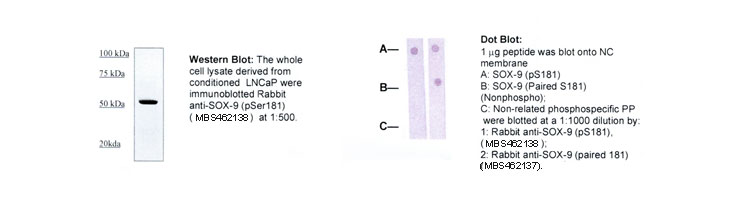
SOX-9 (pS181), Antibody (Cat# AAA71414)
 Application Data
STAT6 (pY641), Antibody (Cat# AAA71425)
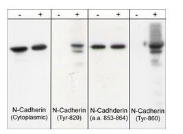
 ICC (Immunocytochemistry)
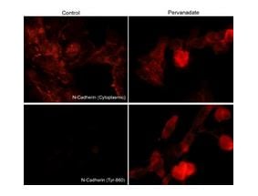
(Immunocytochemical labeling of phosphorylated N-Cadherin in pervanadate-treated mouse C2C12. The cells were labeled with mouse monoclonal N-Cadherin (Cytoplasmic) and rabbit polyclonal N-Cadherin(Tyr-860) antibodies, then the antibodies were detected using appropriate secondary antibodies conjugated to Cy3.)
ICC (Immunocytochemistry)
(Immunocytochemical labeling of phosphorylated N-Cadherin in pervanadate-treated mouse C2C12. The cells were labeled with mouse monoclonal N-Cadherin (Cytoplasmic) and rabbit polyclonal N-Cadherin(Tyr-860) antibodies, then the antibodies were detected using appropriate secondary antibodies conjugated to Cy3.)
N-Cadherin, Polyclonal Antibody (Cat# AAA71602)
 ICC (Immunocytochemistry)
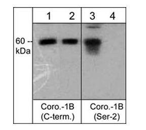
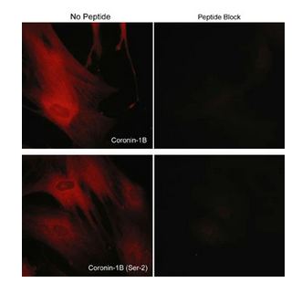
(Immunocytochemical labeling of coronin-1B in rabbit spleen fibroblasts treated with Calyculin A. The cells were labeled with rabbit polyclonal Coronin-1B (C-terminus) and Coronin-1B (Ser-2) antibodies, then detected using appropriate secondary antibodies conjugated to Cy3. The antibodies were used in the absence (left) or presence (right) of their respective blocking peptide (CX2585 or CX2625).)
ICC (Immunocytochemistry)
(Immunocytochemical labeling of coronin-1B in rabbit spleen fibroblasts treated with Calyculin A. The cells were labeled with rabbit polyclonal Coronin-1B (C-terminus) and Coronin-1B (Ser-2) antibodies, then detected using appropriate secondary antibodies conjugated to Cy3. The antibodies were used in the absence (left) or presence (right) of their respective blocking peptide (CX2585 or CX2625).)
Coronin-1B, Polyclonal Antibody (Cat# AAA71607)

 ICC (Immunocytochemistry)
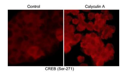
(Immunocytochemical labeling of phosphorylated CREB in control and calyculin A-treated A431 cells. The cells were fixed in paraformaldehyde and permeabilized using NP-40 before labeling with rabbit polyclonal CREB (Ser-271). The antibody was detected using goat anti-rabbit DyLight 594.)
ICC (Immunocytochemistry)
(Immunocytochemical labeling of phosphorylated CREB in control and calyculin A-treated A431 cells. The cells were fixed in paraformaldehyde and permeabilized using NP-40 before labeling with rabbit polyclonal CREB (Ser-271). The antibody was detected using goat anti-rabbit DyLight 594.)
CREB, Polyclonal Antibody (Cat# AAA71612)
 ICC (Immunocytochemistry)
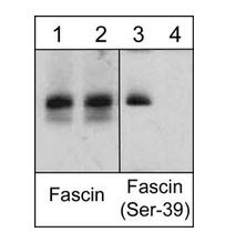
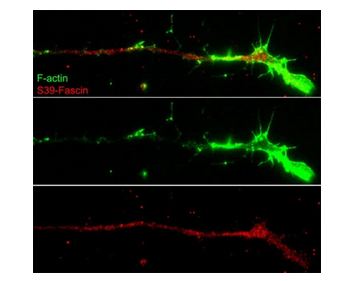
(Immunocytochemical labeling of fascin phosphorylation relative to F-actin in chick E9 DRG neurons. The cells were labeled with rabbit polyclonal Fascin (Ser-39) antibody, then detected using appropriate secondary antibody (Red). Fascin (Ser-39) labeling is compared (Top) to F-actin staining (Green). (Image provided by Dr. Gianluca Gallo at Drexel University).)
ICC (Immunocytochemistry)
(Immunocytochemical labeling of fascin phosphorylation relative to F-actin in chick E9 DRG neurons. The cells were labeled with rabbit polyclonal Fascin (Ser-39) antibody, then detected using appropriate secondary antibody (Red). Fascin (Ser-39) labeling is compared (Top) to F-actin staining (Green). (Image provided by Dr. Gianluca Gallo at Drexel University).)
Fascin, Polyclonal Antibody (Cat# AAA71641)
 WB (Western Blot)
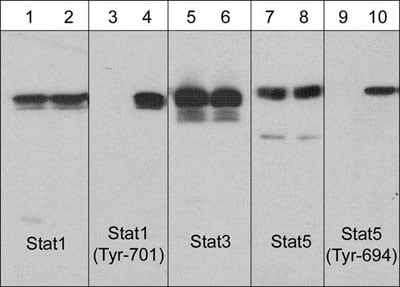
(Western blot analysis of human A431 cells untreated (lanes 1, 3, 5, 7 & 9) or treated with EGF (100 nM) for 60 min (lanes 2, 4, 6, 8 & 10). The blots were probed with anti-Stat1 (lanes 1 & 2), anti-Stat1 (Tyr-701) (lanes 3 & 4), anti-Stat3 (lanes 5 & 6), anti-Stat5 (lanes 7 & 8), and anti-Stat5 (Tyr-694) (lanes 9 & 10).)
WB (Western Blot)
(Western blot analysis of human A431 cells untreated (lanes 1, 3, 5, 7 & 9) or treated with EGF (100 nM) for 60 min (lanes 2, 4, 6, 8 & 10). The blots were probed with anti-Stat1 (lanes 1 & 2), anti-Stat1 (Tyr-701) (lanes 3 & 4), anti-Stat3 (lanes 5 & 6), anti-Stat5 (lanes 7 & 8), and anti-Stat5 (Tyr-694) (lanes 9 & 10).)
Stat5 (Tyr-694), Monoclonal Antibody (Cat# AAA71509)
 ICC (Immunocytochemistry)
b-Catenin (Tyr-86), Polyclonal Antibody (Cat# AAA71512)
 Application Data
(Western blot analysis of purified brain tubulin untreated (lanes 1, 3, 5) or treated with ERK2 kinase to phosphorylate Ser-172 (lanes 2, 4, 6).The blot was probed with anti-beta-Tubulin (a.a.168-177)(lanes 1 & 2), anti-beta-Tubulin (Ser-172)(lanes 3 & 4), and anti-beta-Tubulin (lanes 5 & 6).)
Application Data
(Western blot analysis of purified brain tubulin untreated (lanes 1, 3, 5) or treated with ERK2 kinase to phosphorylate Ser-172 (lanes 2, 4, 6).The blot was probed with anti-beta-Tubulin (a.a.168-177)(lanes 1 & 2), anti-beta-Tubulin (Ser-172)(lanes 3 & 4), and anti-beta-Tubulin (lanes 5 & 6).)
beta-Tubulin (Ser-172), Polyclonal Antibody (Cat# AAA71514)
 ICC (Immunocytochemistry)
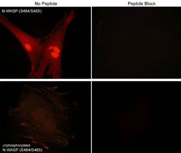
(Immunocytometrical labeling of phosphorylated and unphosphorylated N-WASP in rabbit spleen fibroblasts. The cells were probed with N-WASP phospho-specific and N-WASP unphosphorylated antibodies, then the antibodies were detected using appropriate secondary antibodies conjugated to Cy3. The antibodies were in the absence (left) or presence (right) or their respective blocking peptide.)
ICC (Immunocytochemistry)
(Immunocytometrical labeling of phosphorylated and unphosphorylated N-WASP in rabbit spleen fibroblasts. The cells were probed with N-WASP phospho-specific and N-WASP unphosphorylated antibodies, then the antibodies were detected using appropriate secondary antibodies conjugated to Cy3. The antibodies were in the absence (left) or presence (right) or their respective blocking peptide.)
N-WASP (Ser-484/Ser-485), Polyclonal Antibody (Cat# AAA71525)
 Application Data
Paxillin (Ser-178), Polyclonal Antibody (Cat# AAA71526)
phospho-b-Tubulin (Ser-172) Peptide, Peptide (Cat# AAA71540)
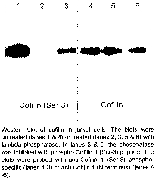 Application Data
phospho-Cofilin 1 (Ser-3) Peptide, Peptide (Cat# AAA71541)
 Application Data
S6K (pS434), Antibody (Cat# AAA71399)
 Application Data
PAK4 (pS474), Antibody (Cat# AAA71400)
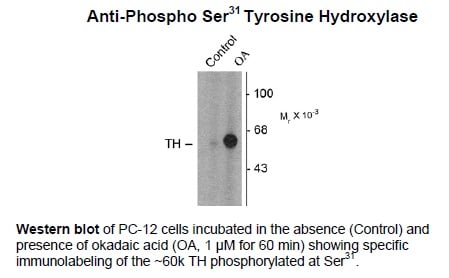 Application Data
Tyrosine Hydroxylase (Ser31), Polyclonal Antibody (Cat# AAA72805)
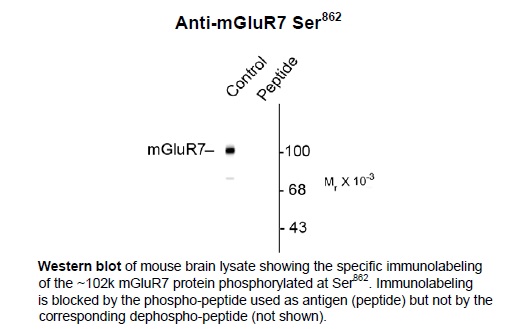 Application Data
Metabotropic Glutamate Receptor 7 (Ser862), Polyclonal Antibody (Cat# AAA72814)
 Application Data
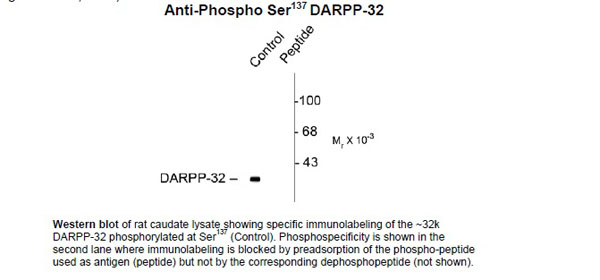
DARPP-32 (Ser137), Polyclonal Antibody (Cat# AAA72815)
 Application Data
Potassium Chloride Cotransporter (KCC2) (Ser940), Polyclonal Antibody (Cat# AAA72823)
(Prepared from pooled rabbit serum by affinity purification via sequential chromatography on phospho and non-phosphopeptide affinity columns.)
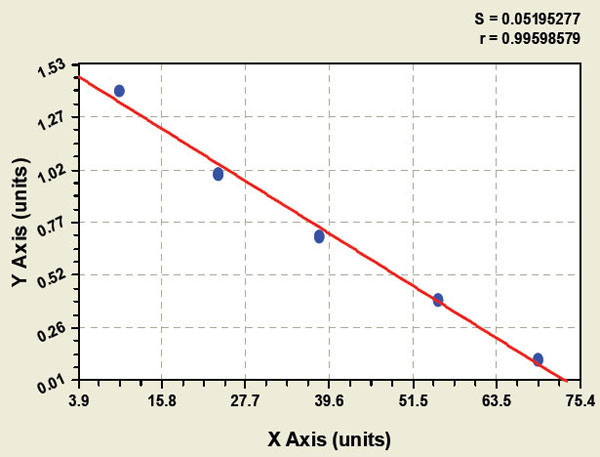 Standard Curve (Sample)
Phospho Spleen tyosine kinase, ELISA Kit (Cat# AAA96475)
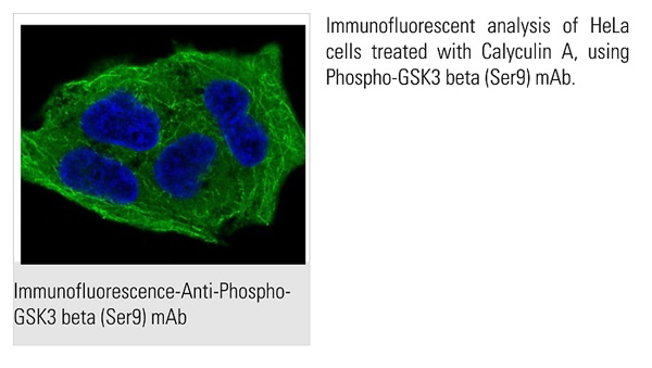 IF (Immunofluorescence)
GSK3 beta, Monoclonal Antibody (Cat# AAA62480)
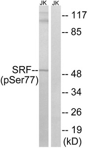
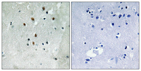 IHC (Immunohiostchemistry)
(Immunohistochemistry analysis of paraffin-embedded human brain tissue using SRF (Phospho-Ser77) antibody. The picture on the right is treated with the synthesized peptide.)
IHC (Immunohiostchemistry)
(Immunohistochemistry analysis of paraffin-embedded human brain tissue using SRF (Phospho-Ser77) antibody. The picture on the right is treated with the synthesized peptide.)
SRF, Polyclonal Antibody (Cat# AAA243249)
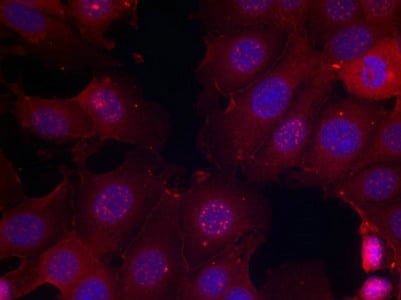 IF (Immunofluorescence)
(Immunofluorescence staining of methanol-fixed MCF cells using Estrogen Receptor-a(Phospho-Ser167) Antibody.)
IF (Immunofluorescence)
(Immunofluorescence staining of methanol-fixed MCF cells using Estrogen Receptor-a(Phospho-Ser167) Antibody.)
ESR1, Polyclonal Antibody (Cat# AAA243266)
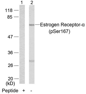
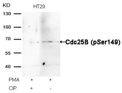 WB (Western Blot)
(Western blot analysis of extracts from HT29 cells, treated with PMA or calf intestinal phosphatase (CIP), using Cdc25B (Phospho-Ser149) Antibody.)
WB (Western Blot)
(Western blot analysis of extracts from HT29 cells, treated with PMA or calf intestinal phosphatase (CIP), using Cdc25B (Phospho-Ser149) Antibody.)
Cdc25b, Polyclonal Antibody (Cat# AAA243163)
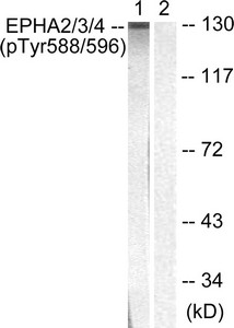
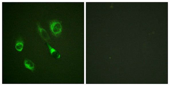 IF (Immunofluorescence)
(Immunofluorescence staining of methanol-fixed HeLa cells using EPHA2/3/4 (Phospho-Tyr588/596) Antibody.)
IF (Immunofluorescence)
(Immunofluorescence staining of methanol-fixed HeLa cells using EPHA2/3/4 (Phospho-Tyr588/596) Antibody.)
EPHA2/EPHA3/EPHA4, Polyclonal Antibody (Cat# AAA243198)
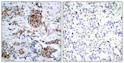
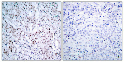 IHC (Immunohiostchemistry)
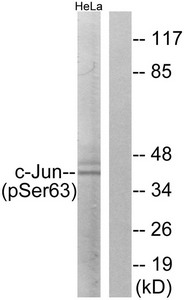
(Immunohistochemical analysis of paraffin-embedded human breast carcinoma tissue, using c-Jun (Phospho-Ser63) antibody (left)or the same antibody preincubated with blocking peptide (right).)
IHC (Immunohiostchemistry)
(Immunohistochemical analysis of paraffin-embedded human breast carcinoma tissue, using c-Jun (Phospho-Ser63) antibody (left)or the same antibody preincubated with blocking peptide (right).)
JUN, Polyclonal Antibody (Cat# AAA243203)
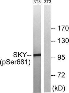
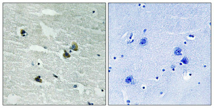 IHC (Immunohiostchemistry)
(Immunohistochemical analysis of paraffin-embedded human brain tissue using MER/SKY (Phospho-Tyr749/681) antibody (left)or the same antibody preincubated with blocking peptide (right).)
IHC (Immunohiostchemistry)
(Immunohistochemical analysis of paraffin-embedded human brain tissue using MER/SKY (Phospho-Tyr749/681) antibody (left)or the same antibody preincubated with blocking peptide (right).)
MERTK/TYRO3, Polyclonal Antibody (Cat# AAA243208)


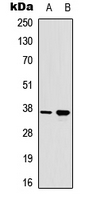
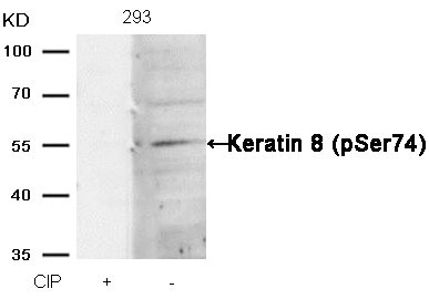 WB (Western Blot)
(Western blot analysis of extracts from 293 cells, treated with calf intestinal phosphatase (CIP), using Keratin 8 (Phospho-Ser74) Antibody.)
WB (Western Blot)
(Western blot analysis of extracts from 293 cells, treated with calf intestinal phosphatase (CIP), using Keratin 8 (Phospho-Ser74) Antibody.)
KRT8, Polyclonal Antibody (Cat# AAA243317)


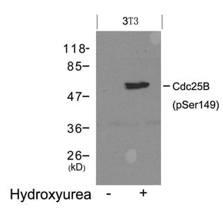
 WB (Western Blot)
(Western blot analysis of extracts from 3T3 cells, treated with serum or calf intestinal phosphatase (CIP), using MARCKS (phospho-Ser170) Antibody.)
WB (Western Blot)
(Western blot analysis of extracts from 3T3 cells, treated with serum or calf intestinal phosphatase (CIP), using MARCKS (phospho-Ser170) Antibody.)
MARCKS, Polyclonal Antibody (Cat# AAA243326)


 IF (Immunofluorescence)
(Immunofluorescence staining of methanol-fixed Hela cells using Cyclin B1(phospho-Ser147) Antibody.)
IF (Immunofluorescence)
(Immunofluorescence staining of methanol-fixed Hela cells using Cyclin B1(phospho-Ser147) Antibody.)
CCNB1, Polyclonal Antibody (Cat# AAA243327)
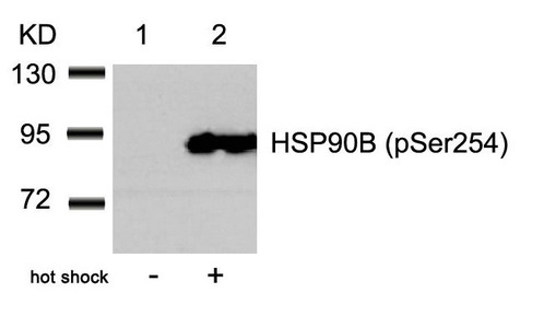
 WB (Western Blot)
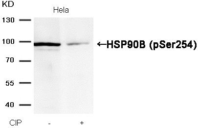
(Western blot analysis of extracts from Hela cells, treated with calf intestinal phosphatase (CIP), using HSP90B (Phospho-Ser254) Antibody.)
WB (Western Blot)
(Western blot analysis of extracts from Hela cells, treated with calf intestinal phosphatase (CIP), using HSP90B (Phospho-Ser254) Antibody.)
HSP90AB1, Polyclonal Antibody (Cat# AAA243111)

 IHC (Immunohiostchemistry)
(Immunohistochemical analysis of paraffin-embedded human tonsil tumor tissue using Bcr(Phospho-Tyr177) Antibody(left) or the same antibody preincubated with blocking peptide(right).)
IHC (Immunohiostchemistry)
(Immunohistochemical analysis of paraffin-embedded human tonsil tumor tissue using Bcr(Phospho-Tyr177) Antibody(left) or the same antibody preincubated with blocking peptide(right).)
BCR, Polyclonal Antibody (Cat# AAA243113)
 IHC (Immunohiostchemistry)
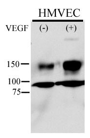
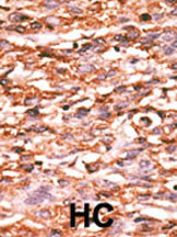
(Formalin-fixed and paraffin-embedded human cancer tissue reacted with the primary antibody, which was peroxidase-conjugated to the secondary antibody, followed by AEC staining. This data demonstrates the use of this antibody for immunohistochemistry; clinical relevance has not been evaluated. BC = breast carcinoma; HC = hepatocarcinoma.)
IHC (Immunohiostchemistry)
(Formalin-fixed and paraffin-embedded human cancer tissue reacted with the primary antibody, which was peroxidase-conjugated to the secondary antibody, followed by AEC staining. This data demonstrates the use of this antibody for immunohistochemistry; clinical relevance has not been evaluated. BC = breast carcinoma; HC = hepatocarcinoma.)
Phospho-KDR/FLK1 (Y996), Polyclonal Antibody (Cat# AAA283770)
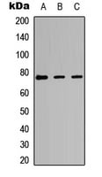
 WB (Western Blot)
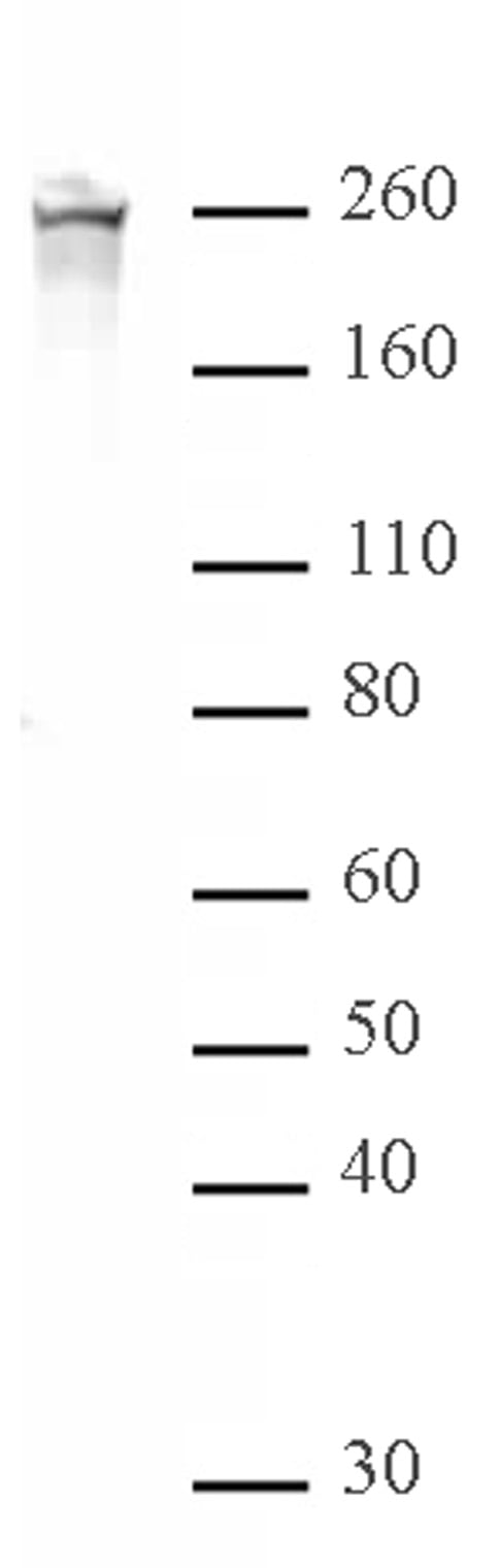
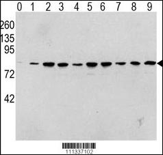
(Western blot analysis of Phospho-RAF1-pS338 Antibody in human TPA activated Hela cell line lysates. Phospho-RAF1 (arrow) was detected using the purified PAb. (0: without TPA; 1: 60ug/ml TPA, 15min; 2: 60ug/ml TPA, 30min; 3: 60ug/ml TPA, 45min; 4: 125ug/ml TPA, 15min; 5: 125ug/ml TPA, 30min; 6: 125ug/ml TPA, 45min; 7: 250ug/ml TPA, 15min; 8: 250ug/ml TPA, 30min; 9: 250ug/ml, 45min))
WB (Western Blot)
(Western blot analysis of Phospho-RAF1-pS338 Antibody in human TPA activated Hela cell line lysates. Phospho-RAF1 (arrow) was detected using the purified PAb. (0: without TPA; 1: 60ug/ml TPA, 15min; 2: 60ug/ml TPA, 30min; 3: 60ug/ml TPA, 45min; 4: 125ug/ml TPA, 15min; 5: 125ug/ml TPA, 30min; 6: 125ug/ml TPA, 45min; 7: 250ug/ml TPA, 15min; 8: 250ug/ml TPA, 30min; 9: 250ug/ml, 45min))
Phospho-RAF1 (S338), Polyclonal Antibody (Cat# AAA284142)
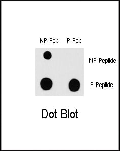
 DB (Dot Blot)
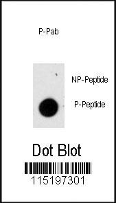
(Dot blot analysis of anti-Phospho-GFAP-S8 Antibody on nitrocellulose membrane. 50ng of Phospho-peptide or Non Phospho-peptide per dot were adsorbed. Antibody working concentrations are 0.5ug per ml.)
DB (Dot Blot)
(Dot blot analysis of anti-Phospho-GFAP-S8 Antibody on nitrocellulose membrane. 50ng of Phospho-peptide or Non Phospho-peptide per dot were adsorbed. Antibody working concentrations are 0.5ug per ml.)
Phospho-GFAP (S8), Polyclonal Antibody (Cat# AAA283958)
 DB (Dot Blot)
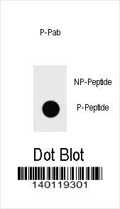
(Dot blot analysis of Phospho-IKKB-S672 Antibody Phospho-specific Pab on nitrocellulose membrane. 50ng of Phospho-peptide or Non Phospho-peptide per dot were adsorbed. Antibody working concentrations are 0.6ug per ml.)
DB (Dot Blot)
(Dot blot analysis of Phospho-IKKB-S672 Antibody Phospho-specific Pab on nitrocellulose membrane. 50ng of Phospho-peptide or Non Phospho-peptide per dot were adsorbed. Antibody working concentrations are 0.6ug per ml.)
Phospho-IKKB (S672), Polyclonal Antibody (Cat# AAA283856)
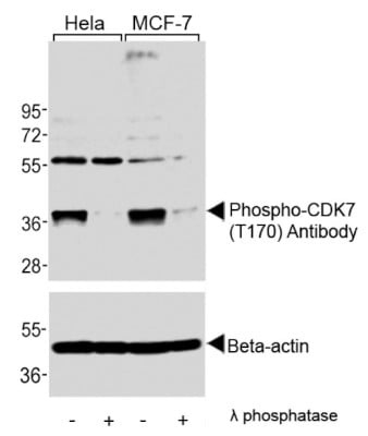
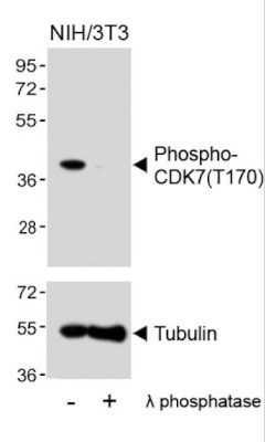
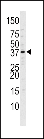
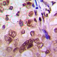
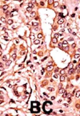 IHC (Immunohistochemistry)
(Formalin-fixed and paraffin-embedded human canccer tissue reacted with the primary antbody, which was peroxidase-conjugated to the secondary antibody, which followed by AEC staining. This data demonstrates the use of this antibody for immunohistochemisty; clinical relevance has not been evaluated. BC= Breast Carcinoma; HC= Hepatocarcinoma.)
IHC (Immunohistochemistry)
(Formalin-fixed and paraffin-embedded human canccer tissue reacted with the primary antbody, which was peroxidase-conjugated to the secondary antibody, which followed by AEC staining. This data demonstrates the use of this antibody for immunohistochemisty; clinical relevance has not been evaluated. BC= Breast Carcinoma; HC= Hepatocarcinoma.)
Phospho-CDK7 (T170), Polyclonal Antibody (Cat# AAA283874)
 DB (Dot Blot)
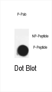
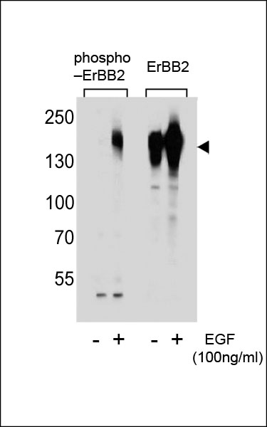
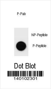
(Dot blot analysis of Phospho-ERBB2-S1107 Antibody Phospho-specific Pab on nitrocellulose membrane. 50ng of Phospho-peptide or Non Phospho-peptide per dot were adsorbed. Antibody working concentrations are 0.6ug per ml.)
DB (Dot Blot)
(Dot blot analysis of Phospho-ERBB2-S1107 Antibody Phospho-specific Pab on nitrocellulose membrane. 50ng of Phospho-peptide or Non Phospho-peptide per dot were adsorbed. Antibody working concentrations are 0.6ug per ml.)
Phospho-ERBB2 (S1107), Polyclonal Antibody (Cat# AAA284285)
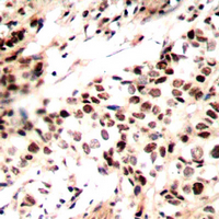
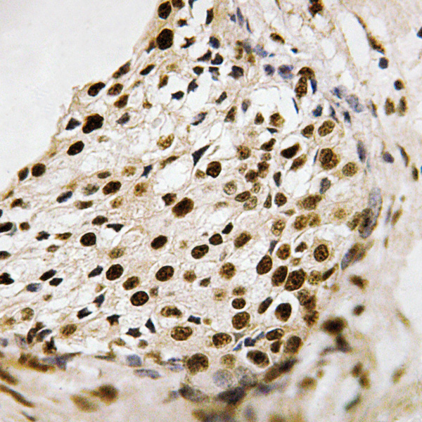 IHC (Immunohiostchemistry)
(Immunohistochemical analysis of PRAK (phospho-Thr182) staining in human breast carcinoma formalin fixed paraffin embedded tissue section. The section was then incubated with the antibody at room temperature and detected using an HRP conjugated compact polymer system. DAB was used as the chromogen. The section was then counterstained with haematoxylin and mounted with DPX.)
IHC (Immunohiostchemistry)
(Immunohistochemical analysis of PRAK (phospho-Thr182) staining in human breast carcinoma formalin fixed paraffin embedded tissue section. The section was then incubated with the antibody at room temperature and detected using an HRP conjugated compact polymer system. DAB was used as the chromogen. The section was then counterstained with haematoxylin and mounted with DPX.)
PRAK (phospho-Thr182), Polyclonal Antibody (Cat# AAA310501)
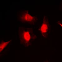 IF (Immunofluorescence)
(Immunofluorescent analysis of PKC iota/zeta (phospho-Thr412/410) staining in MCF7 cells. Formalin-fixed cells were permeabilized with 0.1% Triton X-100 in TBS for 5-10 minutes and blocked with 3% BSA-PBS for 30 minutes at room temperature. Cells were probed with the primary antibody in 3% BSA-PBS and incubated overnight at 4 C in a hidified chamber. Cells were washed with PBST and incubated with a DyLight 594-conjugated secondary antibody (red) in PBS at room temperature in the dark.)
IF (Immunofluorescence)
(Immunofluorescent analysis of PKC iota/zeta (phospho-Thr412/410) staining in MCF7 cells. Formalin-fixed cells were permeabilized with 0.1% Triton X-100 in TBS for 5-10 minutes and blocked with 3% BSA-PBS for 30 minutes at room temperature. Cells were probed with the primary antibody in 3% BSA-PBS and incubated overnight at 4 C in a hidified chamber. Cells were washed with PBST and incubated with a DyLight 594-conjugated secondary antibody (red) in PBS at room temperature in the dark.)
PKC iota/zeta (phospho-Thr412/410), Polyclonal Antibody (Cat# AAA310512)
 IHC (Immunohiostchemistry)
(Immunohistochemical analysis of RAD51A (phospho-Tyr315) staining in human breast cancer formalin fixed paraffin embedded tissue section. The section was then incubated with the antibody at room temperature and detected using an HRP conjugated compact polymer system. DAB was used as the chromogen. The section was then counterstained with haematoxylin and mounted with DPX.)
IHC (Immunohiostchemistry)
(Immunohistochemical analysis of RAD51A (phospho-Tyr315) staining in human breast cancer formalin fixed paraffin embedded tissue section. The section was then incubated with the antibody at room temperature and detected using an HRP conjugated compact polymer system. DAB was used as the chromogen. The section was then counterstained with haematoxylin and mounted with DPX.)
RAD51A (phospho-Tyr315), Polyclonal Antibody (Cat# AAA310516)
What Are Phospho Antibodies?
Protein phosphorylation is a process where a phosphate group is added to certain amino acid residues of a protein – usually serine (S), threonine (T), or tyrosine (Y) - by enzymes called kinases. This process is integral in controlling cellular signaling, cellular growth, and other biological functions.
Our catalog includes a wide range of phospho-specific antibodies that can accurately detect this important marker. They perform strongly in widely-used laboratory applications such as Western blot, flow cytometry, immunohistochemistry, and immunofluorescence microscopy. We value your trust in us and are committed to providing top-quality products and services. All of our antibodies are guaranteed to work for the applications and species indicated on our website & associated product pages.
What Are The Key Applications of Phospho Antibodies?
1. Western Blotting
One of the first steps a researcher can take in utilizing these phospho-specific antibodies, is to check if the antibody works using a technique referred to as “Western blot”. For those unfamiliar, Western Blot aids in showing whether the protein that the antibody recognizes is appearing at the correct/expected size. These phospho-specific antibodies should also be able to detect changes in the target protein’s phosphorylation (on/off state) when cells are stimulated in certain ways.
2. Staining of Fixed Cells (Immunocytochemistry)
Another routine use of these phospho-specific antibodies, is to test if the antibody is able to demonstrate similar performance when used on fixed cells (intact cells that have been preserved) as it did in the Western blot tests. It is an important aspect in many cases to confirm that the antibody works in actual intact cell samples. Ideally, the method used for cellular fixation should be the same as what is used in pathology labs (like using 10% formalin). To check if the antibody works well in tissue sections (FFPE), researchers will often test it on fixed cells that are processed similar to tissue samples.
3. Specificity Tests Using Peptides
In order to make sure that the antibody is only binding to the right target:
- Laboratory technicians will mix the antibody with phospho-peptides (short segments of the protein containing the phosphate group modification).
- If the antibody signal disappears, it is confirmation that it is binding to the correct phosphorylated location.
- A more robust test is to use both the phosphorylated and non-phosphorylated (dephosphorylated) versions of the protein. The antibody should react only with the phosphorylated one.
- Another method sometimes utilized is to treat the sample with an enzyme, such as alkaline phosphatase, that specifically removes phosphate groups. If the antibody signal disappears after this, it also confirms specificity.
4. Genetic Confirmation
As a final step, scientists can genetically manipulate the nucleotide sequence and alter the target protein by removing the exact site where phosphorylation happens. If the antibody no longer appears to detect the modified protein, it is strong evidence supporting the antibody being specific for that phosphorylated site.
Why Buy Phospho Antibodies Through Us?
- The production laboratory adheres to strict and consistent protocols prior to releasing any of these phospho-specific antibodies:
- Standard methods and proper controls in all tests to ensure high quality.
- These antibodies are tested and validated in different cell types and species.
- High quality control criterion to ensure each batch is consistent, so you will obtain reliable results every time.
FAQ
1. What Are Phospho-Specific Antibodies?
Phospho-specific antibodies are made to detect proteins only when they have a phosphate group linked to a specific amino acid residue. This empowers scientists understand if a protein is "turned on" or active, based on its phosphorylation state.
2. How to Detect Phosphorylated Proteins in a Western Blot?
To find out if a protein is phosphorylated using Western blot:
- Use a phospho-specific antibody that binds only to the phosphorylated form of the protein.
- You can also use a “regular” antibody for the same amino acid sequence of the protein that the phospho-specific antibody is binding to (but in this case, this antibody will not bind if there is a phosphate group present) in order to compare how much of it is phosphorylated versus how much is non-phosphorylated (or “total” protein, if the “normal” antibody’s epitopes are non-phospho-site-specific).
3. How to Choose the Best Antibody?
Here are some simple tips to help you pick the right antibody:
- Know your target
- Match your sample characteristics
- Confirm the intended use is appropriate
- Check “host” and “type”
- Check the “quality” of the presented data/images
- Appraise whether the available validation meets your needs