Filters
▼Clonality
▼Type
▼Reactivity
▼Gene Name
▼Isotype
▼Host
▼Application
▼Clone
▼Phospho Antibodies
Phospho-specific antibodies’ typical purpose is to enable researchers to detect changes in proteins. They will exclusively bind to the amino acid sequence on a protein that has been phosphorylated (which is both a physical & chemical change) and do not bind to the same amino acid sequence on said protein if it lacks said phosphorylation. This aids in being able to clearly see and understand the data produced from this particular protein modification.
Viewing 2600-2650 of 5298 product results
 ICC (Immunocytochemistry)
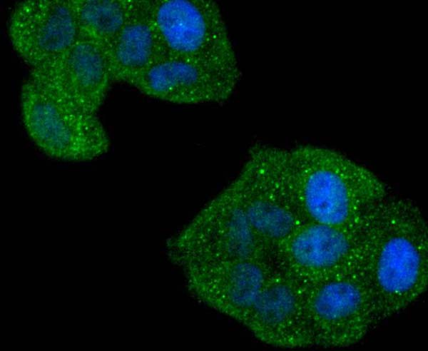
(ICC staining Phospho-p95/NBS1 (S343) in PC-3M cells (green). The nuclear counter stain is DAPI (blue). Cells were fixed in paraformaldehyde, permeabilised with 0.25% Triton X100/PBS.)
ICC (Immunocytochemistry)
(ICC staining Phospho-p95/NBS1 (S343) in PC-3M cells (green). The nuclear counter stain is DAPI (blue). Cells were fixed in paraformaldehyde, permeabilised with 0.25% Triton X100/PBS.)
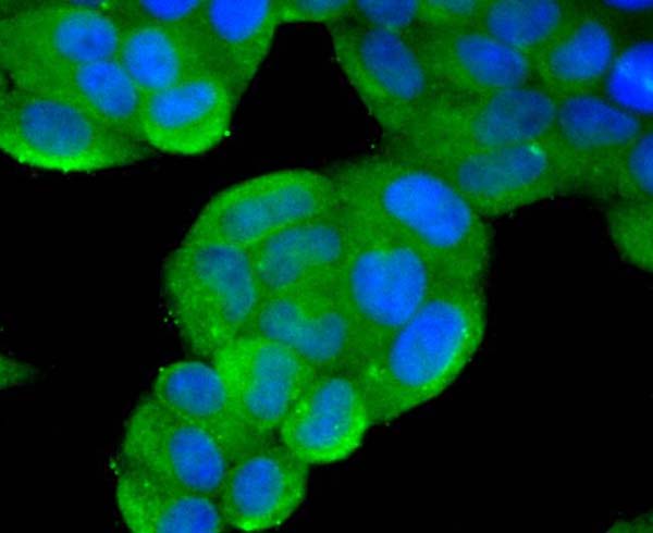
p95/NBS1, Monoclonal Antibody (Cat# AAA311002)
 ICC (Immunocytochemistry)
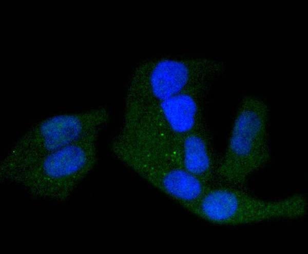
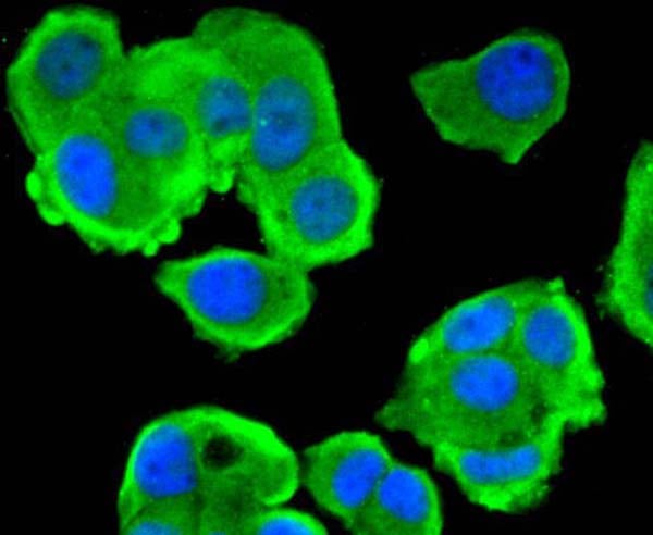
(ICC staining Phospho-c-Myc (S62) in Hela cells (green). The nuclear counter stain is DAPI (blue). Cells were fixed in paraformaldehyde, permeabilised with 0.25% Triton X100/PBS.)
ICC (Immunocytochemistry)
(ICC staining Phospho-c-Myc (S62) in Hela cells (green). The nuclear counter stain is DAPI (blue). Cells were fixed in paraformaldehyde, permeabilised with 0.25% Triton X100/PBS.)
c-Myc, Monoclonal Antibody (Cat# AAA311008)
 ICC (Immunocytochemistry)
(ICC staining phospho-PKA R2 (S99) in MCF-7 cells (green). The nuclear counter stain is DAPI (blue). Cells were fixed in paraformaldehyde, permeabilised with 0.25% Triton X100/PBS.)
ICC (Immunocytochemistry)
(ICC staining phospho-PKA R2 (S99) in MCF-7 cells (green). The nuclear counter stain is DAPI (blue). Cells were fixed in paraformaldehyde, permeabilised with 0.25% Triton X100/PBS.)
PKA R2, Monoclonal Antibody (Cat# AAA311011)
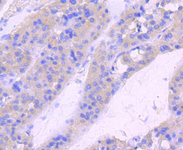
 IHC (Immunohistochemistry)
(Immunohistochemical analysis of paraffin-embedded human breast carcinoma tissue using anti- phospho -SMC1 (S957) antibody. Counter stained with hematoxylin.)
IHC (Immunohistochemistry)
(Immunohistochemical analysis of paraffin-embedded human breast carcinoma tissue using anti- phospho -SMC1 (S957) antibody. Counter stained with hematoxylin.)
SMC1, Monoclonal Antibody (Cat# AAA311015)
 ICC (Immunocytochemistry)
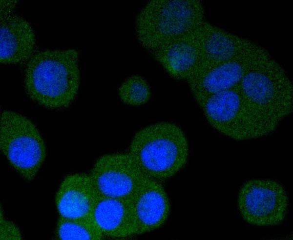
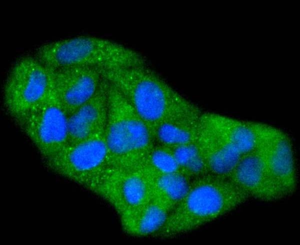
(ICC staining phospho -Tau (S396) in PC12 cells (green). The nuclear counter stain is DAPI (blue). Cells were fixed in paraformaldehyde, permeabilised with 0.25% Triton X100/PBS.)
ICC (Immunocytochemistry)
(ICC staining phospho -Tau (S396) in PC12 cells (green). The nuclear counter stain is DAPI (blue). Cells were fixed in paraformaldehyde, permeabilised with 0.25% Triton X100/PBS.)
Tau, Monoclonal Antibody (Cat# AAA311025)
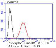 FCM/FACS (Flow Cytometry)
(Flow cytometric analysis of Hela cells with Phospho-Smad2 (S250) antibody at 1/50 dilution (red) compared with an unlabelled control (cells without incubation with primary antibody; black). Alexa Fluor 488-conjugated goat anti rabbit IgG was used as the secondary antibody)
FCM/FACS (Flow Cytometry)
(Flow cytometric analysis of Hela cells with Phospho-Smad2 (S250) antibody at 1/50 dilution (red) compared with an unlabelled control (cells without incubation with primary antibody; black). Alexa Fluor 488-conjugated goat anti rabbit IgG was used as the secondary antibody)
SMAD2, Monoclonal Antibody (Cat# AAA311031)
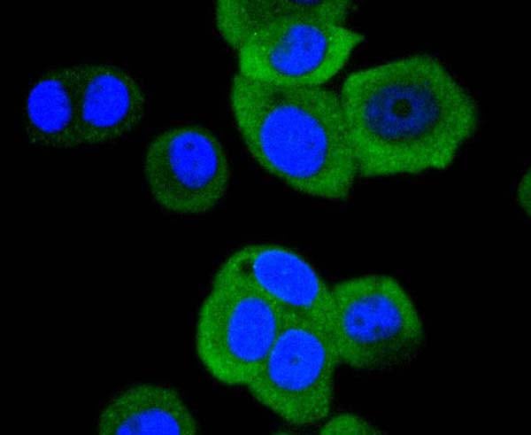
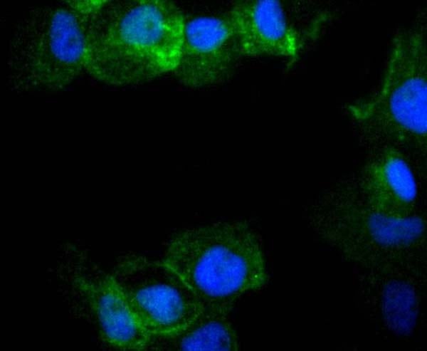
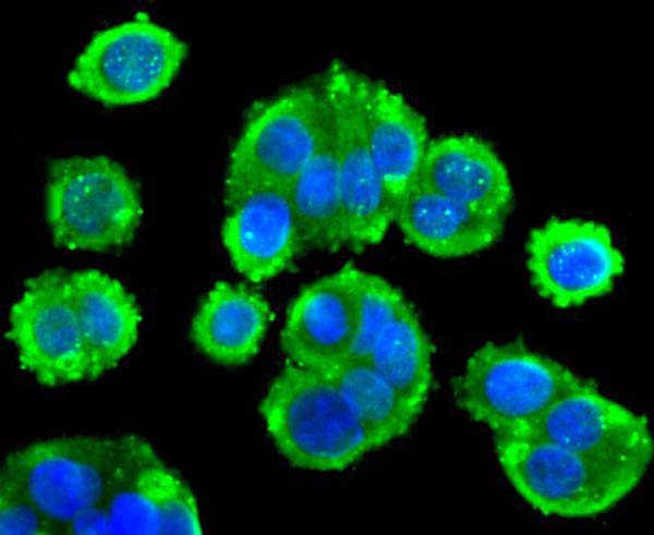
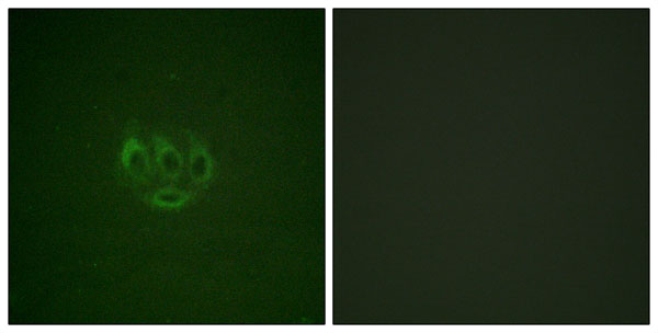
 ICC (Immunocytochemistry)
(ICC staining Phospho-Hsp27 (S78) in NIH/3T3 cells (green). The nuclear counter stain is DAPI (blue). Cells were fixed in paraformaldehyde, permeabilised with 0.25% Triton X100/PBS.)
ICC (Immunocytochemistry)
(ICC staining Phospho-Hsp27 (S78) in NIH/3T3 cells (green). The nuclear counter stain is DAPI (blue). Cells were fixed in paraformaldehyde, permeabilised with 0.25% Triton X100/PBS.)
Hsp27, Monoclonal Antibody (Cat# AAA311037)
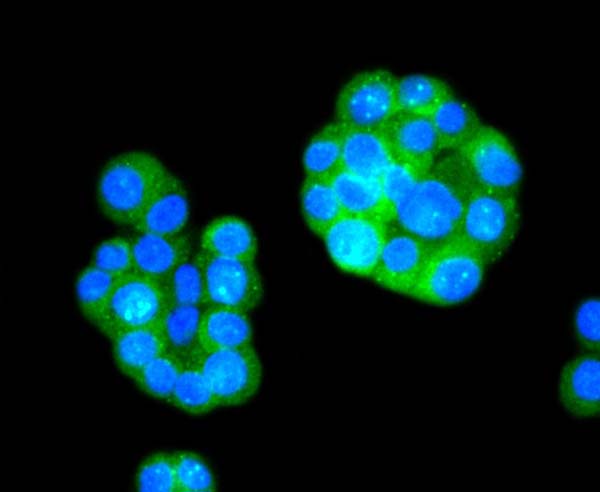
 ICC (Immunocytochemistry)
(ICC staining Phospho-Rac1+Cdc42 (Ser71) in Hela cells (green). The nuclear counter stain is DAPI (blue). Cells were fixed in paraformaldehyde, permeabilised with 0.25% Triton X100/PBS.)
ICC (Immunocytochemistry)
(ICC staining Phospho-Rac1+Cdc42 (Ser71) in Hela cells (green). The nuclear counter stain is DAPI (blue). Cells were fixed in paraformaldehyde, permeabilised with 0.25% Triton X100/PBS.)
Rac1+Cdc42, Monoclonal Antibody (Cat# AAA311043)
 ICC (Immunocytochemistry)
(ICC staining Phospho-PTEN (S380) in SW480 cells (green). The nuclear counter stain is DAPI (blue). Cells were fixed in paraformaldehyde, permeabilised with 0.25% Triton X100/PBS.)
ICC (Immunocytochemistry)
(ICC staining Phospho-PTEN (S380) in SW480 cells (green). The nuclear counter stain is DAPI (blue). Cells were fixed in paraformaldehyde, permeabilised with 0.25% Triton X100/PBS.)
PTEN, Monoclonal Antibody (Cat# AAA311044)

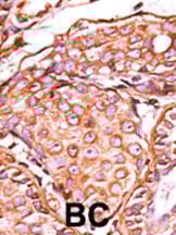
 IHC (Immunohistochemisry)
(Formalin-fixed and paraffin-embedded human cancer tissue reacted with the primary antibody, which was peroxidase-conjugated to the secondary antibody, followed by DAB staining. This data demonstrates the use of this antibody for immunohistochemistry; clinical relevance has not been evaluated. BC = breast carcinoma; HC = hepatocarcinoma.)
IHC (Immunohistochemisry)
(Formalin-fixed and paraffin-embedded human cancer tissue reacted with the primary antibody, which was peroxidase-conjugated to the secondary antibody, followed by DAB staining. This data demonstrates the use of this antibody for immunohistochemistry; clinical relevance has not been evaluated. BC = breast carcinoma; HC = hepatocarcinoma.)
Phospho-STAT5a (Y694), Polyclonal Antibody (Cat# AAA288354)
 IHC (Immunohistochemisry)
(Formalin-fixed and paraffin-embedded human cancer tissue reacted with the primary antibody, which was peroxidase-conjugated to the secondary antibody, followed by AEC staining. This data demonstrates the use of this antibody for immunohistochemistry; clinical relevance has not been evaluated. BC = breast carcinoma; HC = hepatocarcinoma.)
IHC (Immunohistochemisry)
(Formalin-fixed and paraffin-embedded human cancer tissue reacted with the primary antibody, which was peroxidase-conjugated to the secondary antibody, followed by AEC staining. This data demonstrates the use of this antibody for immunohistochemistry; clinical relevance has not been evaluated. BC = breast carcinoma; HC = hepatocarcinoma.)
Phospho-cJun (S63), Polyclonal Antibody (Cat# AAA289769)
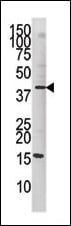
 WB (Western Blot)
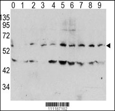
(Western blot analysis of Phospho-MYC-T58 Antibody in human TPA activated Hela cell line lysates. Phospho-MYC (arrow) was detected using the purified PAb. (0: without TPA; 1: 60ug/ml TPA, 15min; 2: 60ug/ml TPA, 30min; 3: 60ug/ml TPA, 45min; 4: 125ug/ml TPA, 15min; 5: 125ug/ml TPA, 30min; 6: 125ug/ml TPA, 45min; 7: 250ug/ml TPA, 15min; 8: 250ug/ml TPA, 30min; 9: 250ug/ml, 45min))
WB (Western Blot)
(Western blot analysis of Phospho-MYC-T58 Antibody in human TPA activated Hela cell line lysates. Phospho-MYC (arrow) was detected using the purified PAb. (0: without TPA; 1: 60ug/ml TPA, 15min; 2: 60ug/ml TPA, 30min; 3: 60ug/ml TPA, 45min; 4: 125ug/ml TPA, 15min; 5: 125ug/ml TPA, 30min; 6: 125ug/ml TPA, 45min; 7: 250ug/ml TPA, 15min; 8: 250ug/ml TPA, 30min; 9: 250ug/ml, 45min))
Phospho-MYC (T58), Polyclonal Antibody (Cat# AAA289788)

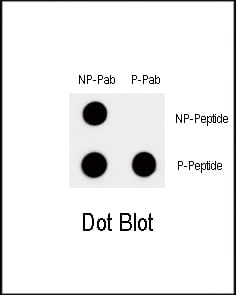
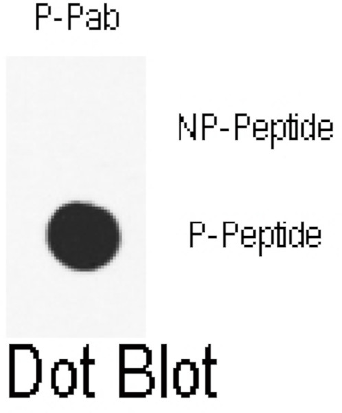
 DB (Dot Blot)
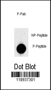
(Dot blot analysis of anti-Phospho-LIN28-S134 Phospho-specific Pab on nitrocellulose membrane. 50ng of Phospho-peptide or Non Phospho-peptide per dot were adsorbed. Antibody working concentrations are 0.5ug per ml.)
DB (Dot Blot)
(Dot blot analysis of anti-Phospho-LIN28-S134 Phospho-specific Pab on nitrocellulose membrane. 50ng of Phospho-peptide or Non Phospho-peptide per dot were adsorbed. Antibody working concentrations are 0.5ug per ml.)
Phospho-LIN28 (S134), Polyclonal Antibody (Cat# AAA289649)
 IHC (Immunohiostchemistry)
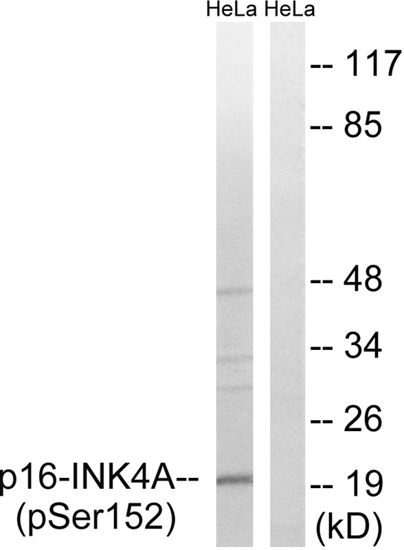
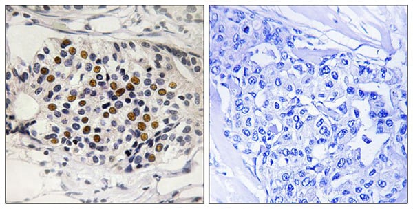
(Immunohistochemistry analysis of paraffin-embedded human breast carcinoma tissue using p16-INK4a (Phospho-Ser152) antibody. The picture on the right is treated with the synthesized peptide.)
IHC (Immunohiostchemistry)
(Immunohistochemistry analysis of paraffin-embedded human breast carcinoma tissue using p16-INK4a (Phospho-Ser152) antibody. The picture on the right is treated with the synthesized peptide.)
p16-INK4a, Polyclonal Antibody (Cat# AAA306318)
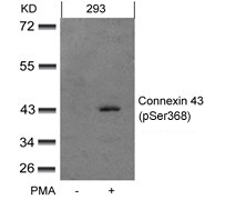
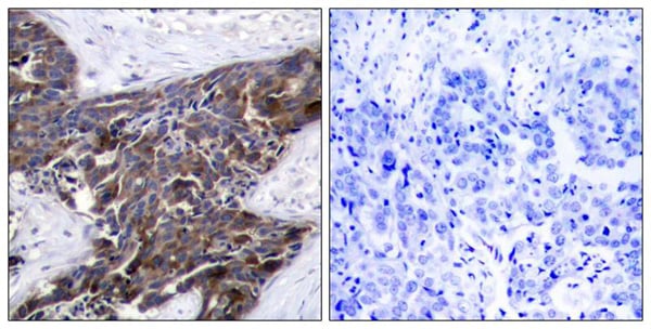 IHC (Immunohiostchemistry)
(Immunohistochemical analysis of paraffin-embedded human breast carcinoma tissue using Connexin 43(Phospho-Ser367) Antibody (left) or the same antibody preincubated with blocking peptide(right).)
IHC (Immunohiostchemistry)
(Immunohistochemical analysis of paraffin-embedded human breast carcinoma tissue using Connexin 43(Phospho-Ser367) Antibody (left) or the same antibody preincubated with blocking peptide(right).)
Connexin 43, Polyclonal Antibody (Cat# AAA303209)
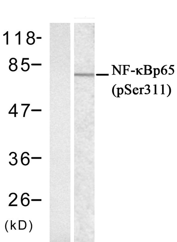
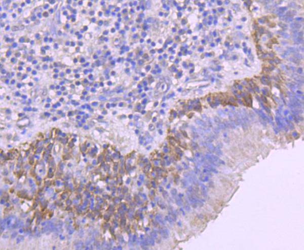
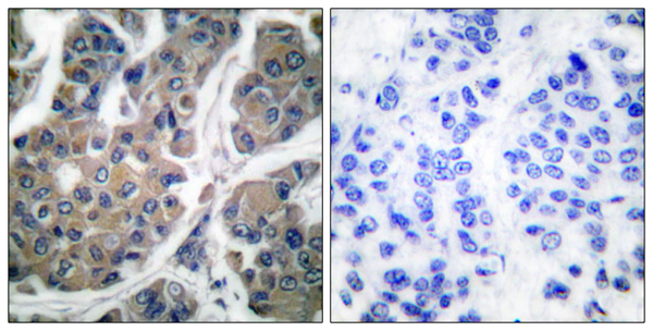
 IHC (Immunohistochemisry)
(Immunohistochemical analysis of paraffin-embedded human breast carcinoma tissue, using NFkappaB-p65 (phospho-Ser311) antibody ().)
IHC (Immunohistochemisry)
(Immunohistochemical analysis of paraffin-embedded human breast carcinoma tissue, using NFkappaB-p65 (phospho-Ser311) antibody ().)
NFkappaB-p65, Polyclonal Antibody (Cat# AAA307237)
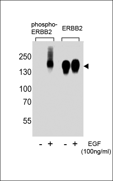
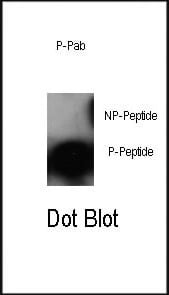
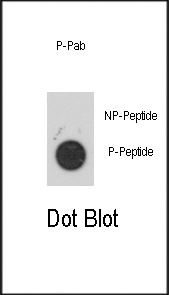
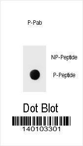 DB (Dot Blot)
(Dot blot analysis of Phospho-ERBB2-Y1139 Antibody Phospho-specific Pab on nitrocellulose membrane. 50ng of Phospho-peptide or Non Phospho-peptide per dot were adsorbed. Antibody working concentrations are 0.6ug per ml.)
DB (Dot Blot)
(Dot blot analysis of Phospho-ERBB2-Y1139 Antibody Phospho-specific Pab on nitrocellulose membrane. 50ng of Phospho-peptide or Non Phospho-peptide per dot were adsorbed. Antibody working concentrations are 0.6ug per ml.)
Phospho-ERBB2 (Y1139), Polyclonal Antibody (Cat# AAA284073)
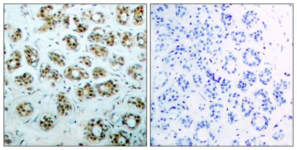
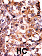
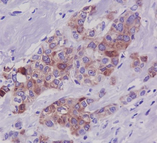
 IHC (Immunohiostchemistry)
(Formalin-fixed and paraffin-embedded human cancer tissue reacted with the primary antibody, which was peroxidase-conjugated to the secondary antibody, followed by AEC staining. This data demonstrates the use of this antibody for immunohistochemistry; clinical relevance has not been evaluated. BC = breast carcinoma; HC = hepatocarcinoma.)
IHC (Immunohiostchemistry)
(Formalin-fixed and paraffin-embedded human cancer tissue reacted with the primary antibody, which was peroxidase-conjugated to the secondary antibody, followed by AEC staining. This data demonstrates the use of this antibody for immunohistochemistry; clinical relevance has not been evaluated. BC = breast carcinoma; HC = hepatocarcinoma.)
Phospho-STAT1 (S727), Polyclonal Antibody (Cat# AAA284815)
c-Abl, Polyclonal Antibody (Cat# AAA312722)
RIP3, Polyclonal Antibody (Cat# AAA312933)
PDE4B/C/D, Polyclonal Antibody (Cat# AAA312934)
IFN-gammaRalpha, Polyclonal Antibody (Cat# AAA312936)
Cyclin B1, Polyclonal Antibody (Cat# AAA312937)
CHKA, Polyclonal Antibody (Cat# AAA312984)
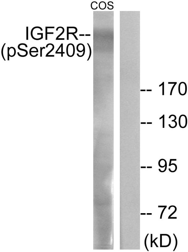
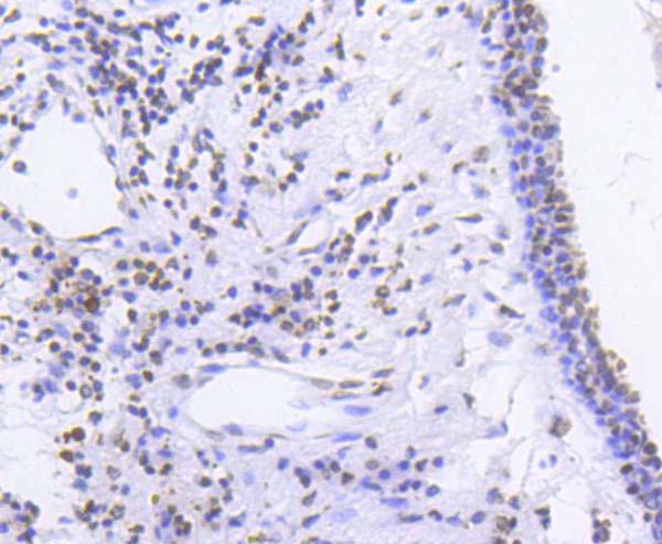
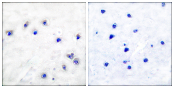
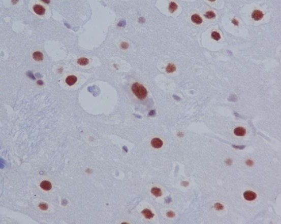
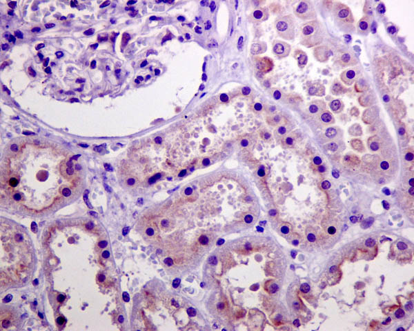
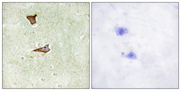 IHC (Immunohiostchemistry)
(Immunohistochemistry analysis of paraffin-embedded human brain tissue using IGF2R (Phospho-Ser2409) antibody.)
IHC (Immunohiostchemistry)
(Immunohistochemistry analysis of paraffin-embedded human brain tissue using IGF2R (Phospho-Ser2409) antibody.)
IGF2R (Phospho-Ser2409), Polyclonal Antibody (Cat# AAA285594)
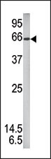
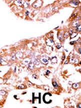
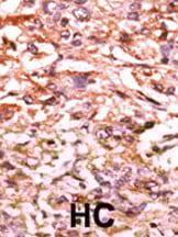
 IHC (Immunohiostchemistry)
(Formalin-fixed and paraffin-embedded human cancer tissue reacted with the primary antibody, which was peroxidase-conjugated to the secondary antibody, followed by AEC staining. This data demonstrates the use of this antibody for immunohistochemistry; clinical relevance has not been evaluated. BC = breast carcinoma; HC = hepatocarcinoma.)
IHC (Immunohiostchemistry)
(Formalin-fixed and paraffin-embedded human cancer tissue reacted with the primary antibody, which was peroxidase-conjugated to the secondary antibody, followed by AEC staining. This data demonstrates the use of this antibody for immunohistochemistry; clinical relevance has not been evaluated. BC = breast carcinoma; HC = hepatocarcinoma.)
Phospho-MAPKAPK2 (S272), Polyclonal Antibody (Cat# AAA285365)

 IHC (Immunohistochemisry)
(Formalin-fixed and paraffin-embedded human cancer tissue reacted with the primary antibody, which was peroxidase-conjugated to the secondary antibody, followed by AEC staining. This data demonstrates the use of this antibody for immunohistochemistry; clinical relevance has not been evaluated. BC = breast carcinoma; HC = hepatocarcinoma.)
IHC (Immunohistochemisry)
(Formalin-fixed and paraffin-embedded human cancer tissue reacted with the primary antibody, which was peroxidase-conjugated to the secondary antibody, followed by AEC staining. This data demonstrates the use of this antibody for immunohistochemistry; clinical relevance has not been evaluated. BC = breast carcinoma; HC = hepatocarcinoma.)
Phospho-CDC25B (S187), Polyclonal Antibody (Cat# AAA285409)
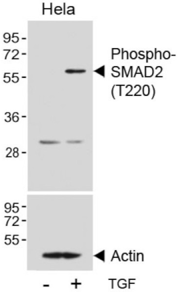
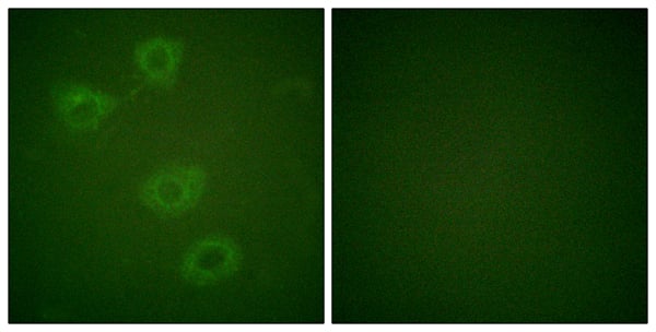
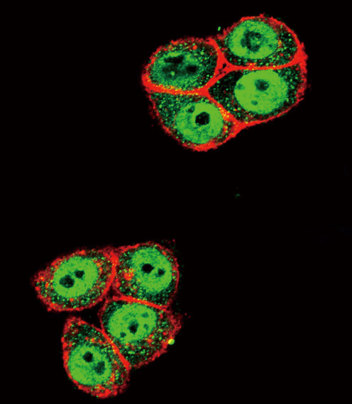 IF (Immunofluorescence)
(Confocal immunofluorescent analysis of Phospho-SMAD2-T220 Antibody with Hela cell followed by Alexa Fluor 488-conjugated goat anti-rabbit lgG (green).Actin filaments have been labeled with Alexa Fluor 555 phalloidin (red).)
IF (Immunofluorescence)
(Confocal immunofluorescent analysis of Phospho-SMAD2-T220 Antibody with Hela cell followed by Alexa Fluor 488-conjugated goat anti-rabbit lgG (green).Actin filaments have been labeled with Alexa Fluor 555 phalloidin (red).)
Phospho-SMAD2 (T220), Polyclonal Antibody (Cat# AAA285432)
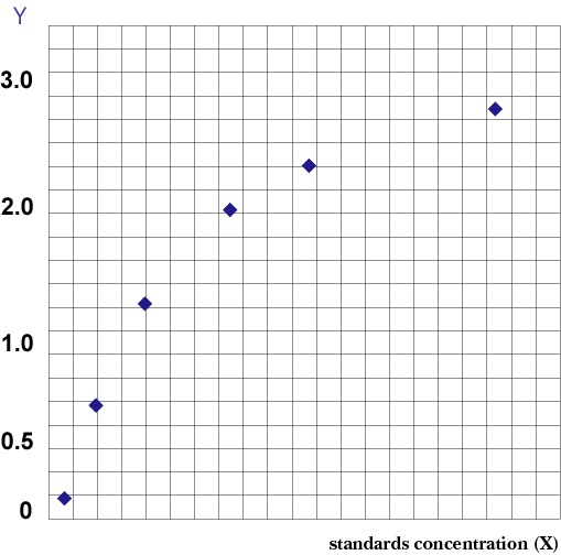 Standard Curve (Sample)
Phospho-Vascular Endothelial Cell Growth Factor Receptor 1, ELISA Kit (Cat# AAA205977)
 Standard Curve (Sample)
Phospho-Vascular Endothelial Cell Growth Factor Receptor 2, ELISA Kit (Cat# AAA204673)
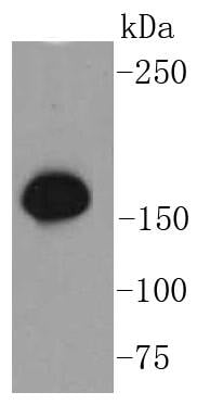
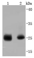
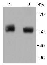
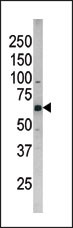
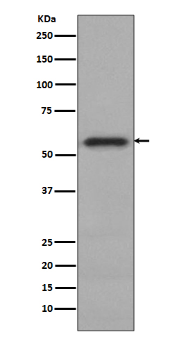
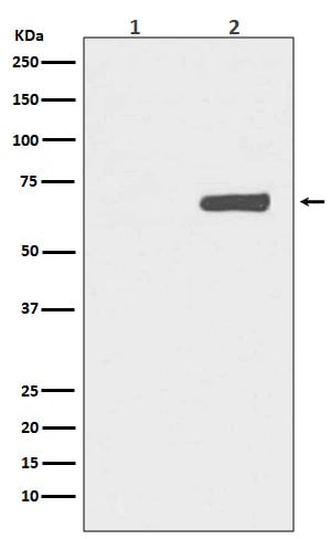
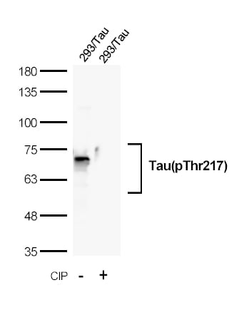 WB (Western Blot)
(Western blot analysis of extracts from 293 cells, transfected with Tau and treated with calf intestinal phosphatase (CIP) using Tau(Phospho-Thr217) Rabbit mAb.)
WB (Western Blot)
(Western blot analysis of extracts from 293 cells, transfected with Tau and treated with calf intestinal phosphatase (CIP) using Tau(Phospho-Thr217) Rabbit mAb.)
Tau, Monoclonal Antibody (Cat# AAA314726)
Tau, ELISA Kit (Cat# AAA315585)

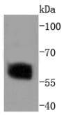
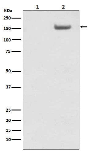
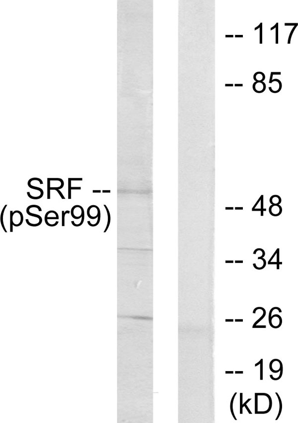 WB (Western Blot)
(Western blot analysis of lysates from LOVO cells treated with Serum 10% 15', using SRF (Phospho-Ser99) Antibody. The lane on the right is blocked with the phospho peptide.)
WB (Western Blot)
(Western blot analysis of lysates from LOVO cells treated with Serum 10% 15', using SRF (Phospho-Ser99) Antibody. The lane on the right is blocked with the phospho peptide.)
SRF, Polyclonal Antibody (Cat# AAA316118)
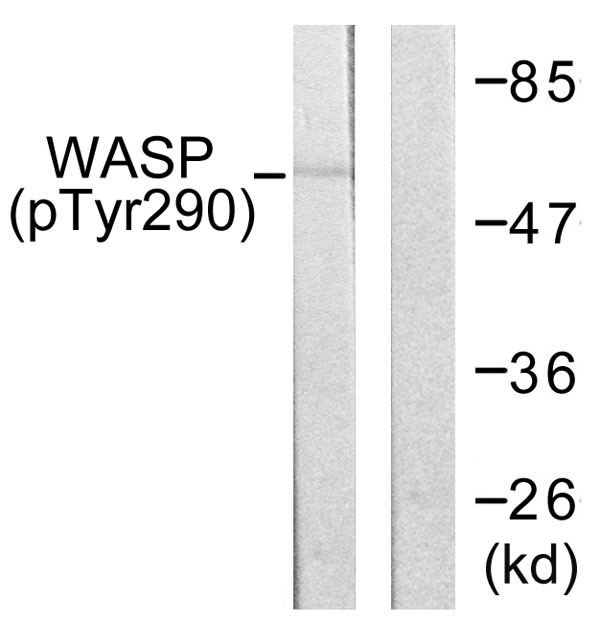 WB (Western Blot)
(Western blot analysis of lysates from HepG2 cells, using WASP (Phospho-Tyr290) Antibody. The lane on the right is blocked with the phospho peptide.)
WB (Western Blot)
(Western blot analysis of lysates from HepG2 cells, using WASP (Phospho-Tyr290) Antibody. The lane on the right is blocked with the phospho peptide.)
WASP, Polyclonal Antibody (Cat# AAA316237)
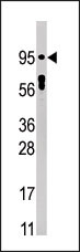
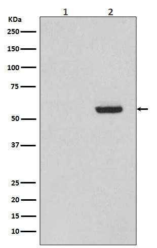
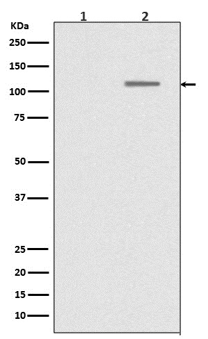
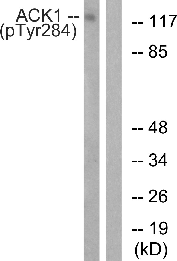 WB (Western Blot)
(Western blot analysis of lysates from HepG2 cells treated with EGF 200ng/ml 30', using ACK1 (Phospho-Tyr284) Antibody. The lane on the right is blocked with the phospho peptide.)
WB (Western Blot)
(Western blot analysis of lysates from HepG2 cells treated with EGF 200ng/ml 30', using ACK1 (Phospho-Tyr284) Antibody. The lane on the right is blocked with the phospho peptide.)
ACK1, Polyclonal Antibody (Cat# AAA316241)
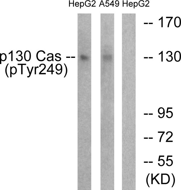 WB (Western Blot)
(Western blot analysis of lysates from HepG2 cells treated with EGF 200ng/ml 30' and A549 cells treated with PMA 125ng/ml 30', using p130 Cas (Phospho-Tyr249) Antibody. The lane on the right is blocked with the phospho peptide.)
WB (Western Blot)
(Western blot analysis of lysates from HepG2 cells treated with EGF 200ng/ml 30' and A549 cells treated with PMA 125ng/ml 30', using p130 Cas (Phospho-Tyr249) Antibody. The lane on the right is blocked with the phospho peptide.)
p130 Cas, Polyclonal Antibody (Cat# AAA316270)
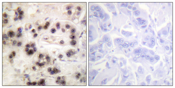
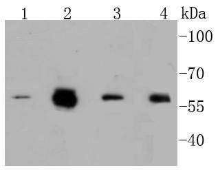
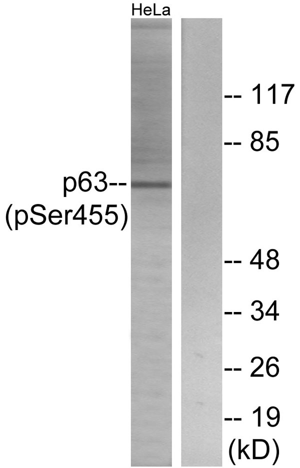 WB (Western Blot)
(Western blot analysis of lysates from HeLa cells treated with TNF 2500U/ML 30', using p63 (Phospho-Ser455) Antibody. The lane on the right is blocked with the phospho peptide.)
WB (Western Blot)
(Western blot analysis of lysates from HeLa cells treated with TNF 2500U/ML 30', using p63 (Phospho-Ser455) Antibody. The lane on the right is blocked with the phospho peptide.)
p63, Polyclonal Antibody (Cat# AAA316271)
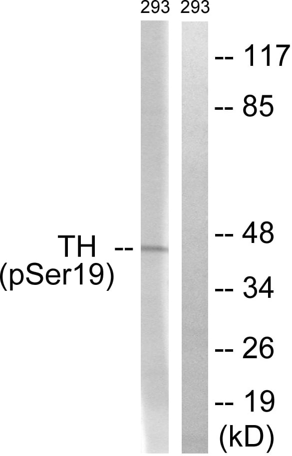 WB (Western Blot)
(Western blot analysis of lysates from 293 cells treated with Insulin 0.01U/ml 30', using Tyrosine Hydroxylase (Phospho-Ser19) Antibody. The lane on the right is blocked with the phospho peptide.)
WB (Western Blot)
(Western blot analysis of lysates from 293 cells treated with Insulin 0.01U/ml 30', using Tyrosine Hydroxylase (Phospho-Ser19) Antibody. The lane on the right is blocked with the phospho peptide.)
Tyrosine Hydroxylase, Polyclonal Antibody (Cat# AAA316085)
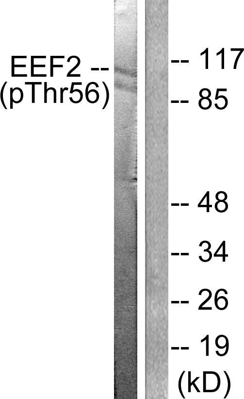 WB (Western Blot)
(Western blot analysis of lysates from NIH/3T3 cells treated with Serum 10% 30', using eEF2 (Phospho-Thr56) Antibody. The lane on the right is blocked with the phospho peptide.)
WB (Western Blot)
(Western blot analysis of lysates from NIH/3T3 cells treated with Serum 10% 30', using eEF2 (Phospho-Thr56) Antibody. The lane on the right is blocked with the phospho peptide.)
eEF2, Polyclonal Antibody (Cat# AAA316101)
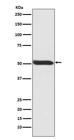
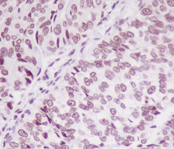
 IHC (Immunohiostchemistry)
(Immunohistochemical analysis of paraffin-embedded human breast, using Phospho-p53 (S392) Antibody.)
IHC (Immunohiostchemistry)
(Immunohistochemical analysis of paraffin-embedded human breast, using Phospho-p53 (S392) Antibody.)
p53, Monoclonal Recombinant Antibody (Cat# AAA314846)
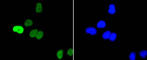
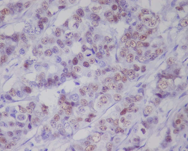
 IHC (Immunohiostchemistry)
(Immunohistochemical analysis of paraffin-embedded human breast cancer, using Phospho-EGFR (Y1068) Antibody .)
IHC (Immunohiostchemistry)
(Immunohistochemical analysis of paraffin-embedded human breast cancer, using Phospho-EGFR (Y1068) Antibody .)
EGFR, Monoclonal Recombinant Antibody (Cat# AAA314847)
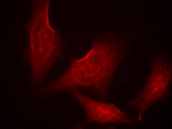
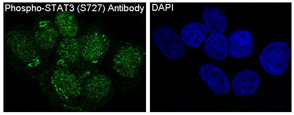 IF (Immunofluorescence)
(Immunofluorescent analysis of A431 cells treated with EGF, using Phospho-STAT3 (S727) Antibody .)
IF (Immunofluorescence)
(Immunofluorescent analysis of A431 cells treated with EGF, using Phospho-STAT3 (S727) Antibody .)
STAT3, Monoclonal Recombinant Antibody (Cat# AAA314849)
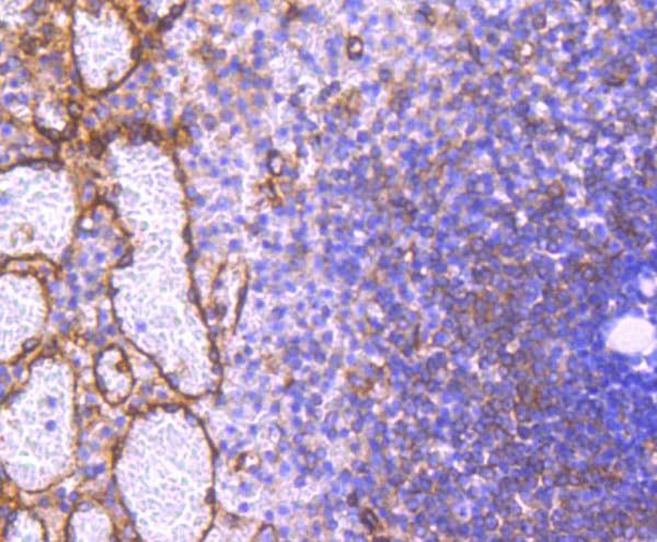
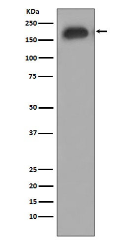
 IHC (Immunohiostchemistry)
(Immunohistochemical analysis of paraffin-embedded rat spleen, using Phospho-Cdk1/2 (T14) Antibody.)
IHC (Immunohiostchemistry)
(Immunohistochemical analysis of paraffin-embedded rat spleen, using Phospho-Cdk1/2 (T14) Antibody.)
Cdk1/2, Monoclonal Recombinant Antibody (Cat# AAA314852)
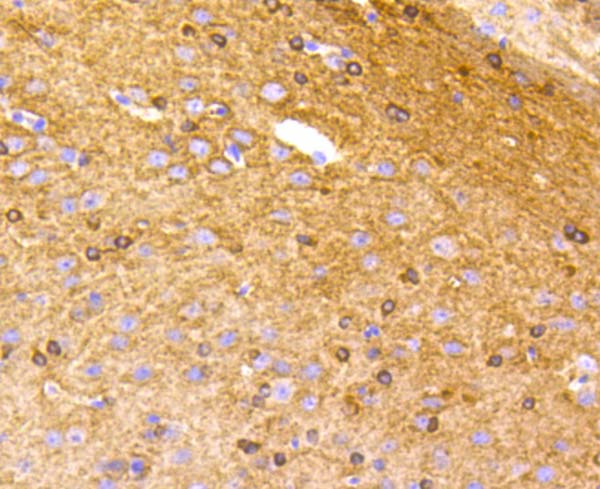
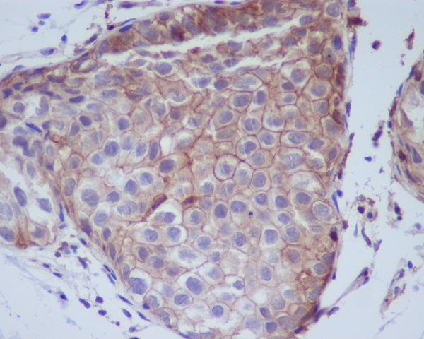
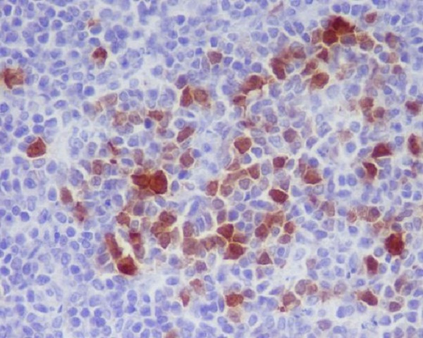
 IHC (Immunohiostchemistry)
(Immunohistochemical analysis of paraffin-embedded human lung carcinoma, using Phospho-c-Myc (S62) Antibody .)
IHC (Immunohiostchemistry)
(Immunohistochemical analysis of paraffin-embedded human lung carcinoma, using Phospho-c-Myc (S62) Antibody .)
c-Myc, Monoclonal Recombinant Antibody (Cat# AAA314856)
 IHC (Immunohiostchemistry)
(Immunohistochemical analysis of paraffin-embedded human breast carcinoma, using Phospho-Tau (S396) Antibody.)
IHC (Immunohiostchemistry)
(Immunohistochemical analysis of paraffin-embedded human breast carcinoma, using Phospho-Tau (S396) Antibody.)
Tau, Monoclonal Recombinant Antibody (Cat# AAA314863)
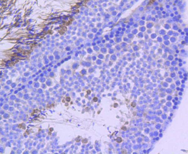
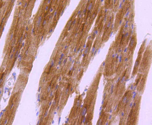
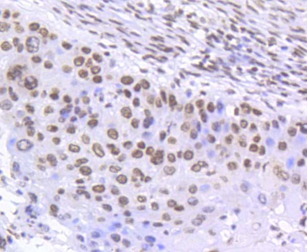
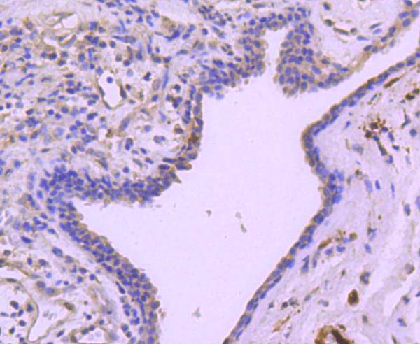
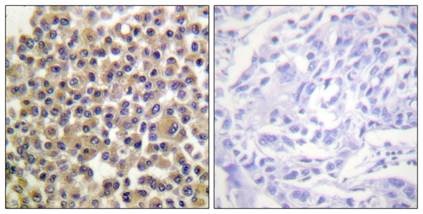
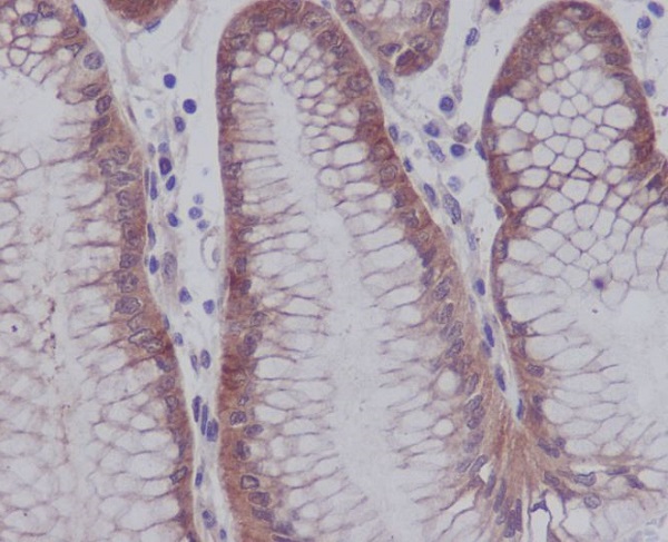
 IHC (Immunohiostchemistry)
(Immunohistochemical analysis of paraffin-embedded human bladder cancer, using Phospho-SMC1 (S957) Antibody.)
IHC (Immunohiostchemistry)
(Immunohistochemical analysis of paraffin-embedded human bladder cancer, using Phospho-SMC1 (S957) Antibody.)
SMC1, Monoclonal Recombinant Antibody (Cat# AAA314865)
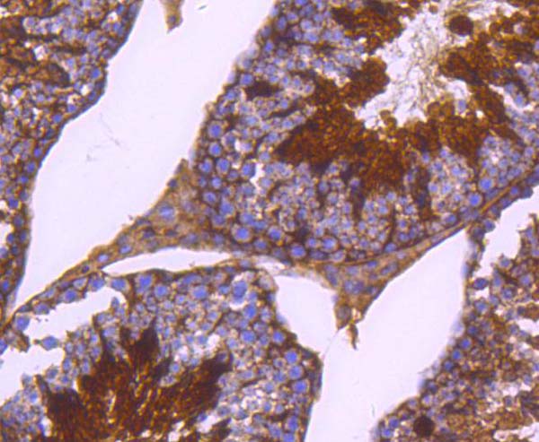
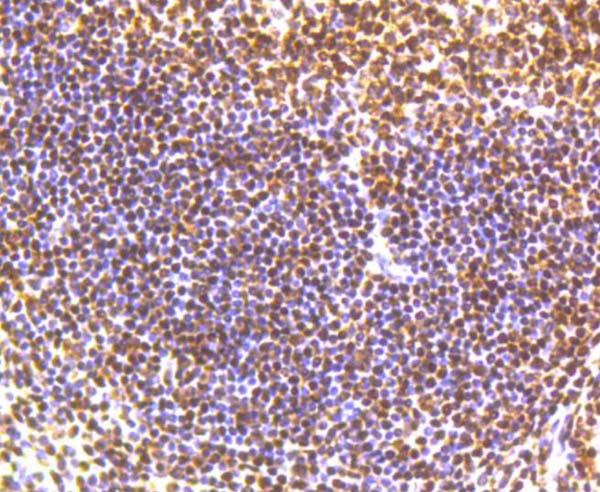
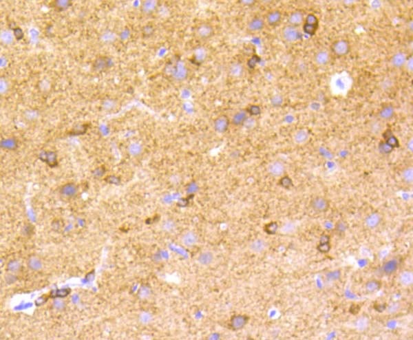
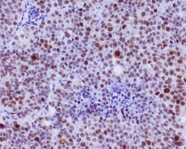
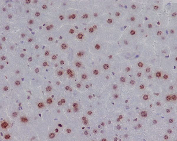
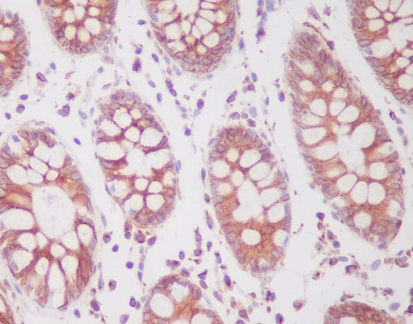 IHC (Immunohiostchemistry)
(Immunohistochemical analysis of paraffin-embedded human colon, using Phospho-SIRT1 (S47) Antibody.)
IHC (Immunohiostchemistry)
(Immunohistochemical analysis of paraffin-embedded human colon, using Phospho-SIRT1 (S47) Antibody.)
SIRT1, Monoclonal Recombinant Antibody (Cat# AAA314866)
 IHC (Immunohiostchemistry)
(Immunohistochemical analysis of paraffin-embedded human stomach, using Phospho-YAP1 (S127) Antibody.)
IHC (Immunohiostchemistry)
(Immunohistochemical analysis of paraffin-embedded human stomach, using Phospho-YAP1 (S127) Antibody.)
YAP1, Monoclonal Recombinant Antibody (Cat# AAA314868)
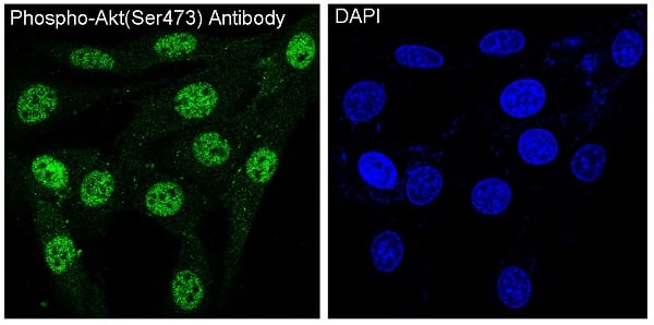 IF (Immunofluorescence)
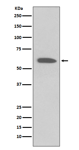
(Immunofluorescent analysis of NIH/3T3 cells treated with PDGF, using Phospho-Akt(Ser473) Antibody.)
IF (Immunofluorescence)
(Immunofluorescent analysis of NIH/3T3 cells treated with PDGF, using Phospho-Akt(Ser473) Antibody.)
Akt, Monoclonal Recombinant Antibody (Cat# AAA314873)
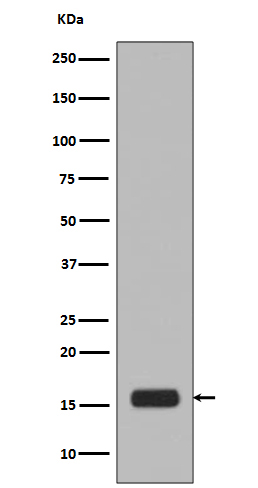
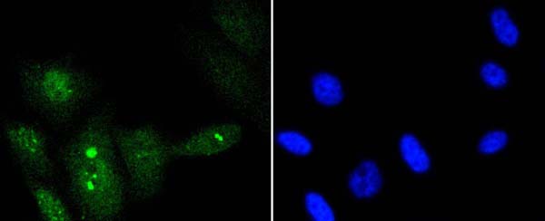
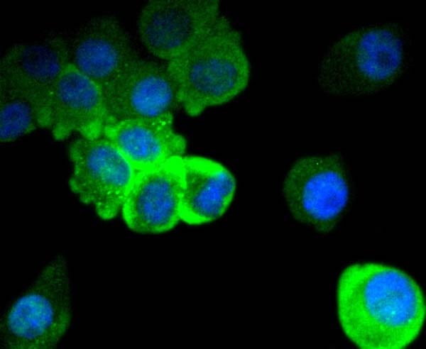
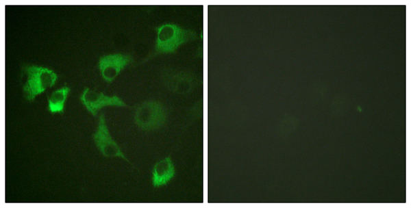
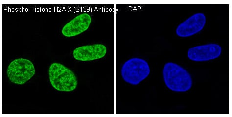 IF (Immunofluorescence)
(Immunofluorescent analysis of HeLa cells treated with H2O2, using Phospho-Histone H2A.X (S139) Antibody.)
IF (Immunofluorescence)
(Immunofluorescent analysis of HeLa cells treated with H2O2, using Phospho-Histone H2A.X (S139) Antibody.)
Histone H2A.X, Monoclonal Recombinant Antibody (Cat# AAA314876)
What Are Phospho Antibodies?
Protein phosphorylation is a process where a phosphate group is added to certain amino acid residues of a protein – usually serine (S), threonine (T), or tyrosine (Y) - by enzymes called kinases. This process is integral in controlling cellular signaling, cellular growth, and other biological functions.
Our catalog includes a wide range of phospho-specific antibodies that can accurately detect this important marker. They perform strongly in widely-used laboratory applications such as Western blot, flow cytometry, immunohistochemistry, and immunofluorescence microscopy. We value your trust in us and are committed to providing top-quality products and services. All of our antibodies are guaranteed to work for the applications and species indicated on our website & associated product pages.
What Are The Key Applications of Phospho Antibodies?
1. Western Blotting
One of the first steps a researcher can take in utilizing these phospho-specific antibodies, is to check if the antibody works using a technique referred to as “Western blot”. For those unfamiliar, Western Blot aids in showing whether the protein that the antibody recognizes is appearing at the correct/expected size. These phospho-specific antibodies should also be able to detect changes in the target protein’s phosphorylation (on/off state) when cells are stimulated in certain ways.
2. Staining of Fixed Cells (Immunocytochemistry)
Another routine use of these phospho-specific antibodies, is to test if the antibody is able to demonstrate similar performance when used on fixed cells (intact cells that have been preserved) as it did in the Western blot tests. It is an important aspect in many cases to confirm that the antibody works in actual intact cell samples. Ideally, the method used for cellular fixation should be the same as what is used in pathology labs (like using 10% formalin). To check if the antibody works well in tissue sections (FFPE), researchers will often test it on fixed cells that are processed similar to tissue samples.
3. Specificity Tests Using Peptides
In order to make sure that the antibody is only binding to the right target:
- Laboratory technicians will mix the antibody with phospho-peptides (short segments of the protein containing the phosphate group modification).
- If the antibody signal disappears, it is confirmation that it is binding to the correct phosphorylated location.
- A more robust test is to use both the phosphorylated and non-phosphorylated (dephosphorylated) versions of the protein. The antibody should react only with the phosphorylated one.
- Another method sometimes utilized is to treat the sample with an enzyme, such as alkaline phosphatase, that specifically removes phosphate groups. If the antibody signal disappears after this, it also confirms specificity.
4. Genetic Confirmation
As a final step, scientists can genetically manipulate the nucleotide sequence and alter the target protein by removing the exact site where phosphorylation happens. If the antibody no longer appears to detect the modified protein, it is strong evidence supporting the antibody being specific for that phosphorylated site.
Why Buy Phospho Antibodies Through Us?
- The production laboratory adheres to strict and consistent protocols prior to releasing any of these phospho-specific antibodies:
- Standard methods and proper controls in all tests to ensure high quality.
- These antibodies are tested and validated in different cell types and species.
- High quality control criterion to ensure each batch is consistent, so you will obtain reliable results every time.
FAQ
1. What Are Phospho-Specific Antibodies?
Phospho-specific antibodies are made to detect proteins only when they have a phosphate group linked to a specific amino acid residue. This empowers scientists understand if a protein is "turned on" or active, based on its phosphorylation state.
2. How to Detect Phosphorylated Proteins in a Western Blot?
To find out if a protein is phosphorylated using Western blot:
- Use a phospho-specific antibody that binds only to the phosphorylated form of the protein.
- You can also use a “regular” antibody for the same amino acid sequence of the protein that the phospho-specific antibody is binding to (but in this case, this antibody will not bind if there is a phosphate group present) in order to compare how much of it is phosphorylated versus how much is non-phosphorylated (or “total” protein, if the “normal” antibody’s epitopes are non-phospho-site-specific).
3. How to Choose the Best Antibody?
Here are some simple tips to help you pick the right antibody:
- Know your target
- Match your sample characteristics
- Confirm the intended use is appropriate
- Check “host” and “type”
- Check the “quality” of the presented data/images
- Appraise whether the available validation meets your needs