Filters
▼Clonality
▼Type
▼Reactivity
▼Gene Name
▼Isotype
▼Host
▼Application
▼Clone
▼Phospho Antibodies
Phospho-specific antibodies’ typical purpose is to enable researchers to detect changes in proteins. They will exclusively bind to the amino acid sequence on a protein that has been phosphorylated (which is both a physical & chemical change) and do not bind to the same amino acid sequence on said protein if it lacks said phosphorylation. This aids in being able to clearly see and understand the data produced from this particular protein modification.
Viewing 2450-2500 of 5298 product results
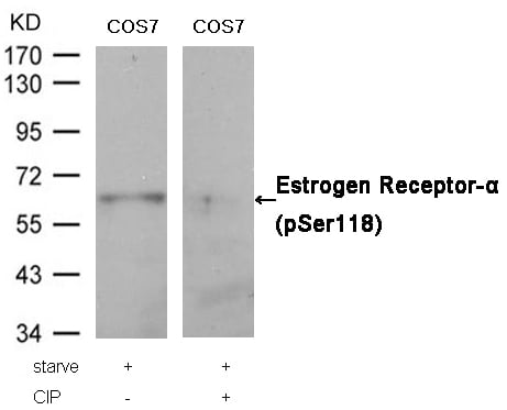 WB (Western Blot)
(Western blot analysis of extracts from COS7 cells, treated with starve or calf intestinal phosphatase (CIP), using Estrogen Receptor-alpha (Phospho-Ser118) Antibody.)
WB (Western Blot)
(Western blot analysis of extracts from COS7 cells, treated with starve or calf intestinal phosphatase (CIP), using Estrogen Receptor-alpha (Phospho-Ser118) Antibody.)
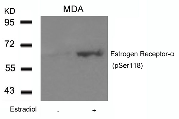
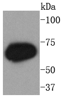
Estrogen Receptor-alpha, Polyclonal Antibody (Cat# AAA305386)
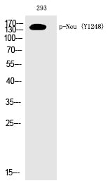
 WB (Western Blot)
(Western Blot analysis of HepG2+PMA cells using Phospho-Neu (Y1248) Polyclonal Antibody)
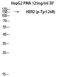
WB (Western Blot)
(Western Blot analysis of HepG2+PMA cells using Phospho-Neu (Y1248) Polyclonal Antibody)
Neu, Polyclonal Antibody (Cat# AAA303699)
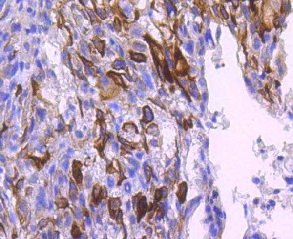
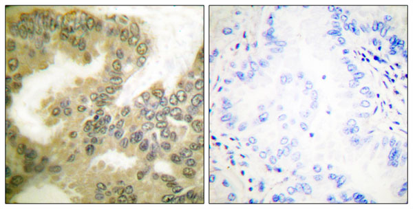
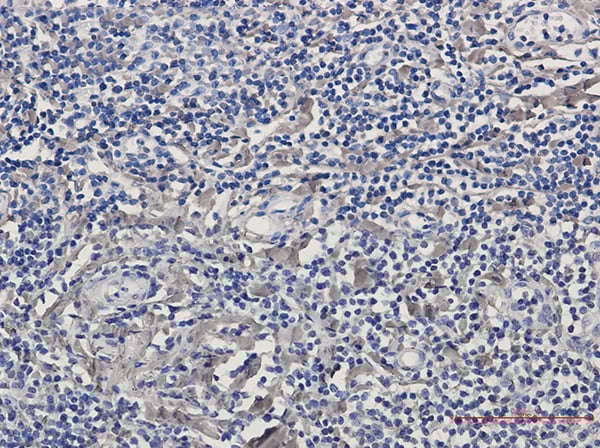
 IHC (Immunohiostchemistry)
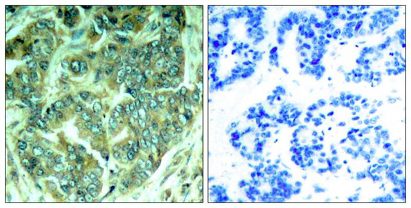
(Immunohistochemical analysis of paraffin-embedded human breast carcinoma tissue using p62Dok(Phospho-Tyr398) Antibody (left) or the same antibody preincubated with blocking peptide(right).)
IHC (Immunohiostchemistry)
(Immunohistochemical analysis of paraffin-embedded human breast carcinoma tissue using p62Dok(Phospho-Tyr398) Antibody (left) or the same antibody preincubated with blocking peptide(right).)
p62Dok, Polyclonal Antibody (Cat# AAA305968)
 WB (Western Blot)
(Western blot analysis of extracts from NIH-3T3 cells treated with TNF using IKK- alpha/ beta (Phospho-Ser176/177) antibody.The lane on the right is treated with the antigen-specific peptide.)
WB (Western Blot)
(Western blot analysis of extracts from NIH-3T3 cells treated with TNF using IKK- alpha/ beta (Phospho-Ser176/177) antibody.The lane on the right is treated with the antigen-specific peptide.)
IKK-alpha/ beta, Polyclonal Antibody (Cat# AAA308010)
Tau, Polyclonal Antibody (Cat# AAA312721)
Cadf, Polyclonal Antibody (Cat# AAA312922)
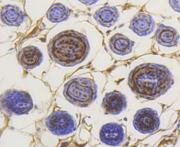
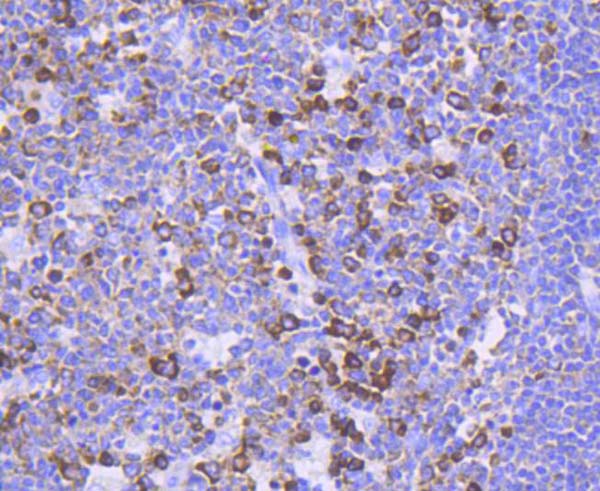
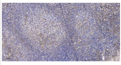 IHC (Immunohiostchemistry)
(Immunohistochemical analysis of paraffin-embedded human tonsil. 1, Antibody was diluted at 1:200(4° overnight). 2, Tris-EDTA,pH9.0 was used for antigen retrieval. 3,Secondary antibody was diluted at 1:200(room temperature, 30min).)
IHC (Immunohiostchemistry)
(Immunohistochemical analysis of paraffin-embedded human tonsil. 1, Antibody was diluted at 1:200(4° overnight). 2, Tris-EDTA,pH9.0 was used for antigen retrieval. 3,Secondary antibody was diluted at 1:200(room temperature, 30min).)
ZFP598, Polyclonal Antibody (Cat# AAA312931)
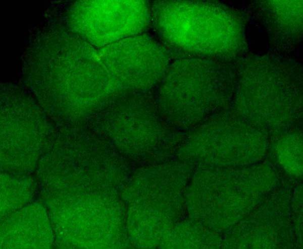
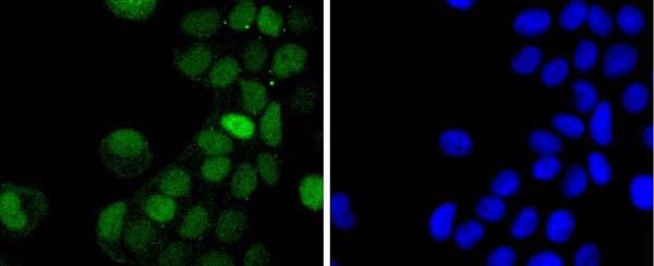
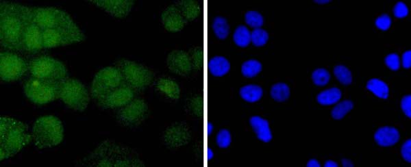
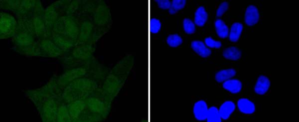
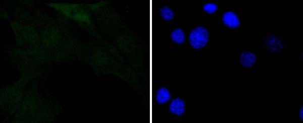
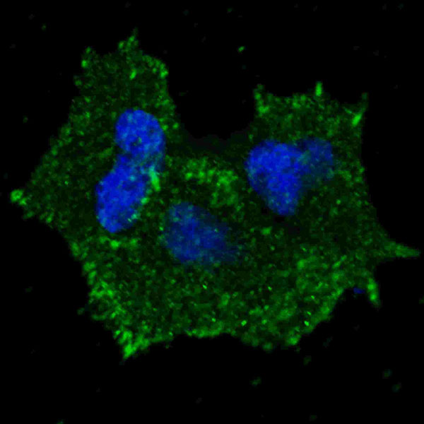
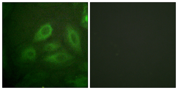
 ICC (Immunocytochemistry)
(ICC staining Phospho-EGFR (pY1173) in untreated B-6F1 cells (green). The nuclear counter stain is DAPI (blue). Cells were fixed in paraformaldehyde, permeabilised with 0.25% Triton X100/PBS.)
ICC (Immunocytochemistry)
(ICC staining Phospho-EGFR (pY1173) in untreated B-6F1 cells (green). The nuclear counter stain is DAPI (blue). Cells were fixed in paraformaldehyde, permeabilised with 0.25% Triton X100/PBS.)
EGFR, Monoclonal Antibody (Cat# AAA311014)
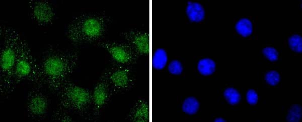
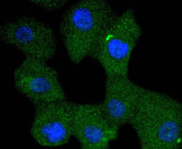
 ICC (Immunocytochemistry)
(ICC staining PDGF Receptor beta (phospho Y740) in Hela cells (green). The nuclear counter stain is DAPI (blue). Cells were fixed in paraformaldehyde, permeabilised with 0.25% Triton X100/PBS.)
ICC (Immunocytochemistry)
(ICC staining PDGF Receptor beta (phospho Y740) in Hela cells (green). The nuclear counter stain is DAPI (blue). Cells were fixed in paraformaldehyde, permeabilised with 0.25% Triton X100/PBS.)
PDGF beta, Monoclonal Antibody (Cat# AAA311021)
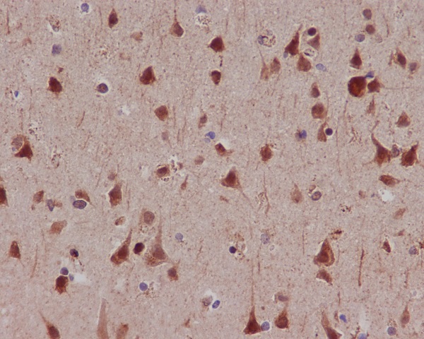
 IHC (Immunohistochemisry)
(Immunohistochemical analysis of paraffin-embedded mouse brain tissue using anti-Phospho-PKC zeta (T560) antibody. Counter stained with hematoxylin.)
IHC (Immunohistochemisry)
(Immunohistochemical analysis of paraffin-embedded mouse brain tissue using anti-Phospho-PKC zeta (T560) antibody. Counter stained with hematoxylin.)
PKC zeta, Monoclonal Antibody (Cat# AAA311028)

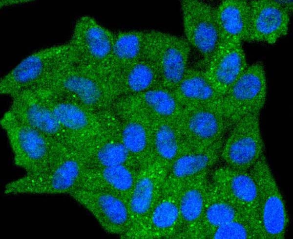
 ICC (Immunocytochemistry)
(ICC staining Phospho-Cyclin E1 (T77) in HepG2 cells (green). The nuclear counter stain is DAPI (blue). Cells were fixed in paraformaldehyde, permeabilised with 0.25% Triton X100/PBS.)
ICC (Immunocytochemistry)
(ICC staining Phospho-Cyclin E1 (T77) in HepG2 cells (green). The nuclear counter stain is DAPI (blue). Cells were fixed in paraformaldehyde, permeabilised with 0.25% Triton X100/PBS.)
Cyclin E1, Monoclonal Antibody (Cat# AAA311030)
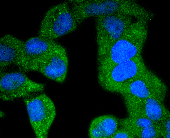
 ICC (Immunocytochemistry)
(ICC staining Phospho-c-Jun (T91) in MCF-7 cells (green). The nuclear counter stain is DAPI (blue). Cells were fixed in paraformaldehyde, permeabilised with 0.25% Triton X100/PBS.)
ICC (Immunocytochemistry)
(ICC staining Phospho-c-Jun (T91) in MCF-7 cells (green). The nuclear counter stain is DAPI (blue). Cells were fixed in paraformaldehyde, permeabilised with 0.25% Triton X100/PBS.)
c-Jun, Monoclonal Antibody (Cat# AAA311040)
 ICC (Immunocytochemistry)
(ICC staining Phospho-AKT1 (S124) in MCF-7 cells (green). The nuclear counter stain is DAPI (blue). Cells were fixed in paraformaldehyde, permeabilised with 0.25% Triton X100/PBS.)
ICC (Immunocytochemistry)
(ICC staining Phospho-AKT1 (S124) in MCF-7 cells (green). The nuclear counter stain is DAPI (blue). Cells were fixed in paraformaldehyde, permeabilised with 0.25% Triton X100/PBS.)
AKT1, Monoclonal Antibody (Cat# AAA311041)

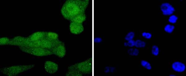
 ICC (Immunocytochemistry)
(ICC staining Phospho-SHIP (Y1020) in HepG2 cells (green). The nuclear counter stain is DAPI (blue). Cells were fixed in paraformaldehyde, permeabilised with 0.25% Triton X100/PBS.)
ICC (Immunocytochemistry)
(ICC staining Phospho-SHIP (Y1020) in HepG2 cells (green). The nuclear counter stain is DAPI (blue). Cells were fixed in paraformaldehyde, permeabilised with 0.25% Triton X100/PBS.)
SHIP, Monoclonal Antibody (Cat# AAA311045)
 ICC (Immunocytochemistry)
(ICC staining Phospho-Smad3 (S423/S425) in A431 cells (green). The nuclear counter stain is DAPI (blue). Cells were fixed in paraformaldehyde, permeabilised with 0.25% Triton X100/PBS.)
ICC (Immunocytochemistry)
(ICC staining Phospho-Smad3 (S423/S425) in A431 cells (green). The nuclear counter stain is DAPI (blue). Cells were fixed in paraformaldehyde, permeabilised with 0.25% Triton X100/PBS.)
SMAD3, Monoclonal Antibody (Cat# AAA311006)
 IHC (Immunohistochemistry)
(Immunohistochemical analysis of paraffin-embedded mouse pancreas tissue using anti-Phospho-FOXO3a (S253) antibody. Counter stained with hematoxylin.)
IHC (Immunohistochemistry)
(Immunohistochemical analysis of paraffin-embedded mouse pancreas tissue using anti-Phospho-FOXO3a (S253) antibody. Counter stained with hematoxylin.)
FOXO3a, Monoclonal Antibody (Cat# AAA311007)

 ICC (Immunocytochemistry)
(ICC staining Phospho-IKB alpha (S32) in NIH/3T3 cells (green). The nuclear counter stain is DAPI (blue). Cells were fixed in paraformaldehyde, permeabilised with 0.25% Triton X100/PBS.)
ICC (Immunocytochemistry)
(ICC staining Phospho-IKB alpha (S32) in NIH/3T3 cells (green). The nuclear counter stain is DAPI (blue). Cells were fixed in paraformaldehyde, permeabilised with 0.25% Triton X100/PBS.)
IKB alpha, Monoclonal Antibody (Cat# AAA311009)
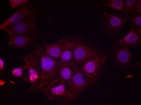
 IF (Immunofluorescence)
(Immunofluorescence staining of methanol-fixed HeLa cells using NF-kB p65(phospho-Thr435) antibody(, Red).)
IF (Immunofluorescence)
(Immunofluorescence staining of methanol-fixed HeLa cells using NF-kB p65(phospho-Thr435) antibody(, Red).)
NFkappaB-p65, Polyclonal Antibody (Cat# AAA307723)
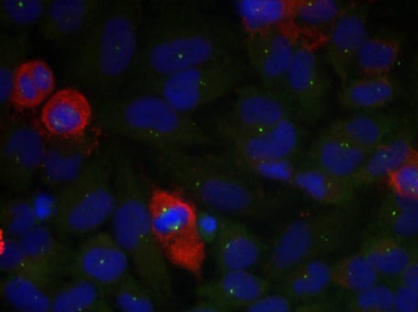
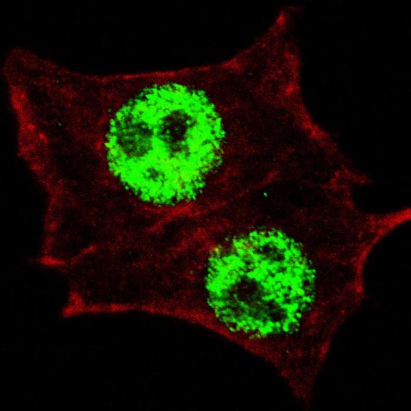
 IF (Immunofluorescence)
(Confocal immunofluorescent analysis of Phospho-Rb-S811 Antibody with HepG2 cell followed by Alexa Fluor 488-conjugated goat anti-rabbit lgG (green).DAPI was used to stain the cell nuclear (blue).)
IF (Immunofluorescence)
(Confocal immunofluorescent analysis of Phospho-Rb-S811 Antibody with HepG2 cell followed by Alexa Fluor 488-conjugated goat anti-rabbit lgG (green).DAPI was used to stain the cell nuclear (blue).)
Phospho-Rb (S811), Polyclonal Antibody (Cat# AAA285351)
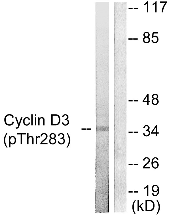
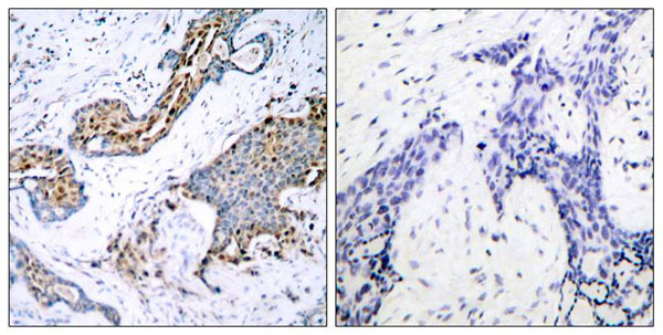
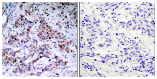
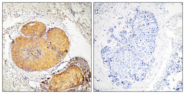
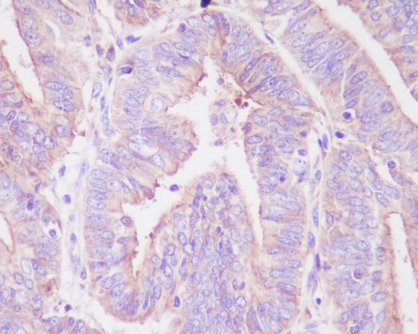
 IHC (Immunohiostchemistry)
(Immunohistochemical analysis of paraffin-embedded human lung carcinoma tissue using Cyclin D3 (Phospho-Thr283) Antibody (#A0418).)
IHC (Immunohiostchemistry)
(Immunohistochemical analysis of paraffin-embedded human lung carcinoma tissue using Cyclin D3 (Phospho-Thr283) Antibody (#A0418).)
Cyclin D3 (Phospho-Thr283), Polyclonal Antibody (Cat# AAA285355)
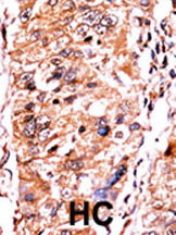
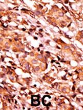
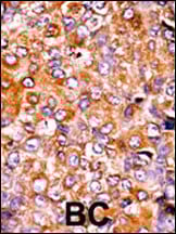
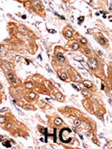
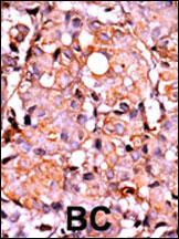 IHC (Immunohiostchemistry)
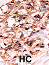
(Formalin-fixed and paraffin-embedded human cancer tissue reacted with the primary antibody, which was peroxidase-conjugated to the secondary antibody, followed by AEC staining. This data demonstrates the use of this antibody for immunohistochemistry; clinical relevance has not been evaluated. BC = breast carcinoma; HC = hepatocarcinoma.)
IHC (Immunohiostchemistry)
(Formalin-fixed and paraffin-embedded human cancer tissue reacted with the primary antibody, which was peroxidase-conjugated to the secondary antibody, followed by AEC staining. This data demonstrates the use of this antibody for immunohistochemistry; clinical relevance has not been evaluated. BC = breast carcinoma; HC = hepatocarcinoma.)
Phospho-RAD9 (S387), Polyclonal Antibody (Cat# AAA285295)
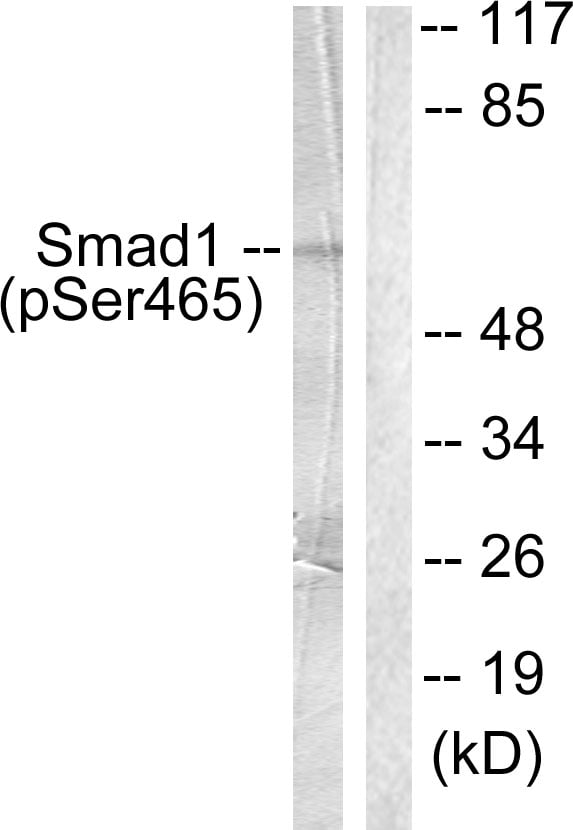
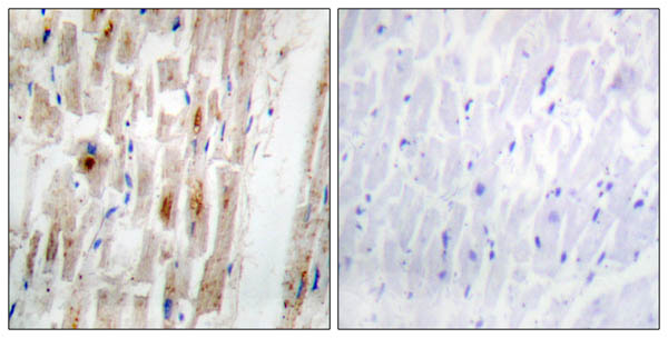 IHC (Immunohiostchemistry)
(Immunohistochemical analysis of paraffin-embedded human heart tissue, using Smad1 (phospho-Ser465) antibody.)
IHC (Immunohiostchemistry)
(Immunohistochemical analysis of paraffin-embedded human heart tissue, using Smad1 (phospho-Ser465) antibody.)
Smad1 (Phospho-Ser465), Polyclonal Antibody (Cat# AAA285118)
 IHC (Immunohiostchemistry)
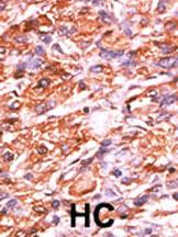
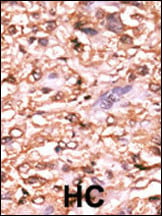
(Formalin-fixed and paraffin-embedded human cancer tissue reacted with the primary antibody, which was peroxidase-conjugated to the secondary antibody, followed by DAB staining. This data demonstrates the use of this antibody for immunohistochemistry; clinical relevance has not been evaluated. BC = breast carcinoma; HC = hepatocarcinoma.)
IHC (Immunohiostchemistry)
(Formalin-fixed and paraffin-embedded human cancer tissue reacted with the primary antibody, which was peroxidase-conjugated to the secondary antibody, followed by DAB staining. This data demonstrates the use of this antibody for immunohistochemistry; clinical relevance has not been evaluated. BC = breast carcinoma; HC = hepatocarcinoma.)
Phospho-p16-INK4A (S140), Polyclonal Antibody (Cat# AAA284679)
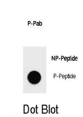
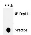
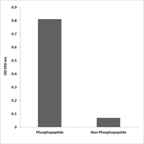
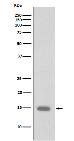
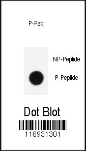 DB (Dot Blot)
(Dot blot analysis of anti-Phospho-KLF4-S245 Antibody on nitrocellulose membrane. 50ng of Phospho-peptide or Non Phospho-peptide per dot were adsorbed. Antibody working concentrations are 0.5ug per ml.)
DB (Dot Blot)
(Dot blot analysis of anti-Phospho-KLF4-S245 Antibody on nitrocellulose membrane. 50ng of Phospho-peptide or Non Phospho-peptide per dot were adsorbed. Antibody working concentrations are 0.5ug per ml.)
Phospho-KLF4 (S245), Polyclonal Antibody (Cat# AAA284718)
 DB (Dot Blot)
(Dot blot analysis of MUC1 Antibody (AAA284599) Pab on nitrocellulose membrane. 50ng of Phospho-peptide or Non Phospho-peptide per dot were adsorbed. Antibody working concentrations are 0.5ug per ml.)
DB (Dot Blot)
(Dot blot analysis of MUC1 Antibody (AAA284599) Pab on nitrocellulose membrane. 50ng of Phospho-peptide or Non Phospho-peptide per dot were adsorbed. Antibody working concentrations are 0.5ug per ml.)
Phospho-MUC1 (Y1229), Polyclonal Antibody (Cat# AAA284599)

 IHC (Immunohistochemisry)
(Formalin-fixed and paraffin-embedded human cancer tissue reacted with the primary antibody, which was peroxidase-conjugated to the secondary antibody, followed by AEC staining. This data demonstrates the use of this antibody for immunohistochemistry; clinical relevance has not been evaluated. BC = breast carcinoma; HC = hepatocarcinoma.)
IHC (Immunohistochemisry)
(Formalin-fixed and paraffin-embedded human cancer tissue reacted with the primary antibody, which was peroxidase-conjugated to the secondary antibody, followed by AEC staining. This data demonstrates the use of this antibody for immunohistochemistry; clinical relevance has not been evaluated. BC = breast carcinoma; HC = hepatocarcinoma.)
Phospho-Bad (S134), Polyclonal Antibody (Cat# AAA284121)
 DB (Dot Blot)
(Dot blot analysis of Phospho-PARP1-S177 Antibody Phospho-specific Pab on nitrocellulose membrane. 50ng of Phospho-peptide or Non Phospho-peptide per dot were adsorbed. Antibody working concentrations are 0.6ug per ml.)
DB (Dot Blot)
(Dot blot analysis of Phospho-PARP1-S177 Antibody Phospho-specific Pab on nitrocellulose membrane. 50ng of Phospho-peptide or Non Phospho-peptide per dot were adsorbed. Antibody working concentrations are 0.6ug per ml.)
Phospho-PARP1 (S177), Polyclonal Antibody (Cat# AAA284168)

 IHC (Immunohistochemisry)
(Formalin-fixed and paraffin-embedded human cancer tissue reacted with the primary antibody, which was peroxidase-conjugated to the secondary antibody, followed by AEC staining. This data demonstrates the use of this antibody for immunohistochemistry; clinical relevance has not been evaluated. BC = breast carcinoma; HC = hepatocarcinoma.)
IHC (Immunohistochemisry)
(Formalin-fixed and paraffin-embedded human cancer tissue reacted with the primary antibody, which was peroxidase-conjugated to the secondary antibody, followed by AEC staining. This data demonstrates the use of this antibody for immunohistochemistry; clinical relevance has not been evaluated. BC = breast carcinoma; HC = hepatocarcinoma.)
Phospho-MYT1 (T495), Polyclonal Antibody (Cat# AAA285563)
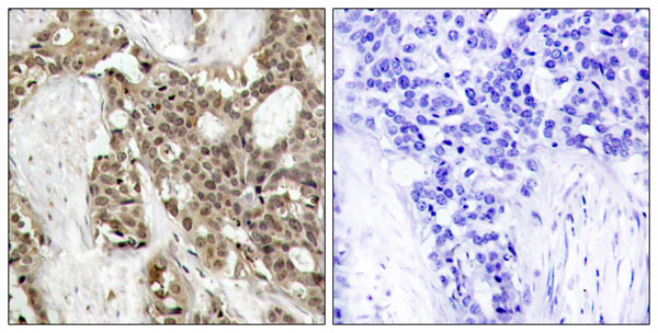
 IHC (Immunohiostchemistry)
(Formalin-fixed and paraffin-embedded human cancer tissue reacted with the primary antibody, which was peroxidase-conjugated to the secondary antibody, followed by AEC staining. This data demonstrates the use of this antibody for immunohistochemistry; clinical relevance has not been evaluated. BC = breast carcinoma; HC = hepatocarcinoma.)
IHC (Immunohiostchemistry)
(Formalin-fixed and paraffin-embedded human cancer tissue reacted with the primary antibody, which was peroxidase-conjugated to the secondary antibody, followed by AEC staining. This data demonstrates the use of this antibody for immunohistochemistry; clinical relevance has not been evaluated. BC = breast carcinoma; HC = hepatocarcinoma.)
Phospho-Bad (S118), Polyclonal Antibody (Cat# AAA285582)
 IHC (Immunohistochemisry)
(Formalin-fixed and paraffin-embedded human cancer tissue reacted with the primary antibody, which was peroxidase-conjugated to the secondary antibody, followed by AEC staining. This data demonstrates the use of this antibody for immunohistochemistry; clinical relevance has not been evaluated. BC = breast carcinoma; HC = hepatocarcinoma.)
IHC (Immunohistochemisry)
(Formalin-fixed and paraffin-embedded human cancer tissue reacted with the primary antibody, which was peroxidase-conjugated to the secondary antibody, followed by AEC staining. This data demonstrates the use of this antibody for immunohistochemistry; clinical relevance has not been evaluated. BC = breast carcinoma; HC = hepatocarcinoma.)
Phospho-p53 (T18), Polyclonal Antibody (Cat# AAA285593)
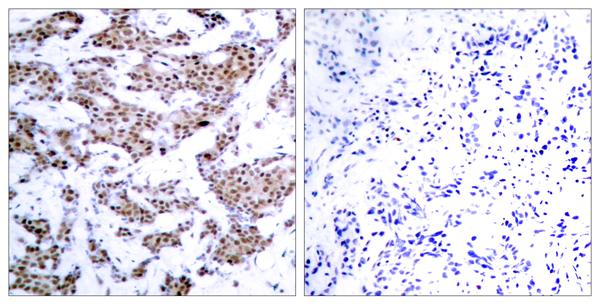
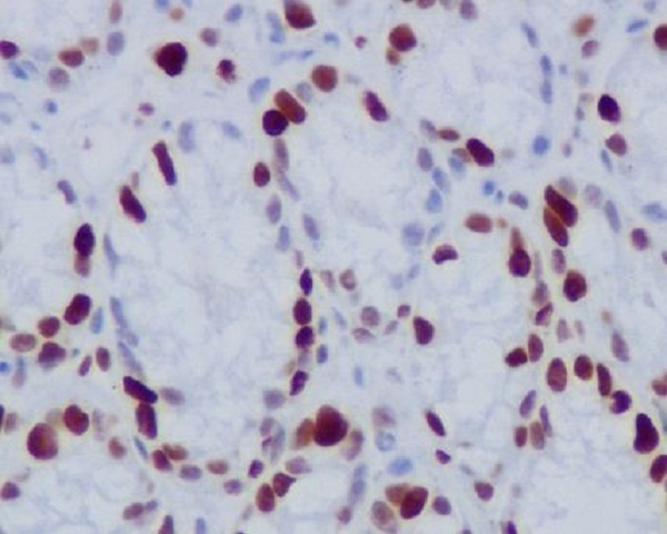
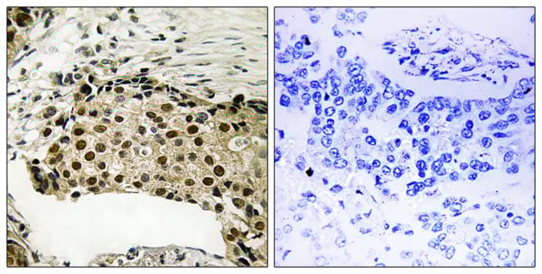 IHC (Immunohistochemisry)
(Immunohistochemistry analysis of paraffin-embedded human breast carcinoma tissue using MSK1 (Phospho-Ser212) antibody.)
IHC (Immunohistochemisry)
(Immunohistochemistry analysis of paraffin-embedded human breast carcinoma tissue using MSK1 (Phospho-Ser212) antibody.)
MSK1 (Phospho-Ser212), Polyclonal Antibody (Cat# AAA288919)
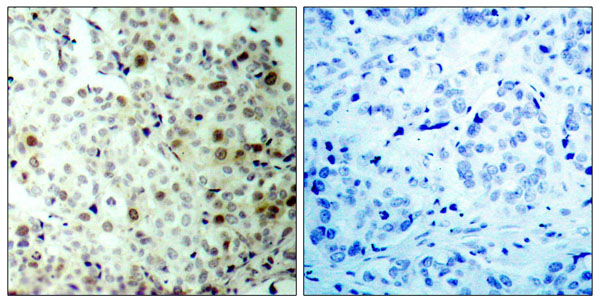
 IHC (Immunohiostchemistry)
(Formalin-fixed and paraffin-embedded human cancer tissue reacted with the primary antibody, which was peroxidase-conjugated to the secondary antibody, followed by AEC staining. This data demonstrates the use of this antibody for immunohistochemistry; clinical relevance has not been evaluated. BC = breast carcinoma; HC = hepatocarcinoma.)
IHC (Immunohiostchemistry)
(Formalin-fixed and paraffin-embedded human cancer tissue reacted with the primary antibody, which was peroxidase-conjugated to the secondary antibody, followed by AEC staining. This data demonstrates the use of this antibody for immunohistochemistry; clinical relevance has not been evaluated. BC = breast carcinoma; HC = hepatocarcinoma.)
Phospho-Bik (T33), Polyclonal Antibody (Cat# AAA288964)
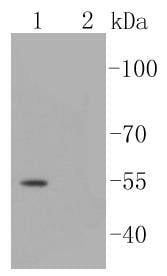
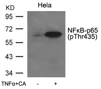
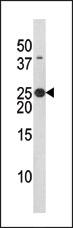
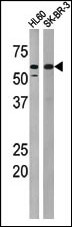
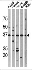
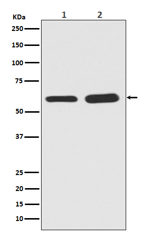
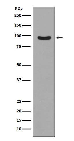
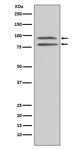
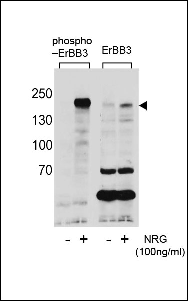 WB (Western Blot)
(Western blot analysis of extracts from T47D cells,untreated or treated with NRG using Phospho-ErBB3(Tyr1289)(left) or ErBB3 antibody(right).)
WB (Western Blot)
(Western blot analysis of extracts from T47D cells,untreated or treated with NRG using Phospho-ErBB3(Tyr1289)(left) or ErBB3 antibody(right).)
Phospho-ERBB3 (Y1289), Polyclonal Antibody (Cat# AAA289231)
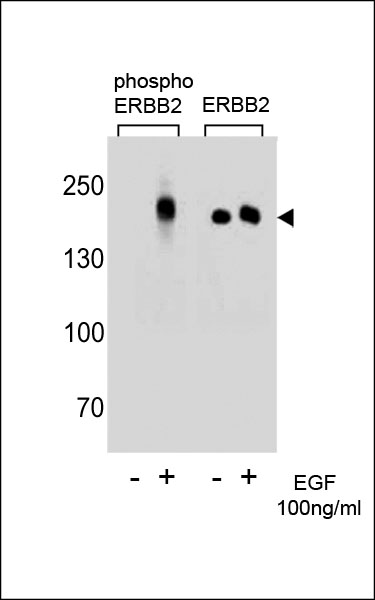
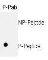
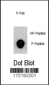
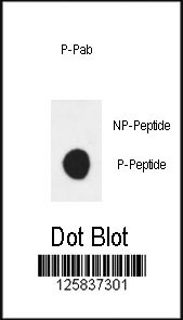
 DB (Dot Blot)
(Dot blot analysis of ERBB2 Antibody (Phospho Y1196) Phospho-specific Pab on nitrocellulose membrane. 50ng of Phospho-peptide or Non Phospho-peptide per dot were adsorbed. Antibody working concentrations are 0.6ug per ml.)
DB (Dot Blot)
(Dot blot analysis of ERBB2 Antibody (Phospho Y1196) Phospho-specific Pab on nitrocellulose membrane. 50ng of Phospho-peptide or Non Phospho-peptide per dot were adsorbed. Antibody working concentrations are 0.6ug per ml.)
Phospho-ERBB2 (Y1196), Polyclonal Antibody (Cat# AAA288697)
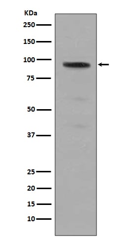
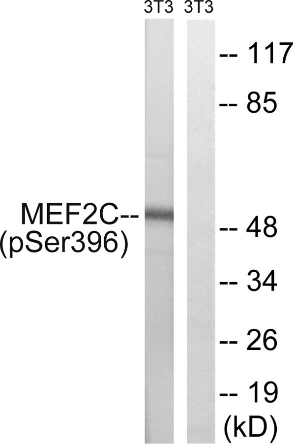 WB (Western Blot)
(Western blot analysis of lysates from NIH/3T3 cells treated with starved 24h, using MEF2C (Phospho-Ser396) Antibody. The lane on the right is blocked with the phospho peptide.)
WB (Western Blot)
(Western blot analysis of lysates from NIH/3T3 cells treated with starved 24h, using MEF2C (Phospho-Ser396) Antibody. The lane on the right is blocked with the phospho peptide.)
MEF2C, Polyclonal Antibody (Cat# AAA316652)
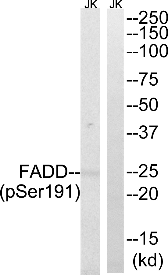 WB (Western Blot)
(Western blot analysis of lysates from Jurkat cells treated with PMA 125ng/ml 30', using FADD (Phospho-Ser191) Antibody. The lane on the right is blocked with the phospho peptide.)
WB (Western Blot)
(Western blot analysis of lysates from Jurkat cells treated with PMA 125ng/ml 30', using FADD (Phospho-Ser191) Antibody. The lane on the right is blocked with the phospho peptide.)
FADD, Polyclonal Antibody (Cat# AAA316666)
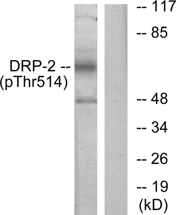 WB (Western Blot)
(Western blot analysis of lysates from NIH/3T3 cells treated with PMA 125ng/ml 30', using DRP-2 (Phospho-Thr514) Antibody. The lane on the right is blocked with the phospho peptide.)
WB (Western Blot)
(Western blot analysis of lysates from NIH/3T3 cells treated with PMA 125ng/ml 30', using DRP-2 (Phospho-Thr514) Antibody. The lane on the right is blocked with the phospho peptide.)
DRP-2, Polyclonal Antibody (Cat# AAA316435)
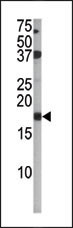
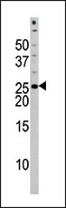
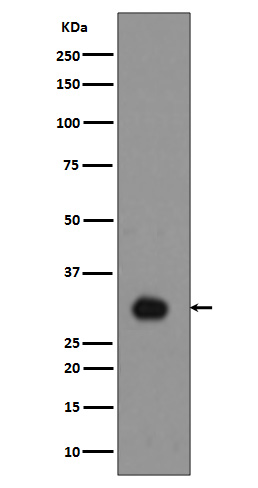
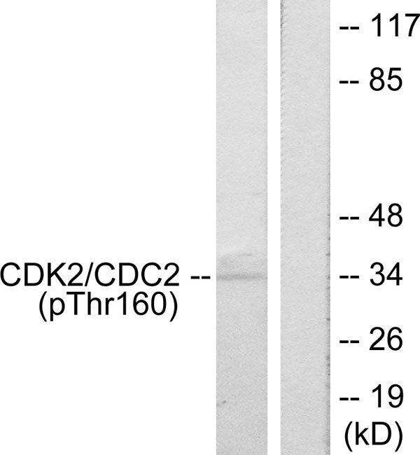 WB (Western Blot)
(Western blot analysis of lysates from A2780 cells, using CDK2 (Phospho-Thr160) Antibody. The lane on the right is blocked with the phospho peptide.)
WB (Western Blot)
(Western blot analysis of lysates from A2780 cells, using CDK2 (Phospho-Thr160) Antibody. The lane on the right is blocked with the phospho peptide.)
CDK2, Polyclonal Antibody (Cat# AAA316473)
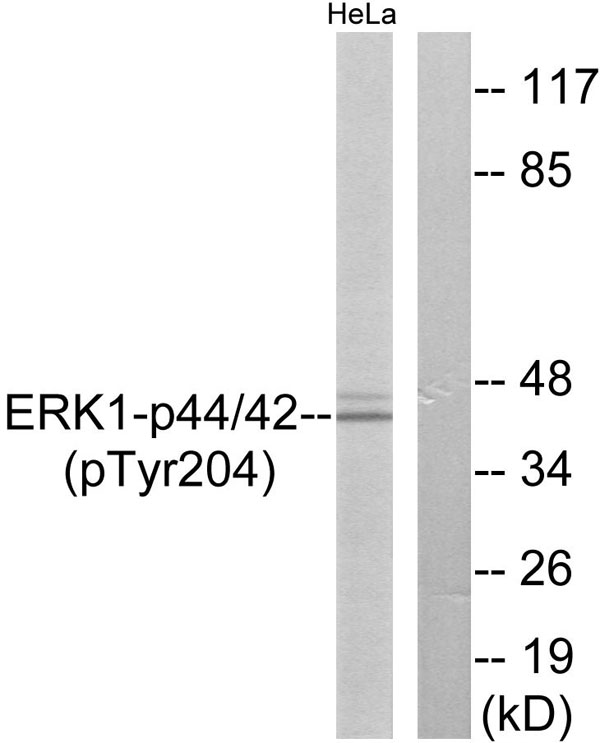 WB (Western Blot)
(Western blot analysis of lysates from HeLa cells treated with EGF 200ng/ml 30', using p44/42 MAP Kinase (Phospho-Tyr204) Antibody. The lane on the right is blocked with the phospho peptide.)
WB (Western Blot)
(Western blot analysis of lysates from HeLa cells treated with EGF 200ng/ml 30', using p44/42 MAP Kinase (Phospho-Tyr204) Antibody. The lane on the right is blocked with the phospho peptide.)
p44/42 MAP Kinase, Polyclonal Antibody (Cat# AAA316489)
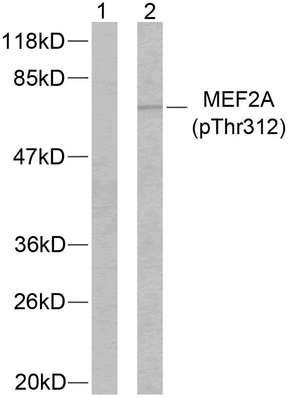 WB (Western Blot)
(Western blot analysis of lysates from NIH/3T3 cells treated with PMA, using MEF2A (Phospho-Thr312) Antibody. The lane on the left is blocked with the phospho peptide.)
WB (Western Blot)
(Western blot analysis of lysates from NIH/3T3 cells treated with PMA, using MEF2A (Phospho-Thr312) Antibody. The lane on the left is blocked with the phospho peptide.)
MEF2A, Polyclonal Antibody (Cat# AAA316532)
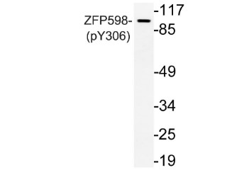
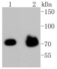
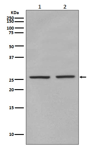
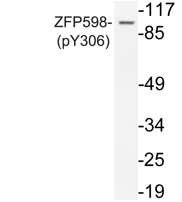 WB (Western Blot)
(Western blot analysis of lysate from Jurkat cells, uisng phospho-ZFP598 (Phospho-Tyr306) antibody.)
WB (Western Blot)
(Western blot analysis of lysate from Jurkat cells, uisng phospho-ZFP598 (Phospho-Tyr306) antibody.)
ZFP598, Polyclonal Antibody (Cat# AAA318279)
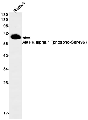
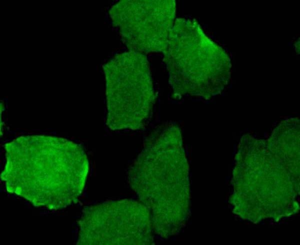
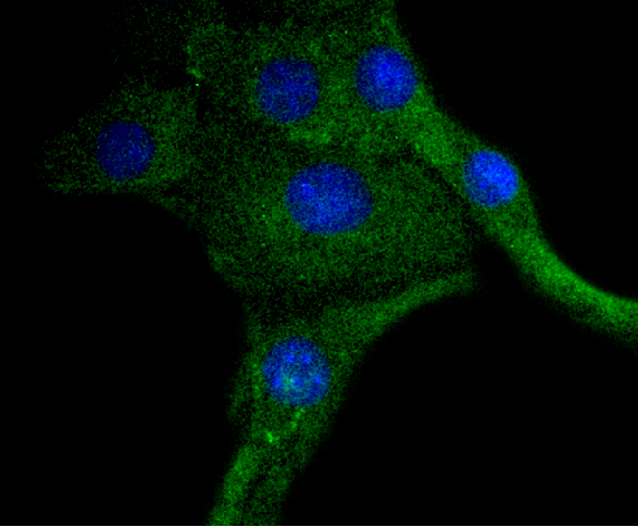
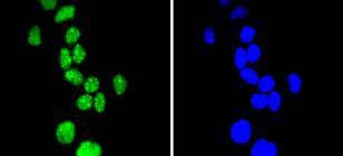
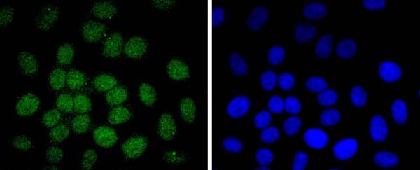
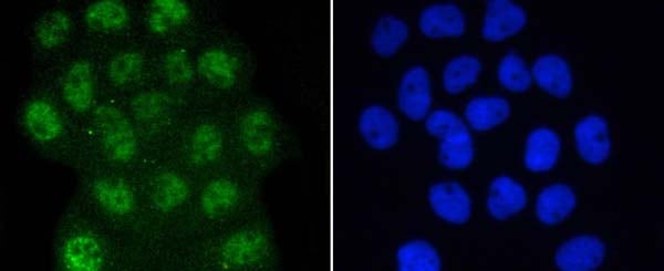
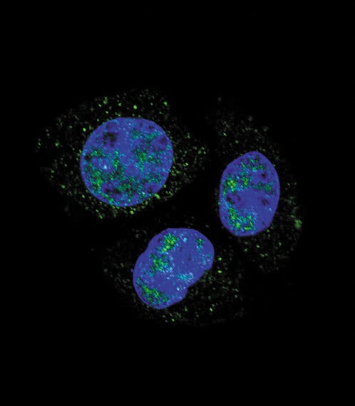
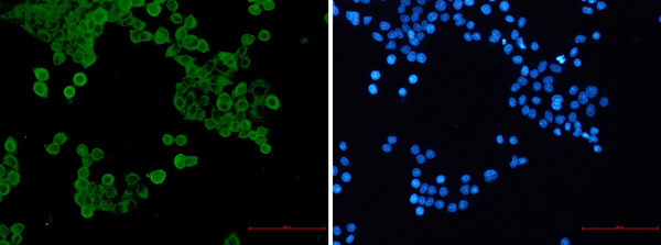 ICC (Immunocytochemistry)
(Immunocytochemistry of AMPK alpha 1 (phospho-Ser496)(green) in hela using AMPK alpha 1 (phospho-Ser496) Rabbit mAb at dilution 1/50, and DAPI(blue))
ICC (Immunocytochemistry)
(Immunocytochemistry of AMPK alpha 1 (phospho-Ser496)(green) in hela using AMPK alpha 1 (phospho-Ser496) Rabbit mAb at dilution 1/50, and DAPI(blue))
AMPK alpha 1, Monoclonal Antibody (Cat# AAA314497)
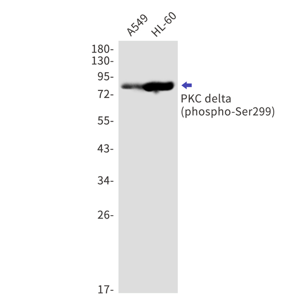
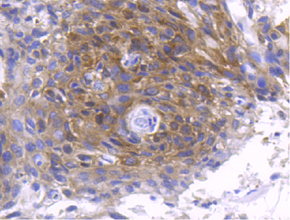
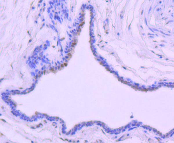
 IHC (Immunohiostchemistry)
(Immunohistochemistry of PKC delta (phospho-Ser299) in paraffin-embedded Human tonsil using PKC delta (phospho-Ser299) Rabbit mAb at dilution 1/50)
IHC (Immunohiostchemistry)
(Immunohistochemistry of PKC delta (phospho-Ser299) in paraffin-embedded Human tonsil using PKC delta (phospho-Ser299) Rabbit mAb at dilution 1/50)
PKC delta, Monoclonal Antibody (Cat# AAA314504)
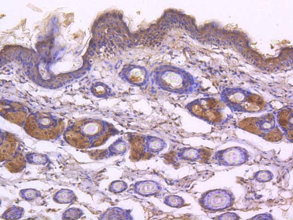
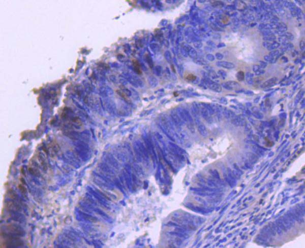
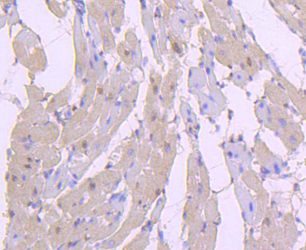
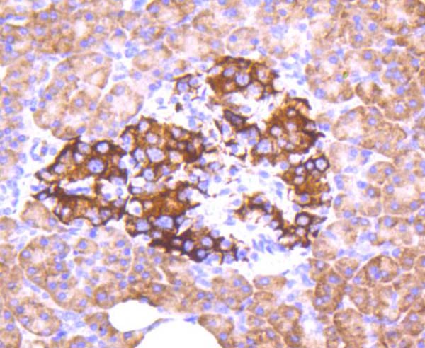
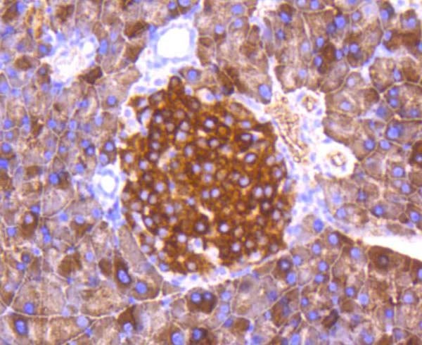
 IHC (Immunohiostchemistry)
(Immunohistochemical analysis of paraffin-embedded human pancreas, using Phospho-Smad5 (S463/465) Antibody.)
IHC (Immunohiostchemistry)
(Immunohistochemical analysis of paraffin-embedded human pancreas, using Phospho-Smad5 (S463/465) Antibody.)
Smad5, Monoclonal Recombinant Antibody (Cat# AAA314842)
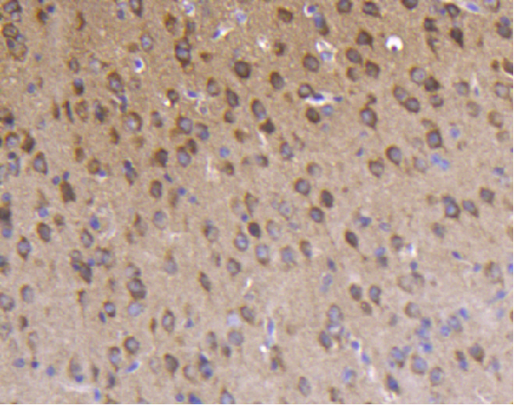
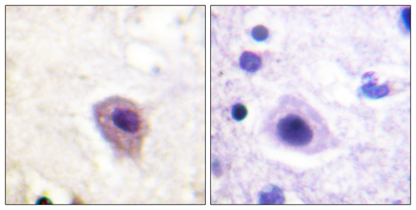
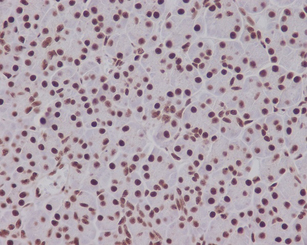
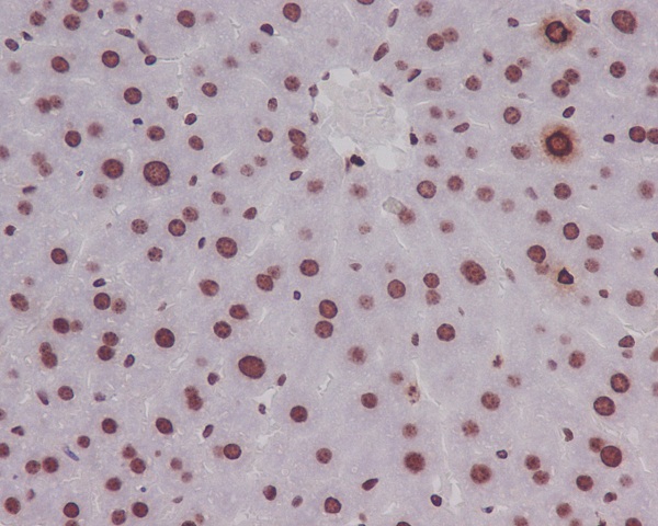 IHC (Immunohiostchemistry)
(Immunohistochemical analysis of paraffin-embedded mouse liver, using Phospho-Histone H3 (S10) Antibody.)
IHC (Immunohiostchemistry)
(Immunohistochemical analysis of paraffin-embedded mouse liver, using Phospho-Histone H3 (S10) Antibody.)
Histone H3, Monoclonal Recombinant Antibody (Cat# AAA314845)
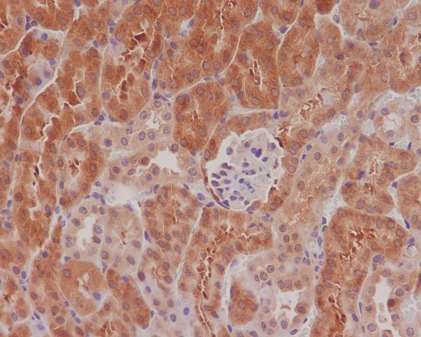 IHC (Immunohiostchemistry)
(Immunohistochemical analysis of paraffin-embedded mouse kidney, using Phospho-eIF4E (S209) antibody.)
IHC (Immunohiostchemistry)
(Immunohistochemical analysis of paraffin-embedded mouse kidney, using Phospho-eIF4E (S209) antibody.)
eIF4E, Monoclonal Recombinant Antibody (Cat# AAA314851)
 IHC (Immunohiostchemistry)
(Immunohistochemical analysis of paraffin-embedded mouse colon, using Phospho-Histone H1.4 (T17) Antibody.)
IHC (Immunohiostchemistry)
(Immunohistochemical analysis of paraffin-embedded mouse colon, using Phospho-Histone H1.4 (T17) Antibody.)
Histone H1.4, Monoclonal Recombinant Antibody (Cat# AAA314854)
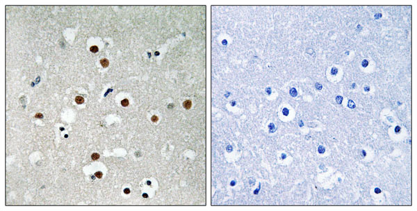
 IHC (Immunohiostchemistry)
(Immunohistochemical analysis of paraffin-embedded human uterus cancer, using Phospho-FoxO3a (S253) Antibody.)
IHC (Immunohiostchemistry)
(Immunohistochemical analysis of paraffin-embedded human uterus cancer, using Phospho-FoxO3a (S253) Antibody.)
FoxO3a, Monoclonal Recombinant Antibody (Cat# AAA314858)
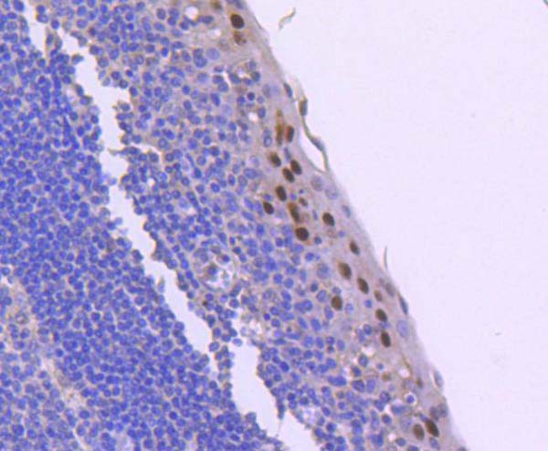
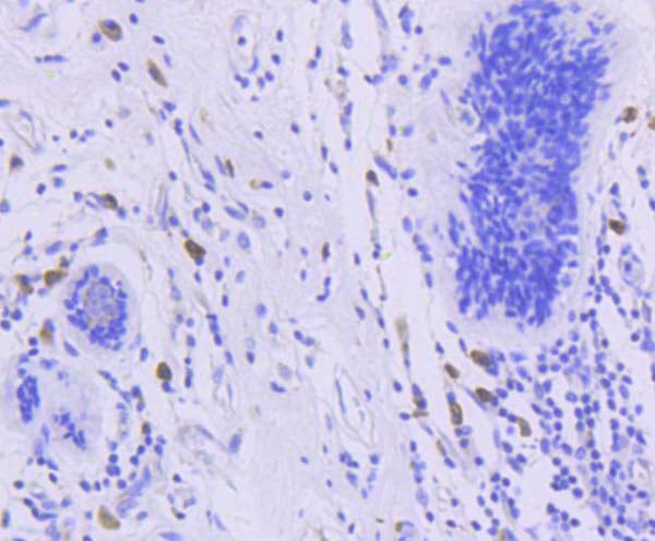
 IHC (Immunohiostchemistry)
(Immunohistochemical analysis of paraffin-embedded human thyroid, using Phospho-Stat5 (Y694) Antibody.)
IHC (Immunohiostchemistry)
(Immunohistochemical analysis of paraffin-embedded human thyroid, using Phospho-Stat5 (Y694) Antibody.)
Stat5, Monoclonal Recombinant Antibody (Cat# AAA314859)
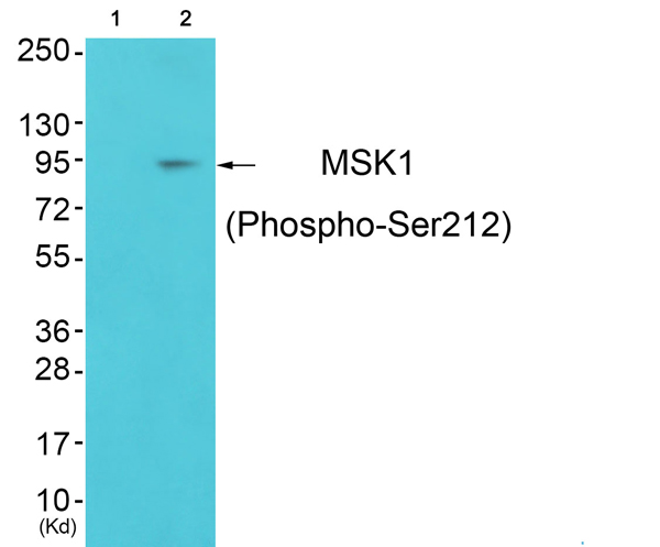
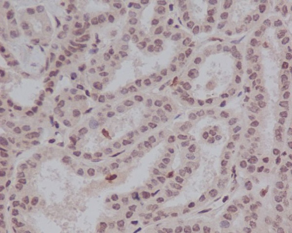
 IHC (Immunohiostchemistry)
(Immunohistochemical analysis of paraffin-embedded human lung cancer, using Phospho-MSK1 (S376) Antibody.)
IHC (Immunohiostchemistry)
(Immunohistochemical analysis of paraffin-embedded human lung cancer, using Phospho-MSK1 (S376) Antibody.)
MSK1, Monoclonal Recombinant Antibody (Cat# AAA314864)
What Are Phospho Antibodies?
Protein phosphorylation is a process where a phosphate group is added to certain amino acid residues of a protein – usually serine (S), threonine (T), or tyrosine (Y) - by enzymes called kinases. This process is integral in controlling cellular signaling, cellular growth, and other biological functions.
Our catalog includes a wide range of phospho-specific antibodies that can accurately detect this important marker. They perform strongly in widely-used laboratory applications such as Western blot, flow cytometry, immunohistochemistry, and immunofluorescence microscopy. We value your trust in us and are committed to providing top-quality products and services. All of our antibodies are guaranteed to work for the applications and species indicated on our website & associated product pages.
What Are The Key Applications of Phospho Antibodies?
1. Western Blotting
One of the first steps a researcher can take in utilizing these phospho-specific antibodies, is to check if the antibody works using a technique referred to as “Western blot”. For those unfamiliar, Western Blot aids in showing whether the protein that the antibody recognizes is appearing at the correct/expected size. These phospho-specific antibodies should also be able to detect changes in the target protein’s phosphorylation (on/off state) when cells are stimulated in certain ways.
2. Staining of Fixed Cells (Immunocytochemistry)
Another routine use of these phospho-specific antibodies, is to test if the antibody is able to demonstrate similar performance when used on fixed cells (intact cells that have been preserved) as it did in the Western blot tests. It is an important aspect in many cases to confirm that the antibody works in actual intact cell samples. Ideally, the method used for cellular fixation should be the same as what is used in pathology labs (like using 10% formalin). To check if the antibody works well in tissue sections (FFPE), researchers will often test it on fixed cells that are processed similar to tissue samples.
3. Specificity Tests Using Peptides
In order to make sure that the antibody is only binding to the right target:
- Laboratory technicians will mix the antibody with phospho-peptides (short segments of the protein containing the phosphate group modification).
- If the antibody signal disappears, it is confirmation that it is binding to the correct phosphorylated location.
- A more robust test is to use both the phosphorylated and non-phosphorylated (dephosphorylated) versions of the protein. The antibody should react only with the phosphorylated one.
- Another method sometimes utilized is to treat the sample with an enzyme, such as alkaline phosphatase, that specifically removes phosphate groups. If the antibody signal disappears after this, it also confirms specificity.
4. Genetic Confirmation
As a final step, scientists can genetically manipulate the nucleotide sequence and alter the target protein by removing the exact site where phosphorylation happens. If the antibody no longer appears to detect the modified protein, it is strong evidence supporting the antibody being specific for that phosphorylated site.
Why Buy Phospho Antibodies Through Us?
- The production laboratory adheres to strict and consistent protocols prior to releasing any of these phospho-specific antibodies:
- Standard methods and proper controls in all tests to ensure high quality.
- These antibodies are tested and validated in different cell types and species.
- High quality control criterion to ensure each batch is consistent, so you will obtain reliable results every time.
FAQ
1. What Are Phospho-Specific Antibodies?
Phospho-specific antibodies are made to detect proteins only when they have a phosphate group linked to a specific amino acid residue. This empowers scientists understand if a protein is "turned on" or active, based on its phosphorylation state.
2. How to Detect Phosphorylated Proteins in a Western Blot?
To find out if a protein is phosphorylated using Western blot:
- Use a phospho-specific antibody that binds only to the phosphorylated form of the protein.
- You can also use a “regular” antibody for the same amino acid sequence of the protein that the phospho-specific antibody is binding to (but in this case, this antibody will not bind if there is a phosphate group present) in order to compare how much of it is phosphorylated versus how much is non-phosphorylated (or “total” protein, if the “normal” antibody’s epitopes are non-phospho-site-specific).
3. How to Choose the Best Antibody?
Here are some simple tips to help you pick the right antibody:
- Know your target
- Match your sample characteristics
- Confirm the intended use is appropriate
- Check “host” and “type”
- Check the “quality” of the presented data/images
- Appraise whether the available validation meets your needs