Filters
▼Clonality
▼Type
▼Reactivity
▼Gene Name
▼Isotype
▼Host
▼Application
▼Clone
▼Phospho Antibodies
Phospho-specific antibodies’ typical purpose is to enable researchers to detect changes in proteins. They will exclusively bind to the amino acid sequence on a protein that has been phosphorylated (which is both a physical & chemical change) and do not bind to the same amino acid sequence on said protein if it lacks said phosphorylation. This aids in being able to clearly see and understand the data produced from this particular protein modification.
Viewing 2750-2800 of 5298 product results

 Application Data
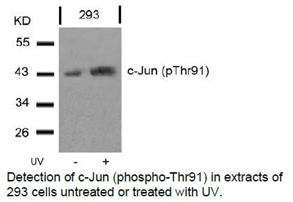
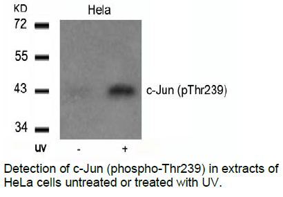
c-Jun, Polyclonal Antibody (Cat# AAA47804)
 Application Data
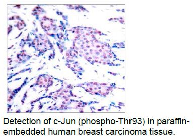
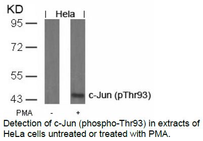
c-Jun, Polyclonal Antibody (Cat# AAA47805)
 Application Data
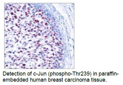
c-Jun, Polyclonal Antibody (Cat# AAA47806)
 Application Data
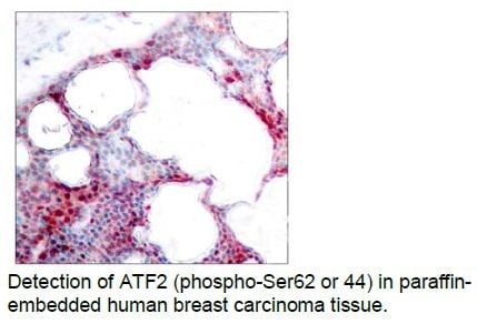
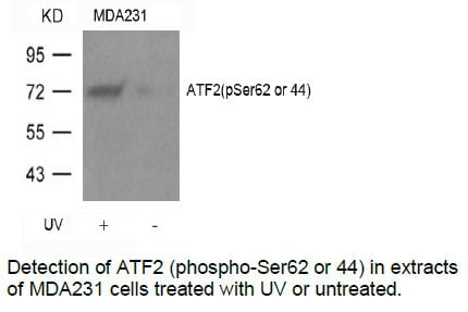
ATF2, Polyclonal Antibody (Cat# AAA47811)
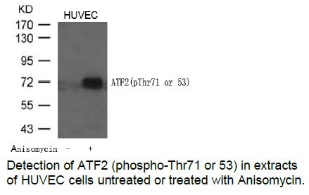 Application Data
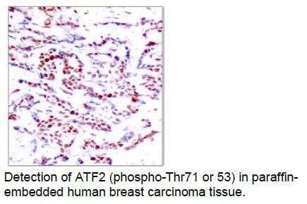
ATF2, Polyclonal Antibody (Cat# AAA47813)
 Application Data
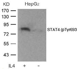
STAT4, Polyclonal Antibody (Cat# AAA47826)
 Application Data
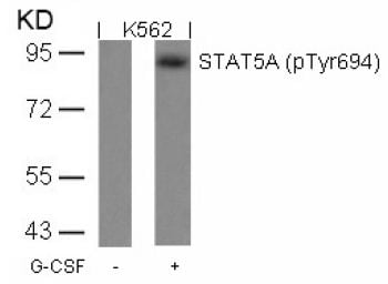
STAT5A, Polyclonal Antibody (Cat# AAA47827)
Phospho-TICAM2, Positive Control (Cat# AAA75985)
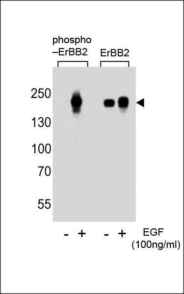
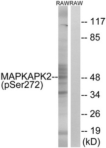
 WB (Western Blot)
Phospho-TICAM2, Polyclonal Antibody (Cat# AAA75753)
Phospho-NT5C2, Polyclonal Antibody (Cat# AAA75785)
 WB (Western Blot)
Phospho-BTK, Polyclonal Antibody (Cat# AAA75800)
 WB (Western Blot)
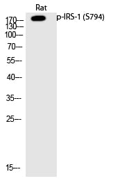
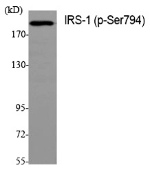
(Western Blot analysis of various cells using Phospho-IRS-1 (S794) Polyclonal Antibody)
WB (Western Blot)
(Western Blot analysis of various cells using Phospho-IRS-1 (S794) Polyclonal Antibody)
IRS-1, Polyclonal Antibody (Cat# AAA305781)

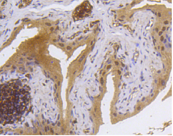
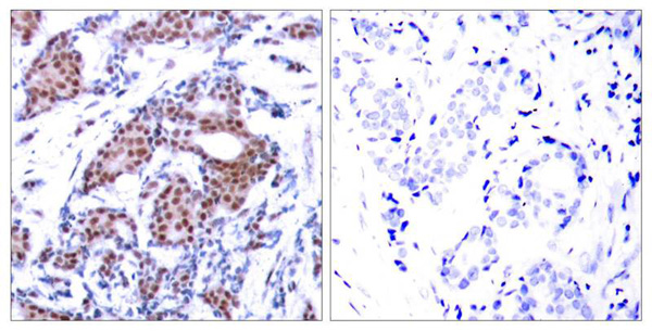 IHC (Immunohiostchemistry)
(Immunohistochemical analysis of paraffin-embedded human breast carcinoma tissue using Elk-1(Phospho-Ser383) Antibody (left) or the same antibody preincubated with blocking peptide(right).)
IHC (Immunohiostchemistry)
(Immunohistochemical analysis of paraffin-embedded human breast carcinoma tissue using Elk-1(Phospho-Ser383) Antibody (left) or the same antibody preincubated with blocking peptide(right).)
Elk-1, Polyclonal Antibody (Cat# AAA306167)
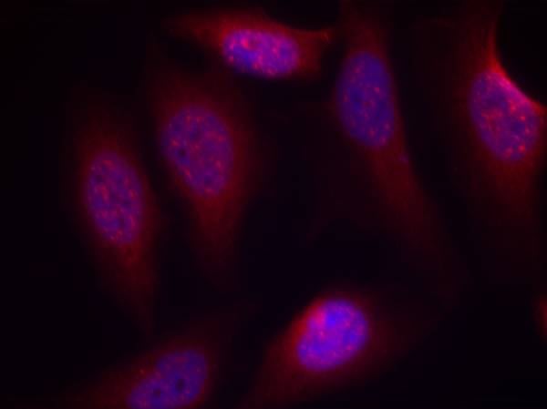 IF (Immunofluorescence)
(Immunofluorescence staining of methanol-fixed Hela cells using SEK1/MKK4(Phospho-Ser80) Antibody.)
IF (Immunofluorescence)
(Immunofluorescence staining of methanol-fixed Hela cells using SEK1/MKK4(Phospho-Ser80) Antibody.)
SEK1/MKK4, Polyclonal Antibody (Cat# AAA305933)
Non-phospho (Active) beta-Catenin (Ser33/37/Thr41), ELISA Kit (Cat# AAA247675)
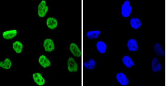
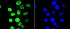
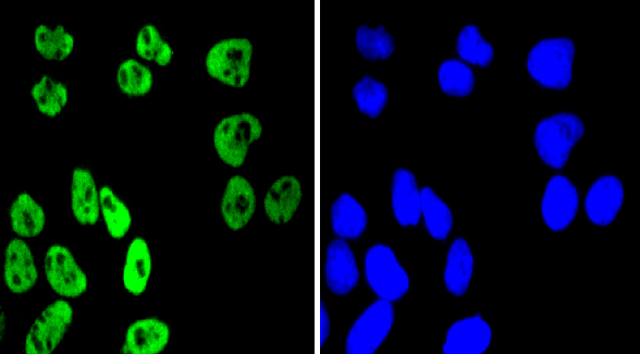
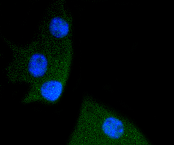
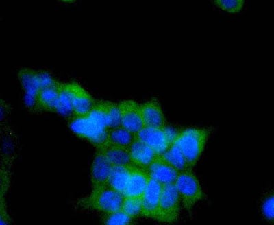
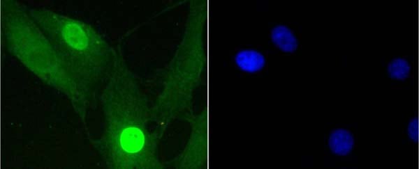
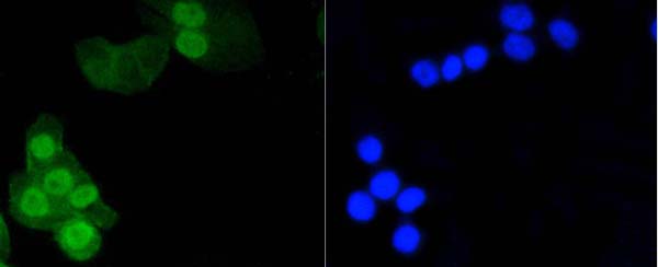
 ICC (Immunocytochemistry)
(ICC staining MSK1 (phospho S376) in 293 cells (green). The nuclear counter stain is DAPI (blue). Cells were fixed in paraformaldehyde, permeabilised with 0.25% Triton X100/PBS.)
ICC (Immunocytochemistry)
(ICC staining MSK1 (phospho S376) in 293 cells (green). The nuclear counter stain is DAPI (blue). Cells were fixed in paraformaldehyde, permeabilised with 0.25% Triton X100/PBS.)
MSK1, Monoclonal Antibody (Cat# AAA311016)
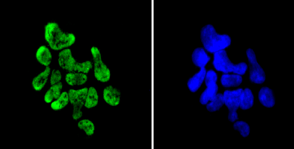
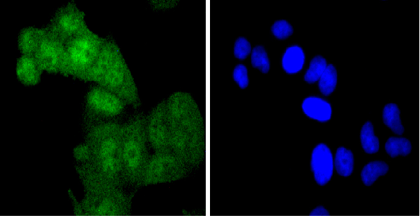
 ICC (Immunocytochemistry)
(ICC staining phospho -SHP2 (Y542) in B-6F1 cells (green). The nuclear counter stain is DAPI (blue). Cells were fixed in paraformaldehyde, permeabilised with 0.25% Triton X100/PBS.)
ICC (Immunocytochemistry)
(ICC staining phospho -SHP2 (Y542) in B-6F1 cells (green). The nuclear counter stain is DAPI (blue). Cells were fixed in paraformaldehyde, permeabilised with 0.25% Triton X100/PBS.)
SHP2, Monoclonal Antibody (Cat# AAA311018)

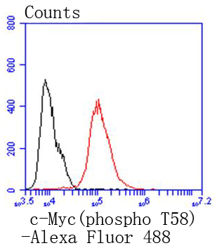 FCM/FACS (Flow Cytometry)
(Flow cytometric analysis of MCF-7 cells with phospho-c-Myc (T58) antibody at 1/50 dilution (red) compared with an unlabelled control (cells without incubation with primary antibody; black). Alexa Fluor 488-conjugated goat anti rabbit IgG was used as the secondary antibody)
FCM/FACS (Flow Cytometry)
(Flow cytometric analysis of MCF-7 cells with phospho-c-Myc (T58) antibody at 1/50 dilution (red) compared with an unlabelled control (cells without incubation with primary antibody; black). Alexa Fluor 488-conjugated goat anti rabbit IgG was used as the secondary antibody)
c-Myc, Monoclonal Antibody (Cat# AAA311019)
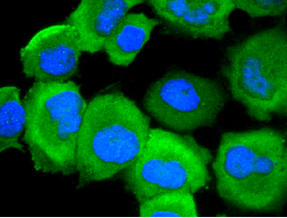
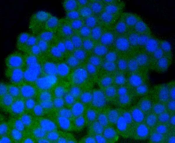
 ICC (Immunocytochemistry)
(ICC staining Phospho-EGFR (S695) in A431 cells (green). The nuclear counter stain is DAPI (blue). Cells were fixed in paraformaldehyde, permeabilised with 0.25% Triton X100/PBS.)
ICC (Immunocytochemistry)
(ICC staining Phospho-EGFR (S695) in A431 cells (green). The nuclear counter stain is DAPI (blue). Cells were fixed in paraformaldehyde, permeabilised with 0.25% Triton X100/PBS.)
EGFR, Monoclonal Antibody (Cat# AAA311023)

 ICC (Immunocytochemistry)
(ICC staining Phospho-AMPK alpha 1 (S496) in 293T cells (green). The nuclear counter stain is DAPI (blue). Cells were fixed in paraformaldehyde, permeabilised with 0.25% Triton X100/PBS.)
ICC (Immunocytochemistry)
(ICC staining Phospho-AMPK alpha 1 (S496) in 293T cells (green). The nuclear counter stain is DAPI (blue). Cells were fixed in paraformaldehyde, permeabilised with 0.25% Triton X100/PBS.)
AMPK alpha 1, Monoclonal Antibody (Cat# AAA311033)
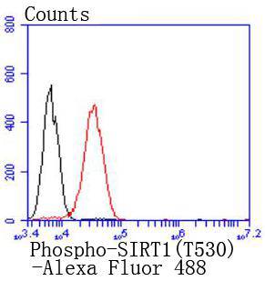 FCM/FACS (Flow Cytometry)
(Flow cytometric analysis of 293 cells with Phospho-SIRT1 (T530) antibody at 1/50 dilution (red) compared with an unlabelled control (cells without incubation with primary antibody; black). Alexa Fluor 488-conjugated goat anti rabbit IgG was used as the secondary antibody.)
FCM/FACS (Flow Cytometry)
(Flow cytometric analysis of 293 cells with Phospho-SIRT1 (T530) antibody at 1/50 dilution (red) compared with an unlabelled control (cells without incubation with primary antibody; black). Alexa Fluor 488-conjugated goat anti rabbit IgG was used as the secondary antibody.)
SIRT1, Monoclonal Antibody (Cat# AAA311039)
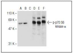
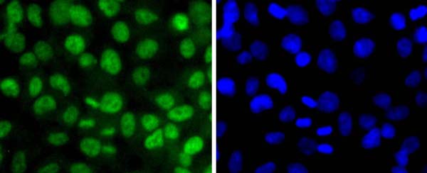
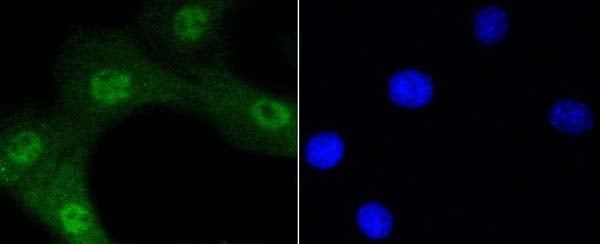
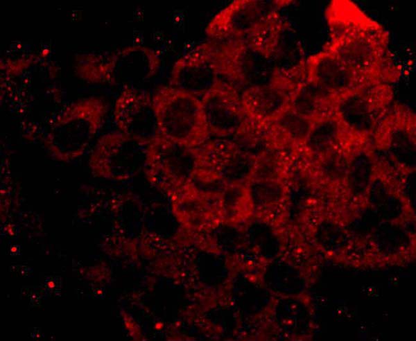 IF (Immunofluorescence)
(Immunofluorescence staining of anisomycin-treated, methanol-fixed NIH/3T3 cells, showing cytoplasmic and nuclear localization of activated p70 S6 kinase alpha (A). Immunoperoxidase staining of formalin fixed, paraffin-embedded human esophagus tissue showing nuclear staining of squamous epithelial cells (B).)
IF (Immunofluorescence)
(Immunofluorescence staining of anisomycin-treated, methanol-fixed NIH/3T3 cells, showing cytoplasmic and nuclear localization of activated p70 S6 kinase alpha (A). Immunoperoxidase staining of formalin fixed, paraffin-embedded human esophagus tissue showing nuclear staining of squamous epithelial cells (B).)
p70S6 Kinase, Monoclonal Antibody (Cat# AAA310991)
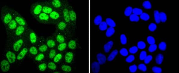
 ICC (Immunocytochemistry)
(ICC staining Phospho-Histone H1.3 (T17)+Histone H1.4 (T17) in CRC cells (green). Cells were fixed in paraformaldehyde, permeabilised with 0.25% Triton X100/PBS.)
ICC (Immunocytochemistry)
(ICC staining Phospho-Histone H1.3 (T17)+Histone H1.4 (T17) in CRC cells (green). Cells were fixed in paraformaldehyde, permeabilised with 0.25% Triton X100/PBS.)
Histone H1.3+Histone H1.4, Monoclonal Antibody (Cat# AAA310995)
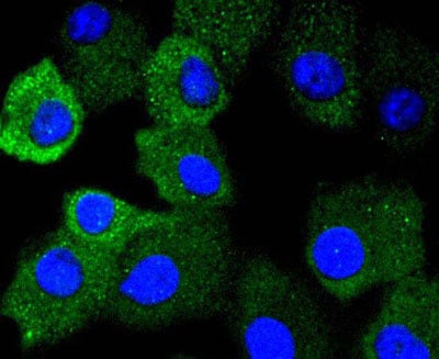
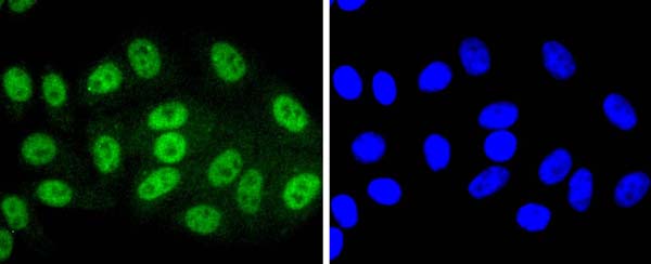
 ICC (Immunocytochemistry)
(ICC staining Phospho-JAK2 (Y1007+Y1008) in Hela cells (green). The nuclear counter stain is DAPI (blue). Cells were fixed in paraformaldehyde, permeabilised with 0.25% Triton X100/PBS.)
ICC (Immunocytochemistry)
(ICC staining Phospho-JAK2 (Y1007+Y1008) in Hela cells (green). The nuclear counter stain is DAPI (blue). Cells were fixed in paraformaldehyde, permeabilised with 0.25% Triton X100/PBS.)
JAK2, Monoclonal Antibody (Cat# AAA311001)
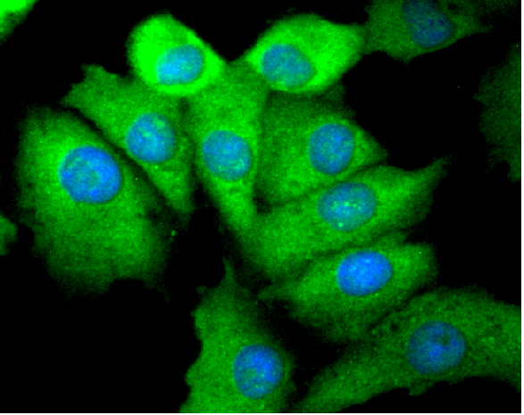
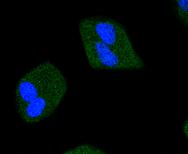
 ICC (Immunocytochemistry)
(ICC staining Phospho-Akt1 (Ser473) in NIH/3T3 cells (green). The nuclear counter stain is DAPI (blue). Cells were fixed in paraformaldehyde, permeabilised with 0.25% Triton X100/PBS.)
ICC (Immunocytochemistry)
(ICC staining Phospho-Akt1 (Ser473) in NIH/3T3 cells (green). The nuclear counter stain is DAPI (blue). Cells were fixed in paraformaldehyde, permeabilised with 0.25% Triton X100/PBS.)
AKT1, Monoclonal Antibody (Cat# AAA311003)
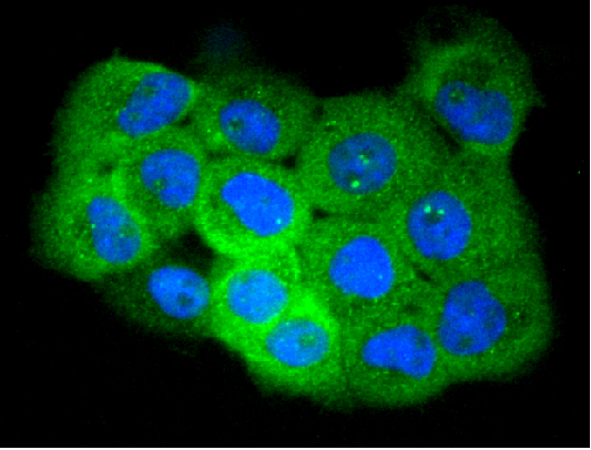
 ICC (Immunocytochemistry)
(ICC staining Phospho-PP2A (pY307) in PC12 cells (green). The nuclear counter stain is DAPI (blue). Cells were fixed in paraformaldehyde, permeabilised with 0.25% Triton X100/PBS.)
ICC (Immunocytochemistry)
(ICC staining Phospho-PP2A (pY307) in PC12 cells (green). The nuclear counter stain is DAPI (blue). Cells were fixed in paraformaldehyde, permeabilised with 0.25% Triton X100/PBS.)
PP2A, Monoclonal Antibody (Cat# AAA311005)
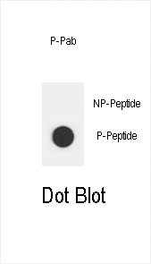
 DB (Dot Blot)
(Dot blot analysis of anti-EGFR-pS1070 Phospho-specific Pab on nitrocellulose membrane. 50ng of Phospho-peptide or Non Phospho-peptide per dot were adsorbed. Antibody working concentrations are 0.5ug per ml.)
DB (Dot Blot)
(Dot blot analysis of anti-EGFR-pS1070 Phospho-specific Pab on nitrocellulose membrane. 50ng of Phospho-peptide or Non Phospho-peptide per dot were adsorbed. Antibody working concentrations are 0.5ug per ml.)
Phospho-EGFR (S1070), Polyclonal Antibody (Cat# AAA285783)
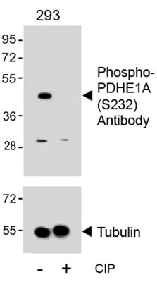
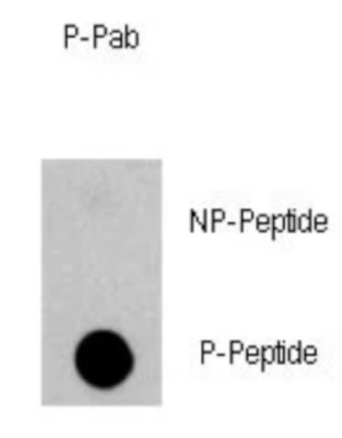
 DB (Dot Blot)
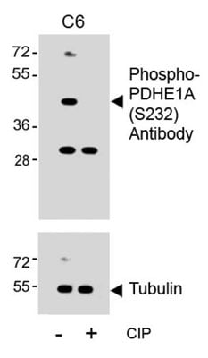
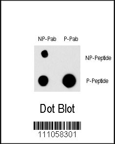
(Dot blot analysis of anti-mPDHE1A-S232 Phospho-specific Pab on nitrocellulose membrane. 50ng of Phospho-peptide or Non Phospho-peptide per dot were adsorbed. Antibody working concentrations are 0.6ug per ml.)
DB (Dot Blot)
(Dot blot analysis of anti-mPDHE1A-S232 Phospho-specific Pab on nitrocellulose membrane. 50ng of Phospho-peptide or Non Phospho-peptide per dot were adsorbed. Antibody working concentrations are 0.6ug per ml.)
Phospho-PDHE1A (S232), Polyclonal Antibody (Cat# AAA285709)
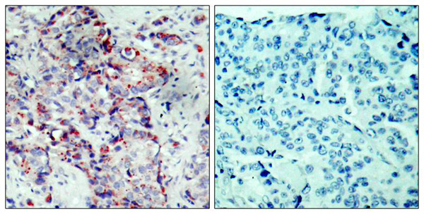
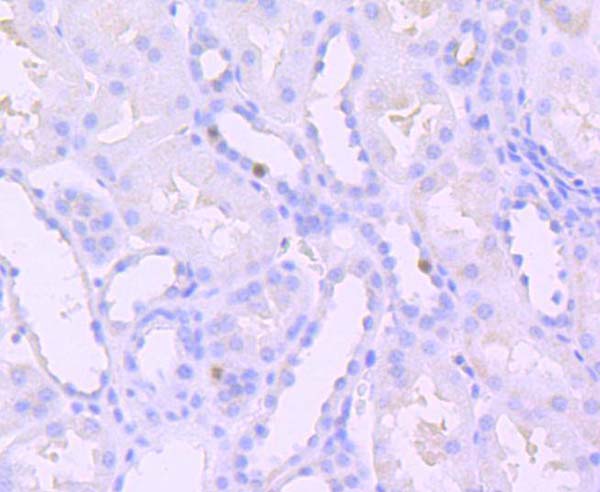
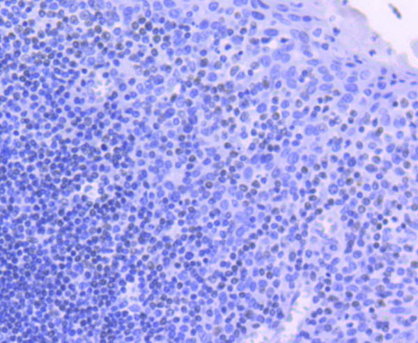
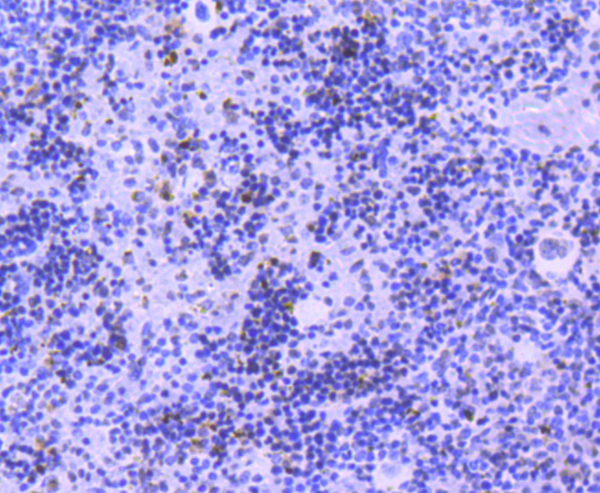
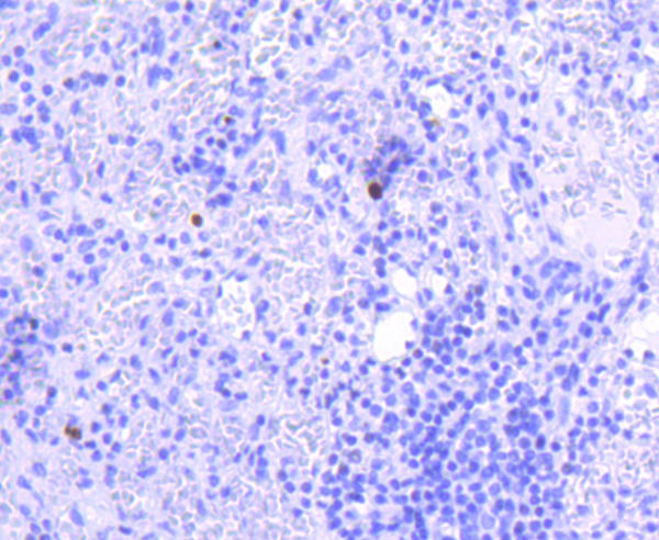
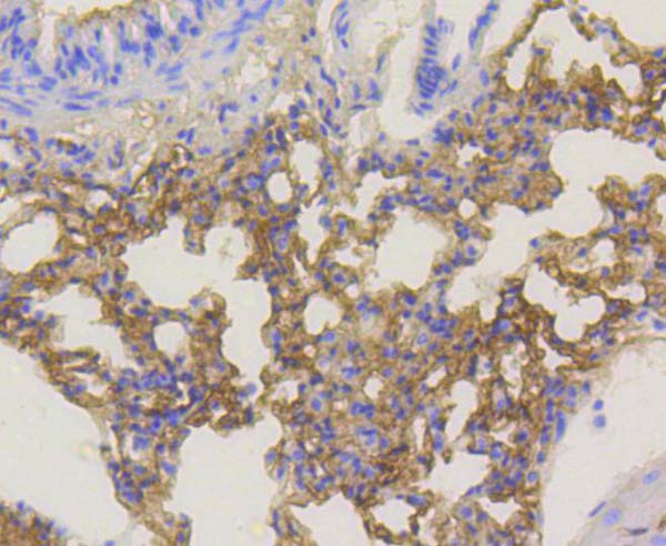
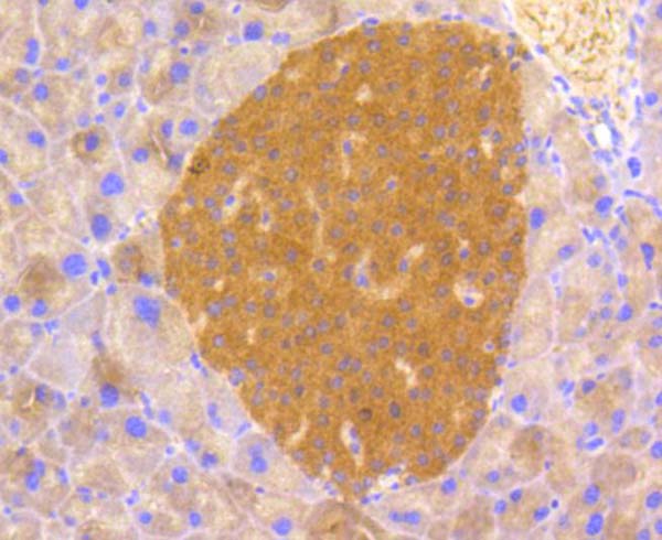
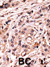 IHC (Immunohiostchemistry)
(Formalin-fixed and paraffin-embedded human cancer tissue reacted with the primary antibody, which was peroxidase-conjugated to the secondary antibody, followed by AEC staining. This data demonstrates the use of this antibody for immunohistochemistry; clinical relevance has not been evaluated. BC = breast carcinoma; HC = hepatocarcinoma.)
IHC (Immunohiostchemistry)
(Formalin-fixed and paraffin-embedded human cancer tissue reacted with the primary antibody, which was peroxidase-conjugated to the secondary antibody, followed by AEC staining. This data demonstrates the use of this antibody for immunohistochemistry; clinical relevance has not been evaluated. BC = breast carcinoma; HC = hepatocarcinoma.)
Phospho-p53 (S20), Polyclonal Antibody (Cat# AAA288717)
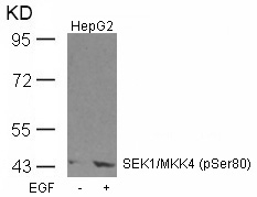
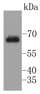
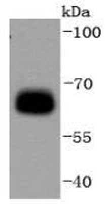
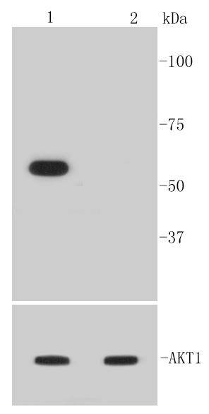
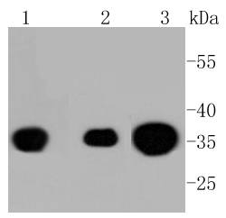
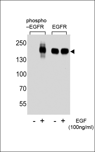
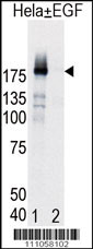 WB (Western Blot)
(Western blot analysis of EGFR (arrow) in Hela cell lysates, either induced (Lane 1) or noninduced with EGF (Lane 2).)
WB (Western Blot)
(Western blot analysis of EGFR (arrow) in Hela cell lysates, either induced (Lane 1) or noninduced with EGF (Lane 2).)
Phospho-EGFR (Y998), Polyclonal Antibody (Cat# AAA288513)
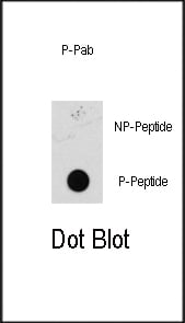
 DB (Dot Blot)
(Dot blot analysis of Phospho-ERBB2-Y1112 Phospho-specific Pab on nitrocellulose membrane. 50ng of Phospho-peptide or Non Phospho-peptide per dot were adsorbed. Antibody working concentrations are 0.6ug per ml.)
DB (Dot Blot)
(Dot blot analysis of Phospho-ERBB2-Y1112 Phospho-specific Pab on nitrocellulose membrane. 50ng of Phospho-peptide or Non Phospho-peptide per dot were adsorbed. Antibody working concentrations are 0.6ug per ml.)
Phospho-ErbB2 (Y1112), Polyclonal Antibody (Cat# AAA285456)
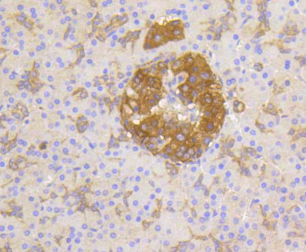
 IHC (Immunohiostchemistry)
(Formalin-fixed and paraffin-embedded human cancer tissue reacted with the primary antibody, which was peroxidase-conjugated to the secondary antibody, followed by AEC staining. This data demonstrates the use of this antibody for immunohistochemistry; clinical relevance has not been evaluated. BC = breast carcinoma; HC = hepatocarcinoma.)
IHC (Immunohiostchemistry)
(Formalin-fixed and paraffin-embedded human cancer tissue reacted with the primary antibody, which was peroxidase-conjugated to the secondary antibody, followed by AEC staining. This data demonstrates the use of this antibody for immunohistochemistry; clinical relevance has not been evaluated. BC = breast carcinoma; HC = hepatocarcinoma.)
Bi-Phospho-MEK1 (S218/222), Polyclonal Antibody (Cat# AAA288138)
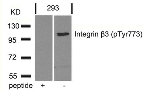
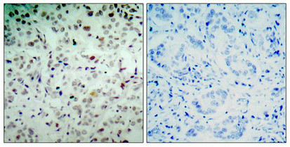
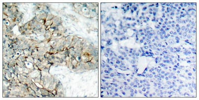 IHC (Immunohiostchemistry)
(Immunohistochemical analysis of paraffin-embedded human breast carcinoma tissue using Integrin b3(Phospho-Tyr773) Antibody(left) or the same antibody preincubated with blocking peptide(right).)
IHC (Immunohiostchemistry)
(Immunohistochemical analysis of paraffin-embedded human breast carcinoma tissue using Integrin b3(Phospho-Tyr773) Antibody(left) or the same antibody preincubated with blocking peptide(right).)
ITGB3, Polyclonal Antibody (Cat# AAA243063)
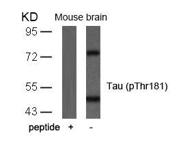
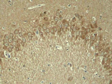 IHC (Immunohiostchemistry)
(Immunohistochemical analysis of paraffin-embedded rat hippocampal region tissue from a model with Alzheimer)
IHC (Immunohiostchemistry)
(Immunohistochemical analysis of paraffin-embedded rat hippocampal region tissue from a model with Alzheimer)
MAPT, Polyclonal Antibody (Cat# AAA243080)

 IHC (Immunohiostchemistry)
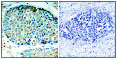
(Immunohistochemical analysis of paraffin-embedded human breast carcinoma tissue using CREB(Phospho-Ser129) Antibody(left) or the same antibody preincubated with blocking peptide(right).)
IHC (Immunohiostchemistry)
(Immunohistochemical analysis of paraffin-embedded human breast carcinoma tissue using CREB(Phospho-Ser129) Antibody(left) or the same antibody preincubated with blocking peptide(right).)
CREB1, Polyclonal Antibody (Cat# AAA243136)
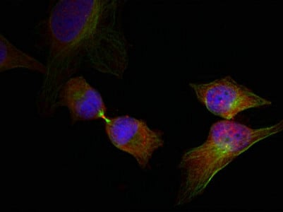 IF (Immunofluorescence)
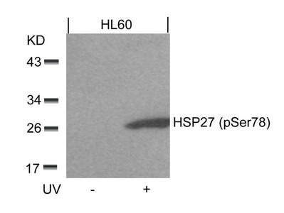
(Immunofluorescence staining of methanol-fixed Hela cells showing cytoplasmic, nuclear, centrosomal, midbody staining using HSP27(Phospho-Ser78) Antibody.)
IF (Immunofluorescence)
(Immunofluorescence staining of methanol-fixed Hela cells showing cytoplasmic, nuclear, centrosomal, midbody staining using HSP27(Phospho-Ser78) Antibody.)
HSPB1, Polyclonal Antibody (Cat# AAA243304)
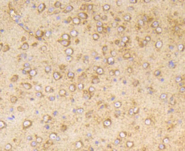
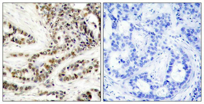
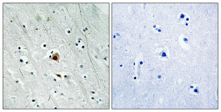 IHC (Immunohiostchemistry)
(Immunohistochemical analysis of paraffin-embedded human brain tissue using MAPKAPK2 (Phospho-Ser272) antibody (left)or the same antibody preincubated with blocking peptide (right).)
IHC (Immunohiostchemistry)
(Immunohistochemical analysis of paraffin-embedded human brain tissue using MAPKAPK2 (Phospho-Ser272) antibody (left)or the same antibody preincubated with blocking peptide (right).)
MAPKAPK2, Polyclonal Antibody (Cat# AAA243225)


 IF (Immunofluorescence)
(Immunofluorescence staining of methanol-fixed Hela cells using cdc25C(Phospho-Ser216) Antibody.)
IF (Immunofluorescence)
(Immunofluorescence staining of methanol-fixed Hela cells using cdc25C(Phospho-Ser216) Antibody.)
CDC25C, Polyclonal Antibody (Cat# AAA243278)
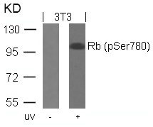
 IF (Immunofluorescence)
(Immunofluorescence staining of methanol-fixed Hela cells using Rb(Phospho-Ser780) Antibody.)
IF (Immunofluorescence)
(Immunofluorescence staining of methanol-fixed Hela cells using Rb(Phospho-Ser780) Antibody.)
RB1, Polyclonal Antibody (Cat# AAA243284)

 IF (Immunofluorescence)
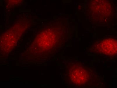
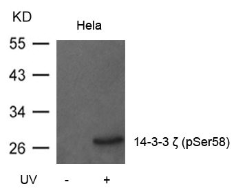
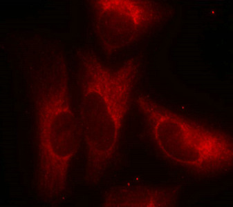
(Immunofluorescence staining of methanol-fixed Hela cells using 14-3-3z(Phospho-Ser58) Antibody.)
IF (Immunofluorescence)
(Immunofluorescence staining of methanol-fixed Hela cells using 14-3-3z(Phospho-Ser58) Antibody.)
YWHAZ, Polyclonal Antibody (Cat# AAA243295)
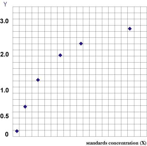 Standard Curve (Sample)
Phospho-Vascular Endothelial Cell Growth Factor Receptor 2 (p-VEGFR2), ELISA Kit (Cat# AAA206808)
 Standard Curve (Sample)
Phospho-Nuclear Factor kappa B, ELISA Kit (Cat# AAA83059)
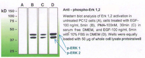
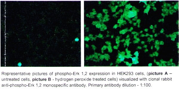 ICC (Immunocytochemistry)
(1. Coat coverslips with 1 % gelatin-coating solution for 2 hours at room temperature (RT): rinse with distilled water, and let to dry overnight. Before plating the cells, wash the coated coverslips briefly with PBS.2. Fix the cells with 4% paraformaldehyde solution (in PBS, pH 7.2), for 15 min at RT.3. Wash 2 x 3 min with PBS.4. Permeabilize the cells with 0.1% Triton X-100 solution (in PBS. pH 7.2) for 5 min on ice.5. Wash 2 x 3 min with PBS.6. Incubate the cells in blocking buffer (0.3M glycine in PBS, 2% BSA) for 30 min at RT.7. Incubate the cells with primary antibody: anti-phospho Erk 1,2 clonal antibody at the dilution of 1:100 - 1 :400 in antibody dilution buffer (PBS, 1 % BSA) for 1 hour at RT in humid chamber.8. Wash 2 x 3 min with PBS.9. Apply the secondary antibody (in this case, the goat anti-rabbit IgG-FITC from Jackson Immunoresearch, cat. # 111-095-003, was used at 1 :300 in antibody dilution buffer, and cells were incubated for 1 hour at RT in dark).10. Wash 3 x 3 min with PBS.11. Rinse once with distilled water.12. Mount the slide for observation, with a drop of anti-fade mounting medium.)
ICC (Immunocytochemistry)
(1. Coat coverslips with 1 % gelatin-coating solution for 2 hours at room temperature (RT): rinse with distilled water, and let to dry overnight. Before plating the cells, wash the coated coverslips briefly with PBS.2. Fix the cells with 4% paraformaldehyde solution (in PBS, pH 7.2), for 15 min at RT.3. Wash 2 x 3 min with PBS.4. Permeabilize the cells with 0.1% Triton X-100 solution (in PBS. pH 7.2) for 5 min on ice.5. Wash 2 x 3 min with PBS.6. Incubate the cells in blocking buffer (0.3M glycine in PBS, 2% BSA) for 30 min at RT.7. Incubate the cells with primary antibody: anti-phospho Erk 1,2 clonal antibody at the dilution of 1:100 - 1 :400 in antibody dilution buffer (PBS, 1 % BSA) for 1 hour at RT in humid chamber.8. Wash 2 x 3 min with PBS.9. Apply the secondary antibody (in this case, the goat anti-rabbit IgG-FITC from Jackson Immunoresearch, cat. # 111-095-003, was used at 1 :300 in antibody dilution buffer, and cells were incubated for 1 hour at RT in dark).10. Wash 3 x 3 min with PBS.11. Rinse once with distilled water.12. Mount the slide for observation, with a drop of anti-fade mounting medium.)
PHOSPHO-Erk 1,2, Monoclonal Antibody (Cat# AAA79001)
Tested: Rat
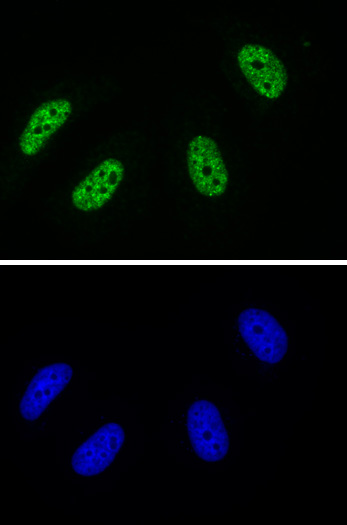
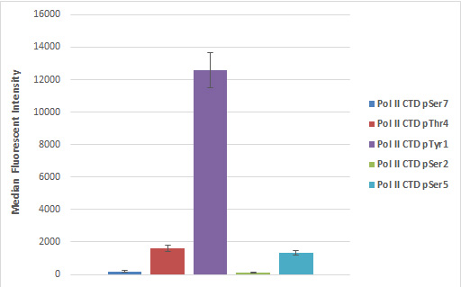
 ChIP (Chromatin Immunoprecipitation)
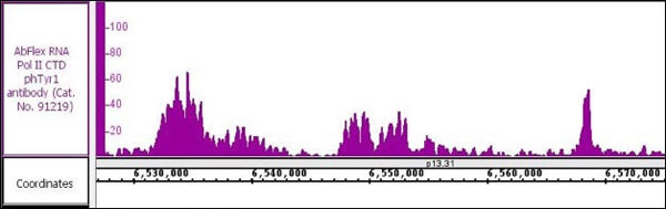
(AbFlex RNA Pol II CTD phospho Tyr1 recombinant antibody (rAb) tested by ChIP-Seq Chromatin immunoprecipitation (ChIP) was performed using the ChIP-IT High Sensitivity Kit with 30 ug of Raji cell chromatin and 10 ug of antibody. ChIP DNA was sequenced on the Illumina NextSeq and 17.3 million sequence tags were mapped to identify RNA Pol II CTD phosphor Tyr1 binding sites on chromosome 12.)
ChIP (Chromatin Immunoprecipitation)
(AbFlex RNA Pol II CTD phospho Tyr1 recombinant antibody (rAb) tested by ChIP-Seq Chromatin immunoprecipitation (ChIP) was performed using the ChIP-IT High Sensitivity Kit with 30 ug of Raji cell chromatin and 10 ug of antibody. ChIP DNA was sequenced on the Illumina NextSeq and 17.3 million sequence tags were mapped to identify RNA Pol II CTD phosphor Tyr1 binding sites on chromosome 12.)
RNA Pol II CTD phospho Tyr1, Antibody (Cat# AAA60276)
mDia Phospho-Regulation, Antibody Sampler Kit (Cat# AAA71616)
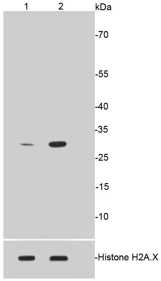
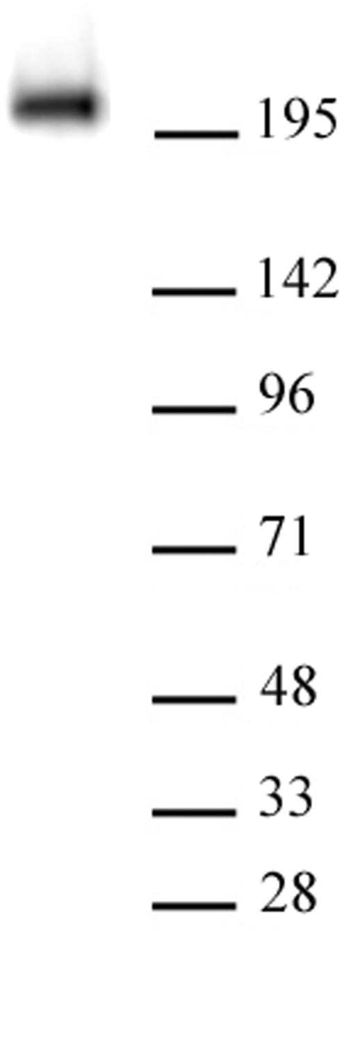
 WB (Western Blot)
(The whole cell lysate derived from human umbilical vein endothelial cell was stimulated by VEGF-A for 20 min, then immunoprecipitated by Rabbit anti-VEGFR-2 followed by immune-probing with Rabbit anti phosphor-VEGFR-2 (pY951) (AAA71420) at 1:500. An immunoreactive band is observed around ~240kDa(lane 1). The lane 2 is a negative control.)
WB (Western Blot)
(The whole cell lysate derived from human umbilical vein endothelial cell was stimulated by VEGF-A for 20 min, then immunoprecipitated by Rabbit anti-VEGFR-2 followed by immune-probing with Rabbit anti phosphor-VEGFR-2 (pY951) (AAA71420) at 1:500. An immunoreactive band is observed around ~240kDa(lane 1). The lane 2 is a negative control.)
VEGFR-2 (pY951) (CD309), Antibody (Cat# AAA71420)
Phospho-Tyrosine, Serine, Threonine, Antibody Sampler Kit (Cat# AAA71678)
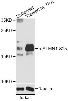
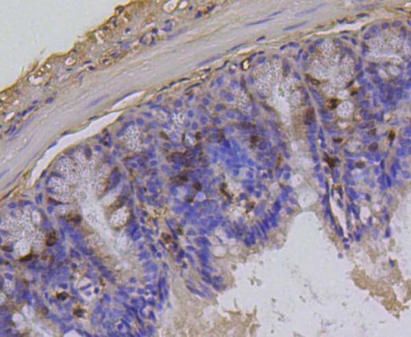
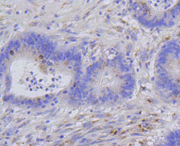
 IHC (Immunohiostchemistry)
(Immunohistochemistry of paraffin-embedded human breast carcinoma using Phospho-STMN1-S25 antibody.)
IHC (Immunohiostchemistry)
(Immunohistochemistry of paraffin-embedded human breast carcinoma using Phospho-STMN1-S25 antibody.)
STMN1-S25, Antibody (Cat# AAA37416)
Phospho-STAT1, Positive Control (Cat# AAA75983)
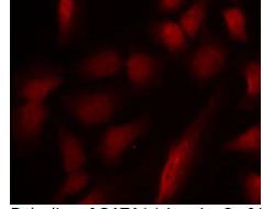
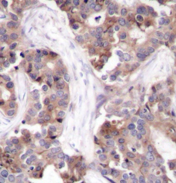
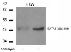 Application Data
GATA1, Polyclonal Antibody (Cat# AAA47822)
What Are Phospho Antibodies?
Protein phosphorylation is a process where a phosphate group is added to certain amino acid residues of a protein – usually serine (S), threonine (T), or tyrosine (Y) - by enzymes called kinases. This process is integral in controlling cellular signaling, cellular growth, and other biological functions.
Our catalog includes a wide range of phospho-specific antibodies that can accurately detect this important marker. They perform strongly in widely-used laboratory applications such as Western blot, flow cytometry, immunohistochemistry, and immunofluorescence microscopy. We value your trust in us and are committed to providing top-quality products and services. All of our antibodies are guaranteed to work for the applications and species indicated on our website & associated product pages.
What Are The Key Applications of Phospho Antibodies?
1. Western Blotting
One of the first steps a researcher can take in utilizing these phospho-specific antibodies, is to check if the antibody works using a technique referred to as “Western blot”. For those unfamiliar, Western Blot aids in showing whether the protein that the antibody recognizes is appearing at the correct/expected size. These phospho-specific antibodies should also be able to detect changes in the target protein’s phosphorylation (on/off state) when cells are stimulated in certain ways.
2. Staining of Fixed Cells (Immunocytochemistry)
Another routine use of these phospho-specific antibodies, is to test if the antibody is able to demonstrate similar performance when used on fixed cells (intact cells that have been preserved) as it did in the Western blot tests. It is an important aspect in many cases to confirm that the antibody works in actual intact cell samples. Ideally, the method used for cellular fixation should be the same as what is used in pathology labs (like using 10% formalin). To check if the antibody works well in tissue sections (FFPE), researchers will often test it on fixed cells that are processed similar to tissue samples.
3. Specificity Tests Using Peptides
In order to make sure that the antibody is only binding to the right target:
- Laboratory technicians will mix the antibody with phospho-peptides (short segments of the protein containing the phosphate group modification).
- If the antibody signal disappears, it is confirmation that it is binding to the correct phosphorylated location.
- A more robust test is to use both the phosphorylated and non-phosphorylated (dephosphorylated) versions of the protein. The antibody should react only with the phosphorylated one.
- Another method sometimes utilized is to treat the sample with an enzyme, such as alkaline phosphatase, that specifically removes phosphate groups. If the antibody signal disappears after this, it also confirms specificity.
4. Genetic Confirmation
As a final step, scientists can genetically manipulate the nucleotide sequence and alter the target protein by removing the exact site where phosphorylation happens. If the antibody no longer appears to detect the modified protein, it is strong evidence supporting the antibody being specific for that phosphorylated site.
Why Buy Phospho Antibodies Through Us?
- The production laboratory adheres to strict and consistent protocols prior to releasing any of these phospho-specific antibodies:
- Standard methods and proper controls in all tests to ensure high quality.
- These antibodies are tested and validated in different cell types and species.
- High quality control criterion to ensure each batch is consistent, so you will obtain reliable results every time.
FAQ
1. What Are Phospho-Specific Antibodies?
Phospho-specific antibodies are made to detect proteins only when they have a phosphate group linked to a specific amino acid residue. This empowers scientists understand if a protein is "turned on" or active, based on its phosphorylation state.
2. How to Detect Phosphorylated Proteins in a Western Blot?
To find out if a protein is phosphorylated using Western blot:
- Use a phospho-specific antibody that binds only to the phosphorylated form of the protein.
- You can also use a “regular” antibody for the same amino acid sequence of the protein that the phospho-specific antibody is binding to (but in this case, this antibody will not bind if there is a phosphate group present) in order to compare how much of it is phosphorylated versus how much is non-phosphorylated (or “total” protein, if the “normal” antibody’s epitopes are non-phospho-site-specific).
3. How to Choose the Best Antibody?
Here are some simple tips to help you pick the right antibody:
- Know your target
- Match your sample characteristics
- Confirm the intended use is appropriate
- Check “host” and “type”
- Check the “quality” of the presented data/images
- Appraise whether the available validation meets your needs