Filters
▼Clonality
▼Type
▼Reactivity
▼Gene Name
▼Isotype
▼Host
▼Application
▼Clone
▼Monoclonal Antibodies
Get accurate results in your research with our Monoclonal Antibodies, which are specially made to target exactly what you require for your research, and will produce consistent, reliable performance in lab tests.
Viewing 6700-6750 of 27597 product results
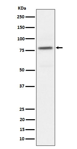
 WB (Western Blot)
(HepG2 cells were subjected to SDS PAGE followed by western blot with AAA247979 (ARG1 Antibody) at dilution of 1:4000)
WB (Western Blot)
(HepG2 cells were subjected to SDS PAGE followed by western blot with AAA247979 (ARG1 Antibody) at dilution of 1:4000)
liver Arginase, Monoclonal Antibody (Cat# AAA247979)
Protein A+G purification
 WB (Western Blot)
(PC-3 cells were subjected to SDS PAGE followed by western blot with AAA247990 (MEST antibody) at dilution of 1:1000)
WB (Western Blot)
(PC-3 cells were subjected to SDS PAGE followed by western blot with AAA247990 (MEST antibody) at dilution of 1:1000)
MEST, Monoclonal Antibody (Cat# AAA247990)
Protein A+G purification
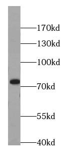
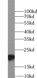
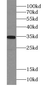
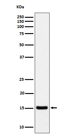
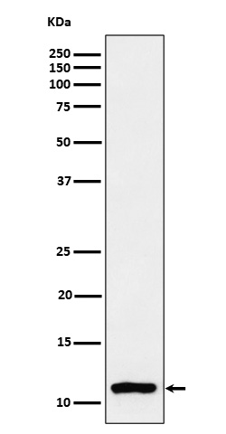
 WB (Western Blot)
(HL-60 cells were subjected to SDS PAGE followed by western blot with AAA247995 (MPO Antibody) at dilution of 1:1000)
WB (Western Blot)
(HL-60 cells were subjected to SDS PAGE followed by western blot with AAA247995 (MPO Antibody) at dilution of 1:1000)
MPO, Monoclonal Antibody (Cat# AAA247995)
Protein A+G purification

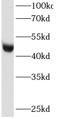
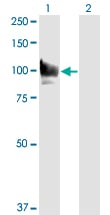
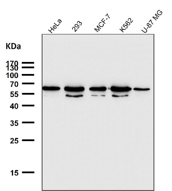
 WB (Western Blot)
(HeLa cells were subjected to SDS PAGE followed by western blot with AAA247998 (MTA2 Antibody) at dilution of 1:1000)
WB (Western Blot)
(HeLa cells were subjected to SDS PAGE followed by western blot with AAA247998 (MTA2 Antibody) at dilution of 1:1000)
MTA2, Monoclonal Antibody (Cat# AAA247998)
Protein A+G purification
 WB (Western Blot)
(HeLa cells were subjected to SDS PAGE followed by western blot with AAA248008 (NDUFA4L2 antibody) at dilution of 1:1000)
WB (Western Blot)
(HeLa cells were subjected to SDS PAGE followed by western blot with AAA248008 (NDUFA4L2 antibody) at dilution of 1:1000)
NDUFA4L2, Monoclonal Antibody (Cat# AAA248008)
Protein A+G purification
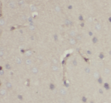
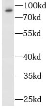
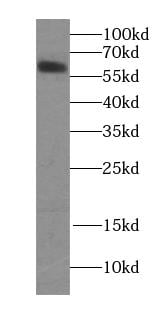
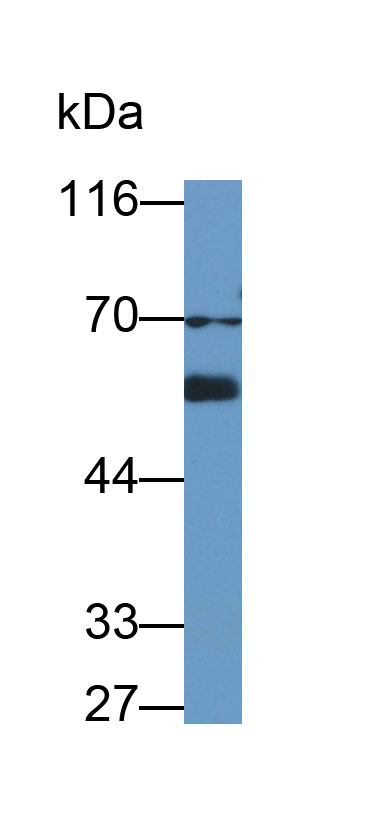
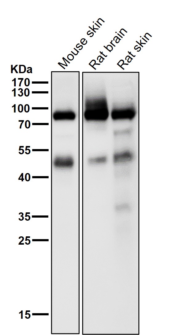
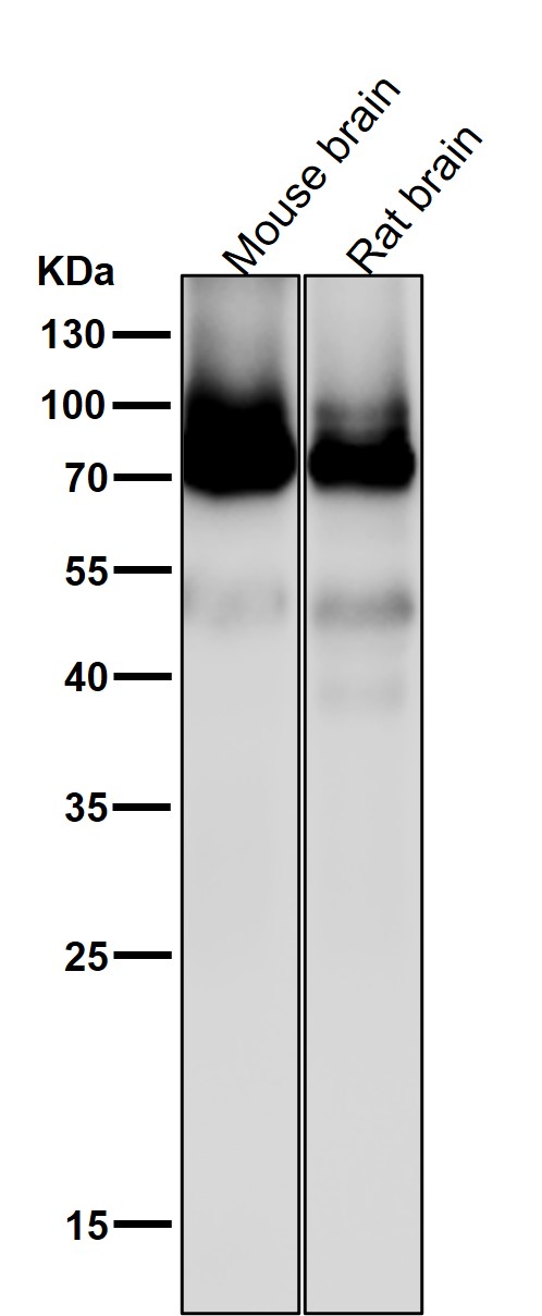
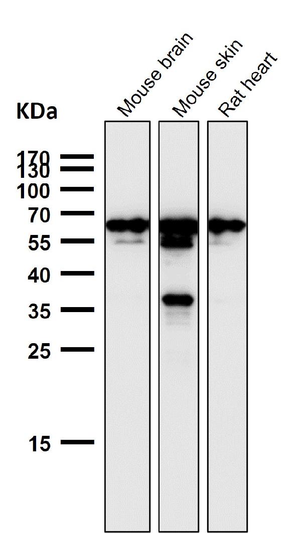
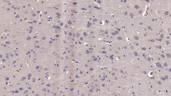
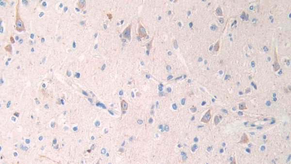
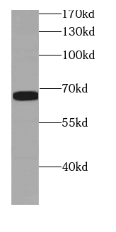 WB (Western Blot)
(human brain tissue were subjected to SDS PAGE followed by western blot with AAA248021 (NUMB antibody) at dilution of 1:300)
WB (Western Blot)
(human brain tissue were subjected to SDS PAGE followed by western blot with AAA248021 (NUMB antibody) at dilution of 1:300)
NUMB, Monoclonal Antibody (Cat# AAA248021)
Protein A+G purification
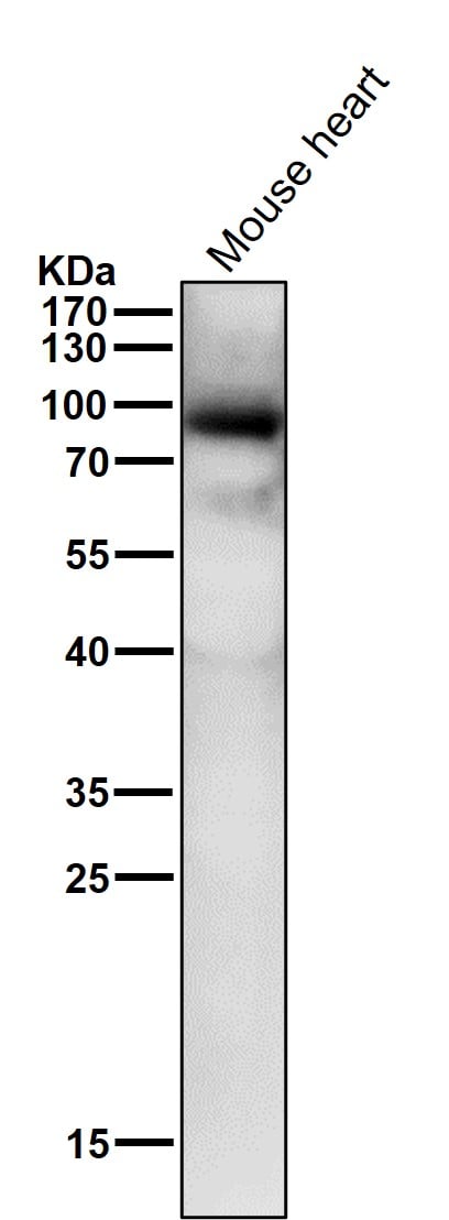
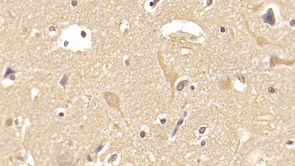
 WB (Western Blot)
(fetal human brain tissue were subjected to SDS PAGE followed by western blot with AAA248029 (p120 Catenin antibody at dilution of 1:1000)
WB (Western Blot)
(fetal human brain tissue were subjected to SDS PAGE followed by western blot with AAA248029 (p120 Catenin antibody at dilution of 1:1000)
p120 Catenin, Monoclonal Antibody (Cat# AAA248029)
Protein A+G purification
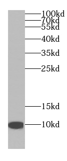
 WB (Western Blot)
(human saliva tissue were subjected to SDS PAGE followed by western blot with AAA247940 (Human IgA Antibody) at dilution of 1:16000)
WB (Western Blot)
(human saliva tissue were subjected to SDS PAGE followed by western blot with AAA247940 (Human IgA Antibody) at dilution of 1:16000)
IgA, Monoclonal Antibody (Cat# AAA247940)
Protein A+G purification
 WB (Western Blot)
(HeLa cells were subjected to SDS PAGE followed by western blot with AAA248040 (PRDX2 Antibody) at dilution of 1:2000)
WB (Western Blot)
(HeLa cells were subjected to SDS PAGE followed by western blot with AAA248040 (PRDX2 Antibody) at dilution of 1:2000)
peroxiredoxin 2, Monoclonal Antibody (Cat# AAA248040)
Protein A+G purification
 WB (Western Blot)
(Neuro-2a cells were subjected to SDS PAGE followed by western blot with AAA248044 (PHOX2B Antibody) at dilution of 1:2500)
WB (Western Blot)
(Neuro-2a cells were subjected to SDS PAGE followed by western blot with AAA248044 (PHOX2B Antibody) at dilution of 1:2500)
PHOX2B, Monoclonal Antibody (Cat# AAA248044)
Protein A+G purification
 WB (Western Blot)
(Hela cells were subjected to SDS PAGE followed by western blot with AAA248071 (RNH1 antibody) at dilution of 1:2000)
WB (Western Blot)
(Hela cells were subjected to SDS PAGE followed by western blot with AAA248071 (RNH1 antibody) at dilution of 1:2000)
RNH1, Monoclonal Antibody (Cat# AAA248071)
Protein A+G purification
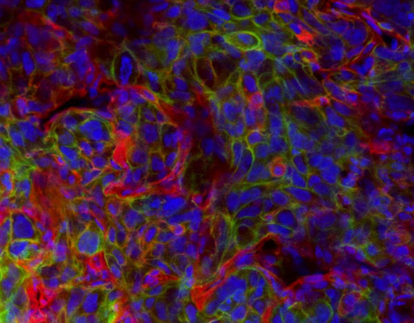
 Application Data
ICAM-1, Monoclonal Antibody (Cat# AAA197030)
This monoclonal antibody was purified using multi-step affinity chromatography methods such as Protein A or G depending on the species and isotype.
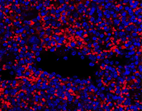
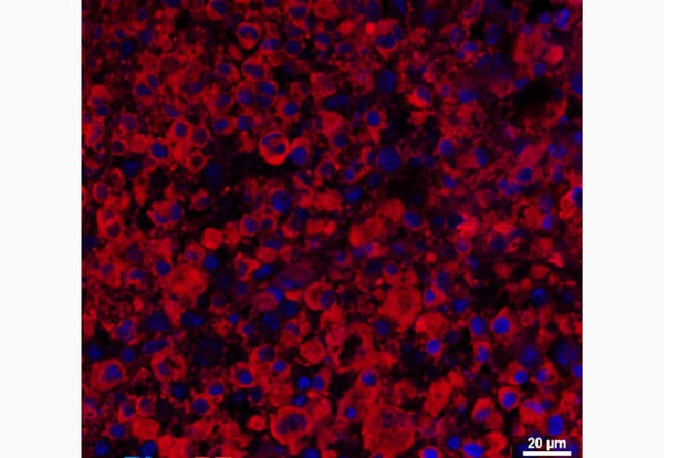
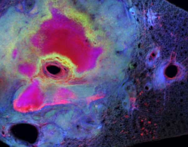
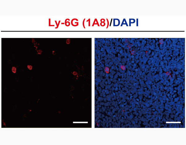 IF (Immunofluorescence)
Ly6G, Monoclonal Antibody (Cat# AAA197036)
This monoclonal antibody was purified using multi-step affinity chromatography methods such as Protein A or G depending on the species and isotype.
MHC Class II, Monoclonal Antibody (Cat# AAA197041)
This monoclonal antibody was purified using multi-step affinity chromatography methods such as Protein A or G depending on the species and isotype.

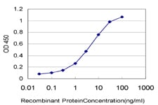 ELISA
(Detection limit for recombinant GST tagged STAT6 is approximately 0.1 ng/ml as a capture antibody.)
ELISA
(Detection limit for recombinant GST tagged STAT6 is approximately 0.1 ng/ml as a capture antibody.)
STAT6, Monoclonal Antibody (Cat# AAA162238)
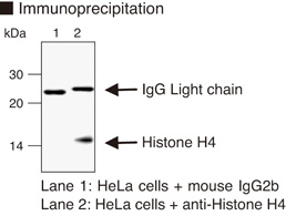
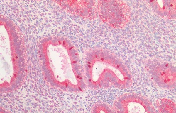
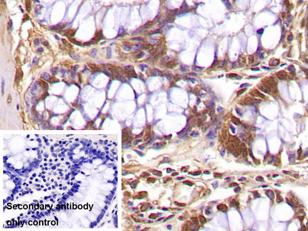
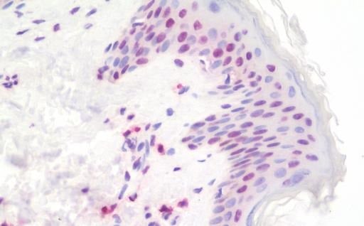 IHC (Immunohiostchemistry)
(Histone H4 Antibody-Human Skin: Formalin-Fixed, Paraffin-Embedded (FFPE))
IHC (Immunohiostchemistry)
(Histone H4 Antibody-Human Skin: Formalin-Fixed, Paraffin-Embedded (FFPE))
Histone H4, Monoclonal Antibody (Cat# AAA162592)
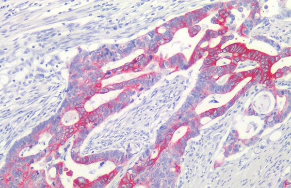
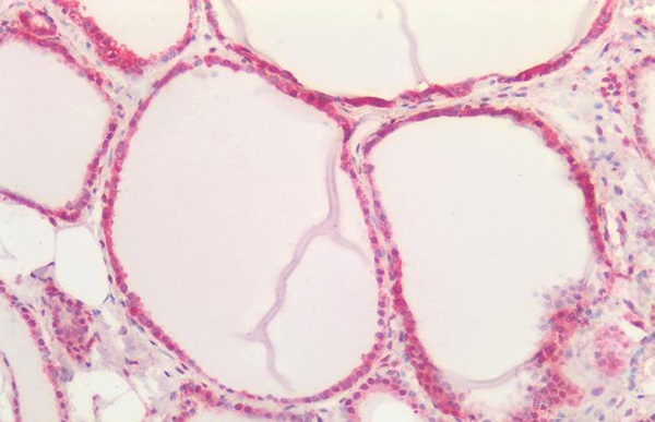
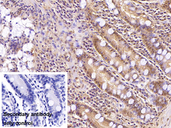
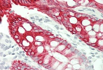 IHC (Immunohiostchemistry)
(KRT20/CK20/Cytokeratin 20 Antibody-Human Colon: Formalin-Fixed, Paraffin-Embedded (FFPE))
IHC (Immunohiostchemistry)
(KRT20/CK20/Cytokeratin 20 Antibody-Human Colon: Formalin-Fixed, Paraffin-Embedded (FFPE))
KRT20/CK20/Cytokeratin 20, Monoclonal Antibody (Cat# AAA162682)
CD11a, Monoclonal Antibody (Cat# AAA129152)
CD68, Monoclonal Antibody (Cat# AAA129067)
CD122, Monoclonal Antibody (Cat# AAA129003)
CD305, Monoclonal Antibody (Cat# AAA128903)
CD49d, Monoclonal Antibody (Cat# AAA128944)
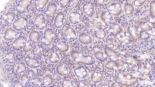
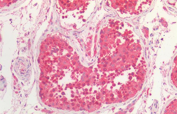 IHC (Immunohistochemistry)
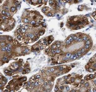
(P4HA1 Antibody-Human Testis: Formalin-Fixed, Paraffin-Embedded (FFPE))
IHC (Immunohistochemistry)
(P4HA1 Antibody-Human Testis: Formalin-Fixed, Paraffin-Embedded (FFPE))
P4HA1, Monoclonal Antibody (Cat# AAA163205)
CD266, Monoclonal Antibody (Cat# AAA128630)
CD18, Monoclonal Antibody (Cat# AAA128692)
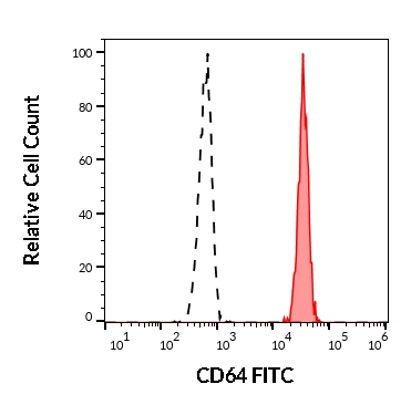
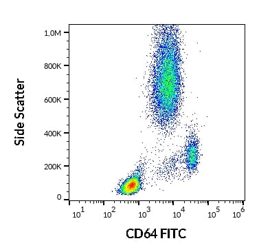 FCM/FACS (Flow Cytometry)
(Flow cytometry surface staining pattern of human peripheral whole blood stained using anti-human CD64 (10.1) FITC antibody (4 ul reagent / 100 ul of peripheral whole blood).)
FCM/FACS (Flow Cytometry)
(Flow cytometry surface staining pattern of human peripheral whole blood stained using anti-human CD64 (10.1) FITC antibody (4 ul reagent / 100 ul of peripheral whole blood).)
CD64, Monoclonal Antibody (Cat# AAA128248)
CD45, Monoclonal Antibody (Cat# AAA128307)
CD36, Monoclonal Antibody (Cat# AAA128479)
CD106, Monoclonal Antibody (Cat# AAA128342)
CD34, Monoclonal Antibody (Cat# AAA128371)
CD49b, Monoclonal Antibody (Cat# AAA128417)
TCR gamma/delta, Monoclonal Antibody (Cat# AAA128422)
 IHC (Immunohistochemistry)
(DAB staining on IHC-P;Sample: Porcine Pancreas TissuePrimary Ab: 40ug/ml Mouse Anti-ulti-species Ub AntibodyControl: Used PBS instead of primary antibodySecond Ab: 2 ug/ml HRP-Linked Caprine Anti-Mouse IgG Polyclonal Antibody)
IHC (Immunohistochemistry)
(DAB staining on IHC-P;Sample: Porcine Pancreas TissuePrimary Ab: 40ug/ml Mouse Anti-ulti-species Ub AntibodyControl: Used PBS instead of primary antibodySecond Ab: 2 ug/ml HRP-Linked Caprine Anti-Mouse IgG Polyclonal Antibody)
Ubiquitin (Ub), Monoclonal Antibody (Cat# AAA152871)
 IHC (Immunohistochemisry)
(DAB staining on IHC-P;Sample: Porcine Liver TissuePrimary Ab: 30ug/ml Mouse Anti-human VEGF165 AntibodyControl: Used PBS instead of primary antibodySecond Ab: 2 ug/ml HRP-Linked Caprine Anti-Mouse IgG Polyclonal Antibody)
IHC (Immunohistochemisry)
(DAB staining on IHC-P;Sample: Porcine Liver TissuePrimary Ab: 30ug/ml Mouse Anti-human VEGF165 AntibodyControl: Used PBS instead of primary antibodySecond Ab: 2 ug/ml HRP-Linked Caprine Anti-Mouse IgG Polyclonal Antibody)
Vasular Endothelial Growth Factor 165 (VEGF165), Monoclonal Antibody (Cat# AAA152873)

 WB (Western Blot)
(Western Blot; Samples: Lane1: Porcine CerebuM lysate; Lane2: Mouse CerebuM lysate; Lane3: Rat CerebuM lysate; Primary Ab: 2ug/ml Mouse Anti-human NRGN Antibody Second Ab: 0.2 ug/ml HRP-Linked Caprine Anti-Mouse IgG Polyclonal Antibody)
WB (Western Blot)
(Western Blot; Samples: Lane1: Porcine CerebuM lysate; Lane2: Mouse CerebuM lysate; Lane3: Rat CerebuM lysate; Primary Ab: 2ug/ml Mouse Anti-human NRGN Antibody Second Ab: 0.2 ug/ml HRP-Linked Caprine Anti-Mouse IgG Polyclonal Antibody)
Neurogranin (NRGN), Monoclonal Antibody (Cat# AAA152880)
 WB (Western Blot)
(Western Blot; Samples: Lane1: Bovine Kidney lysate; Lane2: Rabbit Lung lysate; Lane3: Ovine Lung lysate;Primary Ab: 2ug/ml Mouse Anti-human LMNB1 AntibodySecond Ab: 0.2 ug/ml HRP-Linked Caprine Anti-Mouse IgG Polyclonal Antibody)
WB (Western Blot)
(Western Blot; Samples: Lane1: Bovine Kidney lysate; Lane2: Rabbit Lung lysate; Lane3: Ovine Lung lysate;Primary Ab: 2ug/ml Mouse Anti-human LMNB1 AntibodySecond Ab: 0.2 ug/ml HRP-Linked Caprine Anti-Mouse IgG Polyclonal Antibody)
Lamin B1 (LMNB1), Monoclonal Antibody (Cat# AAA152899)
 WB (Western Blot)
(All lanes use the Antibody at 1:1K dilution for 1 hour at room temperature.)
WB (Western Blot)
(All lanes use the Antibody at 1:1K dilution for 1 hour at room temperature.)
PKC alpha, Monoclonal Antibody (Cat# AAA128118)
 WB (Western Blot)
(All lanes use the Antibody at 1:2K dilution for 1 hour at room temperature.)
WB (Western Blot)
(All lanes use the Antibody at 1:2K dilution for 1 hour at room temperature.)
PKC, Monoclonal Antibody (Cat# AAA128124)
 IF (Immunofluorescence)
(Immunofluorescent analysis using the Antibody at 1:150 dilution.)
IF (Immunofluorescence)
(Immunofluorescent analysis using the Antibody at 1:150 dilution.)
TFIIE alpha, Monoclonal Antibody (Cat# AAA128188)
 WB (Western Blot)
(All lanes use the Antibody at 1:1K dilution for 1 hour at room temperature.)
WB (Western Blot)
(All lanes use the Antibody at 1:1K dilution for 1 hour at room temperature.)
Histone H3, Monoclonal Antibody (Cat# AAA128191)
 WB (Western Blot)
(All lanes use the Antibody at 1:3K dilution for 1 hour at room temperature.)
WB (Western Blot)
(All lanes use the Antibody at 1:3K dilution for 1 hour at room temperature.)
Histone H4, Monoclonal Antibody (Cat# AAA128197)
 IHC (Immunohistochemisry)
(DAB staining on IHC-P;Samples: Rat Testis Tissue; Primary Ab: 30ug/ml Mouse Anti-Rat Bcl2L AntibodySecond Ab: 2ug/mL HRP-Linked Caprine Anti-Mouse IgG Polyclonal Antibody)
IHC (Immunohistochemisry)
(DAB staining on IHC-P;Samples: Rat Testis Tissue; Primary Ab: 30ug/ml Mouse Anti-Rat Bcl2L AntibodySecond Ab: 2ug/mL HRP-Linked Caprine Anti-Mouse IgG Polyclonal Antibody)
B-Cell CLL/Lymphoma 2 Like Protein (Bcl2L), Monoclonal Antibody (Cat# AAA152710)
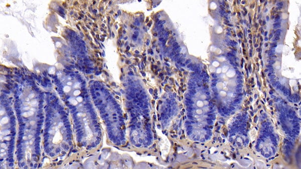
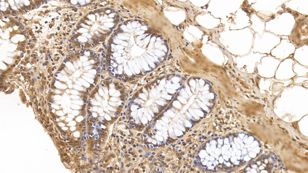 IHC (Immunohistochemisry)
(DAB staining on IHC-P;Samples: human Colon Tissue; Primary Ab: 30ug/ml Mouse Anti-human GATA3 AntibodySecond Ab: 2ug/mL HRP-Linked Caprine Anti-Mouse IgG Polyclonal Antibody)
IHC (Immunohistochemisry)
(DAB staining on IHC-P;Samples: human Colon Tissue; Primary Ab: 30ug/ml Mouse Anti-human GATA3 AntibodySecond Ab: 2ug/mL HRP-Linked Caprine Anti-Mouse IgG Polyclonal Antibody)
GATA Binding Protein 3 (GATA3), Monoclonal Antibody (Cat# AAA152765)
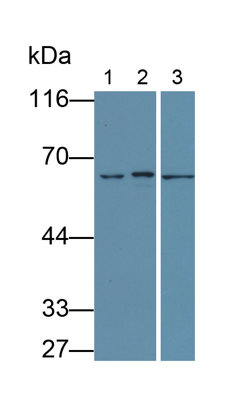
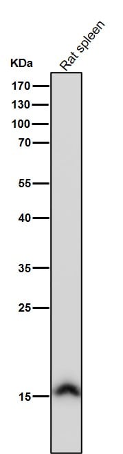
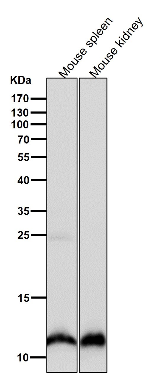
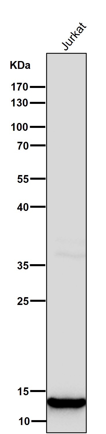
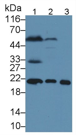
 WB (Western Blot)
(Western Blot; Sample: Lane1: Rat Liver lysate; Lane2: Rat Spleen lysate; Lane3: Rat CerebuM lysate; Lane4: Jurkat cell lysate Primary Ab: 3 ug/ml Mouse Anti-Mouse FTH Antibody Second Ab: 0.2ug/mL HRP-Linked Caprine Anti-Mouse IgG Polyclonal Antibody)
WB (Western Blot)
(Western Blot; Sample: Lane1: Rat Liver lysate; Lane2: Rat Spleen lysate; Lane3: Rat CerebuM lysate; Lane4: Jurkat cell lysate Primary Ab: 3 ug/ml Mouse Anti-Mouse FTH Antibody Second Ab: 0.2ug/mL HRP-Linked Caprine Anti-Mouse IgG Polyclonal Antibody)
Ferritin, Heavy Polypeptide (FTH), Monoclonal Antibody (Cat# AAA152776)
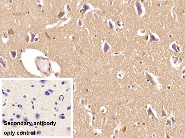
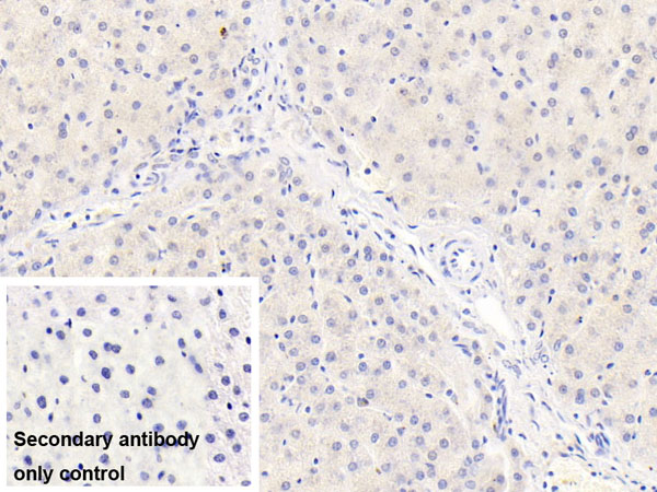
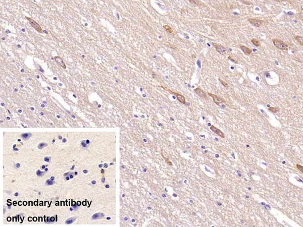
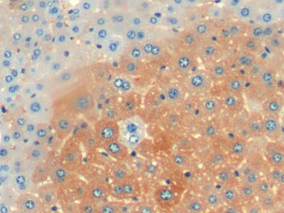
 IHC (Immunohiostchemistry)
(DAB staining on IHCP;Samples: Human Cerebrum Tissue; Primary Ab: 10ug/ml Mouse AntiHuman NRG1 AntibodySecond Ab: 2ug/mL HRPLinked Caprine AntiMouse IgG Polyclonal Antibody(Catalog: SAA544Mu19))
IHC (Immunohiostchemistry)
(DAB staining on IHCP;Samples: Human Cerebrum Tissue; Primary Ab: 10ug/ml Mouse AntiHuman NRG1 AntibodySecond Ab: 2ug/mL HRPLinked Caprine AntiMouse IgG Polyclonal Antibody(Catalog: SAA544Mu19))
Neuregulin 1 (NRG1), Monoclonal Antibody (Cat# AAA151763)
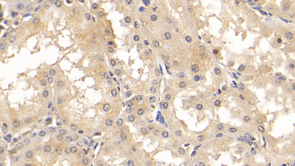
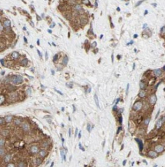
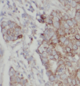
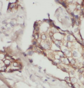
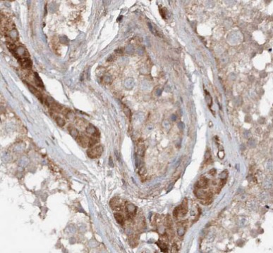
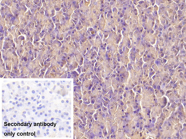
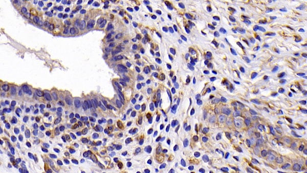
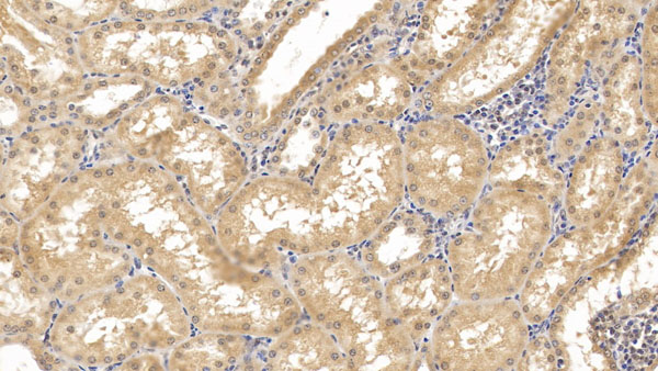
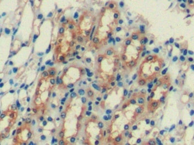
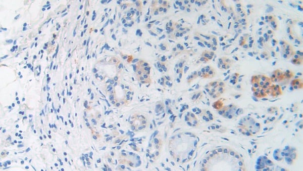
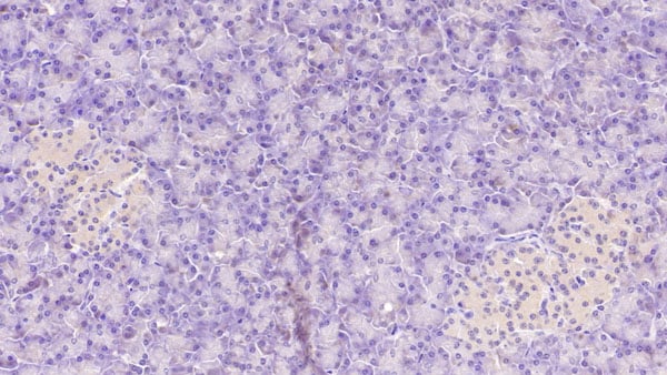
 IHC (Immunohistochemisry)
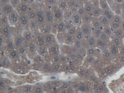
(DAB staining on IHCP;Sample: Human Liver Tissue; Primary Ab: 30ug/ml Mouse AntiHuman FAPa AntibodySecond Ab: 2ug/mL HRPLinked Caprine AntiMouse IgG Polyclonal Antibody(Catalog: SAA544Mu19))
IHC (Immunohistochemisry)
(DAB staining on IHCP;Sample: Human Liver Tissue; Primary Ab: 30ug/ml Mouse AntiHuman FAPa AntibodySecond Ab: 2ug/mL HRPLinked Caprine AntiMouse IgG Polyclonal Antibody(Catalog: SAA544Mu19))
Fibroblast Activation Protein Alpha (FAPa), Monoclonal Antibody (Cat# AAA151808)
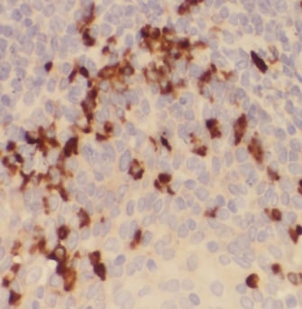
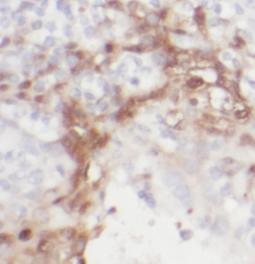
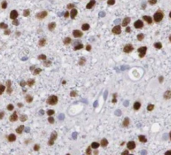
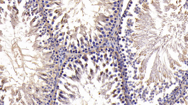
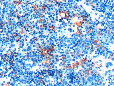
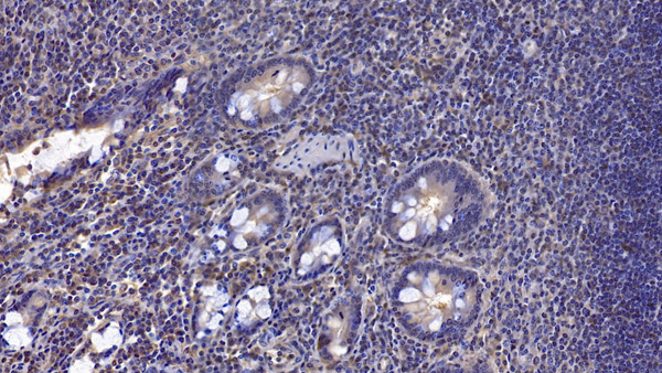
 IHC (Immunohiostchemistry)
(DAB staining on IHCP;Samples: Human Pancreatic cancer Tissue;Primary Ab: 30ug/ml Mouse AntiMultispecies BNP AntibodySecond Ab: 2ug/mL HRPLinked Caprine AntiMouse IgG Polyclonal Antibody(Catalog: SAA544Mu19))
IHC (Immunohiostchemistry)
(DAB staining on IHCP;Samples: Human Pancreatic cancer Tissue;Primary Ab: 30ug/ml Mouse AntiMultispecies BNP AntibodySecond Ab: 2ug/mL HRPLinked Caprine AntiMouse IgG Polyclonal Antibody(Catalog: SAA544Mu19))
Brain Natriuretic Peptide (BNP), Monoclonal Antibody (Cat# AAA151557)

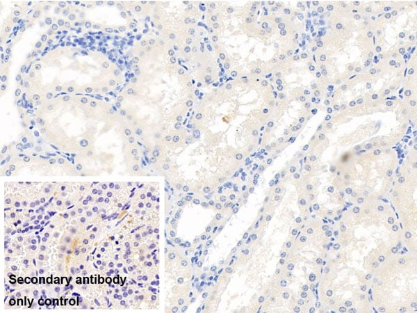
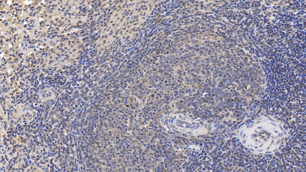
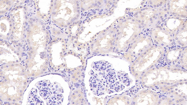
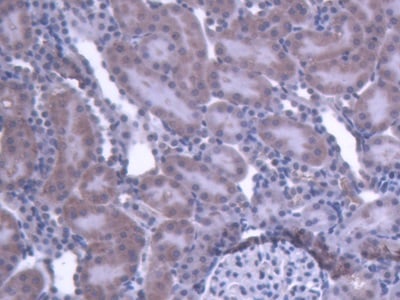 IHC (Immunohiostchemistry)
(DAB staining on IHCP;Sample: Rat Kidney Tissue; Primary Ab: 20ug/ml Mouse AntiRat IL1b AntibodySecond Ab: 2ug/mL HRPLinked Caprine AntiMouse IgG Polyclonal Antibody(Catalog: SAA544Mu19))
IHC (Immunohiostchemistry)
(DAB staining on IHCP;Sample: Rat Kidney Tissue; Primary Ab: 20ug/ml Mouse AntiRat IL1b AntibodySecond Ab: 2ug/mL HRPLinked Caprine AntiMouse IgG Polyclonal Antibody(Catalog: SAA544Mu19))
Interleukin 1 Beta (IL1b), Monoclonal Antibody (Cat# AAA151570)
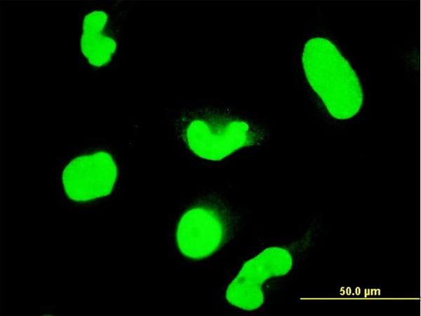
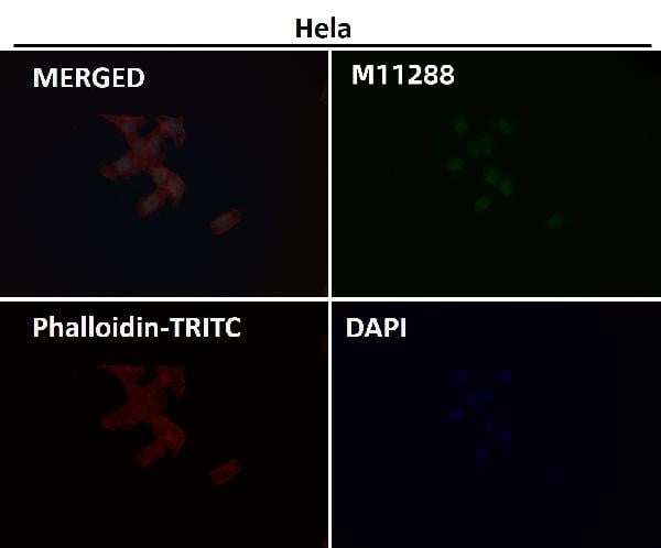
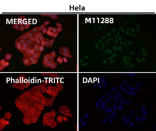
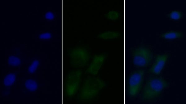
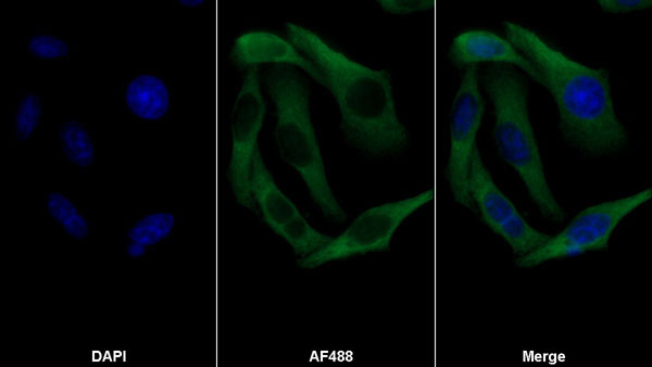 IF (Immunofluorescence)
(AF488 staining on IF;Sample: Hela cellPrimary Ab: 30ug/ml Mouse Anti-human ASPH AntibodySecond Ab: 2 ug/ml AF488-Linked Caprine Anti-Mouse IgG Polyclonal Antibody)
IF (Immunofluorescence)
(AF488 staining on IF;Sample: Hela cellPrimary Ab: 30ug/ml Mouse Anti-human ASPH AntibodySecond Ab: 2 ug/ml AF488-Linked Caprine Anti-Mouse IgG Polyclonal Antibody)
Aspartate Beta Hydroxylase (ASPH), Monoclonal Antibody (Cat# AAA152701)
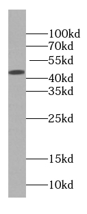
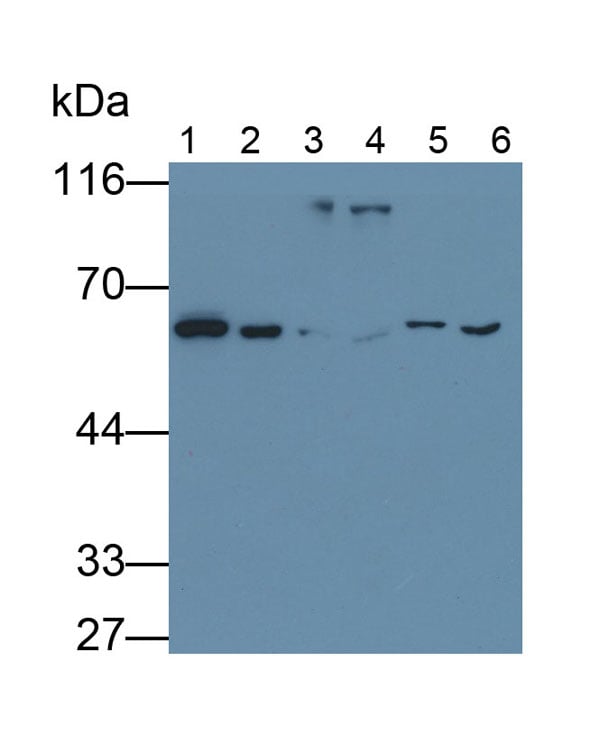
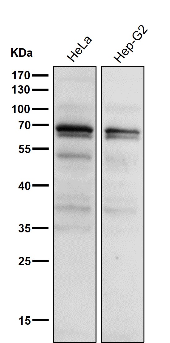
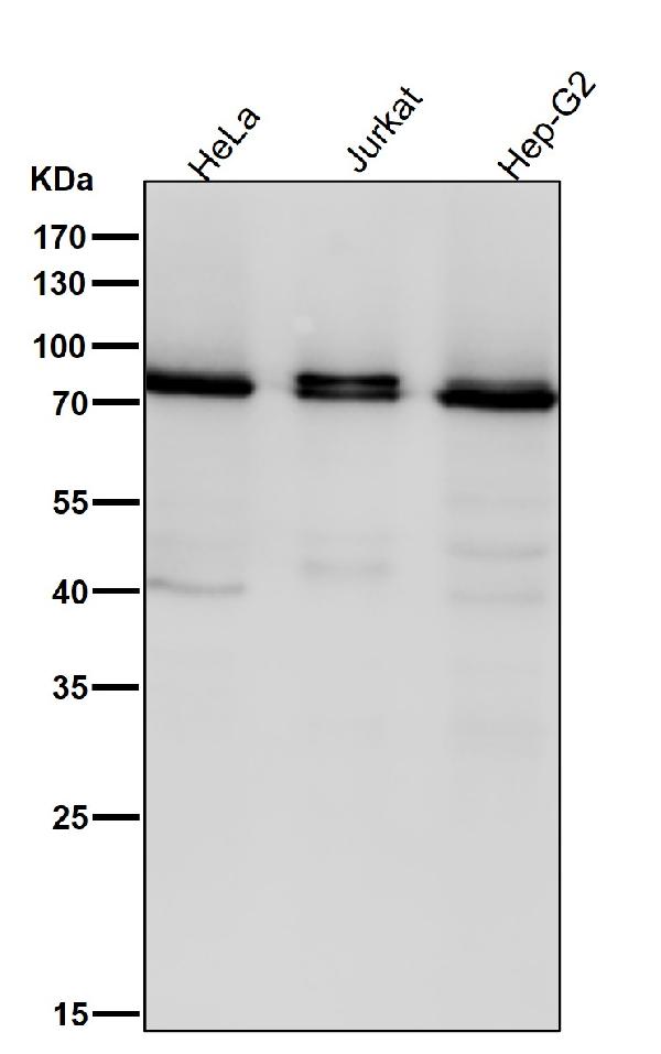
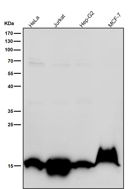
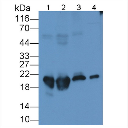
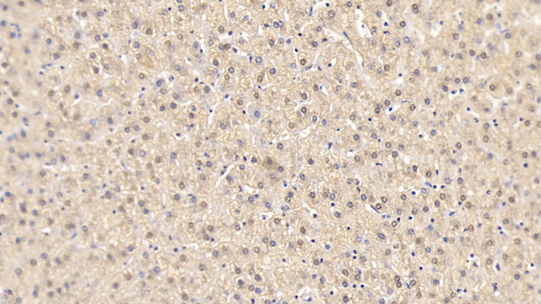
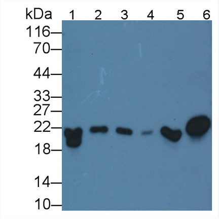 WB (Western Blot)
(Western Blot; Sample: Lane1: human Liver lysate; Lane2: human Lung lysate; Lane3: human Placenta lysate; Lane4: 293T cell lysate; Lane5: HepG2 cell lysate; Lane6: U87MG cell lysatePrimary Ab: 3ug/ml Mouse Anti-Mouse FTH AntibodySecond Ab: 0.2ug/mL HRP-Linked Caprine Anti-Mouse IgG Polyclonal Antibody)
WB (Western Blot)
(Western Blot; Sample: Lane1: human Liver lysate; Lane2: human Lung lysate; Lane3: human Placenta lysate; Lane4: 293T cell lysate; Lane5: HepG2 cell lysate; Lane6: U87MG cell lysatePrimary Ab: 3ug/ml Mouse Anti-Mouse FTH AntibodySecond Ab: 0.2ug/mL HRP-Linked Caprine Anti-Mouse IgG Polyclonal Antibody)
Ferritin, Heavy Polypeptide (FTH), Monoclonal Antibody (Cat# AAA152607)
What are Monoclonal Antibodies?
Monoclonal antibodies are specialized laboratory-produced proteins developed for binding to specific biological antigens or other molecular targets. Since they come from a single cell (or clone), they are especially consistent and accurate in the data they are involved in producing.
This type of antibody material has been shown to be a powerful tool in finding and subsequently destroying harmful cells in an organism, such as those found in cancers or various autoimmune diseases. This makes them excellent aids in medical testing and research, which is why they are so widely used.
AAA Biotech offers a comprehensive range of high-quality monoclonal antibodies that perform effectively in various laboratory tests, including (amongst others) ELISA, western blotting, immunohistochemistry, and flow cytometry. All of the products in our catalog are thoroughly quality tested to make sure that they are reliable and will consistently perform well in your research.
What Are The Uses of Monoclonal Antibodies
Monoclonal antibodies are used in many lab tests, including (amongst others) ELISA, western blotting, immunohistochemistry, and flow cytometry.
ELISA is a test that helps detect a specific substance/analyte in a sample. It uses antibodies (often monoclonal) bound to a solid surface (such as the well of a microplate) to “capture” the substance/analyte in the sample and immobilize it so that the detection antibody component can then bind to it and produce a signal, which can then be measured.
Western blotting identifies specific proteins in a sample. The sample is first separated on a gel, and then antibodies are applied that will typically bind to the target, which will all be localized to a single band in a lane.
Immunohistochemistry helps locate specific proteins in cells or tissue samples using antibodies.
Flow cytometry looks at and sorts cells. It uses antibodies that are conjugated to reporter molecules called “fluorophores”, which, under special lights, emit light themselves, which can then be measured by a detector instrument.
How Monoclonal Antibodies Are Used as Medicine?
Please note that all of the products listed in AAA Biotech’s also known as AAA Bio or AAABio catalog are strictly for research-use only (RUO).
Monoclonal antibodies can also be used as therapeutic/medical treatments, particularly in the context of cancers. They are designed to find and bind to specific cells or proteins, helping the immune system recognize and attack the cancer. These treatments work in different ways, such as:
- Radioimmunotherapy attaches a small amount of radioactive molecule to the antibody, so it delivers the radiation directly to the cancer cells that the antibody is specifically binding to.
- Antibody-directed enzyme prodrug therapy uses antibodies that are specifically bound to special enzymes. These enzymes activate a harmless drug in the body and turn it into a cancer-killing drug only near the cancer cells—this helps avoid harming healthy cells.
- Immunoliposomes are tiny “bubbles” filled with medicine/drug and coated with antibodies. They carry the drug straight to the cancer cells.
Why Buy Monoclonal Antibodies From Us?
At AAA Biotech, we provide high-performance monoclonal antibodies designed to support a wide range of research needs.
1. Validated for Versatile Applications
The antibodies in our catalog are extensively validated and compatible with multiple techniques, including (but not limited to) ELISA, flow cytometry (FC), immunocytochemistry (ICC), immunofluorescence (IF), immunohistochemistry (IHC), immunoprecipitation (IP), and western blotting (WB).
2. Wide Selection & Specialized Options
We offer antibodies for common and rare species, that are available in various conjugated forms, and also in recombinant formats. Essentially, there is almost anything one might need to meet their experimental model’s requirements.
3. High-Quality Proteins
Our proteins meet high purity standards—90% or more as confirmed by SDS-PAGE. Many are available with tags like His, Flag, GST, or MBP, and we also supply native and biologically active proteins for functional studies.
Frequently Asked Questions
1. Are your monoclonal antibodies validated for specific applications?
Yes, our antibodies are tested and validated for use in methods such as ELISA, western blot, IHC, flow cytometry, and more. Refer to specific product pages or datasheets for individual product information.
2. How do I choose the right monoclonal antibody for my application?
Review the product details directly for application validation, species reactivity, and target information. You may also contact our support team at any time for help.
3. How quickly can I receive my order?
Most orders are processed and shipped within 1–3 business days, depending on product availability and your shipping location.