Filters
▼Clonality
▼Type
▼Reactivity
▼Gene Name
▼Isotype
▼Host
▼Application
▼Clone
▼Monoclonal Antibodies
Get accurate results in your research with our Monoclonal Antibodies, which are specially made to target exactly what you require for your research, and will produce consistent, reliable performance in lab tests.
Viewing 6850-6900 of 27597 product results
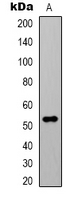
 Application Data
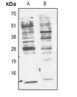
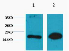
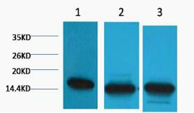
(Dilution: WB 1:1000-2000, IHC 1:100-200)
Application Data
(Dilution: WB 1:1000-2000, IHC 1:100-200)
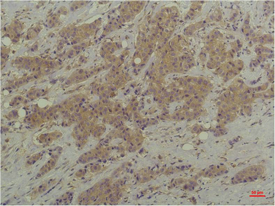
Cyclophilin B, Monoclonal Antibody (Cat# AAA293708)

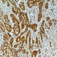
 Application Data
(Dilution: WB 1:1000-2000, IHC 1:50-100)
Application Data
(Dilution: WB 1:1000-2000, IHC 1:50-100)
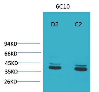
P44/42 MAPK, Monoclonal Antibody (Cat# AAA293709)


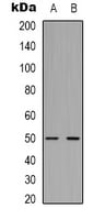
 Application Data
(Dilution: WB 1:1000-2000, IHC 1:50-100)
Application Data
(Dilution: WB 1:1000-2000, IHC 1:50-100)
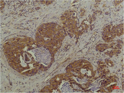
P44/42 MAPK, Monoclonal Antibody (Cat# AAA293711)
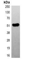
 Application Data
(Dilution: WB 1:1000-2000, IHC 1:50-100)
Application Data
(Dilution: WB 1:1000-2000, IHC 1:50-100)
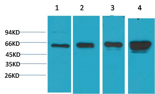
AMPK alpha1, Monoclonal Antibody (Cat# AAA293713)
 Application Data
(Dilution: IHC 1:200 IF 1:50-200)
Application Data
(Dilution: IHC 1:200 IF 1:50-200)
IkappaB beta, Monoclonal Antibody (Cat# AAA293714)
 Application Data
(Dilution: IHC 1:100-200)
Application Data
(Dilution: IHC 1:100-200)
Smad3, Monoclonal Antibody (Cat# AAA293716)
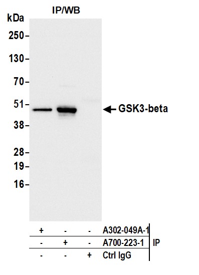
 IP (Immunoprecipitation)
(Dilution: IP WB (1/2000 - 1/5000), IP (1/100 - 1/200)Immunoprecipitation of CBP-tagged protein from HEK293T cells transfected with vector overexpressing CBP tag, using Anti-CBP-tag Antibody.)
IP (Immunoprecipitation)
(Dilution: IP WB (1/2000 - 1/5000), IP (1/100 - 1/200)Immunoprecipitation of CBP-tagged protein from HEK293T cells transfected with vector overexpressing CBP tag, using Anti-CBP-tag Antibody.)
CBP-tag, Monoclonal Antibody (Cat# AAA293367)
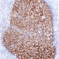
 IHC (Immunohiostchemistry)
(Dilution: WB: (1/1000 - 1/2000), IH (1/100 - 1/200)Immunohistochemical analysis of Alpha-tubulin (AcK40) staining in human breast cancer,mouse brain formalin fixed paraffin embedded tissue section. The section was pre-treated using heat mediated antigen retrieval with sodium citrate buffer (pH 6.0). The section was then incubated with the antibody at room temperature and detected using an HRP conjugated compact polymer system. DAB was used as the chromogen. The section was then counterstained with haematoxylin and mounted with DPX.)
IHC (Immunohiostchemistry)
(Dilution: WB: (1/1000 - 1/2000), IH (1/100 - 1/200)Immunohistochemical analysis of Alpha-tubulin (AcK40) staining in human breast cancer,mouse brain formalin fixed paraffin embedded tissue section. The section was pre-treated using heat mediated antigen retrieval with sodium citrate buffer (pH 6.0). The section was then incubated with the antibody at room temperature and detected using an HRP conjugated compact polymer system. DAB was used as the chromogen. The section was then counterstained with haematoxylin and mounted with DPX.)
Alpha-tubulin, Monoclonal Antibody (Cat# AAA293373)
 IP (Immunoprecipitation)
(Dilution: IP WB (1/50000 - 1/100000), IP (1/100 - 1/200)Immunoprecipitation of Beta2A-tubulin from 0.5mg mouse brain whole cell extract lysate, using Anti-Beta2A-tubulin Antibody.)
IP (Immunoprecipitation)
(Dilution: IP WB (1/50000 - 1/100000), IP (1/100 - 1/200)Immunoprecipitation of Beta2A-tubulin from 0.5mg mouse brain whole cell extract lysate, using Anti-Beta2A-tubulin Antibody.)
Beta2A-tubulin, Monoclonal Antibody (Cat# AAA293374)
 IF (Immunofluorescence)
(Dilution: WB (1/1000 - 1/3000), IH (1/100 - 1/200), IF/IC (1/100 - 1/200))
IF (Immunofluorescence)
(Dilution: WB (1/1000 - 1/3000), IH (1/100 - 1/200), IF/IC (1/100 - 1/200))
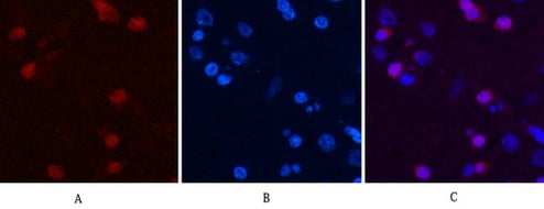
eIF4AI, Monoclonal Antibody (Cat# AAA293380)
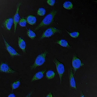 IF (Immunofluorescence)
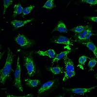
(Dilution: IF WB (1/1000 - 1/3000), IF/IC (1/100 - 1/200)Immunofluorescent analysis of Fumarase staining in Hela cells. Formalin-fixed cells were permeabilized with 0.1% Triton X-100 in TBS for 5-10 minutes and blocked with 3% BSA-PBS for 30 minutes at room temperature. Cells were probed with the primary antibody in 3% BSA-PBS and incubated overnight at 4 degree C in a hidified chamber. Cells were washed with PBST and incubated with a FITC-conjugated secondary antibody (green) in PBS at room temperature in the dark. DAPI was used to stain the cell nuclei (blue).)
IF (Immunofluorescence)
(Dilution: IF WB (1/1000 - 1/3000), IF/IC (1/100 - 1/200)Immunofluorescent analysis of Fumarase staining in Hela cells. Formalin-fixed cells were permeabilized with 0.1% Triton X-100 in TBS for 5-10 minutes and blocked with 3% BSA-PBS for 30 minutes at room temperature. Cells were probed with the primary antibody in 3% BSA-PBS and incubated overnight at 4 degree C in a hidified chamber. Cells were washed with PBST and incubated with a FITC-conjugated secondary antibody (green) in PBS at room temperature in the dark. DAPI was used to stain the cell nuclei (blue).)
Fumarase, Monoclonal Antibody (Cat# AAA293382)

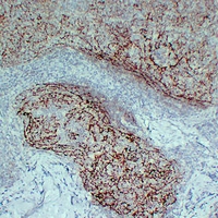
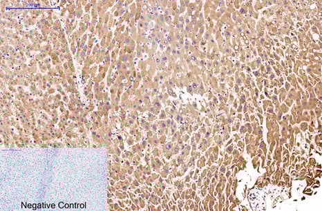
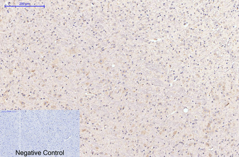
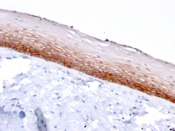
 IHC (Immunohiostchemistry)
(Dilution: WB: (1/1000 - 1/3000), IH (1/200 - 1/500)Immunohistochemical analysis of Histone H3 (TriMethyl K36) staining in human tonsil formalin fixed paraffin embedded tissue section. The section was pre-treated using heat mediated antigen retrieval with sodium citrate buffer (pH 6.0). The section was then incubated with the antibody at room temperature and detected using an HRP conjugated compact polymer system. DAB was used as the chromogen. The section was then counterstained with haematoxylin and mounted with DPX.)
IHC (Immunohiostchemistry)
(Dilution: WB: (1/1000 - 1/3000), IH (1/200 - 1/500)Immunohistochemical analysis of Histone H3 (TriMethyl K36) staining in human tonsil formalin fixed paraffin embedded tissue section. The section was pre-treated using heat mediated antigen retrieval with sodium citrate buffer (pH 6.0). The section was then incubated with the antibody at room temperature and detected using an HRP conjugated compact polymer system. DAB was used as the chromogen. The section was then counterstained with haematoxylin and mounted with DPX.)
Histone H3, Monoclonal Antibody (Cat# AAA293388)
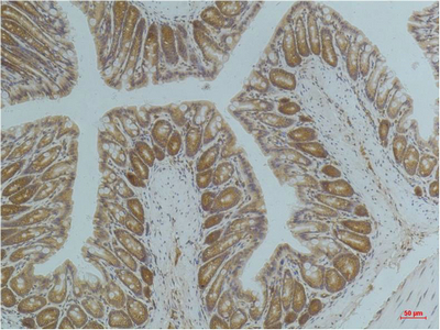
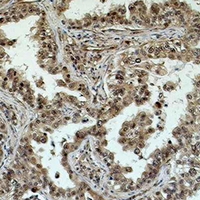
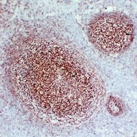
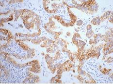
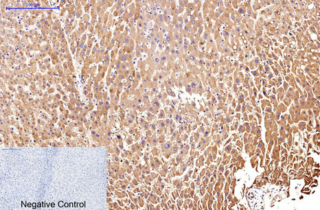
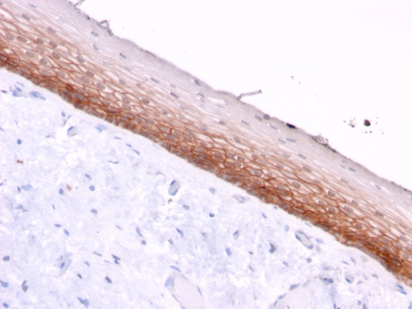
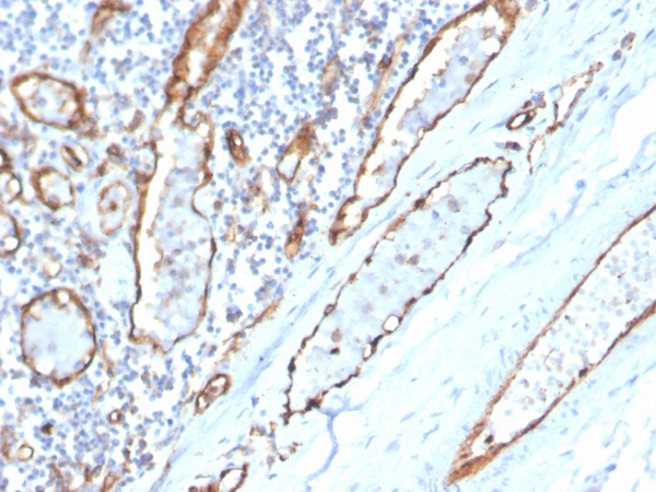
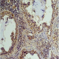 IHC (Immunohiostchemistry)
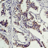
(Dilution: WB: (1/1000 - 1/2000), IH (1/50 - 1/200)Immunohistochemical analysis of Phosphotyrosine staining in human lung cancer formalin fixed paraffin embedded tissue section. The section was pre-treated using heat mediated antigen retrieval with sodium citrate buffer (pH 6.0). The section was then incubated with the antibody at room temperature and detected using an HRP conjugated compact polymer system. DAB was used as the chromogen. The section was then counterstained with haematoxylin and mounted with DPX.)
IHC (Immunohiostchemistry)
(Dilution: WB: (1/1000 - 1/2000), IH (1/50 - 1/200)Immunohistochemical analysis of Phosphotyrosine staining in human lung cancer formalin fixed paraffin embedded tissue section. The section was pre-treated using heat mediated antigen retrieval with sodium citrate buffer (pH 6.0). The section was then incubated with the antibody at room temperature and detected using an HRP conjugated compact polymer system. DAB was used as the chromogen. The section was then counterstained with haematoxylin and mounted with DPX.)
Phosphoserine, Monoclonal Antibody (Cat# AAA293393)
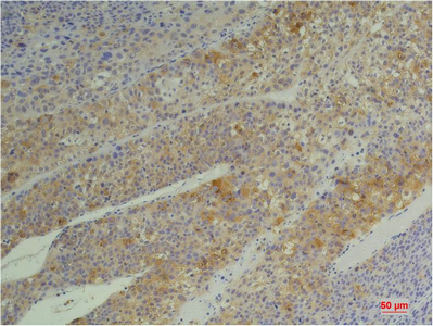
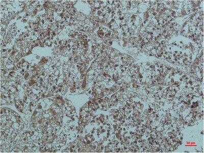
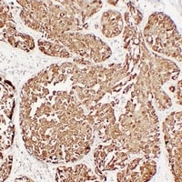
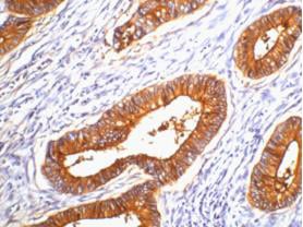
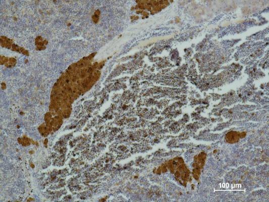
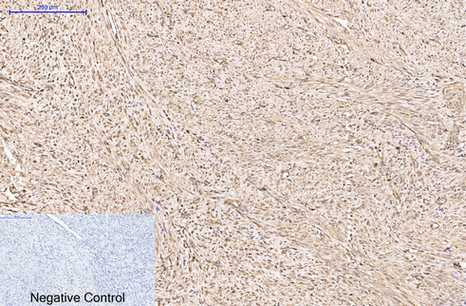
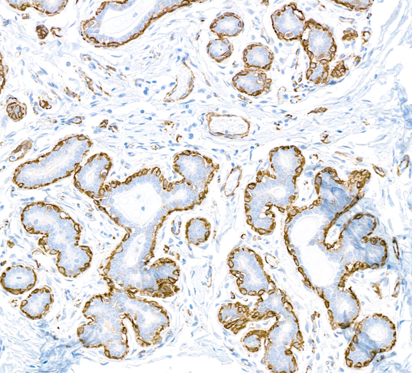
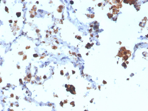
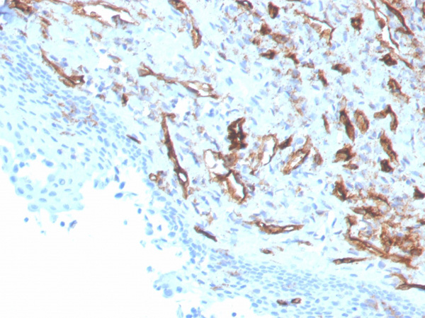
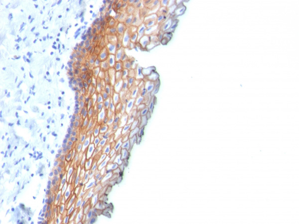
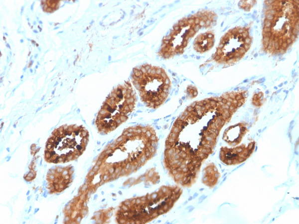
 IHC (Immunohiostchemistry)
(Dilution: WB: (1/1000 - 1/2000), IH (1/50 - 1/200)Immunohistochemical analysis of Phosphotyrosine staining in human breast cancer formalin fixed paraffin embedded tissue section. The section was pre-treated using heat mediated antigen retrieval with sodium citrate buffer (pH 6.0). The section was then incubated with the antibody at room temperature and detected using an HRP conjugated compact polymer system. DAB was used as the chromogen. The section was then counterstained with haematoxylin and mounted with DPX.)
IHC (Immunohiostchemistry)
(Dilution: WB: (1/1000 - 1/2000), IH (1/50 - 1/200)Immunohistochemical analysis of Phosphotyrosine staining in human breast cancer formalin fixed paraffin embedded tissue section. The section was pre-treated using heat mediated antigen retrieval with sodium citrate buffer (pH 6.0). The section was then incubated with the antibody at room temperature and detected using an HRP conjugated compact polymer system. DAB was used as the chromogen. The section was then counterstained with haematoxylin and mounted with DPX.)
Phosphotyrosine, Monoclonal Antibody (Cat# AAA293394)
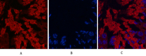
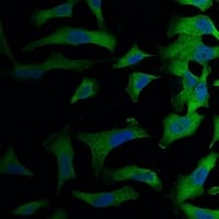
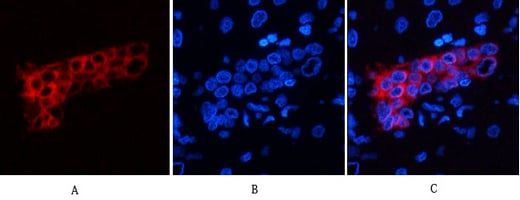
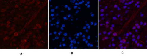
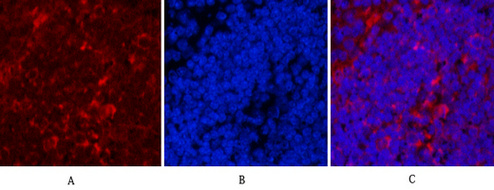
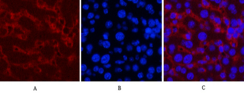
 IF (Immunofluorescence)
(Dilution: IF WB (1/1000 - 1/2000), IF/IC (1/100 - 1/200)Immunofluorescent analysis of Swiprosin-2 staining in Hela cells. Formalin-fixed cells were permeabilized with 0.1% Triton X-100 in TBS for 5-10 minutes and blocked with 3% BSA-PBS for 30 minutes at room temperature. Cells were probed with the primary antibody in 3% BSA-PBS and incubated overnight at 4 degree C in a hidified chamber. Cells were washed with PBST and incubated with a FITC-conjugated secondary antibody (green) in PBS at room temperature in the dark. DAPI was used to stain the cell nuclei (blue).)
IF (Immunofluorescence)
(Dilution: IF WB (1/1000 - 1/2000), IF/IC (1/100 - 1/200)Immunofluorescent analysis of Swiprosin-2 staining in Hela cells. Formalin-fixed cells were permeabilized with 0.1% Triton X-100 in TBS for 5-10 minutes and blocked with 3% BSA-PBS for 30 minutes at room temperature. Cells were probed with the primary antibody in 3% BSA-PBS and incubated overnight at 4 degree C in a hidified chamber. Cells were washed with PBST and incubated with a FITC-conjugated secondary antibody (green) in PBS at room temperature in the dark. DAPI was used to stain the cell nuclei (blue).)
Swiprosin-2, Monoclonal Antibody (Cat# AAA293396)
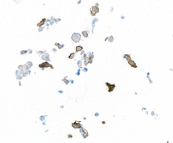
 IP (Immunoprecipitation)
(Dilution: IH (1/100 - 1/300))
IP (Immunoprecipitation)
(Dilution: IH (1/100 - 1/300))
CD21, Monoclonal Antibody (Cat# AAA293400)
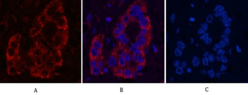
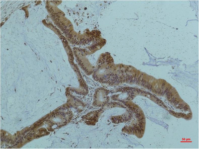
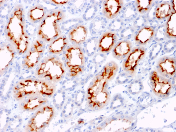
 IF (Immunofluorescence)
(Dilution: IF WB (1/500 - 1/1000), IH (1/100 - 1/300)Immunohistochemical analysis of MUC1 staining in human tonsil formalin fixed paraffin embedded tissue section. The section was pre-treated using heat mediated antigen retrieval with sodium citrate buffer (pH 6.0). The section was then incubated with the antibody at room temperature and detected using an HRP conjugated compact polymer system. DAB was used as the chromogen. The section was then counterstained with haematoxylin and mounted with DPX.)
IF (Immunofluorescence)
(Dilution: IF WB (1/500 - 1/1000), IH (1/100 - 1/300)Immunohistochemical analysis of MUC1 staining in human tonsil formalin fixed paraffin embedded tissue section. The section was pre-treated using heat mediated antigen retrieval with sodium citrate buffer (pH 6.0). The section was then incubated with the antibody at room temperature and detected using an HRP conjugated compact polymer system. DAB was used as the chromogen. The section was then counterstained with haematoxylin and mounted with DPX.)
MUC1, Monoclonal Antibody (Cat# AAA293403)
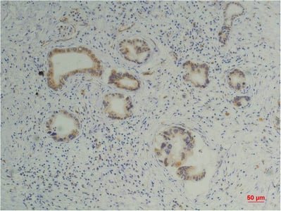
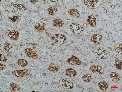
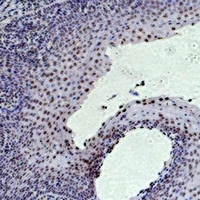
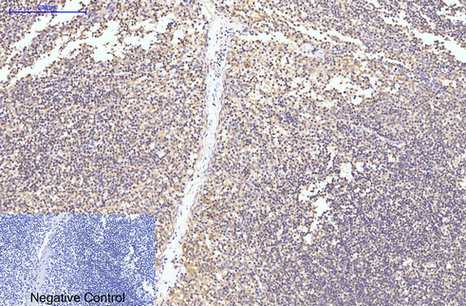
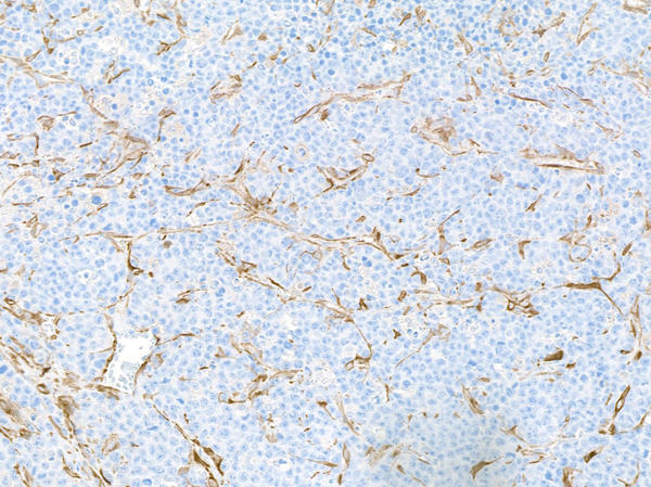
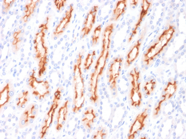
 IF (Immunofluorescence)
(Dilution: IF IH (1/100 - 1/300)Immunohistochemical analysis of CD163 staining in human tonsil formalin fixed paraffin embedded tissue section. The section was pre-treated using heat mediated antigen retrieval with sodium citrate buffer (pH 6.0). The section was then incubated with the antibody at room temperature and detected using an HRP conjugated compact polymer system. DAB was used as the chromogen. The section was then counterstained with haematoxylin and mounted with DPX.)
IF (Immunofluorescence)
(Dilution: IF IH (1/100 - 1/300)Immunohistochemical analysis of CD163 staining in human tonsil formalin fixed paraffin embedded tissue section. The section was pre-treated using heat mediated antigen retrieval with sodium citrate buffer (pH 6.0). The section was then incubated with the antibody at room temperature and detected using an HRP conjugated compact polymer system. DAB was used as the chromogen. The section was then counterstained with haematoxylin and mounted with DPX.)
CD163, Monoclonal Antibody (Cat# AAA293405)
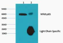

 WB (Western Blot)
(Dilution: WB: 1:1000-3000 IHC: 1:200 IP:1:200The picture was kindly provided by our customer.)
WB (Western Blot)
(Dilution: WB: 1:1000-3000 IHC: 1:200 IP:1:200The picture was kindly provided by our customer.)
NFkB p65, Monoclonal Antibody (Cat# AAA293514)
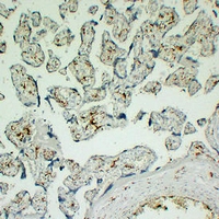
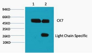 IP (Immunoprecipitation)
(Dilution: IF: 1:50-200 WB: 1:1000-2000 IHC: 1:200 IP:1:200)
IP (Immunoprecipitation)
(Dilution: IF: 1:50-200 WB: 1:1000-2000 IHC: 1:200 IP:1:200)
CK7, Monoclonal Antibody (Cat# AAA293515)

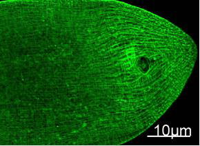 IF (Immunofluorescence)
(Dilution: IF: 1:100)
IF (Immunofluorescence)
(Dilution: IF: 1:100)
Myosin Heavy Chain, Monoclonal Antibody (Cat# AAA293516)
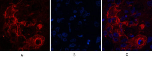
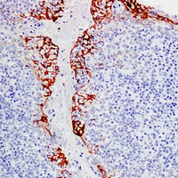
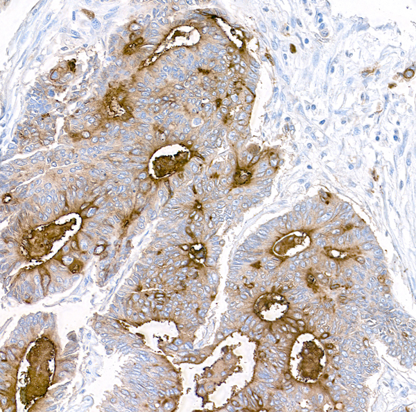
 IHC (Immunohistochemisry)
(Dilution: IHC 1:200 IF 1:50-200)
IHC (Immunohistochemisry)
(Dilution: IHC 1:200 IF 1:50-200)
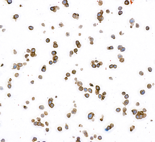
Carcinoembryonic Antigen, Monoclonal Antibody (Cat# AAA293518)
 WB (Western Blot)
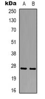
(Dilution: WB: 1:500-2000)
WB (Western Blot)
(Dilution: WB: 1:500-2000)
Histone H3, Monoclonal Antibody (Cat# AAA293531)
 WB (Western Blot)
(Dilution: WB: 1:1000-3000)
WB (Western Blot)
(Dilution: WB: 1:1000-3000)
Histone H3, Monoclonal Antibody (Cat# AAA293534)
 WB (Western Blot)
(Dilution: WB: 1:1000-2000)
WB (Western Blot)
(Dilution: WB: 1:1000-2000)
Histone H3, Monoclonal Antibody (Cat# AAA293538)
 WB (Western Blot)
(Dilution: WB: 1:500-1000)
WB (Western Blot)
(Dilution: WB: 1:500-1000)
Histone H3, Monoclonal Antibody (Cat# AAA293540)

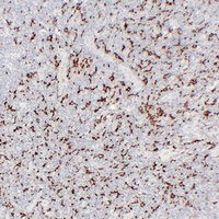
 IHC (Immunohistochemistry)
(Dilution: IHC: 1:200)
IHC (Immunohistochemistry)
(Dilution: IHC: 1:200)
CD25, Monoclonal Antibody (Cat# AAA293542)
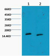
 WB (Western Blot)
(Dilution: WB: 1:1000-3000)
WB (Western Blot)
(Dilution: WB: 1:1000-3000)
Histone H3, Monoclonal Antibody (Cat# AAA293543)
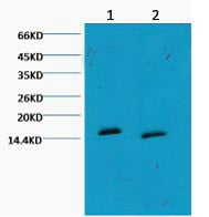
 WB (Western Blot)
(Dilution: WB: 1:1000-3000)
WB (Western Blot)
(Dilution: WB: 1:1000-3000)
Histone H3, Monoclonal Antibody (Cat# AAA293544)
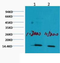
 WB (Western Blot)
(Dilution: WB: 1:1000-3000 IF: 1:100-200 IHC 1:50-300)
WB (Western Blot)
(Dilution: WB: 1:1000-3000 IF: 1:100-200 IHC 1:50-300)
Peroxiredoxin 1, Monoclonal Antibody (Cat# AAA293546)

 WB (Western Blot)
(Dilution: WB: 1:1000-3000)
WB (Western Blot)
(Dilution: WB: 1:1000-3000)
Histone H3, Monoclonal Antibody (Cat# AAA293548)
 WB (Western Blot)
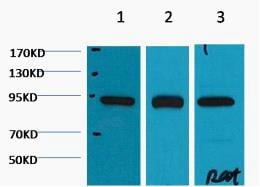
(Dilution: WB: 1:1000-3000 IF 1:200 IHC 1:50-300)
WB (Western Blot)
(Dilution: WB: 1:1000-3000 IF 1:200 IHC 1:50-300)
HSP90beta, Monoclonal Antibody (Cat# AAA293549)
 WB (Western Blot)
(Dilution: WB: 1:1000-3000)
WB (Western Blot)
(Dilution: WB: 1:1000-3000)
PARP, Monoclonal Antibody (Cat# AAA293550)
 WB (Western Blot)
(Dilution: WB: 1:1000-3000)
WB (Western Blot)
(Dilution: WB: 1:1000-3000)
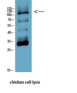
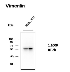
Vimentin, Monoclonal Antibody (Cat# AAA293555)

 WB (Western Blot)
(Dilution: WB: 1:2000-5000)
WB (Western Blot)
(Dilution: WB: 1:2000-5000)
IgG, Monoclonal Antibody (Cat# AAA293558)

 WB (Western Blot)
(Dilution: WB: 1:2000-5000)
WB (Western Blot)
(Dilution: WB: 1:2000-5000)
IgG, Monoclonal Antibody (Cat# AAA293560)
 WB (Western Blot)
(Dilution: WB: 1:1000-2000 IHC:1:200-500 IF 1:200)
WB (Western Blot)
(Dilution: WB: 1:1000-2000 IHC:1:200-500 IF 1:200)
Caspase-8, Monoclonal Antibody (Cat# AAA293653)
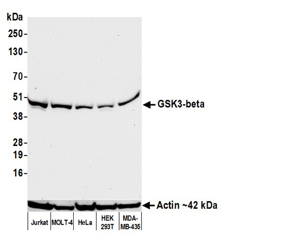
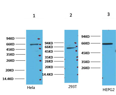
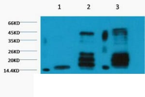
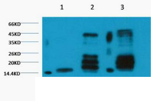
 WB (Western Blot)
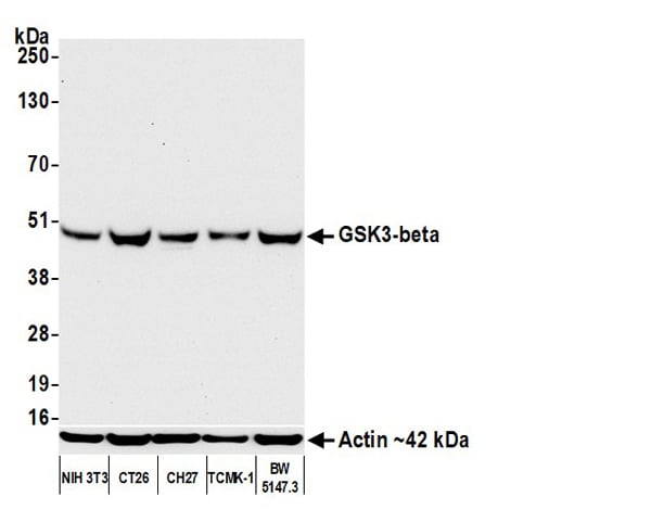
(Detection of mouse GSK3-beta by western blot. Samples: Whole cell lysate (50 ug) from NIH 3T3, CT26, CH27, TCMK-1, and BW5147.3 cells prepared using NETN lysis buffer. Antibody: Rabbit anti-GSK3-beta recombinant monoclonal antibody (AAA213647 lot 1) used at 1:1000. Secondary: HRP-conjugated goat anti-rabbit IgG . Detection: Chemiluminescence with an exposure time of 10 seconds. Lower Panel: Rabbit anti-Actin recombinant monoclonal antibody .)
WB (Western Blot)
(Detection of mouse GSK3-beta by western blot. Samples: Whole cell lysate (50 ug) from NIH 3T3, CT26, CH27, TCMK-1, and BW5147.3 cells prepared using NETN lysis buffer. Antibody: Rabbit anti-GSK3-beta recombinant monoclonal antibody (AAA213647 lot 1) used at 1:1000. Secondary: HRP-conjugated goat anti-rabbit IgG . Detection: Chemiluminescence with an exposure time of 10 seconds. Lower Panel: Rabbit anti-Actin recombinant monoclonal antibody .)
GSK3-beta, Monoclonal Recombinant Antibody (Cat# AAA213647)
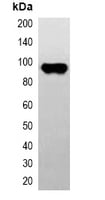
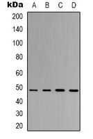
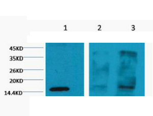
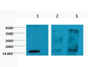
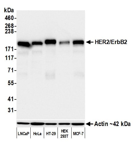 WB (Western Blot)
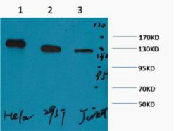
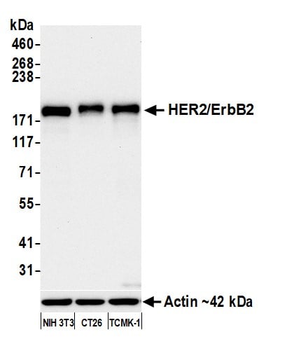
(Detection of human HER2/ErbB2 by western blot. Samples: Whole cell lysate (10 ug) from LNCaP, HeLa, HT-29, HEK293T, and MCF-7 cells prepared using NETN lysis buffer. Antibody: Rabbit anti-HER2/ErbB2 recombinant monoclonal antibody (AAA213657 lot 1) used at 1:1000. Secondary: HRP-conjugated goat anti-rabbit IgG . Detection: Chemiluminescence with an exposure time of 10 seconds. Lower Panel: Rabbit anti-Actin recombinant monoclonal antibody .)
WB (Western Blot)
(Detection of human HER2/ErbB2 by western blot. Samples: Whole cell lysate (10 ug) from LNCaP, HeLa, HT-29, HEK293T, and MCF-7 cells prepared using NETN lysis buffer. Antibody: Rabbit anti-HER2/ErbB2 recombinant monoclonal antibody (AAA213657 lot 1) used at 1:1000. Secondary: HRP-conjugated goat anti-rabbit IgG . Detection: Chemiluminescence with an exposure time of 10 seconds. Lower Panel: Rabbit anti-Actin recombinant monoclonal antibody .)
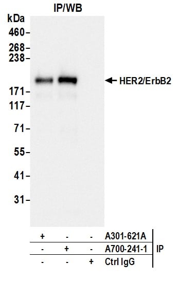
HER2/ErbB2, Monoclonal Recombinant Antibody (Cat# AAA213657)
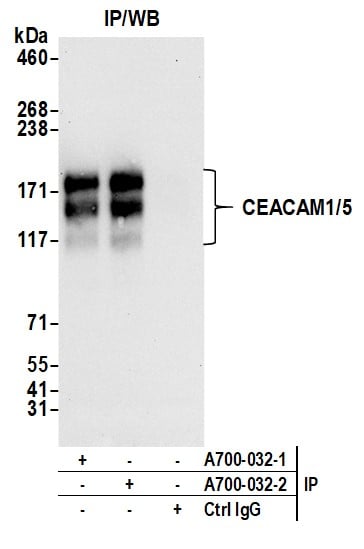
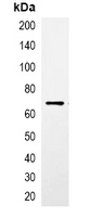
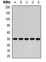
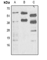
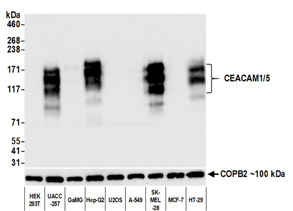 WB (Western Blot)
(Detection of human CEACAM1/5 by western blot. Samples: Whole cell lysate (5 ug) from HEK293T, UACC-257, GaMG, Hep-G2, U2OS, A-549, SK-MEL-28, MCF-7, and HT-29 cells prepared using NETN lysis buffer. Antibody: Rabbit anti-CEACAM1/5 recombinant monoclonal antibody (AAA213541 lot 2) used at 1:1000. Secondary: HRP-conjugated goat anti-rabbit IgG . Detection: Chemiluminescence with an exposure time of 3 seconds.)
WB (Western Blot)
(Detection of human CEACAM1/5 by western blot. Samples: Whole cell lysate (5 ug) from HEK293T, UACC-257, GaMG, Hep-G2, U2OS, A-549, SK-MEL-28, MCF-7, and HT-29 cells prepared using NETN lysis buffer. Antibody: Rabbit anti-CEACAM1/5 recombinant monoclonal antibody (AAA213541 lot 2) used at 1:1000. Secondary: HRP-conjugated goat anti-rabbit IgG . Detection: Chemiluminescence with an exposure time of 3 seconds.)
CEACAM1/5, Monoclonal Recombinant Antibody (Cat# AAA213541)
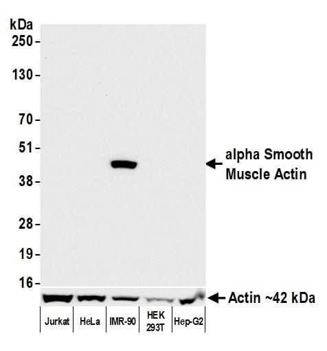
 WB (Western Blot)
(Detection of mouse alpha Smooth Muscle Actin by western blot. Samples: Whole cell lysate (10 ug) from CT26, NIH 3T3, CH27, TCMK-1, and BW5147.3 cells prepared using NETN lysis buffer. Antibody: Rabbit anti-alpha Smooth Muscle Actin recombinant monoclonal antibody (AAA213566 lot 2) used at 1:1000. Secondary: HRP-conjugated goat anti-rabbit IgG . Chemiluminescence with an exposure time of 10 seconds. Lower Panel: Rabbit anti-Actin recombinant monoclonal antibody .)
WB (Western Blot)
(Detection of mouse alpha Smooth Muscle Actin by western blot. Samples: Whole cell lysate (10 ug) from CT26, NIH 3T3, CH27, TCMK-1, and BW5147.3 cells prepared using NETN lysis buffer. Antibody: Rabbit anti-alpha Smooth Muscle Actin recombinant monoclonal antibody (AAA213566 lot 2) used at 1:1000. Secondary: HRP-conjugated goat anti-rabbit IgG . Chemiluminescence with an exposure time of 10 seconds. Lower Panel: Rabbit anti-Actin recombinant monoclonal antibody .)
alpha Smooth Muscle Actin, Monoclonal Recombinant Antibody (Cat# AAA213566)
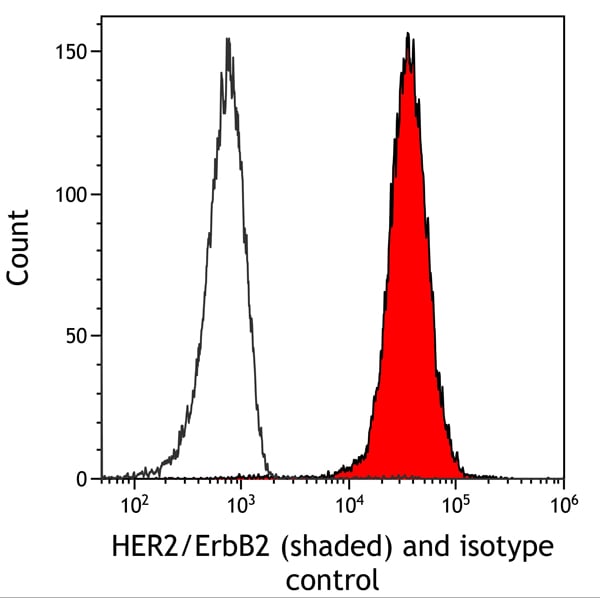
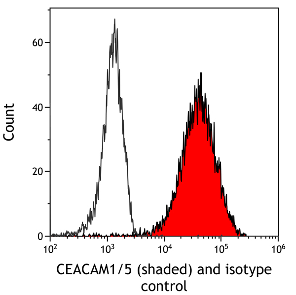
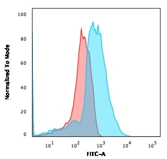 FCM/FACS (Flow Cytometry)
(Flow Cytometric Analysis of PFA-fixed HEK293 cells using CD137L-Monospecific Mouse Monoclonal Antibody (CD137L/1547) followed by goat anti-Mouse IgG-CF488 (Blue); Isotype Control (Red).)
FCM/FACS (Flow Cytometry)
(Flow Cytometric Analysis of PFA-fixed HEK293 cells using CD137L-Monospecific Mouse Monoclonal Antibody (CD137L/1547) followed by goat anti-Mouse IgG-CF488 (Blue); Isotype Control (Red).)
CD137L/4-1BBL/TNFSF9, Monoclonal Antibody (Cat# AAA215273)
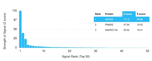 Application Data
(Analysis of Protein Array containing more than 19,000 full-length human proteins using Napsin A Mouse Monoclonal Antibody (NAPSA/3308). Z- and S- Score: The Z-score represents the strength of a signal that a monoclonal antibody (Monoclonal Antibody) (in combination with a fluorescently-tagged anti-IgG secondary antibody) produces when binding to a particular protein on the HuProtTM array. Z-scores are described in units of standard deviations (SD’s) above the mean value of all signals generated on that array. If targets on HuProtTM are arranged in descending order of the Z-score, the S-score is the difference (also in units of SD’s) between the Z-score. S-score therefore represents the relative target specificity of a Monoclonal Antibody to its intended target. A Monoclonal Antibody is considered to specific to its intended target, if the Monoclonal Antibody has an S-score of at least 2.5. For example, if a Monoclonal Antibody binds to protein X with a Z-score of 43 and to protein Y with a Z-score of 14, then the S-score for the binding of that Monoclonal Antibody to protein X is equal to 29.)
Application Data
(Analysis of Protein Array containing more than 19,000 full-length human proteins using Napsin A Mouse Monoclonal Antibody (NAPSA/3308). Z- and S- Score: The Z-score represents the strength of a signal that a monoclonal antibody (Monoclonal Antibody) (in combination with a fluorescently-tagged anti-IgG secondary antibody) produces when binding to a particular protein on the HuProtTM array. Z-scores are described in units of standard deviations (SD’s) above the mean value of all signals generated on that array. If targets on HuProtTM are arranged in descending order of the Z-score, the S-score is the difference (also in units of SD’s) between the Z-score. S-score therefore represents the relative target specificity of a Monoclonal Antibody to its intended target. A Monoclonal Antibody is considered to specific to its intended target, if the Monoclonal Antibody has an S-score of at least 2.5. For example, if a Monoclonal Antibody binds to protein X with a Z-score of 43 and to protein Y with a Z-score of 14, then the S-score for the binding of that Monoclonal Antibody to protein X is equal to 29.)
Napsin A, Monoclonal Antibody (Cat# AAA215301)
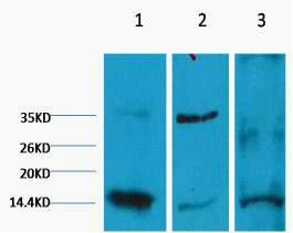
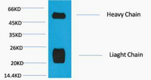
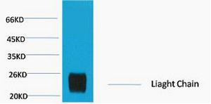
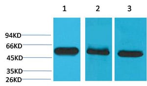
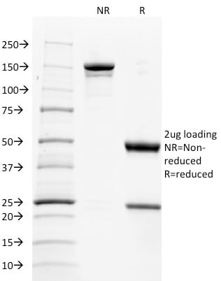
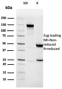
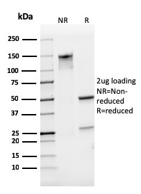
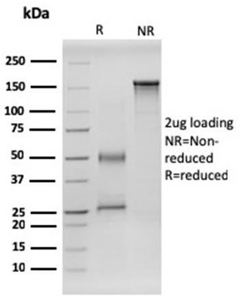
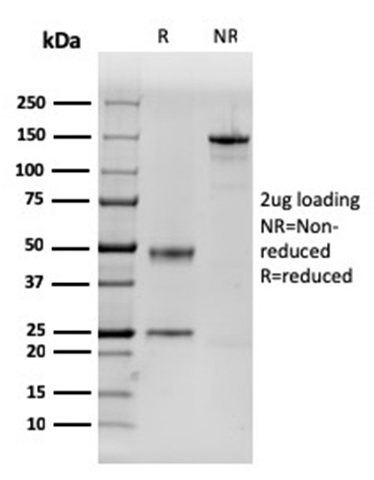
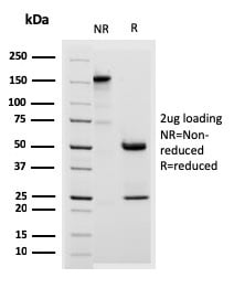
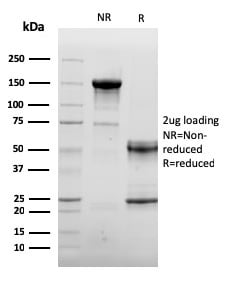
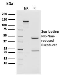 SDS-PAGE
(SDS-PAGE Analysis Purified CD44v4/5 Mouse Monoclonal Antibody (3D2). Confirmation of Purity and Integrity of Antibody.)
SDS-PAGE
(SDS-PAGE Analysis Purified CD44v4/5 Mouse Monoclonal Antibody (3D2). Confirmation of Purity and Integrity of Antibody.)
CD44v4/5, Monoclonal Antibody (Cat# AAA215305)
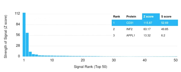 Application Data
(Analysis of Protein Array containing more than 19,000 full-length human proteins using CD31 Mouse Monoclonal Antibody (PECAM1/3529) Z- and S- Score: The Z-score represents the strength of a signal that a monoclonal antibody (MAb) (in combination with a fluorescently-tagged anti-IgG secondary antibody) produces when binding to a particular protein on the HuProtTM array. Z-scores are described in units of standard deviations (SD’s) above the mean value of all signals generated on that array. If targets on HuProtTM are arranged in descending order of the Z-score, the S-score is the difference (also in units of SD’s) between the Z-score. S-score therefore represents the relative target specificity of a MAb to its intended target. A MAb is considered to specific to its intended target, if the MAb has an S-score of at least 2.5. For example, if a MAb binds to protein X with a Z-score of 43 and to protein Y with a Z-score of 14, then the S-score for the binding of that MAb to protein X is equal to 29.)
Application Data
(Analysis of Protein Array containing more than 19,000 full-length human proteins using CD31 Mouse Monoclonal Antibody (PECAM1/3529) Z- and S- Score: The Z-score represents the strength of a signal that a monoclonal antibody (MAb) (in combination with a fluorescently-tagged anti-IgG secondary antibody) produces when binding to a particular protein on the HuProtTM array. Z-scores are described in units of standard deviations (SD’s) above the mean value of all signals generated on that array. If targets on HuProtTM are arranged in descending order of the Z-score, the S-score is the difference (also in units of SD’s) between the Z-score. S-score therefore represents the relative target specificity of a MAb to its intended target. A MAb is considered to specific to its intended target, if the MAb has an S-score of at least 2.5. For example, if a MAb binds to protein X with a Z-score of 43 and to protein Y with a Z-score of 14, then the S-score for the binding of that MAb to protein X is equal to 29.)
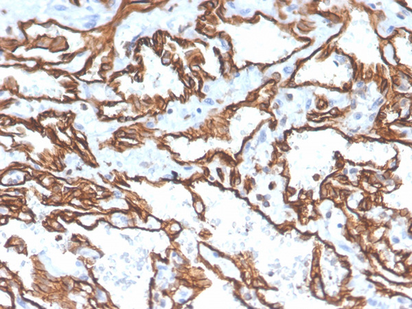
CD31/PECAM-1, Monoclonal Antibody (Cat# AAA215132)
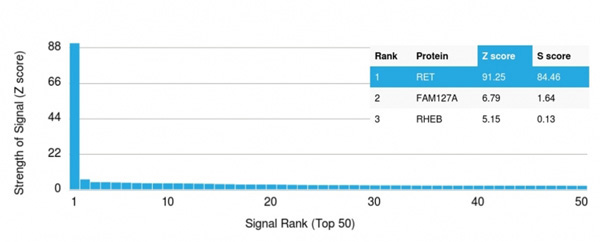 Application Data
(Analysis of Protein Array containing more than 19,000 full-length human proteins using RET-Monospecific Mouse Monoclonal Antibody (RET/2599). Z- and S- Score: The Z-score represents the strength of a signal that a monoclonal antibody (Monoclonal Antibody) (in combination with a fluorescently-tagged anti-IgG secondary antibody) produces when binding to a particular protein on the HuProtTM array. Z-scores are described in units of standard deviations (SD's) above the mean value of all signals generated on that array. If targets on HuProtTM are arranged in descending order of the Z-score, the S-score is the difference (also in units of SD's) between the Z-score. S-score therefore represents the relative target specificity of a Monoclonal Antibody to its intended target. A Monoclonal Antibody is considered to specific to its intended target, if the Monoclonal Antibody has an S-score of at least 2.5. For example, if a Monoclonal Antibody binds to protein X with a Z-score of 43 and to protein Y with a Z-score of 14, then the S-score for the binding of that Monoclonal Antibody to protein X is equal to 29.)
Application Data
(Analysis of Protein Array containing more than 19,000 full-length human proteins using RET-Monospecific Mouse Monoclonal Antibody (RET/2599). Z- and S- Score: The Z-score represents the strength of a signal that a monoclonal antibody (Monoclonal Antibody) (in combination with a fluorescently-tagged anti-IgG secondary antibody) produces when binding to a particular protein on the HuProtTM array. Z-scores are described in units of standard deviations (SD's) above the mean value of all signals generated on that array. If targets on HuProtTM are arranged in descending order of the Z-score, the S-score is the difference (also in units of SD's) between the Z-score. S-score therefore represents the relative target specificity of a Monoclonal Antibody to its intended target. A Monoclonal Antibody is considered to specific to its intended target, if the Monoclonal Antibody has an S-score of at least 2.5. For example, if a Monoclonal Antibody binds to protein X with a Z-score of 43 and to protein Y with a Z-score of 14, then the S-score for the binding of that Monoclonal Antibody to protein X is equal to 29.)
RET Proto-oncogene, Monoclonal Antibody (Cat# AAA215179)

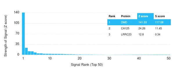 Application Data
(Analysis of Protein Array containing more than 19,000 full-length human proteins using Dystrophin Monospecific Mouse Monoclonal Antibody (DMD/3243). Z- and S- Score: The Z-score represents the strength of a signal that a monoclonal antibody (Monoclonal Antibody) (in combination with a fluorescently-tagged anti-IgG secondary antibody) produces when binding to a particular protein on the HuProtTM array. Z-scores are described in units of standard deviations (SD’s) above the mean value of all signals generated on that array. If targets on HuProtTM are arranged in descending order of the Z-score, the S-score is the difference (also in units of SD’s) between the Z-score. S-score therefore represents the relative target specificity of a Monoclonal Antibody to its intended target. A Monoclonal Antibody is considered to specific to its intended target, if the Monoclonal Antibody has an S-score of at least 2.5. For example, if a Monoclonal Antibody binds to protein X with a Z-score of 43 and to protein Y with a Z-score of 14, then the S-score for the binding of that Monoclonal Antibody to protein X is equal to 29.)
Application Data
(Analysis of Protein Array containing more than 19,000 full-length human proteins using Dystrophin Monospecific Mouse Monoclonal Antibody (DMD/3243). Z- and S- Score: The Z-score represents the strength of a signal that a monoclonal antibody (Monoclonal Antibody) (in combination with a fluorescently-tagged anti-IgG secondary antibody) produces when binding to a particular protein on the HuProtTM array. Z-scores are described in units of standard deviations (SD’s) above the mean value of all signals generated on that array. If targets on HuProtTM are arranged in descending order of the Z-score, the S-score is the difference (also in units of SD’s) between the Z-score. S-score therefore represents the relative target specificity of a Monoclonal Antibody to its intended target. A Monoclonal Antibody is considered to specific to its intended target, if the Monoclonal Antibody has an S-score of at least 2.5. For example, if a Monoclonal Antibody binds to protein X with a Z-score of 43 and to protein Y with a Z-score of 14, then the S-score for the binding of that Monoclonal Antibody to protein X is equal to 29.)
Dystrophin (DMD), Monoclonal Antibody (Cat# AAA214924)
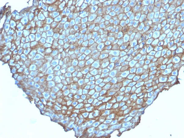
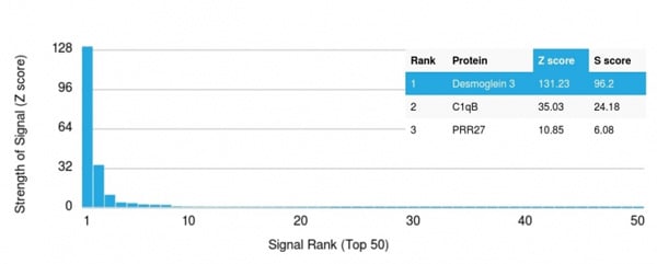 Application Data
(Analysis of Protein Array containing more than 19,000 full-length human proteins using Desmoglein-3 Mouse Monoclonal Antibody (DSG3/2838) Z- and S- Score: The Z-score represents the strength of a signal that a monoclonal antibody (MAb) (in combination with a fluorescently-tagged anti-IgG secondary antibody) produces when binding to a particular protein on the HuProtTM array. Z-scores are described in units of standard deviations (SD's) above the mean value of all signals generated on that array. If targets on HuProtTM are arranged in descending order of the Z-score, the S-score is the difference (also in units of SD's) between the Z-score. S-score therefore represents the relative target specificity of a MAb to its intended target. A MAb is considered to specific to its intended target, if the MAb has an S-score of at least 2.5. For example, if a MAb binds to protein X with a Z-score of 43 and to protein Y with a Z-score of 14, then the S-score for the binding of that MAb to protein X is equal to 29.)
Application Data
(Analysis of Protein Array containing more than 19,000 full-length human proteins using Desmoglein-3 Mouse Monoclonal Antibody (DSG3/2838) Z- and S- Score: The Z-score represents the strength of a signal that a monoclonal antibody (MAb) (in combination with a fluorescently-tagged anti-IgG secondary antibody) produces when binding to a particular protein on the HuProtTM array. Z-scores are described in units of standard deviations (SD's) above the mean value of all signals generated on that array. If targets on HuProtTM are arranged in descending order of the Z-score, the S-score is the difference (also in units of SD's) between the Z-score. S-score therefore represents the relative target specificity of a MAb to its intended target. A MAb is considered to specific to its intended target, if the MAb has an S-score of at least 2.5. For example, if a MAb binds to protein X with a Z-score of 43 and to protein Y with a Z-score of 14, then the S-score for the binding of that MAb to protein X is equal to 29.)
Desmoglein-3, Monoclonal Antibody (Cat# AAA214934)
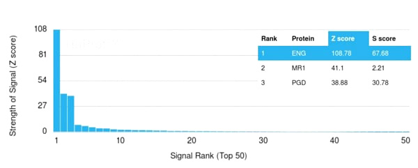 Application Data
(Analysis of Protein Array containing more than 19,000 full-length human proteins using Endoglin/CD105 Mouse Monoclonal Antibody (ENG/3269). Z- and S- Score: The Z-score represents the strength of a signal that a monoclonal antibody (MAb) (in combination with a fluorescently-tagged anti-IgG secondary antibody) produces when binding to a particular protein on the HuProtTM array. Z-scores are described in units of standard deviations (SD’s) above the mean value of all signals generated on that array. If targets on HuProtTM are arranged in descending order of the Z-score, the S-score is the difference (also in units of SD’s) between the Z-score. S-score therefore represents the relative target specificity of a MAb to its intended target. A MAb is considered to specific to its intended target, if the MAb has an S-score of at least 2.5. For example, if a MAb binds to protein X with a Z-score of 43 and to protein Y with a Z-score of 14, then the S-score for the binding of that MAb to protein X is equal to 29.)
Application Data
(Analysis of Protein Array containing more than 19,000 full-length human proteins using Endoglin/CD105 Mouse Monoclonal Antibody (ENG/3269). Z- and S- Score: The Z-score represents the strength of a signal that a monoclonal antibody (MAb) (in combination with a fluorescently-tagged anti-IgG secondary antibody) produces when binding to a particular protein on the HuProtTM array. Z-scores are described in units of standard deviations (SD’s) above the mean value of all signals generated on that array. If targets on HuProtTM are arranged in descending order of the Z-score, the S-score is the difference (also in units of SD’s) between the Z-score. S-score therefore represents the relative target specificity of a MAb to its intended target. A MAb is considered to specific to its intended target, if the MAb has an S-score of at least 2.5. For example, if a MAb binds to protein X with a Z-score of 43 and to protein Y with a Z-score of 14, then the S-score for the binding of that MAb to protein X is equal to 29.)
Endoglin/CD105, Monoclonal Antibody (Cat# AAA214940)
 Application Data
(Analysis of Protein Array containing more than 19,000 full-length human proteins using HER-4 Mouse Monoclonal Antibody (ERBB4/2581). Z- and S- Score: The Z-score represents the strength of a signal that a monoclonal antibody (MAb) (in combination with a fluorescently-tagged anti-IgG secondary antibody) produces when binding to a particular protein on the HuProtTM array. Z-scores are described in units of standard deviations (SD's) above the mean value of all signals generated on that array. If targets on HuProtTM are arranged in descending order of the Z-score, the S-score is the difference (also in units of SD's) between the Z-score. S-score therefore represents the relative target specificity of a MAb to its intended target. A MAb is considered to specific to its intended target, if the MAb has an S-score of at least 2.5. For example, if a MAb binds to protein X with a Z-score of 43 and to protein Y with a Z-score of 14, then the S-score for the binding of that MAb to protein X is equal to 29.)
Application Data
(Analysis of Protein Array containing more than 19,000 full-length human proteins using HER-4 Mouse Monoclonal Antibody (ERBB4/2581). Z- and S- Score: The Z-score represents the strength of a signal that a monoclonal antibody (MAb) (in combination with a fluorescently-tagged anti-IgG secondary antibody) produces when binding to a particular protein on the HuProtTM array. Z-scores are described in units of standard deviations (SD's) above the mean value of all signals generated on that array. If targets on HuProtTM are arranged in descending order of the Z-score, the S-score is the difference (also in units of SD's) between the Z-score. S-score therefore represents the relative target specificity of a MAb to its intended target. A MAb is considered to specific to its intended target, if the MAb has an S-score of at least 2.5. For example, if a MAb binds to protein X with a Z-score of 43 and to protein Y with a Z-score of 14, then the S-score for the binding of that MAb to protein X is equal to 29.)
HER-4/ERBB4, Monoclonal Antibody (Cat# AAA214948)
What are Monoclonal Antibodies?
Monoclonal antibodies are specialized laboratory-produced proteins developed for binding to specific biological antigens or other molecular targets. Since they come from a single cell (or clone), they are especially consistent and accurate in the data they are involved in producing.
This type of antibody material has been shown to be a powerful tool in finding and subsequently destroying harmful cells in an organism, such as those found in cancers or various autoimmune diseases. This makes them excellent aids in medical testing and research, which is why they are so widely used.
AAA Biotech offers a comprehensive range of high-quality monoclonal antibodies that perform effectively in various laboratory tests, including (amongst others) ELISA, western blotting, immunohistochemistry, and flow cytometry. All of the products in our catalog are thoroughly quality tested to make sure that they are reliable and will consistently perform well in your research.
What Are The Uses of Monoclonal Antibodies
Monoclonal antibodies are used in many lab tests, including (amongst others) ELISA, western blotting, immunohistochemistry, and flow cytometry.
ELISA is a test that helps detect a specific substance/analyte in a sample. It uses antibodies (often monoclonal) bound to a solid surface (such as the well of a microplate) to “capture” the substance/analyte in the sample and immobilize it so that the detection antibody component can then bind to it and produce a signal, which can then be measured.
Western blotting identifies specific proteins in a sample. The sample is first separated on a gel, and then antibodies are applied that will typically bind to the target, which will all be localized to a single band in a lane.
Immunohistochemistry helps locate specific proteins in cells or tissue samples using antibodies.
Flow cytometry looks at and sorts cells. It uses antibodies that are conjugated to reporter molecules called “fluorophores”, which, under special lights, emit light themselves, which can then be measured by a detector instrument.
How Monoclonal Antibodies Are Used as Medicine?
Please note that all of the products listed in AAA Biotech’s also known as AAA Bio or AAABio catalog are strictly for research-use only (RUO).
Monoclonal antibodies can also be used as therapeutic/medical treatments, particularly in the context of cancers. They are designed to find and bind to specific cells or proteins, helping the immune system recognize and attack the cancer. These treatments work in different ways, such as:
- Radioimmunotherapy attaches a small amount of radioactive molecule to the antibody, so it delivers the radiation directly to the cancer cells that the antibody is specifically binding to.
- Antibody-directed enzyme prodrug therapy uses antibodies that are specifically bound to special enzymes. These enzymes activate a harmless drug in the body and turn it into a cancer-killing drug only near the cancer cells—this helps avoid harming healthy cells.
- Immunoliposomes are tiny “bubbles” filled with medicine/drug and coated with antibodies. They carry the drug straight to the cancer cells.
Why Buy Monoclonal Antibodies From Us?
At AAA Biotech, we provide high-performance monoclonal antibodies designed to support a wide range of research needs.
1. Validated for Versatile Applications
The antibodies in our catalog are extensively validated and compatible with multiple techniques, including (but not limited to) ELISA, flow cytometry (FC), immunocytochemistry (ICC), immunofluorescence (IF), immunohistochemistry (IHC), immunoprecipitation (IP), and western blotting (WB).
2. Wide Selection & Specialized Options
We offer antibodies for common and rare species, that are available in various conjugated forms, and also in recombinant formats. Essentially, there is almost anything one might need to meet their experimental model’s requirements.
3. High-Quality Proteins
Our proteins meet high purity standards—90% or more as confirmed by SDS-PAGE. Many are available with tags like His, Flag, GST, or MBP, and we also supply native and biologically active proteins for functional studies.
Frequently Asked Questions
1. Are your monoclonal antibodies validated for specific applications?
Yes, our antibodies are tested and validated for use in methods such as ELISA, western blot, IHC, flow cytometry, and more. Refer to specific product pages or datasheets for individual product information.
2. How do I choose the right monoclonal antibody for my application?
Review the product details directly for application validation, species reactivity, and target information. You may also contact our support team at any time for help.
3. How quickly can I receive my order?
Most orders are processed and shipped within 1–3 business days, depending on product availability and your shipping location.