Filters
▼Clonality
▼Type
▼Reactivity
▼Gene Name
▼Isotype
▼Host
▼Application
▼Clone
▼Monoclonal Antibodies
Get accurate results in your research with our Monoclonal Antibodies, which are specially made to target exactly what you require for your research, and will produce consistent, reliable performance in lab tests.
Viewing 5800-5850 of 27597 product results
 IHC (Immunohistochemisry)
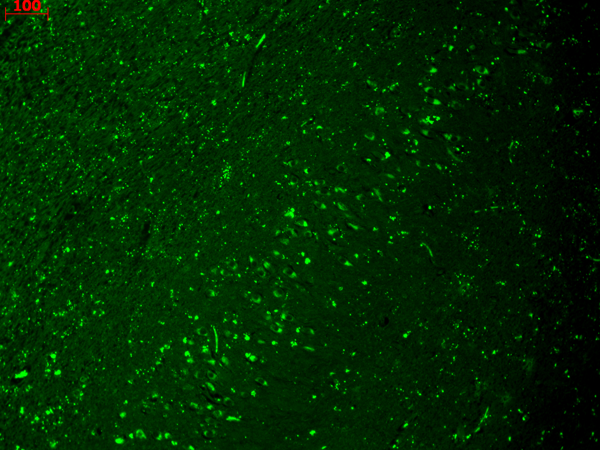
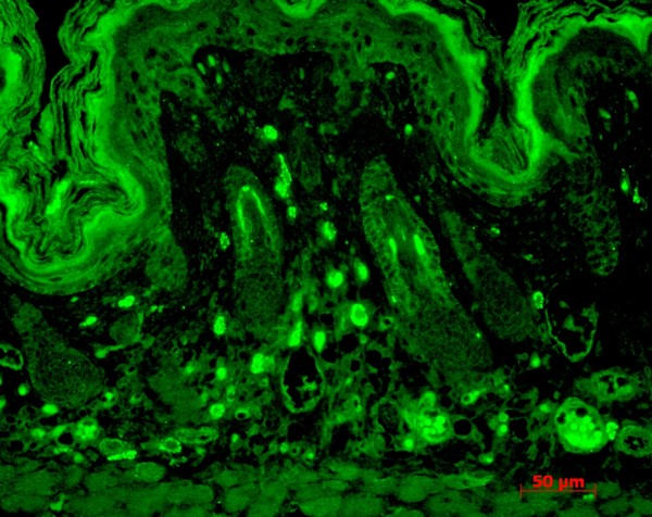
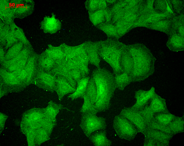
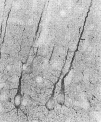
(Immunohistochemistry analysis using Mouse Anti-HCN4 Monoclonal Antibody, Clone S114-10. Tissue: hippocampus. Species: Human. Fixation: Bouin's Fixative and paraffin-embedded. Primary Antibody: Mouse Anti-HCN4 Monoclonal Antibody at 1:100 for 1 hour at RT. Secondary Antibody: FITC Goat Anti-Mouse (green) at 1:50 for 1 hour at RT.)
IHC (Immunohistochemisry)
(Immunohistochemistry analysis using Mouse Anti-HCN4 Monoclonal Antibody, Clone S114-10. Tissue: hippocampus. Species: Human. Fixation: Bouin's Fixative and paraffin-embedded. Primary Antibody: Mouse Anti-HCN4 Monoclonal Antibody at 1:100 for 1 hour at RT. Secondary Antibody: FITC Goat Anti-Mouse (green) at 1:50 for 1 hour at RT.)
HCN4, Monoclonal Antibody (Cat# AAA102879)
 WB (Western Blot)
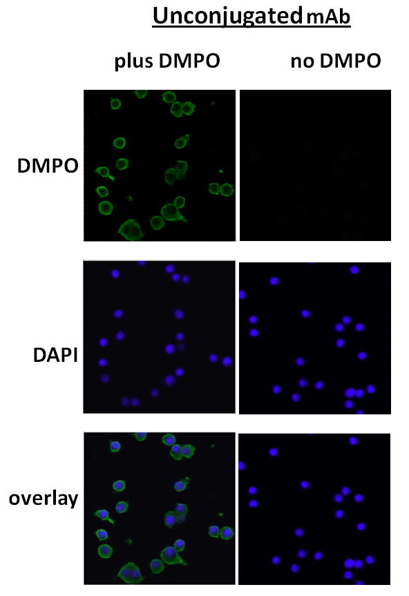
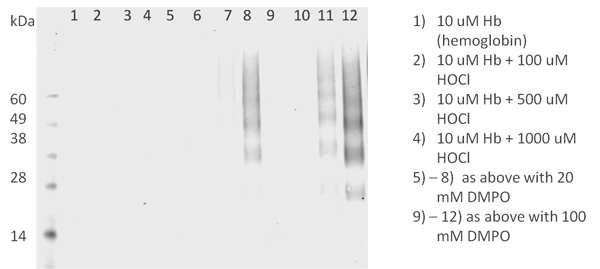
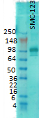
(Western Blot analysis of Human HL 60 clone 15 eosinophils lysates showing detection of DMPO protein using Mouse Anti-DMPO Monoclonal Antibody, Clone N1664A. Primary Antibody: Mouse Anti-DMPO Monoclonal Antibody at 1:200.)
WB (Western Blot)
(Western Blot analysis of Human HL 60 clone 15 eosinophils lysates showing detection of DMPO protein using Mouse Anti-DMPO Monoclonal Antibody, Clone N1664A. Primary Antibody: Mouse Anti-DMPO Monoclonal Antibody at 1:200.)
DMPO, Monoclonal Antibody (Cat# AAA102884)
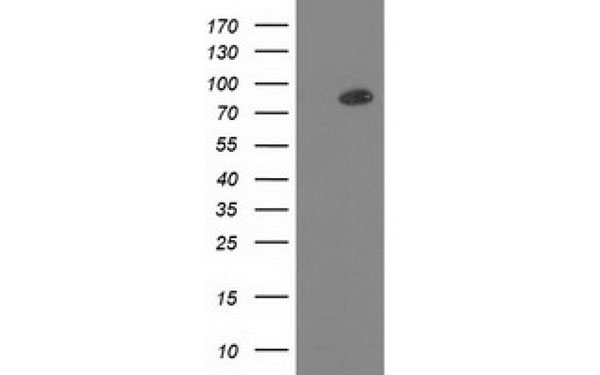
 WB (Western Blot)
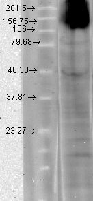
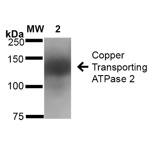
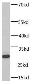
(Western Blot analysis of Rat Brain Membrane showing detection of ~160 kDa Copper Transporting ATPase 2 protein using Mouse Anti-Copper Transporting ATPase 2 Monoclonal Antibody, Clone S62-29 . Lane 1: Molecular Weight Ladder (MW). Lane 2: Rat Brain Membrane cell lysate. Load: 20 ug. Block: 2% BSA and 2% Skim Milk in 1X TBST. Primary Antibody: Mouse Anti-Copper Transporting ATPase 2 Monoclonal Antibody at 1:1000 for 16 hours at 4 degree C. Secondary Antibody: Goat Anti-Mouse IgG: HRP at 1:100 for 60 min at RT. Color Development: ECL solution for 6 min in RT. Predicted/Observed Size: ~160 kDa.)
WB (Western Blot)
(Western Blot analysis of Rat Brain Membrane showing detection of ~160 kDa Copper Transporting ATPase 2 protein using Mouse Anti-Copper Transporting ATPase 2 Monoclonal Antibody, Clone S62-29 . Lane 1: Molecular Weight Ladder (MW). Lane 2: Rat Brain Membrane cell lysate. Load: 20 ug. Block: 2% BSA and 2% Skim Milk in 1X TBST. Primary Antibody: Mouse Anti-Copper Transporting ATPase 2 Monoclonal Antibody at 1:1000 for 16 hours at 4 degree C. Secondary Antibody: Goat Anti-Mouse IgG: HRP at 1:100 for 60 min at RT. Color Development: ECL solution for 6 min in RT. Predicted/Observed Size: ~160 kDa.)
Copper-Transporting ATPase2, Monoclonal Antibody (Cat# AAA102892)
 WB (Western Blot)
(Western Blot analysis of Rat brain membrane lysate showing detection of PSD95 protein using Mouse Anti-PSD95 Monoclonal Antibody, Clone 7E3. Primary Antibody: Mouse Anti-PSD95 Monoclonal Antibody at 1:1000.)
WB (Western Blot)
(Western Blot analysis of Rat brain membrane lysate showing detection of PSD95 protein using Mouse Anti-PSD95 Monoclonal Antibody, Clone 7E3. Primary Antibody: Mouse Anti-PSD95 Monoclonal Antibody at 1:1000.)
PSD95, Monoclonal Antibody (Cat# AAA102917)


 WB (Western Blot)
(human brain tissue were subjected to SDS PAGE followed by western blot with AAA102664 (14-3-3 antibody) at dilution of 1:5000)
WB (Western Blot)
(human brain tissue were subjected to SDS PAGE followed by western blot with AAA102664 (14-3-3 antibody) at dilution of 1:5000)
14-3-3, Monoclonal Antibody (Cat# AAA102664)
Protein A+G purification
 WB (Western Blot)
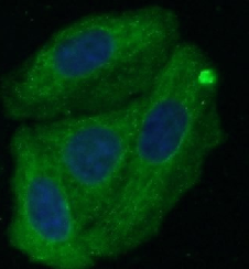
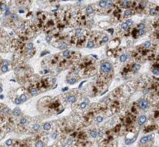
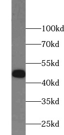
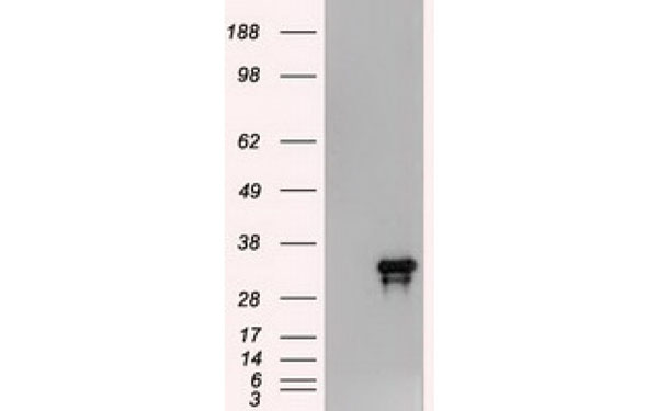
(HepG2 cells were subjected to SDS PAGE followed by western blot with AAA102683 (Alpha-1-Antitrypsin antibody) at dilution of 1:1000)
WB (Western Blot)
(HepG2 cells were subjected to SDS PAGE followed by western blot with AAA102683 (Alpha-1-Antitrypsin antibody) at dilution of 1:1000)
Alpha-1-Antitrypsin, Monoclonal Antibody (Cat# AAA102683)
Protein A+G purification

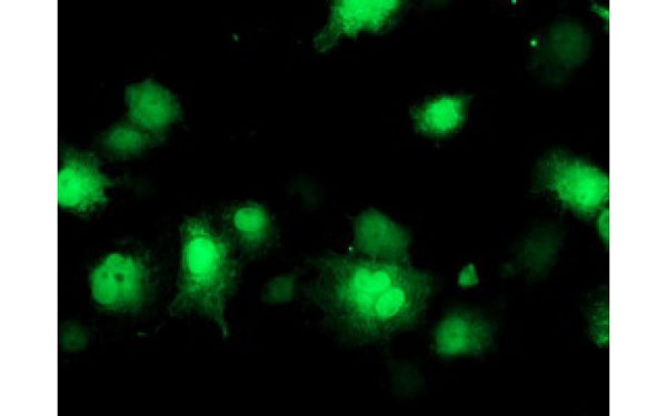
 IF (Immunofluorescence)
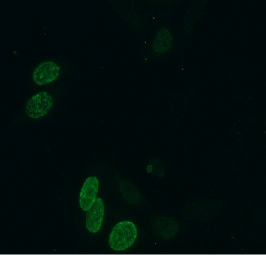
(Immunofluorescent analysis of HeLa cells (treated with 0.03 mg/ml BrdU for 2 hours) using AAA102714 (BrdU Antibody) at dilution of 1:300 and Alexa Fluor 488-congugated AffiniPure Goat Anti-Mouse IgG (H+L))
IF (Immunofluorescence)
(Immunofluorescent analysis of HeLa cells (treated with 0.03 mg/ml BrdU for 2 hours) using AAA102714 (BrdU Antibody) at dilution of 1:300 and Alexa Fluor 488-congugated AffiniPure Goat Anti-Mouse IgG (H+L))
BrdU, Monoclonal Antibody (Cat# AAA102714)
Protein A+G purification
 WB (Western Blot)
(HeLa cells were subjected to SDS PAGE followed by western blot with AAA102722 (Caspase 9 Antibody) at dilution of 1:1000)
WB (Western Blot)
(HeLa cells were subjected to SDS PAGE followed by western blot with AAA102722 (Caspase 9 Antibody) at dilution of 1:1000)
Caspase 9, Monoclonal Antibody (Cat# AAA102722)
Protein A+G purification
 WB (Western Blot)
(Raji cells were subjected to SDS PAGE followed by western blot with AAA102734 (CD23 antibody) at dilution of 1:2000)
WB (Western Blot)
(Raji cells were subjected to SDS PAGE followed by western blot with AAA102734 (CD23 antibody) at dilution of 1:2000)
CD23, Monoclonal Antibody (Cat# AAA102734)
Protein A+G purification
 WB (Western Blot)
(Jurkat cells were subjected to SDS PAGE followed by western blot with AAA102735 (CD3E Antibody) at dilution of 1:10000)
WB (Western Blot)
(Jurkat cells were subjected to SDS PAGE followed by western blot with AAA102735 (CD3E Antibody) at dilution of 1:10000)
CD3 epsilon, Monoclonal Antibody (Cat# AAA102735)
Purity: >95% as determined by SDS-PAGE
 WB (Western Blot)
(HeLa cells were subjected to SDS PAGE followed by western blot with AAA102741 (CD9 antibody) at dilution of 1:1000)
WB (Western Blot)
(HeLa cells were subjected to SDS PAGE followed by western blot with AAA102741 (CD9 antibody) at dilution of 1:1000)
CD9, Monoclonal Antibody (Cat# AAA102741)
Protein A+G purification

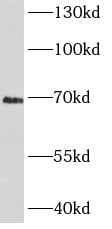
 WB (Western Blot)
(human placenta tissue were subjected to SDS PAGE followed by western blot with AAA102743 (CP Antibody) at dilution of 1:1000)
WB (Western Blot)
(human placenta tissue were subjected to SDS PAGE followed by western blot with AAA102743 (CP Antibody) at dilution of 1:1000)
Ceruloplasmin, Monoclonal Antibody (Cat# AAA102743)
Protein A+G purification
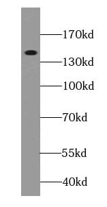
 WB (Western Blot)
(rat pancreas tissue were subjected to SDS PAGE followed by western blot with AAA102746 (CHGA Antibody) at dilution of 1:2000)
WB (Western Blot)
(rat pancreas tissue were subjected to SDS PAGE followed by western blot with AAA102746 (CHGA Antibody) at dilution of 1:2000)
Chromogranin A, Monoclonal Antibody (Cat# AAA102746)
Protein A+G purification
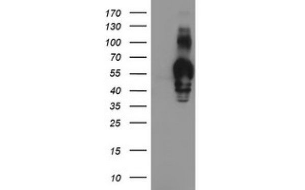
 Application Data
CYB5R3, Monoclonal Antibody (Cat# AAA102769)
Protein A+G purification
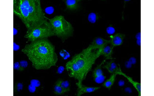
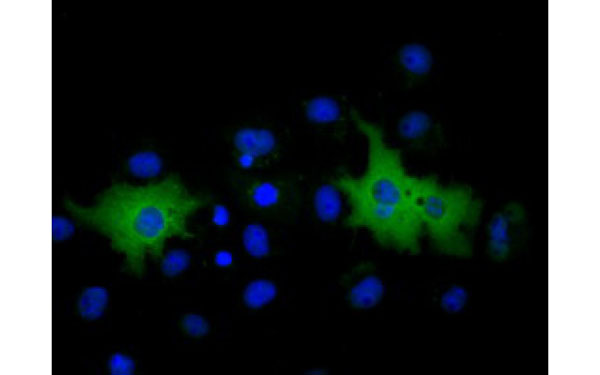
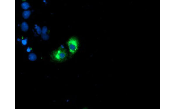
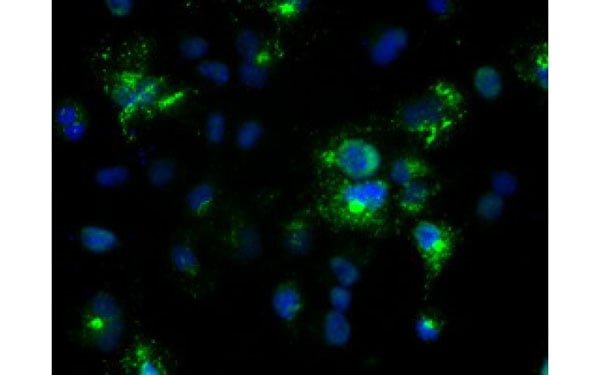
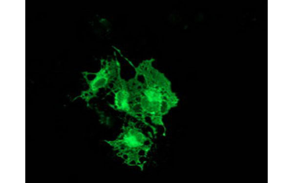
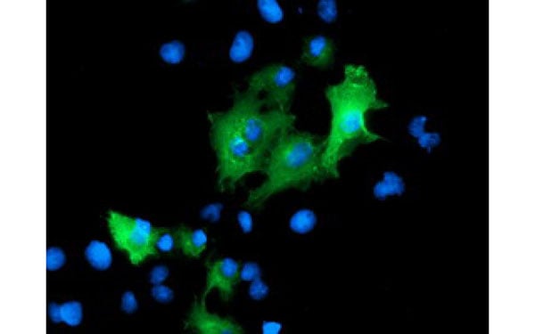
 IF (Immunofluorescence)
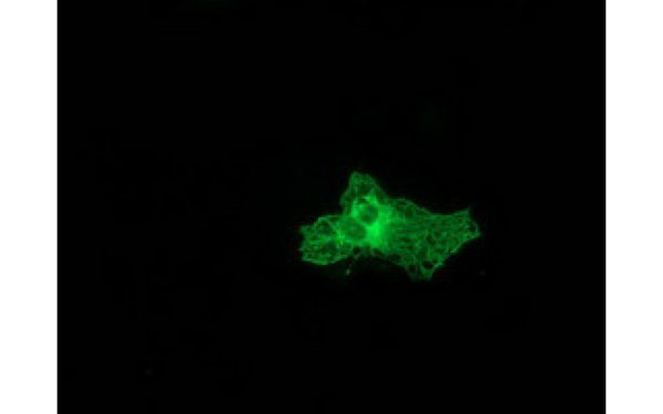
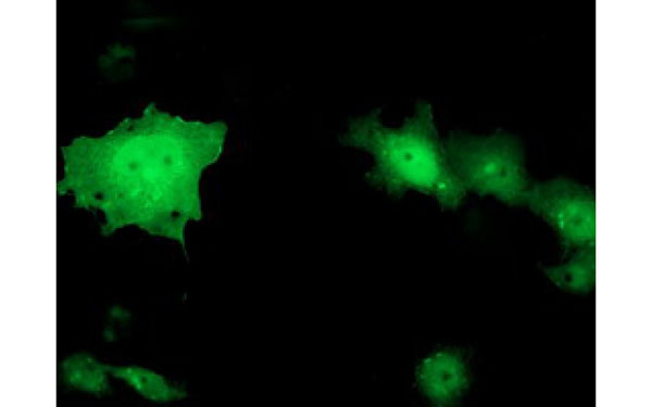
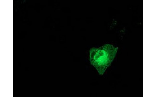
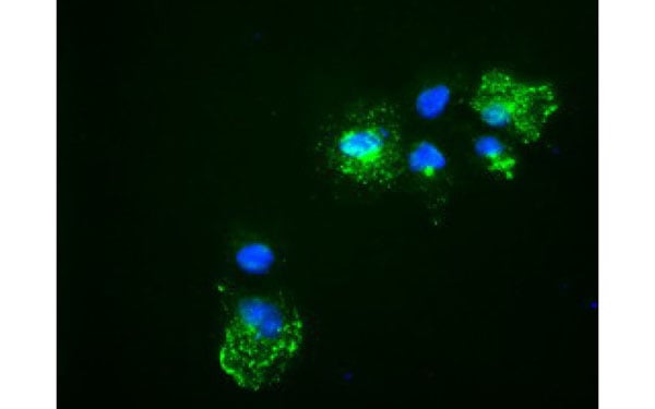
(Immunofluorescent staining of COS7 cells transiently transfected with recombinant PRKCE protein using PRKCE antibody)
IF (Immunofluorescence)
(Immunofluorescent staining of COS7 cells transiently transfected with recombinant PRKCE protein using PRKCE antibody)
PRKCE, Monoclonal Antibody (Cat# AAA108086)
 IF (Immunofluorescence)
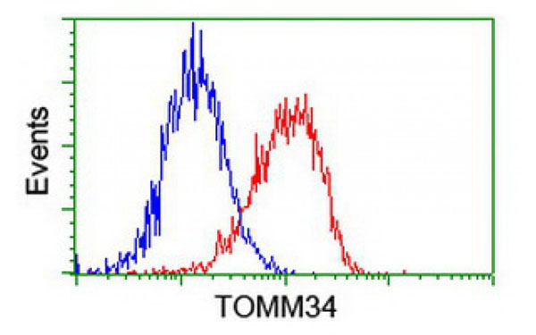
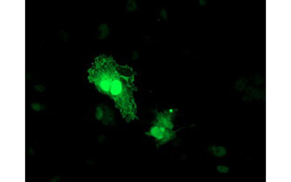
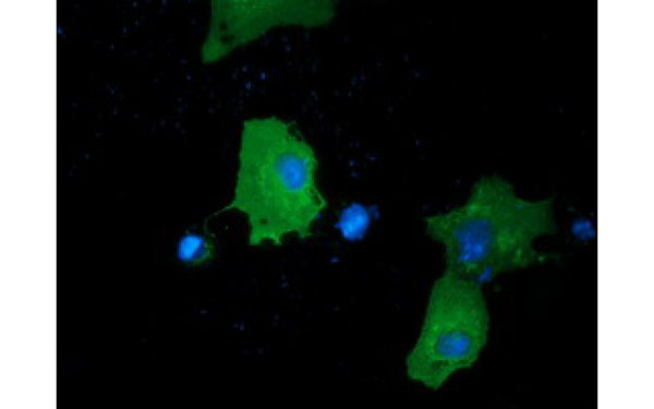
(Immunofluorescent staining of COS7 cells transiently transfected with recombinant TOMM34 protein using TOMM34 antibody)
IF (Immunofluorescence)
(Immunofluorescent staining of COS7 cells transiently transfected with recombinant TOMM34 protein using TOMM34 antibody)
TOMM34, Monoclonal Antibody (Cat# AAA108092)
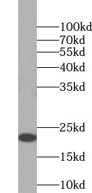
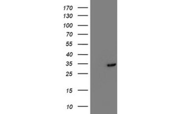
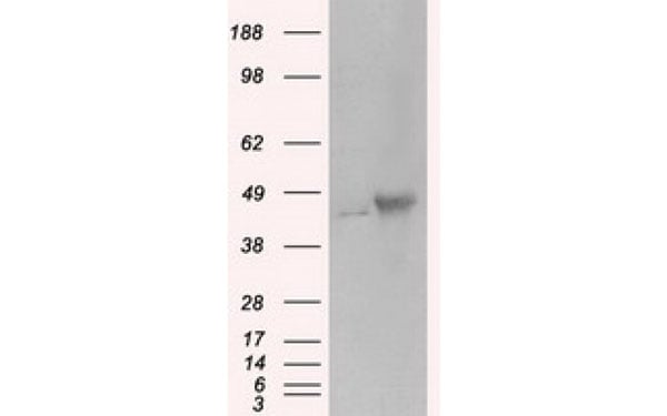
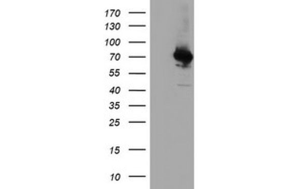
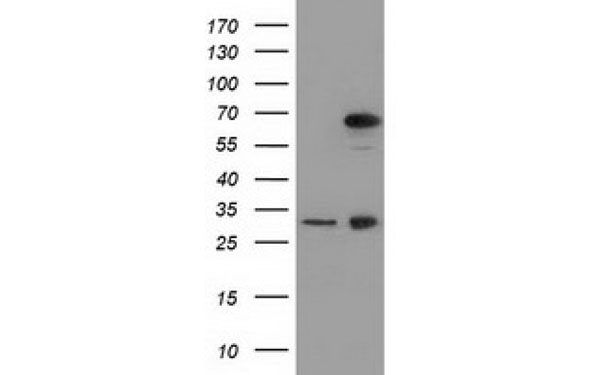
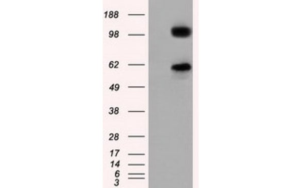
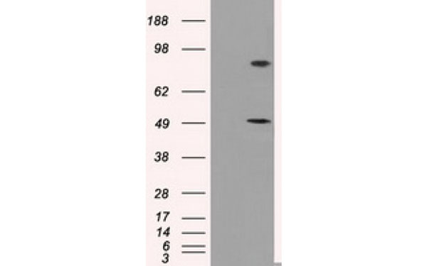
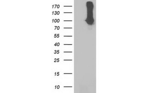
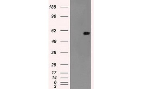
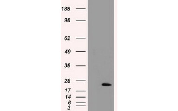 WB (Western Blot)
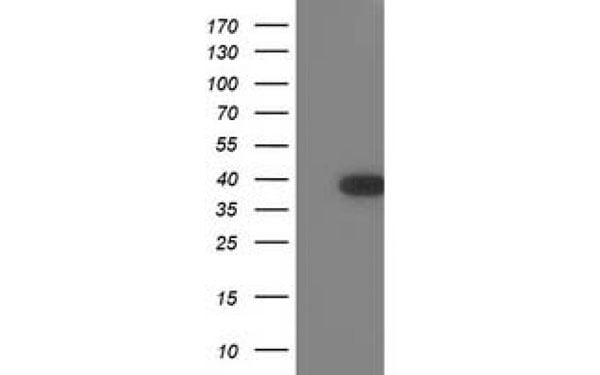
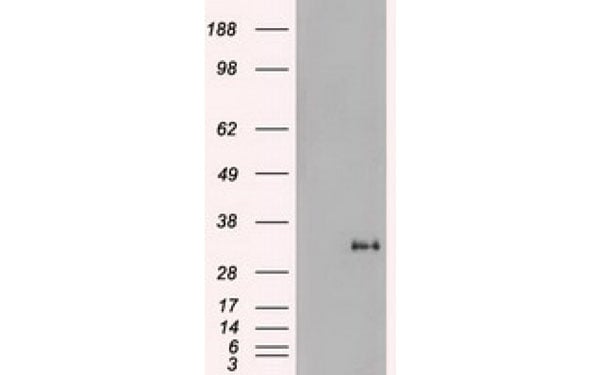
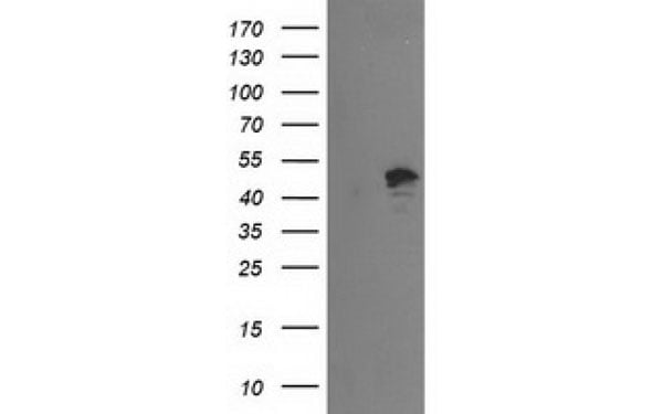
(Western Blot analysis of HEK293T cell lysates (5 ug) transfected with either recombinant TNNI3 protein (Right) or empty vector (Left) detected with TNNI3 antibody)
WB (Western Blot)
(Western Blot analysis of HEK293T cell lysates (5 ug) transfected with either recombinant TNNI3 protein (Right) or empty vector (Left) detected with TNNI3 antibody)
TNNI3, Monoclonal Antibody (Cat# AAA108108)
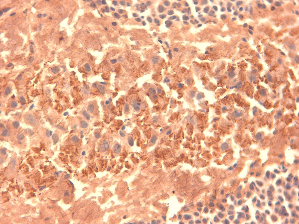
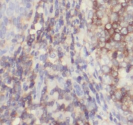
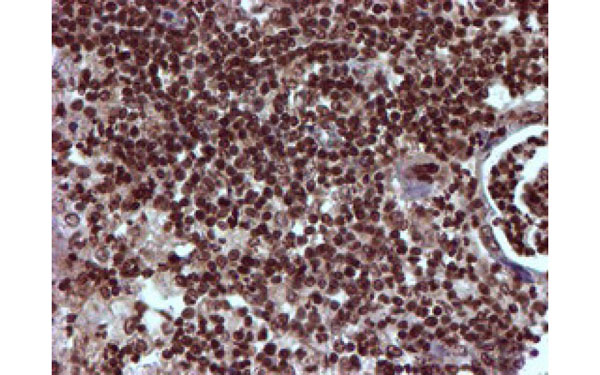
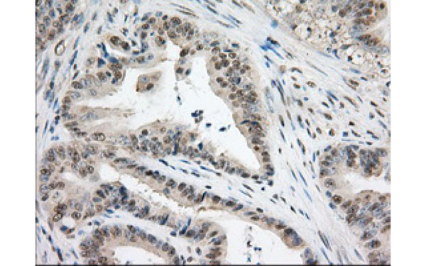
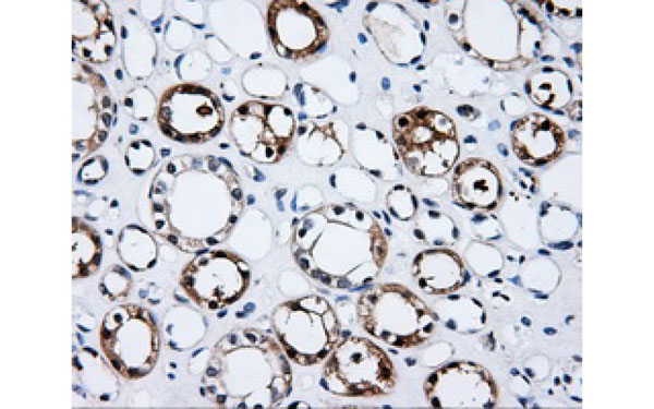
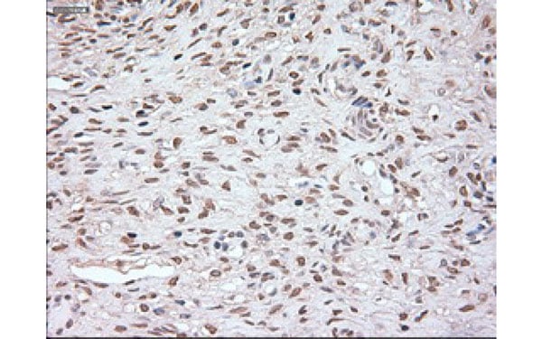
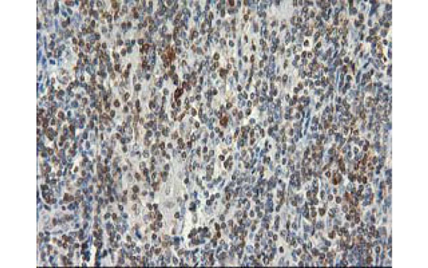
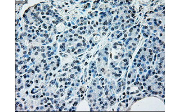
 IHC (Immunohistochemisry)
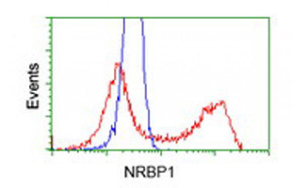
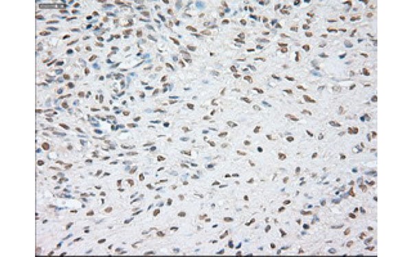
(Immunohistochemical analysis of NRBP1 protein in paraffin embedded Carcinoma of Human thyroid tissue using NRBP1 antibody)
IHC (Immunohistochemisry)
(Immunohistochemical analysis of NRBP1 protein in paraffin embedded Carcinoma of Human thyroid tissue using NRBP1 antibody)
NRBP1, Monoclonal Antibody (Cat# AAA108109)
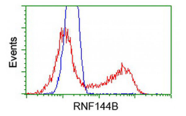
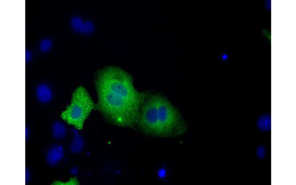
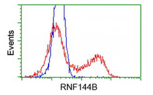
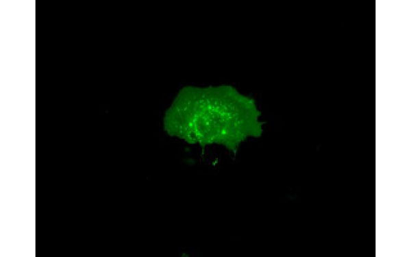 IF (Immunofluorescence)
(Immunofluorescent staining of COS7 cells transiently transfected with recombinant RNF144B protein using RNF144B antibody)
IF (Immunofluorescence)
(Immunofluorescent staining of COS7 cells transiently transfected with recombinant RNF144B protein using RNF144B antibody)
RNF144B, Monoclonal Antibody (Cat# AAA108117)
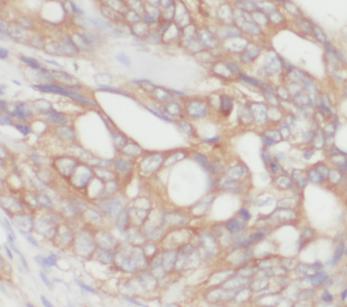
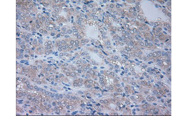
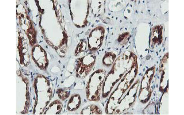
 IHC (Immunohistochemisry)
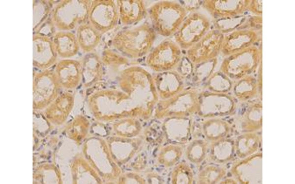
(Immunohistochemical analysis of PRKAR2A protein in paraffin embedded Human kidney tissue using PRKAR2A antibody)
IHC (Immunohistochemisry)
(Immunohistochemical analysis of PRKAR2A protein in paraffin embedded Human kidney tissue using PRKAR2A antibody)
PRKAR2A, Monoclonal Antibody (Cat# AAA108141)
 IF (Immunofluorescence)
(Immunofluorescent staining of COS7 cells transiently transfected with recombinant COASY protein using COASY antibody)
IF (Immunofluorescence)
(Immunofluorescent staining of COS7 cells transiently transfected with recombinant COASY protein using COASY antibody)
COASY, Monoclonal Antibody (Cat# AAA108154)
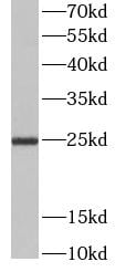
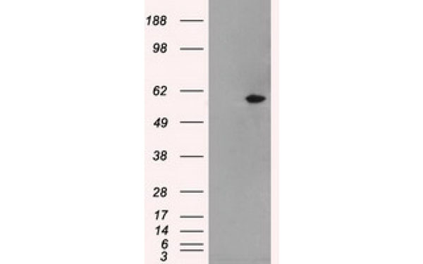
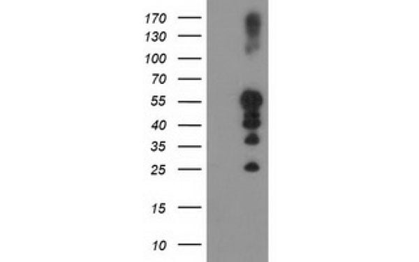
 WB (Western Blot)
(Western Blot analysis of HEK293T cell lysates (5 ug) transfected with either recombinant MPP3 protein (Right) or empty vector (Left) detected with MPP3 antibody)
WB (Western Blot)
(Western Blot analysis of HEK293T cell lysates (5 ug) transfected with either recombinant MPP3 protein (Right) or empty vector (Left) detected with MPP3 antibody)
MPP3, Monoclonal Antibody (Cat# AAA108155)
 IF (Immunofluorescence)
(Immunofluorescent staining of COS7 cells transiently transfected with recombinant TACC3 protein using TACC3 antibody)
IF (Immunofluorescence)
(Immunofluorescent staining of COS7 cells transiently transfected with recombinant TACC3 protein using TACC3 antibody)
TACC3, Monoclonal Antibody (Cat# AAA108168)
hCG alpha, Monoclonal Antibody (Cat# AAA108172)
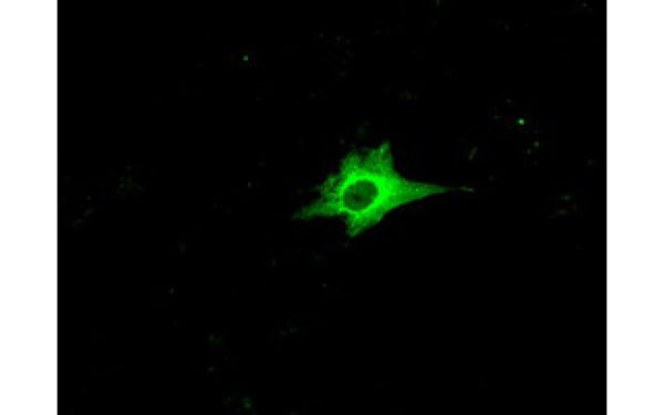 IF (Immunofluorescence)
(Immunofluorescent staining of COS7 cells transiently transfected with recombinant RNF144B protein using RNF144B antibody)
IF (Immunofluorescence)
(Immunofluorescent staining of COS7 cells transiently transfected with recombinant RNF144B protein using RNF144B antibody)
RNF144B, Monoclonal Antibody (Cat# AAA108186)
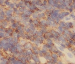
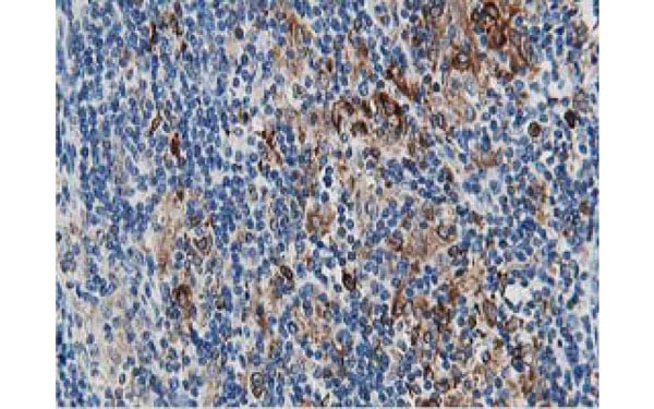
 IHC (Immunohistochemisry)
(Immunohistochemical analysis of GOLM1 protein in paraffin embedded Carcinoma of Human lung tissue using GOLM1 antibody)
IHC (Immunohistochemisry)
(Immunohistochemical analysis of GOLM1 protein in paraffin embedded Carcinoma of Human lung tissue using GOLM1 antibody)
GOLM1, Monoclonal Antibody (Cat# AAA108194)
 IF (Immunofluorescence)
(Immunofluorescent staining of COS7 cells transiently transfected with recombinant PSMC3 protein using PSMC3 antibody)
IF (Immunofluorescence)
(Immunofluorescent staining of COS7 cells transiently transfected with recombinant PSMC3 protein using PSMC3 antibody)
PSMC3, Monoclonal Antibody (Cat# AAA108195)
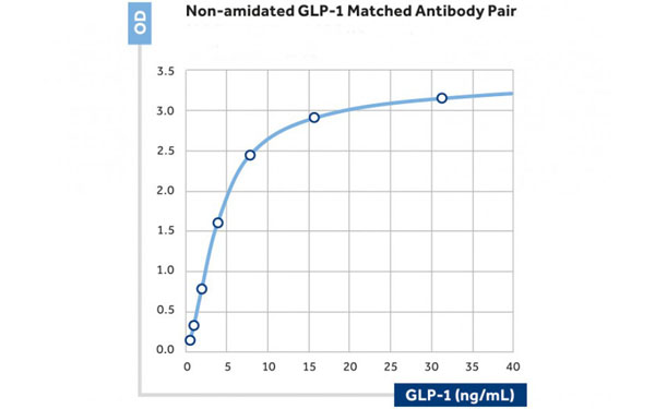
 IA (Immunoassay)
(The calibration curve of a sandwich assay showing measurement of "active" GLP-1(7-36)amide using as capture antibody and AAA108196 as the biotinylated detection antibody.)
IA (Immunoassay)
(The calibration curve of a sandwich assay showing measurement of "active" GLP-1(7-36)amide using as capture antibody and AAA108196 as the biotinylated detection antibody.)
GLP1, Monoclonal Antibody (Cat# AAA108196)
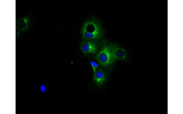
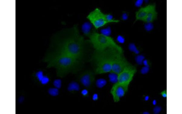
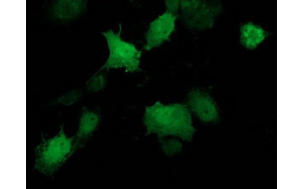
 IF (Immunofluorescence)
(Immunofluorescent staining of COS7 cells transiently transfected with recombinant PTPN1 protein using PTPN1 antibody)
IF (Immunofluorescence)
(Immunofluorescent staining of COS7 cells transiently transfected with recombinant PTPN1 protein using PTPN1 antibody)
PTPN1, Monoclonal Antibody (Cat# AAA108207)
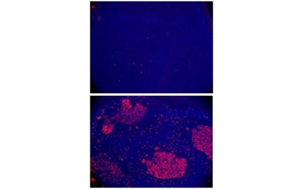
 IF (Immunofluorescence)
(Immunofluorescent staining of AID knock-out (top) or wild type (bottom) mouse intestinal Peyer's patches with AID antibody (AAA107965))
IF (Immunofluorescence)
(Immunofluorescent staining of AID knock-out (top) or wild type (bottom) mouse intestinal Peyer's patches with AID antibody (AAA107965))
AID, Monoclonal Antibody (Cat# AAA107965)
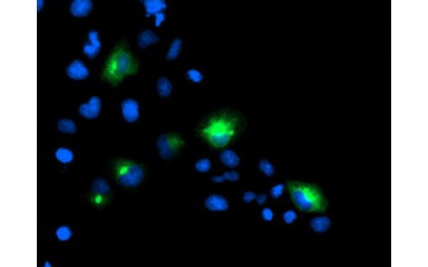
 IF (Immunofluorescence)
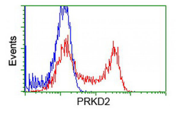
(Immunofluorescent staining of COS7 cells transiently transfected with recombinant PRKD2 protein using PRKD2 antibody)
IF (Immunofluorescence)
(Immunofluorescent staining of COS7 cells transiently transfected with recombinant PRKD2 protein using PRKD2 antibody)
PRKD2, Monoclonal Antibody (Cat# AAA107970)
 IF (Immunofluorescence)
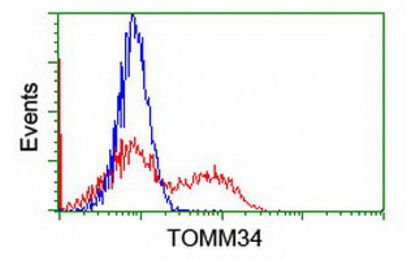
(Immunofluorescent staining of COS7 cells transiently transfected with recombinant TOMM34 protein using TOMM34 antibody)
IF (Immunofluorescence)
(Immunofluorescent staining of COS7 cells transiently transfected with recombinant TOMM34 protein using TOMM34 antibody)
TOMM34, Monoclonal Antibody (Cat# AAA107973)
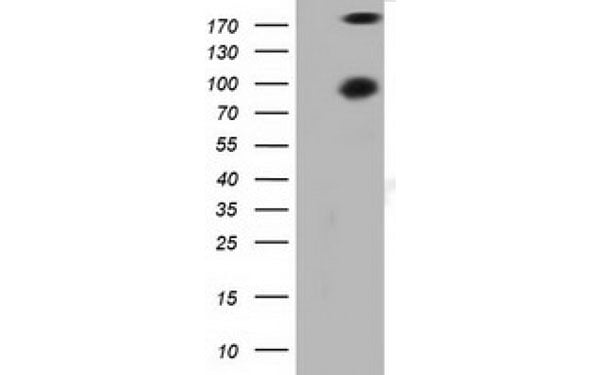
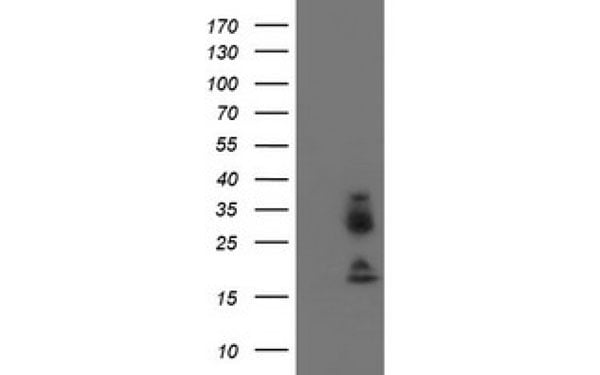
 WB (Western Blot)
(Western Blot analysis of HEK293T cell lysates (5 ug) transfected with either recombinant VASP protein (Right) or empty vector (Left) detected with VASP antibody)
WB (Western Blot)
(Western Blot analysis of HEK293T cell lysates (5 ug) transfected with either recombinant VASP protein (Right) or empty vector (Left) detected with VASP antibody)
VASP, Monoclonal Antibody (Cat# AAA107976)
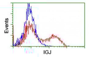
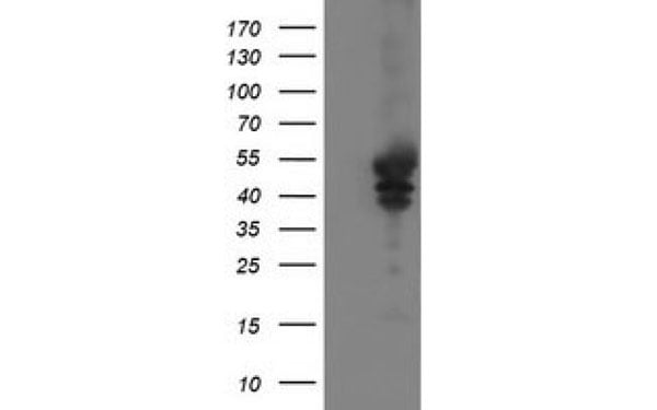
 WB (Western Blot)
(Western Blot analysis of HEK293T cell lysates (5 ug) transfected with either recombinant IGJ protein (Right) or empty vector (Left) detected with IGJ antibody)
WB (Western Blot)
(Western Blot analysis of HEK293T cell lysates (5 ug) transfected with either recombinant IGJ protein (Right) or empty vector (Left) detected with IGJ antibody)
IGJ, Monoclonal Antibody (Cat# AAA107985)
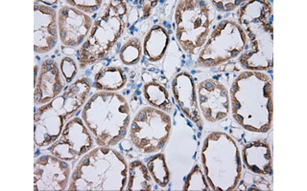
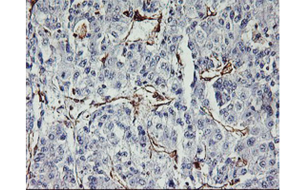
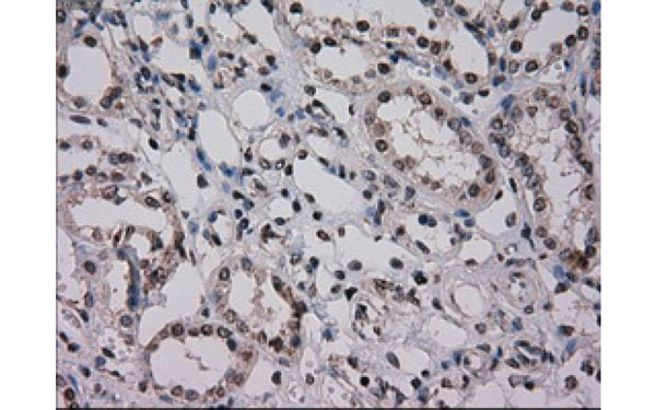 IHC (Immunohistochemisry)
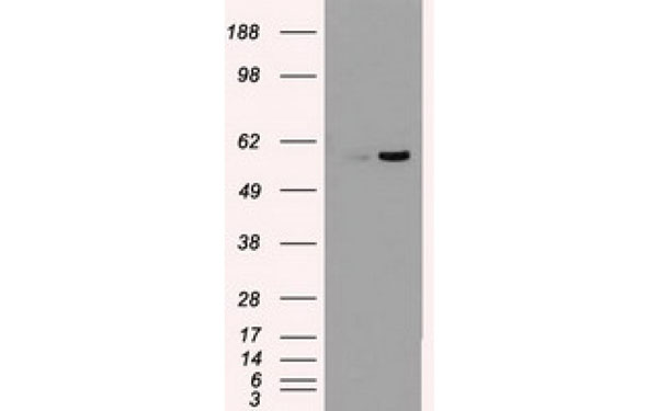
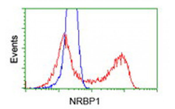
(Immunohistochemical analysis of NRBP1 protein in paraffin embedded Human Kidney tissue using NRBP1 antibody)
IHC (Immunohistochemisry)
(Immunohistochemical analysis of NRBP1 protein in paraffin embedded Human Kidney tissue using NRBP1 antibody)
NRBP1, Monoclonal Antibody (Cat# AAA107993)
 IF (Immunofluorescence)
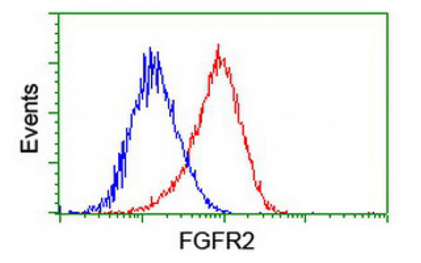
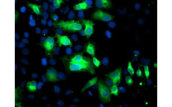
(Immunofluorescent staining of COS7 cells transiently transfected with recombinant FGFR2 protein using FGFR2 antibody)
IF (Immunofluorescence)
(Immunofluorescent staining of COS7 cells transiently transfected with recombinant FGFR2 protein using FGFR2 antibody)
FGFR2, Monoclonal Antibody (Cat# AAA108221)
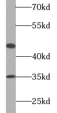
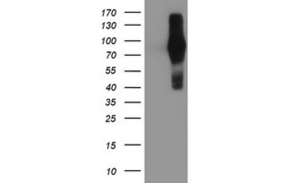
 WB (Western Blot)
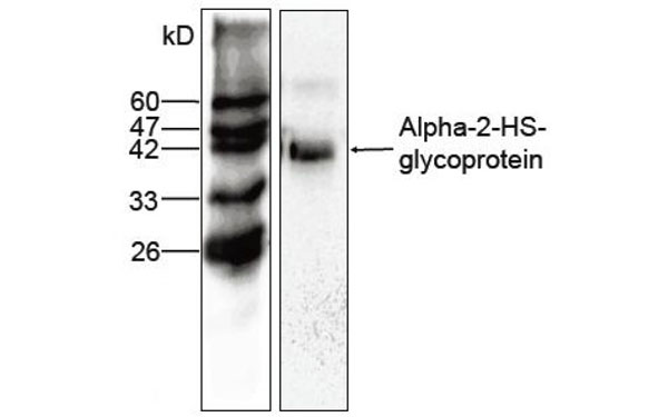
(Western blot (1:1000) analysis of Alpha-2-HS-glycoprotein expression in HepG2 whole cell lysate with Alpha-2-HS-glycoprotein antibody)
WB (Western Blot)
(Western blot (1:1000) analysis of Alpha-2-HS-glycoprotein expression in HepG2 whole cell lysate with Alpha-2-HS-glycoprotein antibody)
AHSG, Monoclonal Antibody (Cat# AAA108228)
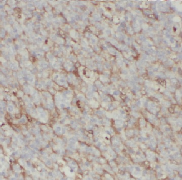
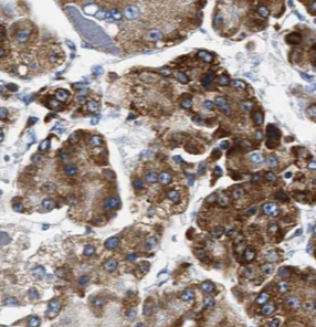
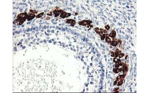
 IHC (Immunohistochemisry)
(Immunohistochemical analysis of NOG protein in paraffin embedded Human ovary tissue using NOG antibody)
IHC (Immunohistochemisry)
(Immunohistochemical analysis of NOG protein in paraffin embedded Human ovary tissue using NOG antibody)
NOG, Monoclonal Antibody (Cat# AAA108244)
Poliovirus Type 1, Monoclonal Antibody (Cat# AAA108254)
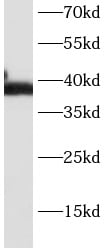
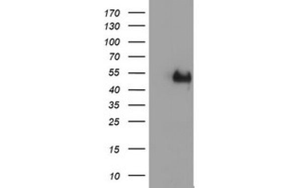
 WB (Western Blot)
(Western Blot analysis of HEK293T cell lysates (5 ug) transfected with either recombinant MAPK7 protein (Right) or empty vector (Left) detected with MAPK7 antibody)
WB (Western Blot)
(Western Blot analysis of HEK293T cell lysates (5 ug) transfected with either recombinant MAPK7 protein (Right) or empty vector (Left) detected with MAPK7 antibody)
MAPK7, Monoclonal Antibody (Cat# AAA108264)
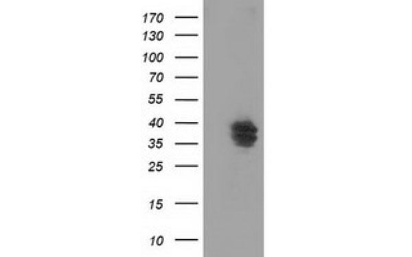
 WB (Western Blot)
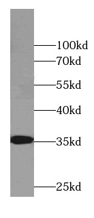
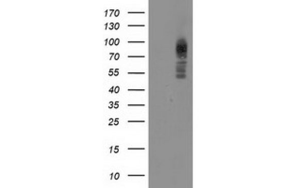
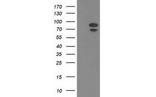
(Western Blot analysis of HEK293T cell lysates (5 ug) transfected with either recombinant PRKCE protein (Right) or empty vector (Left) detected with PRKCE antibody)
WB (Western Blot)
(Western Blot analysis of HEK293T cell lysates (5 ug) transfected with either recombinant PRKCE protein (Right) or empty vector (Left) detected with PRKCE antibody)
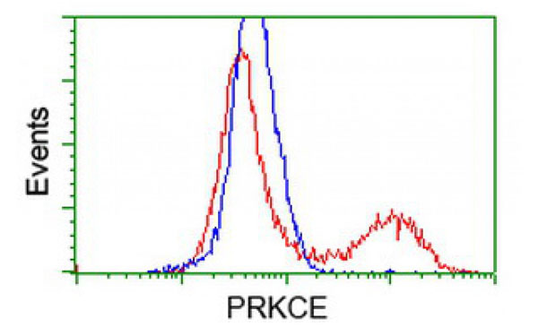
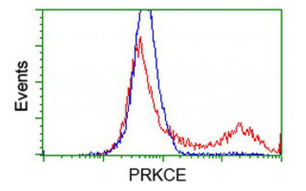
PRKCE, Monoclonal Antibody (Cat# AAA108269)
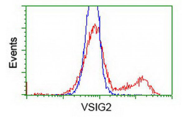
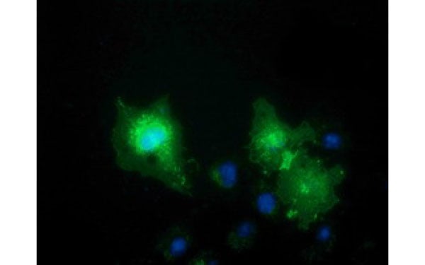 IF (Immunofluorescence)
(Immunofluorescent staining of COS7 cells transiently transfected with recombinant VSIG2 protein using VSIG2 antibody)
IF (Immunofluorescence)
(Immunofluorescent staining of COS7 cells transiently transfected with recombinant VSIG2 protein using VSIG2 antibody)
VSIG2, Monoclonal Antibody (Cat# AAA108274)
 IF (Immunofluorescence)
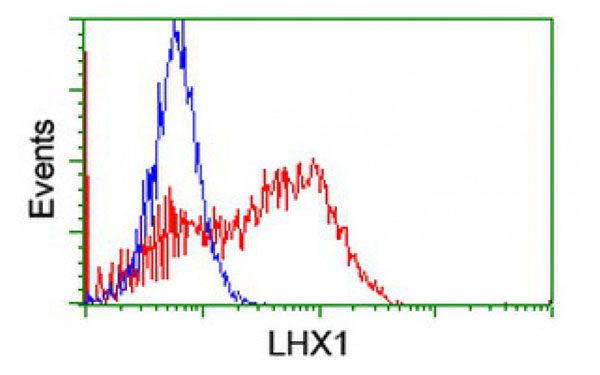
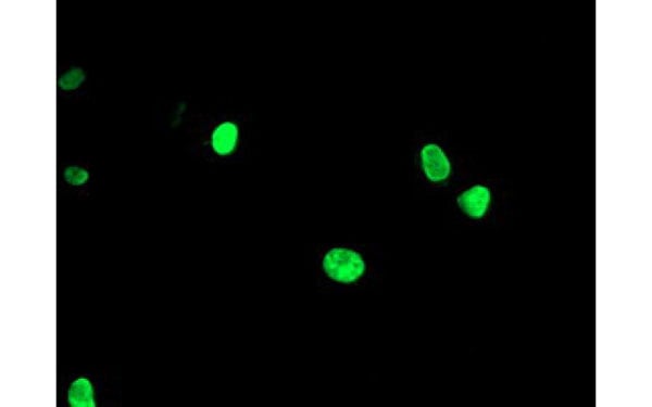
(Immunofluorescent staining of COS7 cells transiently transfected with recombinant LHX1 protein using LHX1 antibody)
IF (Immunofluorescence)
(Immunofluorescent staining of COS7 cells transiently transfected with recombinant LHX1 protein using LHX1 antibody)
LHX1, Monoclonal Antibody (Cat# AAA108275)
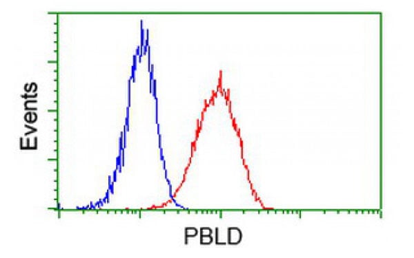
 IF (Immunofluorescence)
(Immunofluorescent staining of COS7 cells transiently transfected with recombinant PBLD protein using PBLD antibody)
IF (Immunofluorescence)
(Immunofluorescent staining of COS7 cells transiently transfected with recombinant PBLD protein using PBLD antibody)
PBLD, Monoclonal Antibody (Cat# AAA108278)
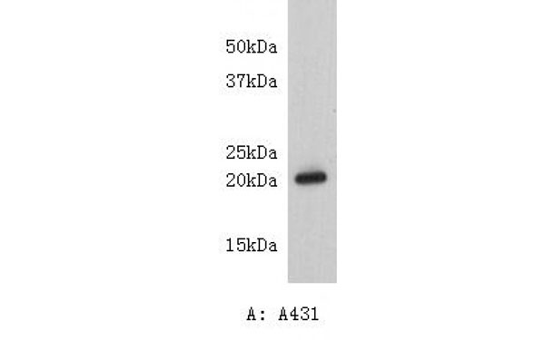
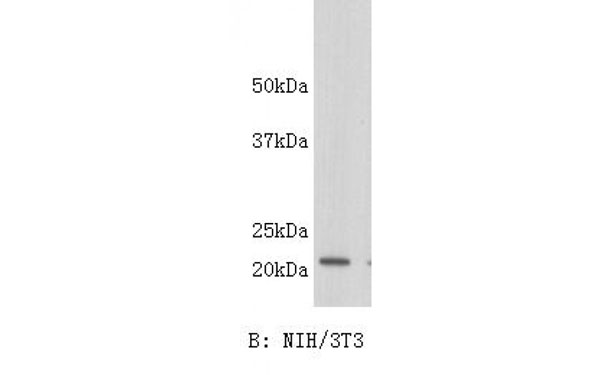 WB (Western Blot)
(Western blot analysis on A431or NIH/3T3 using anti-Caveolin-1 monoclonal antibody.)
WB (Western Blot)
(Western blot analysis on A431or NIH/3T3 using anti-Caveolin-1 monoclonal antibody.)
Caveolin, Monoclonal Antibody (Cat# AAA108292)
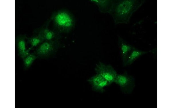
 IF (Immunofluorescence)
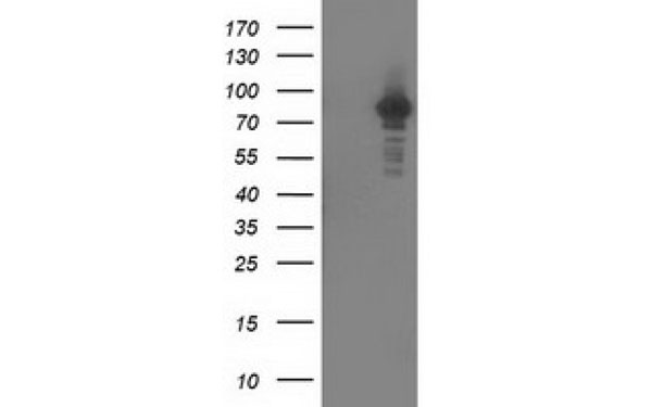
(Immunofluorescent staining of COS7 cells transiently transfected with recombinant PRKCE protein using PRKCE antibody)
IF (Immunofluorescence)
(Immunofluorescent staining of COS7 cells transiently transfected with recombinant PRKCE protein using PRKCE antibody)
PRKCE, Monoclonal Antibody (Cat# AAA108008)
 IF (Immunofluorescence)
(Immunofluorescent staining of COS7 cells transiently transfected with recombinant STAT5A protein using STAT5A antibody)
IF (Immunofluorescence)
(Immunofluorescent staining of COS7 cells transiently transfected with recombinant STAT5A protein using STAT5A antibody)
STAT5A, Monoclonal Antibody (Cat# AAA108009)
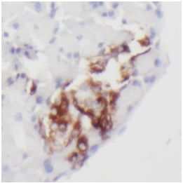
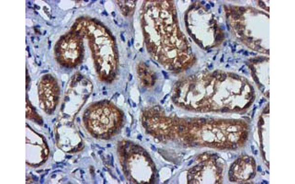
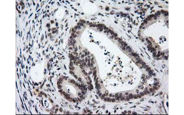
 IHC (Immunohistochemisry)
(Immunohistochemical analysis of NRBP1 protein in paraffin embedded Human pancreas tissue using NRBP1 antibody)
IHC (Immunohistochemisry)
(Immunohistochemical analysis of NRBP1 protein in paraffin embedded Human pancreas tissue using NRBP1 antibody)
NRBP1, Monoclonal Antibody (Cat# AAA108031)
 IHC (Immunohistochemisry)
(Immunohistochemical analysis of NOG protein in paraffin embedded Human ovary tissue using NOG antibody)
IHC (Immunohistochemisry)
(Immunohistochemical analysis of NOG protein in paraffin embedded Human ovary tissue using NOG antibody)
NOG, Monoclonal Antibody (Cat# AAA108050)
 IHC (Immunohistochemisry)
(Immunohistochemical analysis of PTPN7 protein in paraffin embedded Adenocarcinoma of Human ovary tissue using PTPN7 antibody)
IHC (Immunohistochemisry)
(Immunohistochemical analysis of PTPN7 protein in paraffin embedded Adenocarcinoma of Human ovary tissue using PTPN7 antibody)
PTPN7, Monoclonal Antibody (Cat# AAA108053)
What are Monoclonal Antibodies?
Monoclonal antibodies are specialized laboratory-produced proteins developed for binding to specific biological antigens or other molecular targets. Since they come from a single cell (or clone), they are especially consistent and accurate in the data they are involved in producing.
This type of antibody material has been shown to be a powerful tool in finding and subsequently destroying harmful cells in an organism, such as those found in cancers or various autoimmune diseases. This makes them excellent aids in medical testing and research, which is why they are so widely used.
AAA Biotech offers a comprehensive range of high-quality monoclonal antibodies that perform effectively in various laboratory tests, including (amongst others) ELISA, western blotting, immunohistochemistry, and flow cytometry. All of the products in our catalog are thoroughly quality tested to make sure that they are reliable and will consistently perform well in your research.
What Are The Uses of Monoclonal Antibodies
Monoclonal antibodies are used in many lab tests, including (amongst others) ELISA, western blotting, immunohistochemistry, and flow cytometry.
ELISA is a test that helps detect a specific substance/analyte in a sample. It uses antibodies (often monoclonal) bound to a solid surface (such as the well of a microplate) to “capture” the substance/analyte in the sample and immobilize it so that the detection antibody component can then bind to it and produce a signal, which can then be measured.
Western blotting identifies specific proteins in a sample. The sample is first separated on a gel, and then antibodies are applied that will typically bind to the target, which will all be localized to a single band in a lane.
Immunohistochemistry helps locate specific proteins in cells or tissue samples using antibodies.
Flow cytometry looks at and sorts cells. It uses antibodies that are conjugated to reporter molecules called “fluorophores”, which, under special lights, emit light themselves, which can then be measured by a detector instrument.
How Monoclonal Antibodies Are Used as Medicine?
Please note that all of the products listed in AAA Biotech’s also known as AAA Bio or AAABio catalog are strictly for research-use only (RUO).
Monoclonal antibodies can also be used as therapeutic/medical treatments, particularly in the context of cancers. They are designed to find and bind to specific cells or proteins, helping the immune system recognize and attack the cancer. These treatments work in different ways, such as:
- Radioimmunotherapy attaches a small amount of radioactive molecule to the antibody, so it delivers the radiation directly to the cancer cells that the antibody is specifically binding to.
- Antibody-directed enzyme prodrug therapy uses antibodies that are specifically bound to special enzymes. These enzymes activate a harmless drug in the body and turn it into a cancer-killing drug only near the cancer cells—this helps avoid harming healthy cells.
- Immunoliposomes are tiny “bubbles” filled with medicine/drug and coated with antibodies. They carry the drug straight to the cancer cells.
Why Buy Monoclonal Antibodies From Us?
At AAA Biotech, we provide high-performance monoclonal antibodies designed to support a wide range of research needs.
1. Validated for Versatile Applications
The antibodies in our catalog are extensively validated and compatible with multiple techniques, including (but not limited to) ELISA, flow cytometry (FC), immunocytochemistry (ICC), immunofluorescence (IF), immunohistochemistry (IHC), immunoprecipitation (IP), and western blotting (WB).
2. Wide Selection & Specialized Options
We offer antibodies for common and rare species, that are available in various conjugated forms, and also in recombinant formats. Essentially, there is almost anything one might need to meet their experimental model’s requirements.
3. High-Quality Proteins
Our proteins meet high purity standards—90% or more as confirmed by SDS-PAGE. Many are available with tags like His, Flag, GST, or MBP, and we also supply native and biologically active proteins for functional studies.
Frequently Asked Questions
1. Are your monoclonal antibodies validated for specific applications?
Yes, our antibodies are tested and validated for use in methods such as ELISA, western blot, IHC, flow cytometry, and more. Refer to specific product pages or datasheets for individual product information.
2. How do I choose the right monoclonal antibody for my application?
Review the product details directly for application validation, species reactivity, and target information. You may also contact our support team at any time for help.
3. How quickly can I receive my order?
Most orders are processed and shipped within 1–3 business days, depending on product availability and your shipping location.