Filters
▼Clonality
▼Type
▼Reactivity
▼Gene Name
▼Isotype
▼Host
▼Application
▼Clone
▼Monoclonal Antibodies
Get accurate results in your research with our Monoclonal Antibodies, which are specially made to target exactly what you require for your research, and will produce consistent, reliable performance in lab tests.
Viewing 5600-5650 of 27597 product results
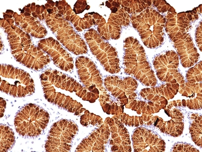
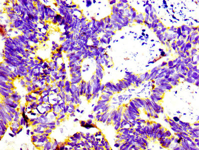
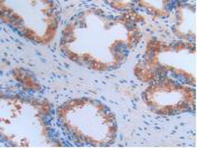
 IHC (Immunohiostchemistry)
(Immunohistochemical analysis of BSG protein in paraffin embedded Adenocarcinoma of Human colon tissue using BSG antibody)
IHC (Immunohiostchemistry)
(Immunohistochemical analysis of BSG protein in paraffin embedded Adenocarcinoma of Human colon tissue using BSG antibody)
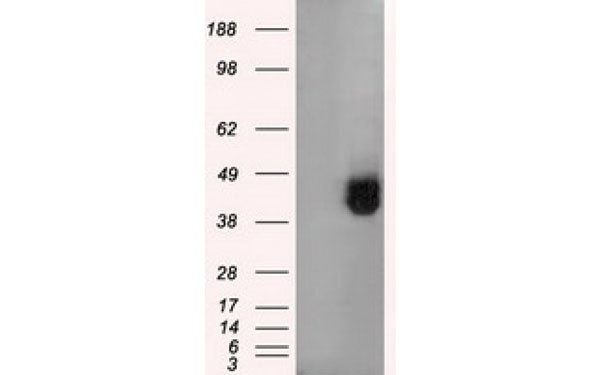
BSG, Monoclonal Antibody (Cat# AAA74716)
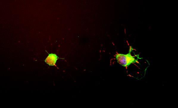
 IF (Immunofluorescence)
(Immunofluorescent staining of COS7 cells transiently transfected with recombinant FHL1 protein using FHL1 antibody)
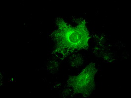
IF (Immunofluorescence)
(Immunofluorescent staining of COS7 cells transiently transfected with recombinant FHL1 protein using FHL1 antibody)
FHL1, Monoclonal Antibody (Cat# AAA74723)
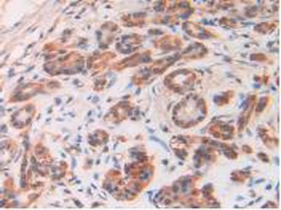
 IHC (Immunohiostchemistry)
(Immunohistochemical analysis of FKBP5 protein in paraffin embedded Human pancreas tissue using FKBP5 antibody)
IHC (Immunohiostchemistry)
(Immunohistochemical analysis of FKBP5 protein in paraffin embedded Human pancreas tissue using FKBP5 antibody)
FKBP5, Monoclonal Antibody (Cat# AAA74907)
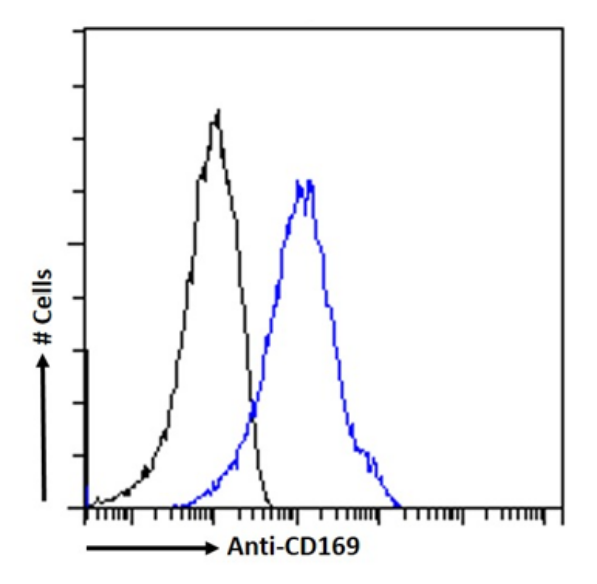
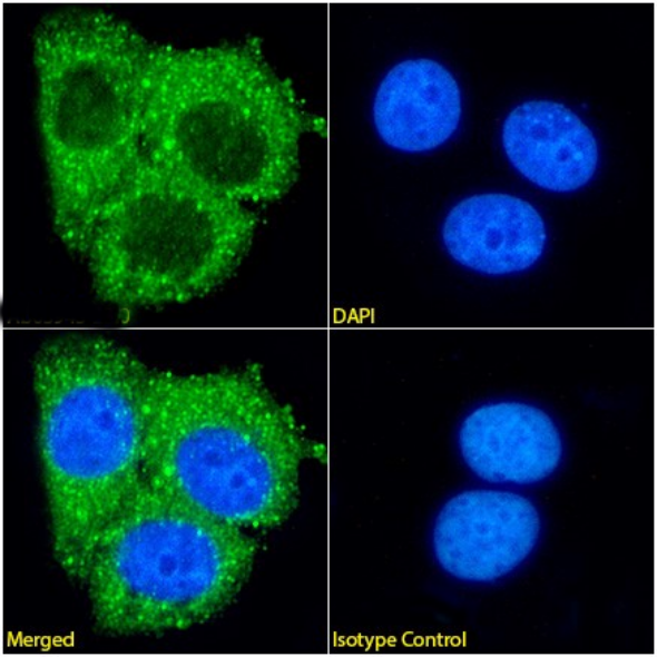
 IF (Immunofluorescence)
(ImmunofluorescencestainingofRAW264.7cellswithanti-CD169(AAA72426)SER-4(recombinantversion) ImmunofluorescenceanalysisofparaformaldehydefixedRAW264.7cellsstainedwiththechimericrabbitIgGversionofSER-4(recombinantversion)(AAA72426)at10ug/mlfor1hfollowedbyAlexaFluor488secondaryantibody(2ug/ml),showingmembranestaining.ThenuclearstainisDAPI(blue).Panelsshowfromleft-right,top-bottomAAA72426,DAPI,mergedchannelsandanisotypecontrol.TheisotypecontrolwasanunknownspecificityantibodyfollowedbystainingwithAlexaFluor488secondaryantibody.)
IF (Immunofluorescence)
(ImmunofluorescencestainingofRAW264.7cellswithanti-CD169(AAA72426)SER-4(recombinantversion) ImmunofluorescenceanalysisofparaformaldehydefixedRAW264.7cellsstainedwiththechimericrabbitIgGversionofSER-4(recombinantversion)(AAA72426)at10ug/mlfor1hfollowedbyAlexaFluor488secondaryantibody(2ug/ml),showingmembranestaining.ThenuclearstainisDAPI(blue).Panelsshowfromleft-right,top-bottomAAA72426,DAPI,mergedchannelsandanisotypecontrol.TheisotypecontrolwasanunknownspecificityantibodyfollowedbystainingwithAlexaFluor488secondaryantibody.)
CD169, Monoclonal Recombinant Antibody (Cat# AAA72426)

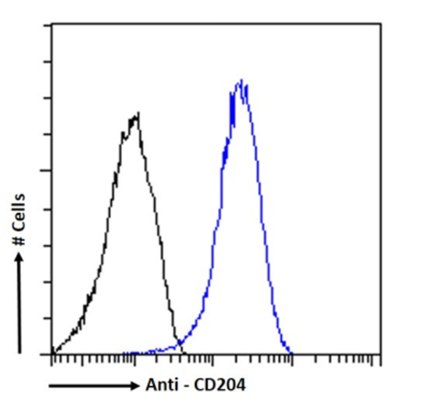

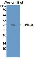
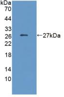
 WB (Western Blot)
(Western Blotusinganti-CD204antibody2F8(recombinantversion)(AAA72428). RAW264.7celllysateof(35ugproteininRIPAbuffer)wereresolvedonaSDSPAGEgelandblotswereprobedwiththechimericrabbitversionof2F8(recombinantversion)(AAA72428)0.3ug/mlbeforedetectionusingananti-rabbitsecondaryantibody.Aprimaryincubationof1hwasusedandproteinwasdetectedbychemiluminescence.)
WB (Western Blot)
(Western Blotusinganti-CD204antibody2F8(recombinantversion)(AAA72428). RAW264.7celllysateof(35ugproteininRIPAbuffer)wereresolvedonaSDSPAGEgelandblotswereprobedwiththechimericrabbitversionof2F8(recombinantversion)(AAA72428)0.3ug/mlbeforedetectionusingananti-rabbitsecondaryantibody.Aprimaryincubationof1hwasusedandproteinwasdetectedbychemiluminescence.)
CD204, Monoclonal Recombinant Antibody (Cat# AAA72428)
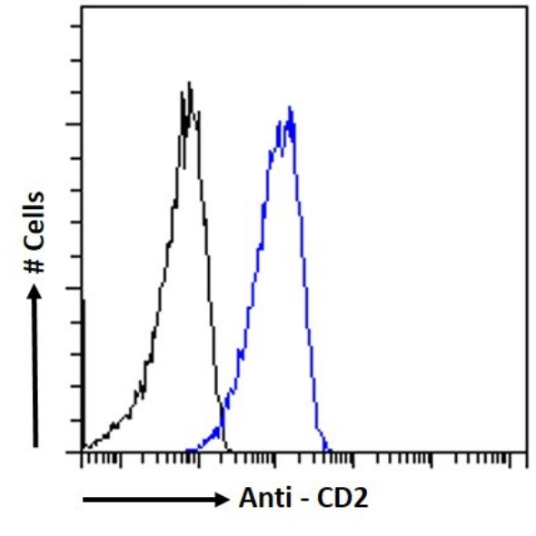
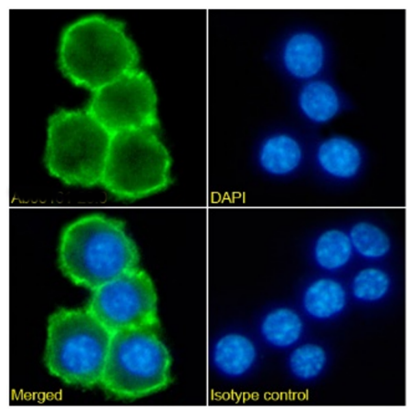
 IF (Immunofluorescence)
(ImmunofluorescencestainingofJurkatcellswithanti-CD2antibodyVIPVIIIC8(AAA72509). ImmunofluorescenceanalysisofparaformaldehydefixedJurkatcellsonShi-fixcoverslipsstainedwiththechimericrabbitIgGversionofVIPVIIIC8(AAA72509)(1:100dilution)for1hfollowedbyAlexaFluor488secondaryantibody(1:1000dilution),showingmembranestaining.ThenuclearstainisDAPI(blue).Panelsshow,fromleft-right,top-bottom,AAA72509,DAPI,mergedchannelsandanisotypecontrol.TheisotypecontrolwasanunknownspecificityantibodyfollowedbystainingwithAlexaFluor488secondaryantibody.)
IF (Immunofluorescence)
(ImmunofluorescencestainingofJurkatcellswithanti-CD2antibodyVIPVIIIC8(AAA72509). ImmunofluorescenceanalysisofparaformaldehydefixedJurkatcellsonShi-fixcoverslipsstainedwiththechimericrabbitIgGversionofVIPVIIIC8(AAA72509)(1:100dilution)for1hfollowedbyAlexaFluor488secondaryantibody(1:1000dilution),showingmembranestaining.ThenuclearstainisDAPI(blue).Panelsshow,fromleft-right,top-bottom,AAA72509,DAPI,mergedchannelsandanisotypecontrol.TheisotypecontrolwasanunknownspecificityantibodyfollowedbystainingwithAlexaFluor488secondaryantibody.)
CD2, Monoclonal Recombinant Antibody (Cat# AAA72509)
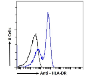
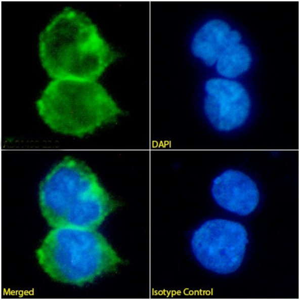
 IF (Immunofluorescence)
(ImmunofluorescencestainingofDaudicellswithanti-HLA-DRantibodyCAT13.3H3(AAA72511). ImmunofluorescenceanalysisofparaformaldehydefixedDaudicellsonShi-fixcoverslipsstainedwiththechimericrabbitIgGversionofCAT13.3H3(AAA72511)(1:100dilution)for1hfollowedbyAlexaFluor488secondaryantibody(1:1000dilution),showingmembraneandcytoplasmicstaining.ThenuclearstainisDAPI(blue).Panelsshow,fromleft-right,top-bottom,AAA72511,DAPI,mergedchannelsandanisotypecontrol.TheisotypecontrolwasanunknownspecificityantibodyfollowedbystainingwithAlexaFluor488secondaryantibody.)
IF (Immunofluorescence)
(ImmunofluorescencestainingofDaudicellswithanti-HLA-DRantibodyCAT13.3H3(AAA72511). ImmunofluorescenceanalysisofparaformaldehydefixedDaudicellsonShi-fixcoverslipsstainedwiththechimericrabbitIgGversionofCAT13.3H3(AAA72511)(1:100dilution)for1hfollowedbyAlexaFluor488secondaryantibody(1:1000dilution),showingmembraneandcytoplasmicstaining.ThenuclearstainisDAPI(blue).Panelsshow,fromleft-right,top-bottom,AAA72511,DAPI,mergedchannelsandanisotypecontrol.TheisotypecontrolwasanunknownspecificityantibodyfollowedbystainingwithAlexaFluor488secondaryantibody.)
HLA-DR, Monoclonal Recombinant Antibody (Cat# AAA72511)
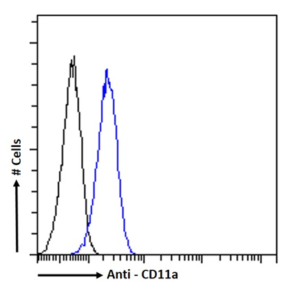

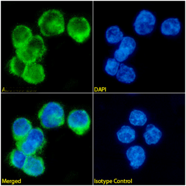
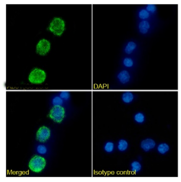 IF (Immunofluorescence)
(Immunofluorescencestainingofhumanperipheralbloodmonocyteswithanti-CD11a(AAA72515)VIPIIIB1 ImmunofluorescenceanalysisofparaformaldehydefixedhumanperipheralbloodmonocytesonShi-fixcoverslipsstainedwiththechimericrabbitIgGversionofVIPIIIB1(AAA72515)at10ug/mlfor1hfollowedbyAlexaFluor488secondaryantibody(2ug/ml),showingmembranestaining.ThenuclearstainisDAPI(blue).Panelsshowfromleft-right,top-bottomAAA72515,DAPI,mergedchannelsandanisotypecontrol.TheisotypecontrolwasanunknownspecificityantibodyfollowedbystainingwithAlexaFluor488secondaryantibody.)
IF (Immunofluorescence)
(Immunofluorescencestainingofhumanperipheralbloodmonocyteswithanti-CD11a(AAA72515)VIPIIIB1 ImmunofluorescenceanalysisofparaformaldehydefixedhumanperipheralbloodmonocytesonShi-fixcoverslipsstainedwiththechimericrabbitIgGversionofVIPIIIB1(AAA72515)at10ug/mlfor1hfollowedbyAlexaFluor488secondaryantibody(2ug/ml),showingmembranestaining.ThenuclearstainisDAPI(blue).Panelsshowfromleft-right,top-bottomAAA72515,DAPI,mergedchannelsandanisotypecontrol.TheisotypecontrolwasanunknownspecificityantibodyfollowedbystainingwithAlexaFluor488secondaryantibody.)
CD11a, Monoclonal Recombinant Antibody (Cat# AAA72515)
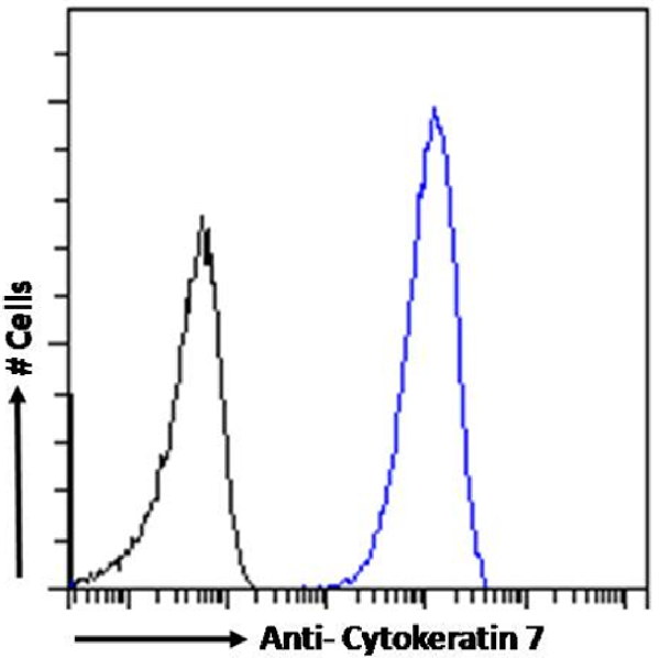 FCM/FACS (Flow Cytometry)
(Flow cytometry using the anti-Cytokeratin 7 antibody OV-TL 12/30 . HeLa cells were fixed using 2% PFA and stained with anti-unknown specificity antibody or the rabbit IgG1 version of OV-TL 12/30 at a dilution of 1:100 for 1h at RT. After washing, the bound antibody was detected using a goat anti-rabbit IgG AlexaFluor 488 antibody at a dilution of 1:1000 and cells analyzed using a FACSCanto flow-cytometer.)
FCM/FACS (Flow Cytometry)
(Flow cytometry using the anti-Cytokeratin 7 antibody OV-TL 12/30 . HeLa cells were fixed using 2% PFA and stained with anti-unknown specificity antibody or the rabbit IgG1 version of OV-TL 12/30 at a dilution of 1:100 for 1h at RT. After washing, the bound antibody was detected using a goat anti-rabbit IgG AlexaFluor 488 antibody at a dilution of 1:1000 and cells analyzed using a FACSCanto flow-cytometer.)
Cytokeratin 7, Monoclonal Antibody (Cat# AAA72181)
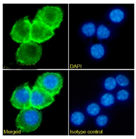
 IF (Immunofluorescence)
( Immunofluorescence staining of fixed mouse splenocytes with anti-PDL1 (CD274) antibody YDC 127.1.1 Immunofluorescence analysis of paraformaldehyde fixed mouse (Mus musculus) splenocytes on Shi-fix coverslips, permeabilized with 0.15% Triton stained with the chimeric rabbit version of YDC 127.1.1 at 10 ug/ml for 1h followed by Alexa Fluor 488 secondary antibody (1 ug/ml), showing membrane and some cytoplasmic staining in a subset of cells. The nuclear stain is DAPI (blue). Panels show from left-right, top-bottom DAPI, merged channels and an isotype control. The isotype control was stained with an anti-Fluorescein antibody followed by Alexa Fluor 488 secondary antibody.)
IF (Immunofluorescence)
( Immunofluorescence staining of fixed mouse splenocytes with anti-PDL1 (CD274) antibody YDC 127.1.1 Immunofluorescence analysis of paraformaldehyde fixed mouse (Mus musculus) splenocytes on Shi-fix coverslips, permeabilized with 0.15% Triton stained with the chimeric rabbit version of YDC 127.1.1 at 10 ug/ml for 1h followed by Alexa Fluor 488 secondary antibody (1 ug/ml), showing membrane and some cytoplasmic staining in a subset of cells. The nuclear stain is DAPI (blue). Panels show from left-right, top-bottom DAPI, merged channels and an isotype control. The isotype control was stained with an anti-Fluorescein antibody followed by Alexa Fluor 488 secondary antibody.)
PDL1, Monoclonal Recombinant Antibody (Cat# AAA71990)
 FCM/FACS (Flow Cytometry)
(Flow-cytometry using anti-CD8alpha/Lyt-2 antibody YTS 169.4 Mouse lymphocytes were stained with an anti-CD44 control or the rabbit-chimeric version of YTS 169.4 (AAA72017, panel B) at a concentration of 1 ug/ml for 30 mins at RT. After washing, bound antibody was detected using a AF488 conjugated donkey anti-rabbit antibody and cells analysed on a FACSCanto flow-cytometer.)
FCM/FACS (Flow Cytometry)
(Flow-cytometry using anti-CD8alpha/Lyt-2 antibody YTS 169.4 Mouse lymphocytes were stained with an anti-CD44 control or the rabbit-chimeric version of YTS 169.4 (AAA72017, panel B) at a concentration of 1 ug/ml for 30 mins at RT. After washing, bound antibody was detected using a AF488 conjugated donkey anti-rabbit antibody and cells analysed on a FACSCanto flow-cytometer.)
CD8 alpha/Lyt-2, Monoclonal Recombinant Antibody (Cat# AAA72017)
 WB (Western Blot)
(Western Blot using anti-Cardiac Troponin I antibody scFv 180 Human heart lysate (35ug protein in RIPA buffer) was resolved on a 10% SDS PAGE gel and blots probed with the chimeric mouse IgG1 version of scFv 180 at 0.001 ug/ml before detection using an anti-mouse secondary antibody. A primary incubation of 1h was used and protein was detected by chemiluminescence. The predicted running size for Cardiac Troponin I is 24.0 kDa. It successfully detected Cardiac Troponin I in human heart lysate.)
WB (Western Blot)
(Western Blot using anti-Cardiac Troponin I antibody scFv 180 Human heart lysate (35ug protein in RIPA buffer) was resolved on a 10% SDS PAGE gel and blots probed with the chimeric mouse IgG1 version of scFv 180 at 0.001 ug/ml before detection using an anti-mouse secondary antibody. A primary incubation of 1h was used and protein was detected by chemiluminescence. The predicted running size for Cardiac Troponin I is 24.0 kDa. It successfully detected Cardiac Troponin I in human heart lysate.)
Cardiac Troponin I, Monoclonal Recombinant Antibody (Cat# AAA72031)

 Application Data
(Binding curve of anti-Phl p 1 antibody Clone 25 to Phl p 1 ELISA Plate coated with Phl p 1 (RayBiotech, 228-22412) at a concentration of 2 ug/ml. A 3-fold serial dilution from 10,000 to 0.1 ng/ml was performed using AAA72039. For detection, a 1:4000 dilution of HRP-labelled goat anti-human kappa antibody (Bio-Rad) was used.)
Application Data
(Binding curve of anti-Phl p 1 antibody Clone 25 to Phl p 1 ELISA Plate coated with Phl p 1 (RayBiotech, 228-22412) at a concentration of 2 ug/ml. A 3-fold serial dilution from 10,000 to 0.1 ng/ml was performed using AAA72039. For detection, a 1:4000 dilution of HRP-labelled goat anti-human kappa antibody (Bio-Rad) was used.)
Phl p 1, Monoclonal Recombinant Antibody (Cat# AAA72039)
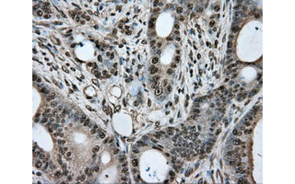
 IHC (Immunohiostchemistry)
(Formalin-fixed, paraffin-embedded human Gastric Carcinoma stained with MUC5AC Monoclonal Antibody (MUC5AC/917 + 45M1).)
IHC (Immunohiostchemistry)
(Formalin-fixed, paraffin-embedded human Gastric Carcinoma stained with MUC5AC Monoclonal Antibody (MUC5AC/917 + 45M1).)
MUC5AC (Mucin 5AC / Gastric Mucin), Monoclonal Antibody (Cat# AAA62900)
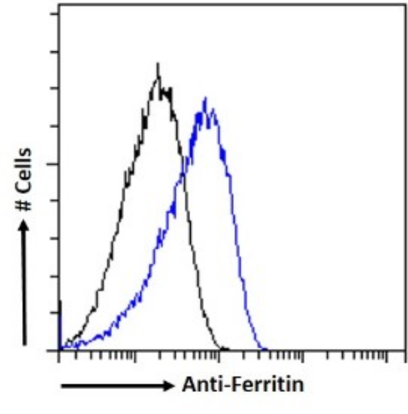 FCM/FACS (Flow Cytometry)
(Flowcytometryusinganti-FerritinantibodyF11(HSF102)(AAA72562). ParaformaldehydefixedMCF7cellspermeabilizedwith0.5%Tritonwerestainedwiththeanti-unknownspecificityantibodyortherabbitIgGversionofF11(HSF102)(AAA72562,blueline)atadilutionof1:100for1hatRT.Afterwashing,theboundantibodywasdetectedusingagoatanti-rabbitIgGAlexaFluor488antibodyatadilutionof1:1000,andthecellswereanalyzedusingaFACSCantoflow-cytometer.)
FCM/FACS (Flow Cytometry)
(Flowcytometryusinganti-FerritinantibodyF11(HSF102)(AAA72562). ParaformaldehydefixedMCF7cellspermeabilizedwith0.5%Tritonwerestainedwiththeanti-unknownspecificityantibodyortherabbitIgGversionofF11(HSF102)(AAA72562,blueline)atadilutionof1:100for1hatRT.Afterwashing,theboundantibodywasdetectedusingagoatanti-rabbitIgGAlexaFluor488antibodyatadilutionof1:1000,andthecellswereanalyzedusingaFACSCantoflow-cytometer.)
Ferritin, Monoclonal Recombinant Antibody (Cat# AAA72562)
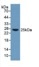
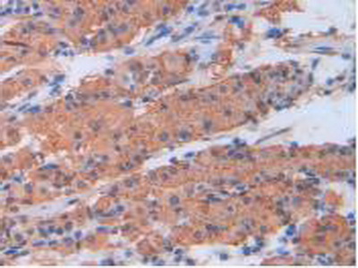
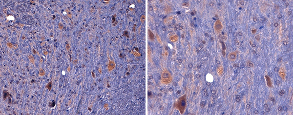 IHC (Immunohiostchemistry)
(CL7778AP (4ug/ml) staining of a rat brain formalin-fixed, paraffin-embedded tissue section; seen at 20x (left) and 40x (right) magnification. Strong staining observed in the cytoplasm of neuronal cells.)
IHC (Immunohiostchemistry)
(CL7778AP (4ug/ml) staining of a rat brain formalin-fixed, paraffin-embedded tissue section; seen at 20x (left) and 40x (right) magnification. Strong staining observed in the cytoplasm of neuronal cells.)
CD8a, Monoclonal Antibody (Cat# AAA74289)
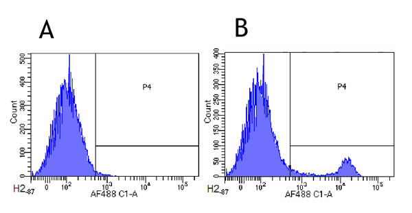
 FCM/FACS (Flow Cytometry)
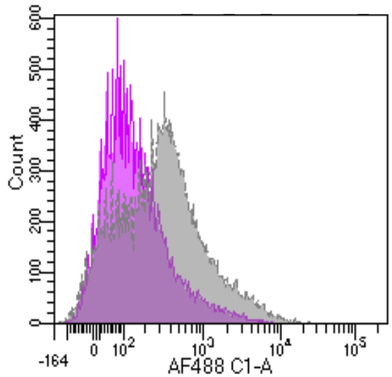
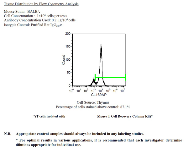
(C3H/HE mouse peritoneal macrophages (left) and bone marrow (right) were stained with anti-CD11b (clone: M1/70.15)(filled histogram) or rat IgG2b isotype control (open histogram).)
FCM/FACS (Flow Cytometry)
(C3H/HE mouse peritoneal macrophages (left) and bone marrow (right) were stained with anti-CD11b (clone: M1/70.15)(filled histogram) or rat IgG2b isotype control (open histogram).)
HLA-ABC, Monoclonal Antibody (Cat# AAA74301)
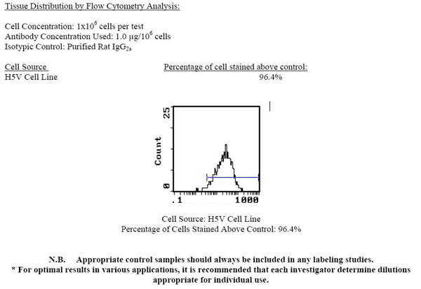
 Application Data
CD8a (Ly 2), Monoclonal Antibody (Cat# AAA74163)
 Application Data
CD34, Monoclonal Antibody (Cat# AAA74214)
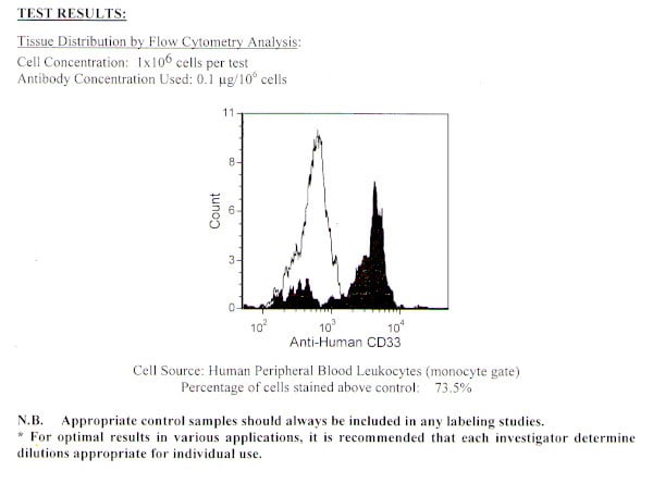 Application Data
CD33, Monoclonal Antibody (Cat# AAA74216)

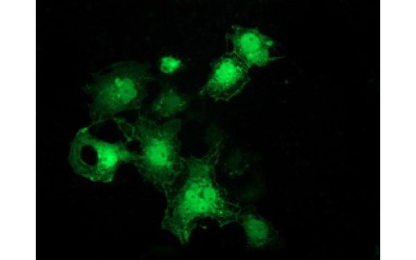
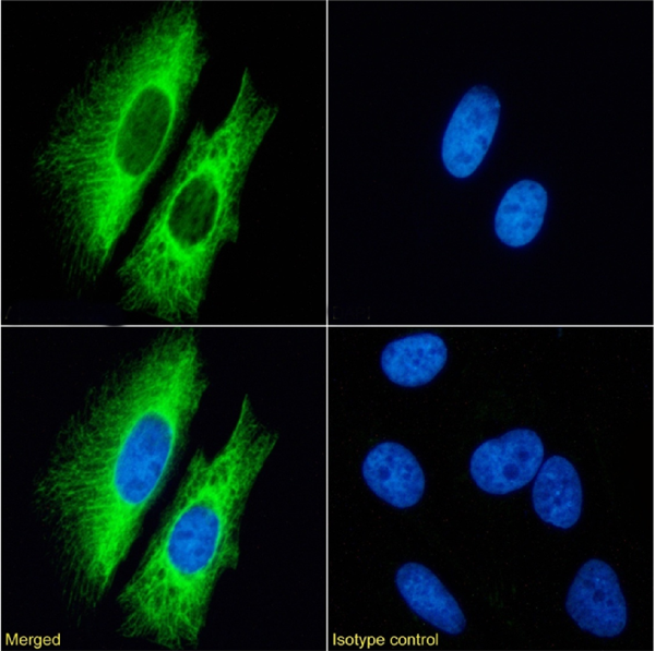
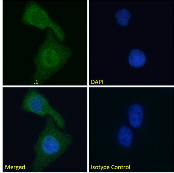
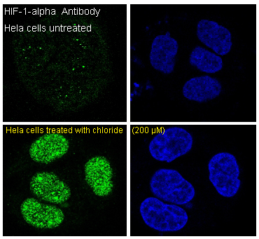 IF (Immunofluorescence)
(Immunofluorescent analysis of HeLa cells, using HIF-1 alpha Antibody.)
IF (Immunofluorescence)
(Immunofluorescent analysis of HeLa cells, using HIF-1 alpha Antibody.)
HIF-1 alpha, Monoclonal Antibody (Cat# AAA126854)
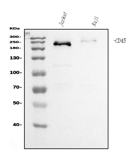
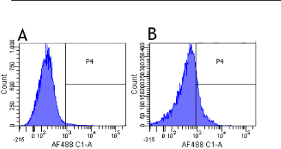
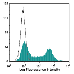
 FCM/FACS (Flow Cytometry)
(Figure 3. Flow Cytometry analysis of Raji cells using anti-CD45 antibody (AAA126872).Overlay histogram showing Raji cells stained with AAA126872 (Blue line). The cells were blocked with 10% normal goat serum. And then incubated with mouse anti-CD45 Antibody (AAA126872, 1 ug/1x10^6 cells) for 30 min at 20 degree C. DyLight488 conjugated goat anti-mouse IgG was used as secondary antibody for 30 minutes at 20 degree C. Isotype control antibody (Green line) was mouse IgG (1 ug/1x10^6) used under the same conditions. Unlabelled sample (Red line) was also used as a control.)
FCM/FACS (Flow Cytometry)
(Figure 3. Flow Cytometry analysis of Raji cells using anti-CD45 antibody (AAA126872).Overlay histogram showing Raji cells stained with AAA126872 (Blue line). The cells were blocked with 10% normal goat serum. And then incubated with mouse anti-CD45 Antibody (AAA126872, 1 ug/1x10^6 cells) for 30 min at 20 degree C. DyLight488 conjugated goat anti-mouse IgG was used as secondary antibody for 30 minutes at 20 degree C. Isotype control antibody (Green line) was mouse IgG (1 ug/1x10^6) used under the same conditions. Unlabelled sample (Red line) was also used as a control.)
CD45, Monoclonal Antibody (Cat# AAA126872)
 FCM/FACS (Flow Cytometry)
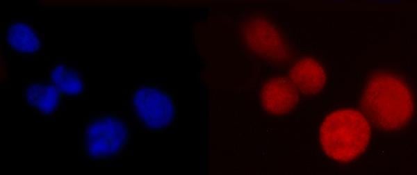
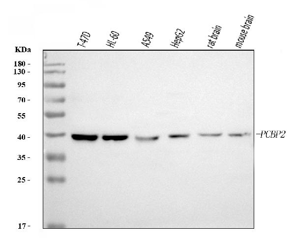
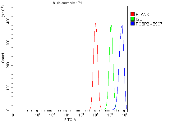
(Figure 3. Flow Cytometry analysis of PC-3 cells using anti-PCBP2/hnRNP E2 antibody (AAA126909).Overlay histogram showing PC-3 cells stained with AAA126909 (Blue line). The cells were blocked with 10% normal goat serum. And then incubated with mouse anti-PCBP2/hnRNP E2 Antibody (AAA126909, 1 ug/1x10^6 cells) for 30 min at 20 degree C. DyLight488 conjugated goat anti-mouse IgG was used as secondary antibody for 30 minutes at 20 degree C. Isotype control antibody (Green line) was mouse IgG (1 ug/1x10^6) used under the same conditions. Unlabelled sample (Red line) was also used as a control.)
FCM/FACS (Flow Cytometry)
(Figure 3. Flow Cytometry analysis of PC-3 cells using anti-PCBP2/hnRNP E2 antibody (AAA126909).Overlay histogram showing PC-3 cells stained with AAA126909 (Blue line). The cells were blocked with 10% normal goat serum. And then incubated with mouse anti-PCBP2/hnRNP E2 Antibody (AAA126909, 1 ug/1x10^6 cells) for 30 min at 20 degree C. DyLight488 conjugated goat anti-mouse IgG was used as secondary antibody for 30 minutes at 20 degree C. Isotype control antibody (Green line) was mouse IgG (1 ug/1x10^6) used under the same conditions. Unlabelled sample (Red line) was also used as a control.)
PCBP2/hnRNP E2, Monoclonal Antibody (Cat# AAA126909)


 WB (Western Blot)
(PCAF monoclonal antibody (M04), clone 5E11. Western Blot analysis of PCAF expression in LNCaP (Cat # L004V1).)
WB (Western Blot)
(PCAF monoclonal antibody (M04), clone 5E11. Western Blot analysis of PCAF expression in LNCaP (Cat # L004V1).)
PCAF, Monoclonal Antibody (Cat# AAA26606)
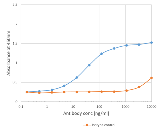
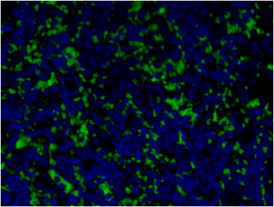
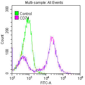 Application Data
(Overlay histogram showing Raji cells stained with AAA235192 (red line) at 1:50. The cells were fixed with 70% Ethylalcohol (18h) and then permeabilized with 0.3% Triton X-100 for 2 min.The cells were then incubated in 1x PBS /10% normal goat serum to block non-specific protein-protein interactions followed by primary antibody for 1 h at 4 degree C.The secondary antibody used was FITC goat anti-rabbit IgG (H+L) at 1/200 dilution for 1 h at 4 degree C. Control antibody (green line) was used under the same conditions. Acquisition of >10, 000 events was performed.)
Application Data
(Overlay histogram showing Raji cells stained with AAA235192 (red line) at 1:50. The cells were fixed with 70% Ethylalcohol (18h) and then permeabilized with 0.3% Triton X-100 for 2 min.The cells were then incubated in 1x PBS /10% normal goat serum to block non-specific protein-protein interactions followed by primary antibody for 1 h at 4 degree C.The secondary antibody used was FITC goat anti-rabbit IgG (H+L) at 1/200 dilution for 1 h at 4 degree C. Control antibody (green line) was used under the same conditions. Acquisition of >10, 000 events was performed.)
CD74, Monoclonal Recombinant Antibody (Cat# AAA235192)

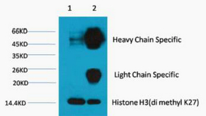
 IP (Immunoprecipitation)
(Dilution: WB: 1:1000-3000 IP:1:200)
IP (Immunoprecipitation)
(Dilution: WB: 1:1000-3000 IP:1:200)
Histone H3, Monoclonal Antibody (Cat# AAA293537)
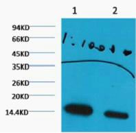
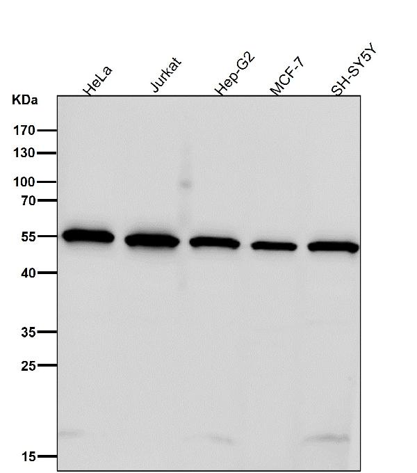
 WB (Western Blot)
(All lanes use the Antibody at 1:1W dilution for 1 hour at room temperature.)
WB (Western Blot)
(All lanes use the Antibody at 1:1W dilution for 1 hour at room temperature.)
GRB2, Monoclonal Antibody (Cat# AAA128115)
 WB (Western Blot)
(All lanes use the Antibody at 1:8K dilution for 1 hour at room temperature.)
WB (Western Blot)
(All lanes use the Antibody at 1:8K dilution for 1 hour at room temperature.)
SHB, Monoclonal Antibody (Cat# AAA128141)
CD64, Monoclonal Antibody (Cat# AAA128249)
CD161, Monoclonal Antibody (Cat# AAA128253)
CD273, Monoclonal Antibody (Cat# AAA128293)
CD274, Monoclonal Antibody (Cat# AAA128298)
CD274, Monoclonal Antibody (Cat# AAA128302)
CD274, Monoclonal Antibody (Cat# AAA128304)
CD5, Monoclonal Antibody (Cat# AAA128370)

 IHC (Immunohiostchemistry)
(LY96/MD2/MD-2 Antibody-Human Small Intestine: Formalin-Fixed, Paraffin-Embedded (FFPE))
IHC (Immunohiostchemistry)
(LY96/MD2/MD-2 Antibody-Human Small Intestine: Formalin-Fixed, Paraffin-Embedded (FFPE))
LY96/MD2/MD-2, Monoclonal Antibody (Cat# AAA162767)
Histamine, Monoclonal Antibody (Cat# AAA162923)

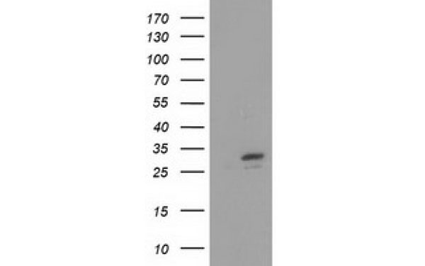
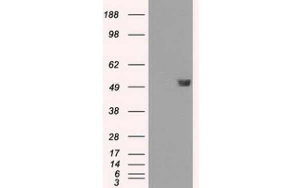
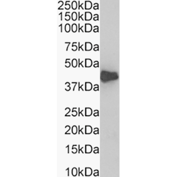
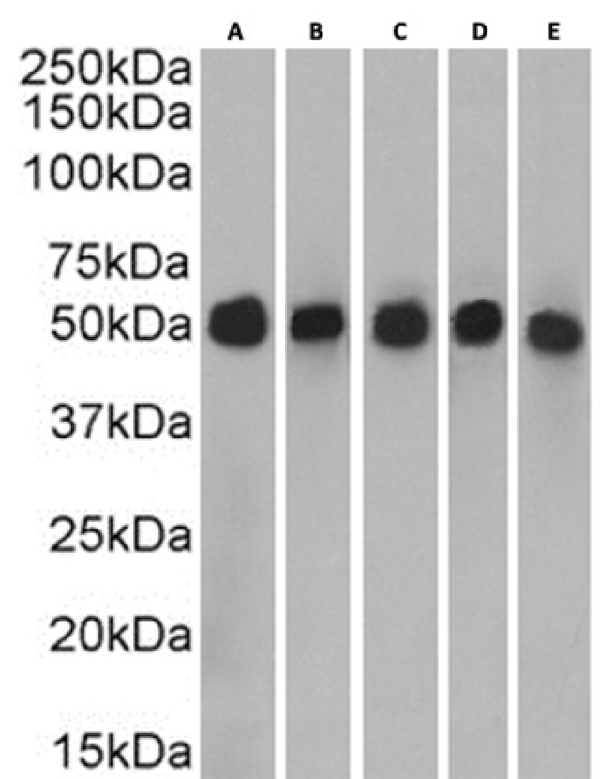
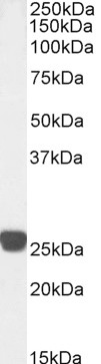
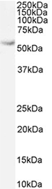
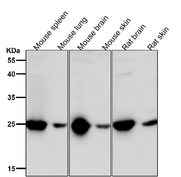
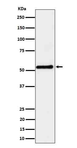
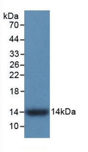
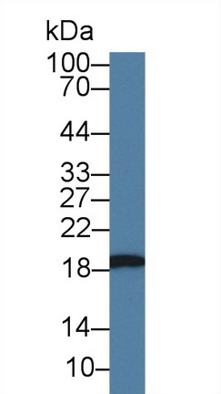
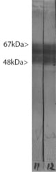 WB (Western Blot)
(Blots of crude rat brain extract stained with MAPT/TAU antibody, revealing, as expected, multiple bands in the range 48-67 kDa.)
WB (Western Blot)
(Blots of crude rat brain extract stained with MAPT/TAU antibody, revealing, as expected, multiple bands in the range 48-67 kDa.)
MAPT/Tau, Monoclonal Antibody (Cat# AAA162349)
Predicted: Mouse, Rat
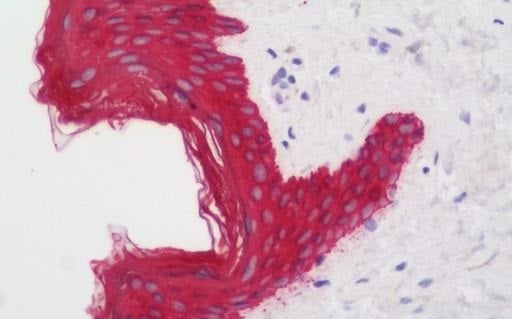 IHC (Immunohistochemistry)
(KRT19/CK19/Cytokeratin 19 Antibody-Anti-KRT19/CK19/Cytokeratin 19 antibody IHC staining of human skin. Immunohistochemistry of formalin-fixed, paraffin-embedded tissue after heat-induced antigen retrieval. Antibody dilution 1:50.)
IHC (Immunohistochemistry)
(KRT19/CK19/Cytokeratin 19 Antibody-Anti-KRT19/CK19/Cytokeratin 19 antibody IHC staining of human skin. Immunohistochemistry of formalin-fixed, paraffin-embedded tissue after heat-induced antigen retrieval. Antibody dilution 1:50.)
KRT19/CK19/Cytokeratin 19, Monoclonal Antibody (Cat# AAA162600)
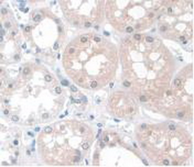
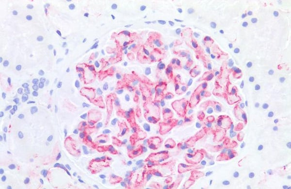 IHC (Immunohistochemisry)
(PECAM-1/CD31 Antibody-Human Kidney: Formalin-Fixed, Paraffin-Embedded (FFPE))
IHC (Immunohistochemisry)
(PECAM-1/CD31 Antibody-Human Kidney: Formalin-Fixed, Paraffin-Embedded (FFPE))
PECAM-1/CD31, Monoclonal Antibody (Cat# AAA163274)
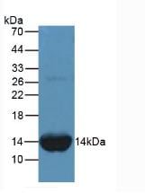
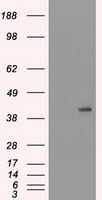
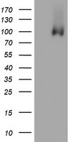
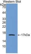
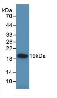
 WB (Western Blot)
(Western Blot. Sample: Recombinant FABP1, Human.)
WB (Western Blot)
(Western Blot. Sample: Recombinant FABP1, Human.)
Fatty Acid Binding Protein 1, Liver (FABP1), Monoclonal Antibody (Cat# AAA130613)
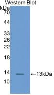
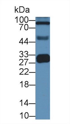
 WB (Western Blot)
(Western Blot: Sample: Recombinant GSTa1, Human.)
WB (Western Blot)
(Western Blot: Sample: Recombinant GSTa1, Human.)
Glutathione S Transferase Alpha 1 (GSTa1), Monoclonal Antibody (Cat# AAA130617)
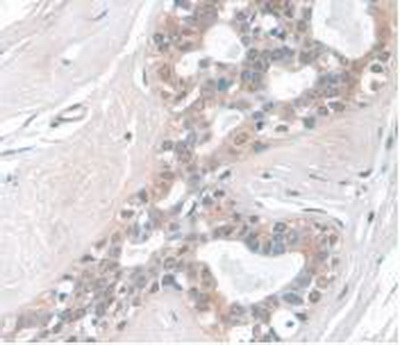
 IHC (Immunohistochemistry)
(DAB staining on IHC-P; Samples: Human Stomach Tissue)
IHC (Immunohistochemistry)
(DAB staining on IHC-P; Samples: Human Stomach Tissue)
C Reactive Protein (CRP), Monoclonal Antibody (Cat# AAA130624)
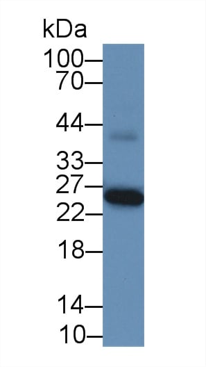
 WB (Western Blot)
(Western Blot: Sample: Recombinant S100A9, Human.)
WB (Western Blot)
(Western Blot: Sample: Recombinant S100A9, Human.)
S100 Calcium Binding Protein A9 (S100A9), Monoclonal Antibody (Cat# AAA130631)
 IHC (Immunohistochemistry)
(DAB staining on IHC-P; Samples: Human Breast Cancer Tissue)
IHC (Immunohistochemistry)
(DAB staining on IHC-P; Samples: Human Breast Cancer Tissue)
Apolipoprotein A1 (APOA1), Monoclonal Antibody (Cat# AAA130647)
 WB (Western Blot)
(Western Blot: Sample: Recombinant HSPb6, Rat.)
WB (Western Blot)
(Western Blot: Sample: Recombinant HSPb6, Rat.)
Heat Shock Protein Beta 6 (HSPb6), Monoclonal Antibody (Cat# AAA130654)
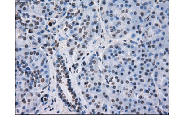
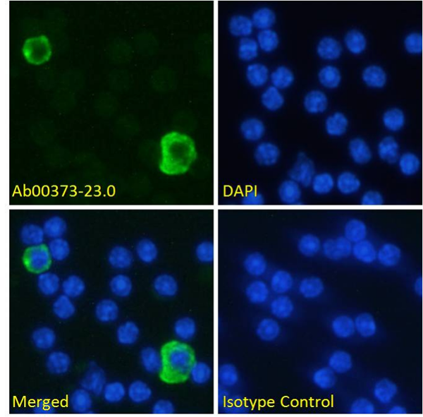
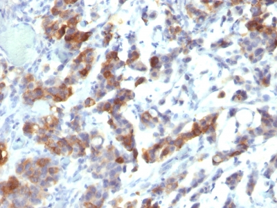
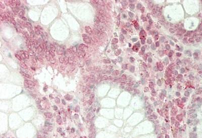
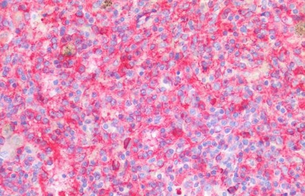
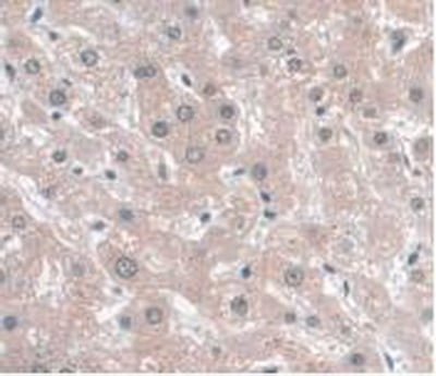
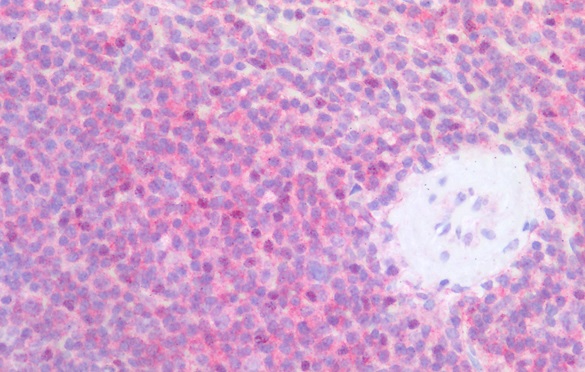 IHC (Immunohistochemistry)
(Immunohistochemistry staining of human spleen (paraffin-embedded sections) with anti-CD79b (CB3-1), 10 ug/ml. Commercially tested by LifeSpan BioSciences.)
IHC (Immunohistochemistry)
(Immunohistochemistry staining of human spleen (paraffin-embedded sections) with anti-CD79b (CB3-1), 10 ug/ml. Commercially tested by LifeSpan BioSciences.)
CD79b, Monoclonal Antibody (Cat# AAA128476)

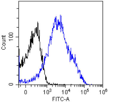
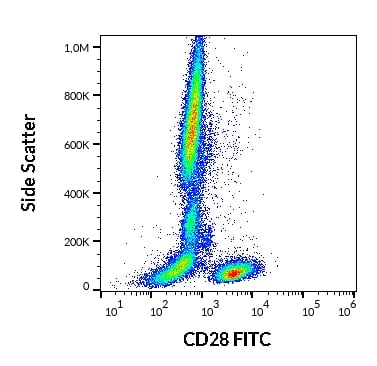 FCM/FACS (Flow Cytometry)
(Flow cytometry surface staining pattern of human peripheral whole blood stained using anti-human CD28 (CD28.2) FITC antibody (20 ul reagent / 100 ul of peripheral whole blood).)
FCM/FACS (Flow Cytometry)
(Flow cytometry surface staining pattern of human peripheral whole blood stained using anti-human CD28 (CD28.2) FITC antibody (20 ul reagent / 100 ul of peripheral whole blood).)
CD28, Monoclonal Antibody (Cat# AAA128485)
CD28, Monoclonal Antibody (Cat# AAA128488)
CD84, Monoclonal Antibody (Cat# AAA128489)
What are Monoclonal Antibodies?
Monoclonal antibodies are specialized laboratory-produced proteins developed for binding to specific biological antigens or other molecular targets. Since they come from a single cell (or clone), they are especially consistent and accurate in the data they are involved in producing.
This type of antibody material has been shown to be a powerful tool in finding and subsequently destroying harmful cells in an organism, such as those found in cancers or various autoimmune diseases. This makes them excellent aids in medical testing and research, which is why they are so widely used.
AAA Biotech offers a comprehensive range of high-quality monoclonal antibodies that perform effectively in various laboratory tests, including (amongst others) ELISA, western blotting, immunohistochemistry, and flow cytometry. All of the products in our catalog are thoroughly quality tested to make sure that they are reliable and will consistently perform well in your research.
What Are The Uses of Monoclonal Antibodies
Monoclonal antibodies are used in many lab tests, including (amongst others) ELISA, western blotting, immunohistochemistry, and flow cytometry.
ELISA is a test that helps detect a specific substance/analyte in a sample. It uses antibodies (often monoclonal) bound to a solid surface (such as the well of a microplate) to “capture” the substance/analyte in the sample and immobilize it so that the detection antibody component can then bind to it and produce a signal, which can then be measured.
Western blotting identifies specific proteins in a sample. The sample is first separated on a gel, and then antibodies are applied that will typically bind to the target, which will all be localized to a single band in a lane.
Immunohistochemistry helps locate specific proteins in cells or tissue samples using antibodies.
Flow cytometry looks at and sorts cells. It uses antibodies that are conjugated to reporter molecules called “fluorophores”, which, under special lights, emit light themselves, which can then be measured by a detector instrument.
How Monoclonal Antibodies Are Used as Medicine?
Please note that all of the products listed in AAA Biotech’s also known as AAA Bio or AAABio catalog are strictly for research-use only (RUO).
Monoclonal antibodies can also be used as therapeutic/medical treatments, particularly in the context of cancers. They are designed to find and bind to specific cells or proteins, helping the immune system recognize and attack the cancer. These treatments work in different ways, such as:
- Radioimmunotherapy attaches a small amount of radioactive molecule to the antibody, so it delivers the radiation directly to the cancer cells that the antibody is specifically binding to.
- Antibody-directed enzyme prodrug therapy uses antibodies that are specifically bound to special enzymes. These enzymes activate a harmless drug in the body and turn it into a cancer-killing drug only near the cancer cells—this helps avoid harming healthy cells.
- Immunoliposomes are tiny “bubbles” filled with medicine/drug and coated with antibodies. They carry the drug straight to the cancer cells.
Why Buy Monoclonal Antibodies From Us?
At AAA Biotech, we provide high-performance monoclonal antibodies designed to support a wide range of research needs.
1. Validated for Versatile Applications
The antibodies in our catalog are extensively validated and compatible with multiple techniques, including (but not limited to) ELISA, flow cytometry (FC), immunocytochemistry (ICC), immunofluorescence (IF), immunohistochemistry (IHC), immunoprecipitation (IP), and western blotting (WB).
2. Wide Selection & Specialized Options
We offer antibodies for common and rare species, that are available in various conjugated forms, and also in recombinant formats. Essentially, there is almost anything one might need to meet their experimental model’s requirements.
3. High-Quality Proteins
Our proteins meet high purity standards—90% or more as confirmed by SDS-PAGE. Many are available with tags like His, Flag, GST, or MBP, and we also supply native and biologically active proteins for functional studies.
Frequently Asked Questions
1. Are your monoclonal antibodies validated for specific applications?
Yes, our antibodies are tested and validated for use in methods such as ELISA, western blot, IHC, flow cytometry, and more. Refer to specific product pages or datasheets for individual product information.
2. How do I choose the right monoclonal antibody for my application?
Review the product details directly for application validation, species reactivity, and target information. You may also contact our support team at any time for help.
3. How quickly can I receive my order?
Most orders are processed and shipped within 1–3 business days, depending on product availability and your shipping location.