Filters
▼Clonality
▼Type
▼Reactivity
▼Gene Name
▼Isotype
▼Host
▼Application
▼Clone
▼Monoclonal Antibodies
Get accurate results in your research with our Monoclonal Antibodies, which are specially made to target exactly what you require for your research, and will produce consistent, reliable performance in lab tests.
Viewing 5650-5700 of 27597 product results
CD84, Monoclonal Antibody (Cat# AAA128490)
CD4, Monoclonal Antibody (Cat# AAA128553)
PRR7, Monoclonal Antibody (Cat# AAA128989)
CD243, Monoclonal Antibody (Cat# AAA129024)
CD87, Monoclonal Antibody (Cat# AAA129035)
IgG1, Monoclonal Isotype Control (Cat# AAA128887)
Lysozyme, Monoclonal Antibody (Cat# AAA128686)
CD158d, Monoclonal Antibody (Cat# AAA128701)
CD154, Monoclonal Antibody (Cat# AAA128892)
CD41, Monoclonal Antibody (Cat# AAA128896)
OPAL1, Monoclonal Antibody (Cat# AAA128918)
CD4, Monoclonal Antibody (Cat# AAA128927)
CD11b, Monoclonal Antibody (Cat# AAA128934)
CD45R, Monoclonal Antibody (Cat# AAA128947)
CD150, Monoclonal Antibody (Cat# AAA128958)
CD8b, Monoclonal Antibody (Cat# AAA128563)
CD229, Monoclonal Antibody (Cat# AAA128594)
CD79a, Monoclonal Antibody (Cat# AAA128597)

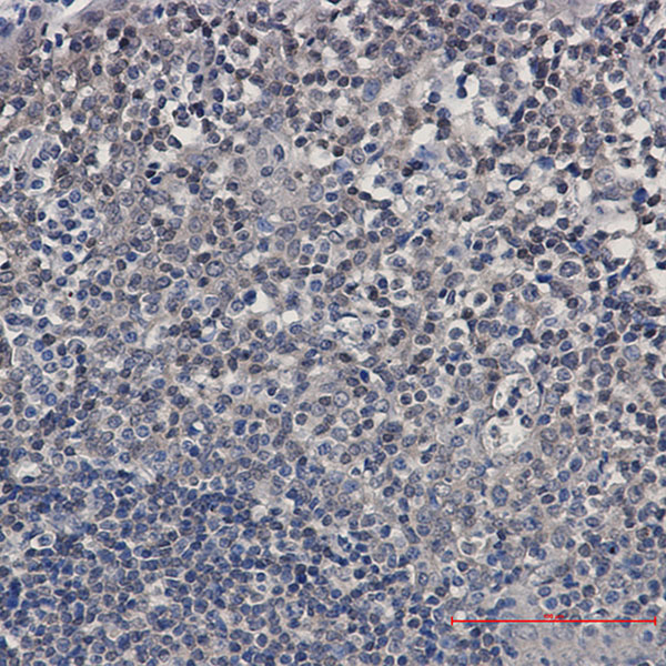
 IHC (Immunohistochemistry)
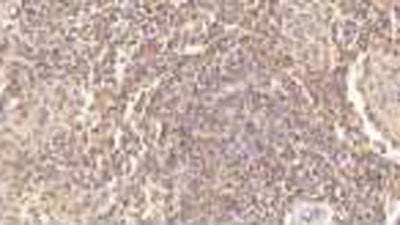
(DAB staining on IHC-P;Samples: Human Placenta Tissue;Primary Ab: 10?g/ml Mouse Anti-Human PF4 AntibodySecond Ab: 2ug/mL HRPLinked Caprine Anti-Mouse IgG Polyclonal Antibody)
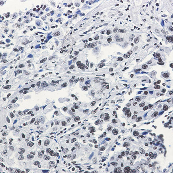
IHC (Immunohistochemistry)
(DAB staining on IHC-P;Samples: Human Placenta Tissue;Primary Ab: 10?g/ml Mouse Anti-Human PF4 AntibodySecond Ab: 2ug/mL HRPLinked Caprine Anti-Mouse IgG Polyclonal Antibody)
Platelet Factor 4 (PF4), Monoclonal Antibody (Cat# AAA134808)
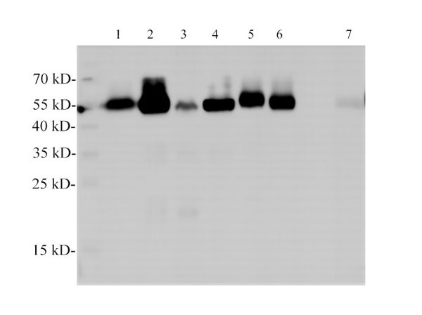
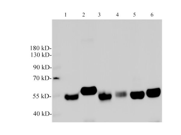
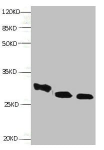
 WB (Western Blot)
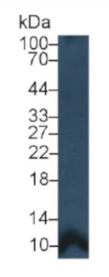
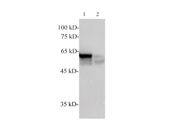
(Western Blot: Sample: Recombinant HGF, Human.)
WB (Western Blot)
(Western Blot: Sample: Recombinant HGF, Human.)
Hepatocyte Growth Factor (HGF), Monoclonal Antibody (Cat# AAA134833)
CD205, Monoclonal Antibody (Cat# AAA129131)
TCR Cbeta1, Monoclonal Antibody (Cat# AAA129159)
CD56, Monoclonal Antibody (Cat# AAA129168)
CD94, Monoclonal Antibody (Cat# AAA129172)
Blood Group ABH, Monoclonal Antibody (Cat# AAA129098)
 FCM/FACS (Flow Cytometry)
IgG, Monoclonal Isotype Control (Cat# AAA174727)
 FCM/FACS (Flow Cytometry)
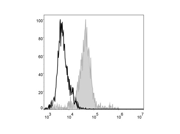
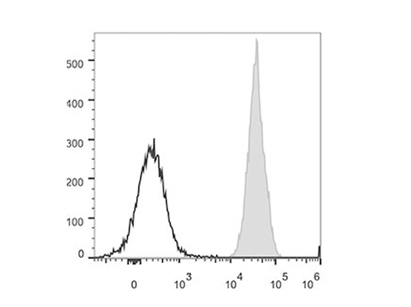
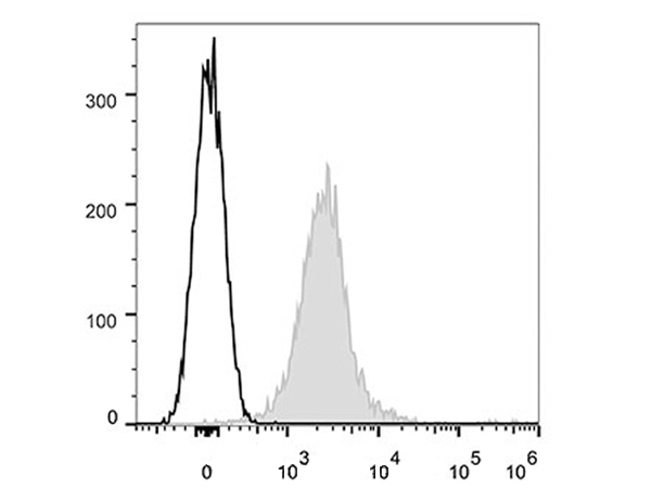
(C57BL/6 murine abdominal macrophages are stained with FITC Anti-Mouse F4/80 Antibody (filled gray histogram). Unstained abdominal macrophages (empty black histogram) are used as control)
FCM/FACS (Flow Cytometry)
(C57BL/6 murine abdominal macrophages are stained with FITC Anti-Mouse F4/80 Antibody (filled gray histogram). Unstained abdominal macrophages (empty black histogram) are used as control)
F4/80, Monoclonal Antibody (Cat# AAA174731)
CD40, Monoclonal Antibody (Cat# AAA174736)
 FCM/FACS (Flow Cytometry)
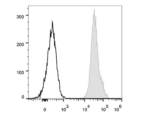
(Human peripheral blood lymphocytes are stained with Anti-Human CD29 Monoclonal Antibody(PerCP/Cy5.5 Conjugated)(filled gray histogram). Unstained lymphocytes (empty black histogram) are used as control.)
FCM/FACS (Flow Cytometry)
(Human peripheral blood lymphocytes are stained with Anti-Human CD29 Monoclonal Antibody(PerCP/Cy5.5 Conjugated)(filled gray histogram). Unstained lymphocytes (empty black histogram) are used as control.)
CD29, Monoclonal Antibody (Cat# AAA174619)
 FCM/FACS (Flow Cytometry)
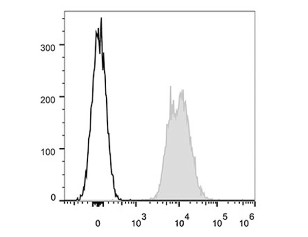
(Human peripheral blood lymphocytes are stained with Anti-Human CD47 Monoclonal Antibody(PerCP/Cy5.5 Conjugated)(filled gray histogram). Unstained lymphocytes (empty black histogram) are used as control.)
FCM/FACS (Flow Cytometry)
(Human peripheral blood lymphocytes are stained with Anti-Human CD47 Monoclonal Antibody(PerCP/Cy5.5 Conjugated)(filled gray histogram). Unstained lymphocytes (empty black histogram) are used as control.)
CD47, Monoclonal Antibody (Cat# AAA174628)
 FCM/FACS (Flow Cytometry)
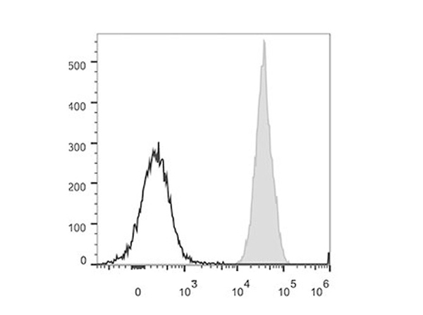
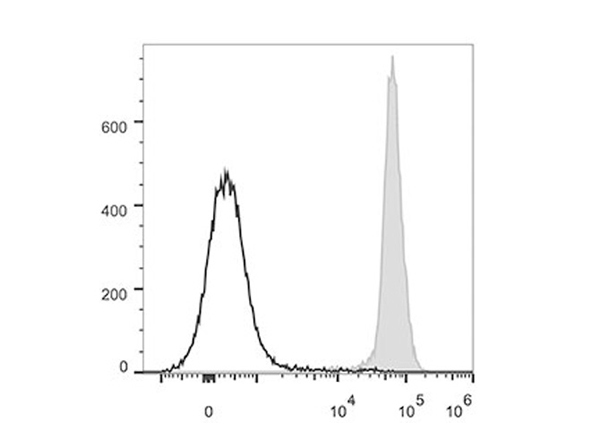
(C57BL/6 murine splenocytes are stained with Anti-Mouse CD45.2 Monoclonal Antibody(PE Conjugated)(filled gray histogram). Unstained splenocytes (empty black histogram) are used as control.)
FCM/FACS (Flow Cytometry)
(C57BL/6 murine splenocytes are stained with Anti-Mouse CD45.2 Monoclonal Antibody(PE Conjugated)(filled gray histogram). Unstained splenocytes (empty black histogram) are used as control.)
CD45.2, Monoclonal Antibody (Cat# AAA174650)

 FCM/FACS (Flow Cytometry)
(C57BL/6 murine splenocytes are stained with Anti-Mouse CD45.2 Monoclonal Antibody(APC Conjugated)(filled gray histogram). Unstained splenocytes (empty black histogram) are used as control.)
FCM/FACS (Flow Cytometry)
(C57BL/6 murine splenocytes are stained with Anti-Mouse CD45.2 Monoclonal Antibody(APC Conjugated)(filled gray histogram). Unstained splenocytes (empty black histogram) are used as control.)
CD45.2, Monoclonal Antibody (Cat# AAA174651)
 FCM/FACS (Flow Cytometry)
(C57BL/6 murine splenocytes are stained with Anti-Mouse CD45.2 Monoclonal Antibody(PerCP/Cy5.5 Conjugated)(filled gray histogram). Unstained splenocytes (empty black histogram) are used as control.)
FCM/FACS (Flow Cytometry)
(C57BL/6 murine splenocytes are stained with Anti-Mouse CD45.2 Monoclonal Antibody(PerCP/Cy5.5 Conjugated)(filled gray histogram). Unstained splenocytes (empty black histogram) are used as control.)
CD45.2, Monoclonal Antibody (Cat# AAA174652)

 FCM/FACS (Flow Cytometry)
(C57BL/6 murine splenocytes are stained with Anti-Mouse PD-L1 Monoclonal Antibody(APC Conjugated)(filled gray histogram). Unstained splenocytes (empty black histogram) are used as control.)
FCM/FACS (Flow Cytometry)
(C57BL/6 murine splenocytes are stained with Anti-Mouse PD-L1 Monoclonal Antibody(APC Conjugated)(filled gray histogram). Unstained splenocytes (empty black histogram) are used as control.)
PD-L1, Monoclonal Antibody (Cat# AAA174666)
 FCM/FACS (Flow Cytometry)
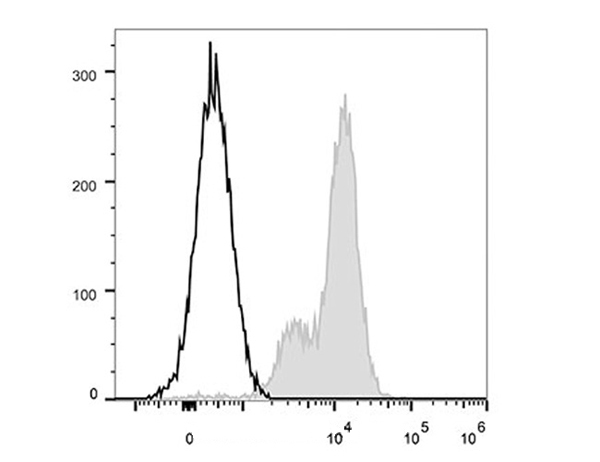
(Mouse abdominal macrophages elicited by starch broth are stained with FITC Anti-Mouse F4/80 Antibody (filled gray histogram). Unstained macrophages (blank black histogram) are used as control.)
FCM/FACS (Flow Cytometry)
(Mouse abdominal macrophages elicited by starch broth are stained with FITC Anti-Mouse F4/80 Antibody (filled gray histogram). Unstained macrophages (blank black histogram) are used as control.)
F4/80, Monoclonal Antibody (Cat# AAA174046)
 IF (Immunofluorescence)
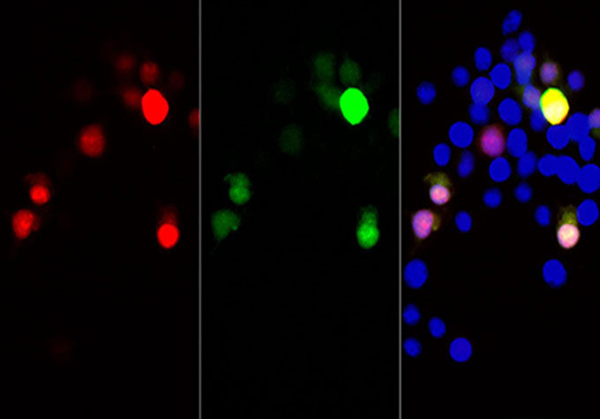
(Immunofluorescent analysis of 293F cells transfected with the mCherry, using anti-mCherry monoclonal antibody at dilution of 1:12000.)
IF (Immunofluorescence)
(Immunofluorescent analysis of 293F cells transfected with the mCherry, using anti-mCherry monoclonal antibody at dilution of 1:12000.)
mCherry-Tag, Monoclonal Antibody (Cat# AAA178008)
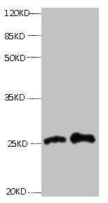
 WB (Western Blot)
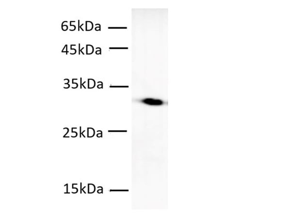
(Western Blotting with anti-TUBA monoclonal antibody at dilution of 1:2000.)
WB (Western Blot)
(Western Blotting with anti-TUBA monoclonal antibody at dilution of 1:2000.)
alpha Tubulin, Monoclonal Antibody (Cat# AAA178663)

 IHC (Immunohiostchemistry)
(Immunohistochemistry of UBE2I/UBC9 in paraffin-embedded Human tonsil using UBE2I/UBC9 Rabbit mAb at dilution 1:50)
IHC (Immunohiostchemistry)
(Immunohistochemistry of UBE2I/UBC9 in paraffin-embedded Human tonsil using UBE2I/UBC9 Rabbit mAb at dilution 1:50)
SUMO Conjugating Enzyme UBC9, Monoclonal Antibody (Cat# AAA178825)
 IF (Immunofluorescence)
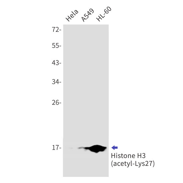
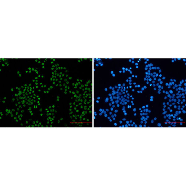
(Immunofluorescence of Histone H3 (acetyl-Lys27)(green) in hela using Histone H3 (acetyl-Lys27) Rabbit mAb at dilution 1:50, and DAPI(blue))
IF (Immunofluorescence)
(Immunofluorescence of Histone H3 (acetyl-Lys27)(green) in hela using Histone H3 (acetyl-Lys27) Rabbit mAb at dilution 1:50, and DAPI(blue))
Acetyl-Histone H3, Monoclonal Antibody (Cat# AAA178829)
 IHC (Immunohiostchemistry)
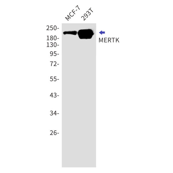
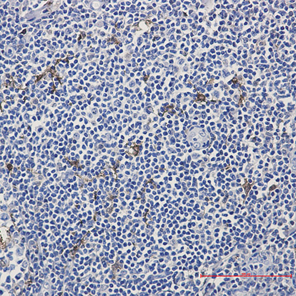
(Immunohistochemistry of MERTK in paraffin-embedded Human tonsil using MERTK Rabbit mAb at dilution 1:50)
IHC (Immunohiostchemistry)
(Immunohistochemistry of MERTK in paraffin-embedded Human tonsil using MERTK Rabbit mAb at dilution 1:50)
MERTK, Monoclonal Antibody (Cat# AAA178865)
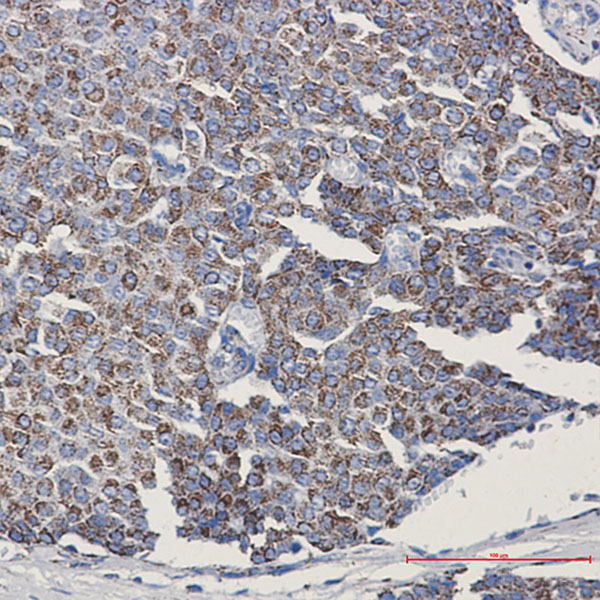
 IHC (Immunohiostchemistry)
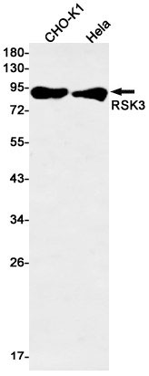
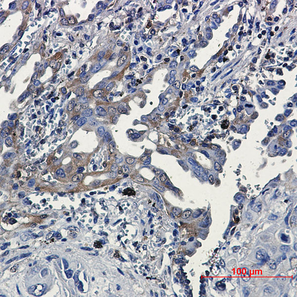
(Immunohistochemical of RSK3 in Human lung cancer tissue using RSK3 antibody at dilution 1?20)
IHC (Immunohiostchemistry)
(Immunohistochemical of RSK3 in Human lung cancer tissue using RSK3 antibody at dilution 1?20)
RSK3, Monoclonal Antibody (Cat# AAA178879)
 IHC (Immunohiostchemistry)
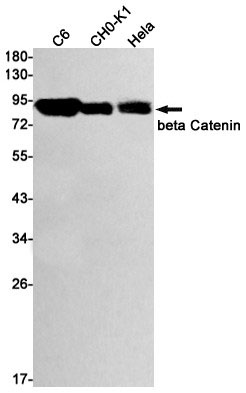
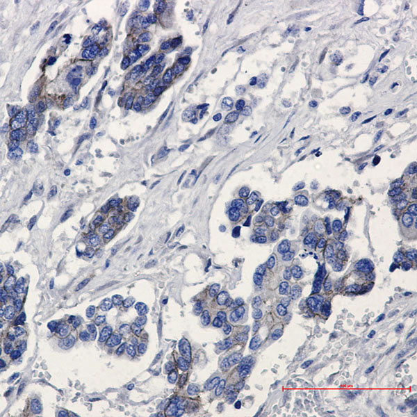
(Immunohistochemistry of beta Catenin in paraffin-embedded Human Cholangiocarcinoma using beta Catenin Rabbit mAb at dilution 1:50)
IHC (Immunohiostchemistry)
(Immunohistochemistry of beta Catenin in paraffin-embedded Human Cholangiocarcinoma using beta Catenin Rabbit mAb at dilution 1:50)
beta Catenin, Monoclonal Antibody (Cat# AAA178778)
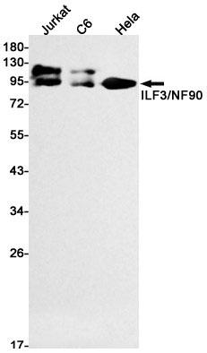
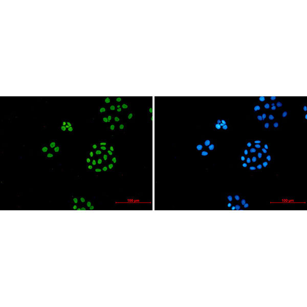 IF (Immunofluorescence)
(Immunofluorescence of ILF3 (green) in hela using ILF3 Rabbit mAb at dilution 1:50, and DAPI(blue))
IF (Immunofluorescence)
(Immunofluorescence of ILF3 (green) in hela using ILF3 Rabbit mAb at dilution 1:50, and DAPI(blue))
ILF3, Monoclonal Antibody (Cat# AAA178801)
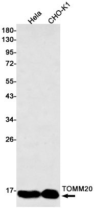
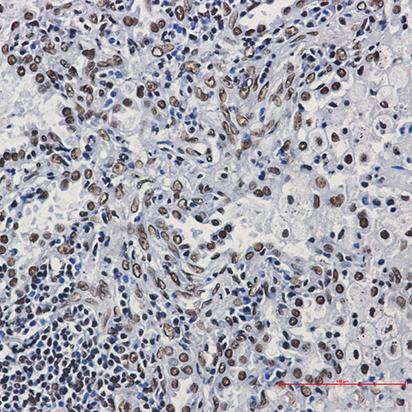
 IHC (Immunohiostchemistry)
(Immunohistochemistry of TOMM20 in paraffin-embedded Human breast cancer tissue using TOMM20 Rabbit mAb at dilution 1:50)
IHC (Immunohiostchemistry)
(Immunohistochemistry of TOMM20 in paraffin-embedded Human breast cancer tissue using TOMM20 Rabbit mAb at dilution 1:50)
TOMM20, Monoclonal Antibody (Cat# AAA178808)
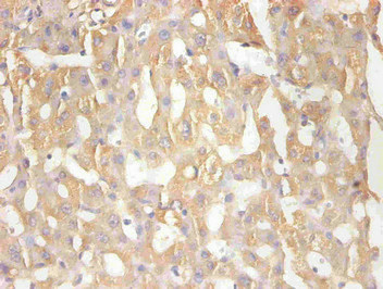
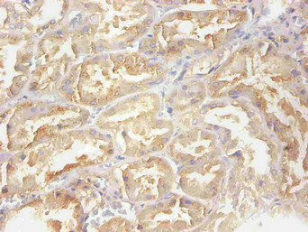 IHC (Immunohistochemistry)
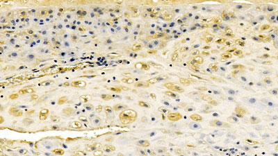
(Immunohistochemical of paraffin-embedded human nephridial tissue using AAA117779 at dilution of 1:200)
IHC (Immunohistochemistry)
(Immunohistochemical of paraffin-embedded human nephridial tissue using AAA117779 at dilution of 1:200)
Retinol-binding protein 4, Monoclonal Antibody (Cat# AAA117779)
ACE2, Monoclonal Antibody (Cat# AAA120177)
IgG, Monoclonal Isotype Control (Cat# AAA120183)
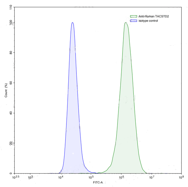
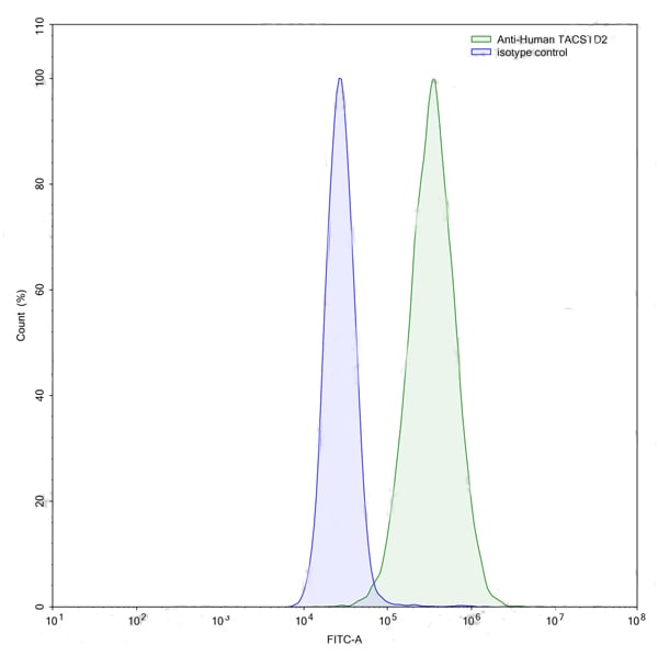
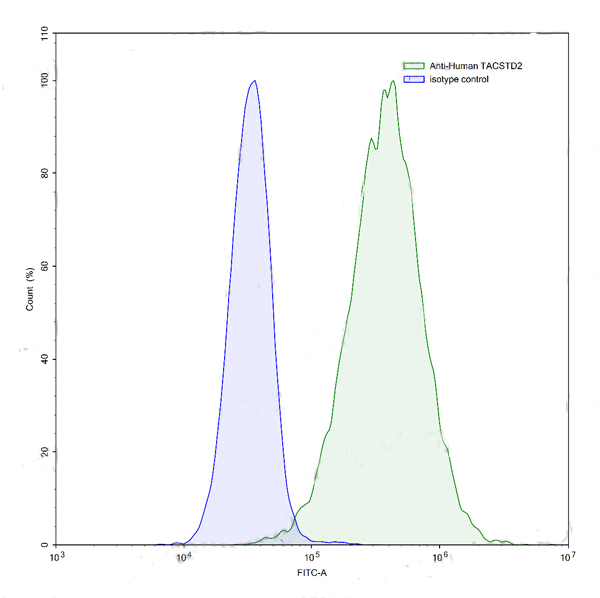
 FCM/FACS (Flow Cytometry)
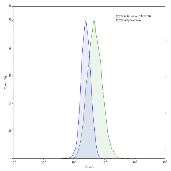
(Flow-cytometry using anti-human TACSTD2 antibody.PC-3 cells were stained with an irrelevant antibody (Blue Histogram) or an anti-human TACSTD2 antibody monoclonal antibody (Catalog # RHC43302 ,Green Histogram) at a concentration of 5 ?ug/ml for 30 mins at RT. After washing, bound antibody was detected using a FITC conjugated goat anti-human antibody (Catalog # PHB96441) and cells analysed on a NovoCyte Flow Cytometer.)
FCM/FACS (Flow Cytometry)
(Flow-cytometry using anti-human TACSTD2 antibody.PC-3 cells were stained with an irrelevant antibody (Blue Histogram) or an anti-human TACSTD2 antibody monoclonal antibody (Catalog # RHC43302 ,Green Histogram) at a concentration of 5 ?ug/ml for 30 mins at RT. After washing, bound antibody was detected using a FITC conjugated goat anti-human antibody (Catalog # PHB96441) and cells analysed on a NovoCyte Flow Cytometer.)
TACSTD2/TROP2, Monoclonal Recombinant Antibody (Cat# AAA120381)
Protein A or G purified from cell culture supernatant.
 FCM/FACS (Flow Cytometry)
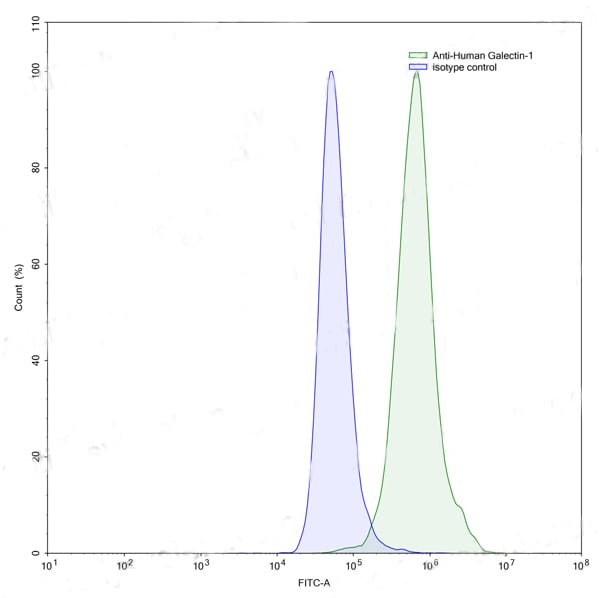
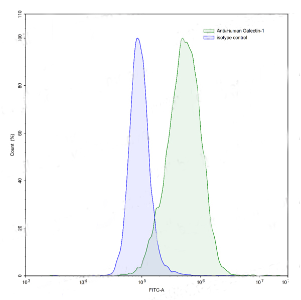
(Flow-cytometry using anti-human Galectin-1 antibody.HeLa cells were stained with an irrelevant antibody (Blue Histogram) or an anti-human Galectin-1 antibody monoclonal antibody (Catalog # RHC40102 ,Green Histogram) at a concentration of 5 ?ug/ml for 30 mins at RT. After washing, bound antibody was detected using a FITC conjugated goat anti-mouse antibody (Catalog # PMB96441) and cells analysed on a NovoCyte Flow Cytometer.)
FCM/FACS (Flow Cytometry)
(Flow-cytometry using anti-human Galectin-1 antibody.HeLa cells were stained with an irrelevant antibody (Blue Histogram) or an anti-human Galectin-1 antibody monoclonal antibody (Catalog # RHC40102 ,Green Histogram) at a concentration of 5 ?ug/ml for 30 mins at RT. After washing, bound antibody was detected using a FITC conjugated goat anti-mouse antibody (Catalog # PMB96441) and cells analysed on a NovoCyte Flow Cytometer.)
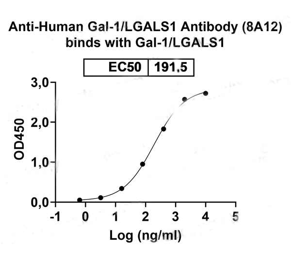
Gal1/LGALS1, Monoclonal Recombinant Antibody (Cat# AAA120402)
Protein A or G purified from cell culture supernatant.
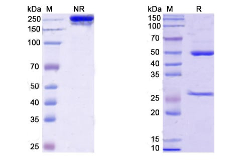 SDS-PAGE
(SDS PAGE for DENV-2 Envelope protein E/EDE1 Antibody)
SDS-PAGE
(SDS PAGE for DENV-2 Envelope protein E/EDE1 Antibody)
DENV-2 Envelope protein E/EDE1, Monoclonal Recombinant Antibody (Cat# AAA120407)
Protein A or G purified from cell culture supernatant.
What are Monoclonal Antibodies?
Monoclonal antibodies are specialized laboratory-produced proteins developed for binding to specific biological antigens or other molecular targets. Since they come from a single cell (or clone), they are especially consistent and accurate in the data they are involved in producing.
This type of antibody material has been shown to be a powerful tool in finding and subsequently destroying harmful cells in an organism, such as those found in cancers or various autoimmune diseases. This makes them excellent aids in medical testing and research, which is why they are so widely used.
AAA Biotech offers a comprehensive range of high-quality monoclonal antibodies that perform effectively in various laboratory tests, including (amongst others) ELISA, western blotting, immunohistochemistry, and flow cytometry. All of the products in our catalog are thoroughly quality tested to make sure that they are reliable and will consistently perform well in your research.
What Are The Uses of Monoclonal Antibodies
Monoclonal antibodies are used in many lab tests, including (amongst others) ELISA, western blotting, immunohistochemistry, and flow cytometry.
ELISA is a test that helps detect a specific substance/analyte in a sample. It uses antibodies (often monoclonal) bound to a solid surface (such as the well of a microplate) to “capture” the substance/analyte in the sample and immobilize it so that the detection antibody component can then bind to it and produce a signal, which can then be measured.
Western blotting identifies specific proteins in a sample. The sample is first separated on a gel, and then antibodies are applied that will typically bind to the target, which will all be localized to a single band in a lane.
Immunohistochemistry helps locate specific proteins in cells or tissue samples using antibodies.
Flow cytometry looks at and sorts cells. It uses antibodies that are conjugated to reporter molecules called “fluorophores”, which, under special lights, emit light themselves, which can then be measured by a detector instrument.
How Monoclonal Antibodies Are Used as Medicine?
Please note that all of the products listed in AAA Biotech’s also known as AAA Bio or AAABio catalog are strictly for research-use only (RUO).
Monoclonal antibodies can also be used as therapeutic/medical treatments, particularly in the context of cancers. They are designed to find and bind to specific cells or proteins, helping the immune system recognize and attack the cancer. These treatments work in different ways, such as:
- Radioimmunotherapy attaches a small amount of radioactive molecule to the antibody, so it delivers the radiation directly to the cancer cells that the antibody is specifically binding to.
- Antibody-directed enzyme prodrug therapy uses antibodies that are specifically bound to special enzymes. These enzymes activate a harmless drug in the body and turn it into a cancer-killing drug only near the cancer cells—this helps avoid harming healthy cells.
- Immunoliposomes are tiny “bubbles” filled with medicine/drug and coated with antibodies. They carry the drug straight to the cancer cells.
Why Buy Monoclonal Antibodies From Us?
At AAA Biotech, we provide high-performance monoclonal antibodies designed to support a wide range of research needs.
1. Validated for Versatile Applications
The antibodies in our catalog are extensively validated and compatible with multiple techniques, including (but not limited to) ELISA, flow cytometry (FC), immunocytochemistry (ICC), immunofluorescence (IF), immunohistochemistry (IHC), immunoprecipitation (IP), and western blotting (WB).
2. Wide Selection & Specialized Options
We offer antibodies for common and rare species, that are available in various conjugated forms, and also in recombinant formats. Essentially, there is almost anything one might need to meet their experimental model’s requirements.
3. High-Quality Proteins
Our proteins meet high purity standards—90% or more as confirmed by SDS-PAGE. Many are available with tags like His, Flag, GST, or MBP, and we also supply native and biologically active proteins for functional studies.
Frequently Asked Questions
1. Are your monoclonal antibodies validated for specific applications?
Yes, our antibodies are tested and validated for use in methods such as ELISA, western blot, IHC, flow cytometry, and more. Refer to specific product pages or datasheets for individual product information.
2. How do I choose the right monoclonal antibody for my application?
Review the product details directly for application validation, species reactivity, and target information. You may also contact our support team at any time for help.
3. How quickly can I receive my order?
Most orders are processed and shipped within 1–3 business days, depending on product availability and your shipping location.