Filters
▼Clonality
▼Type
▼Reactivity
▼Gene Name
▼Isotype
▼Host
▼Application
▼Clone
▼Monoclonal Antibodies
Get accurate results in your research with our Monoclonal Antibodies, which are specially made to target exactly what you require for your research, and will produce consistent, reliable performance in lab tests.
Viewing 5500-5550 of 27597 product results
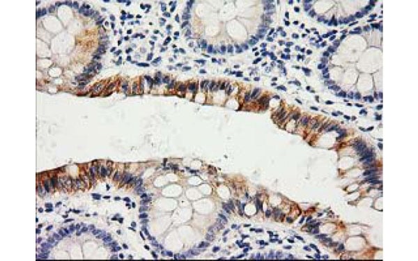
 IHC (Immunohistochemisry)
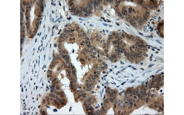
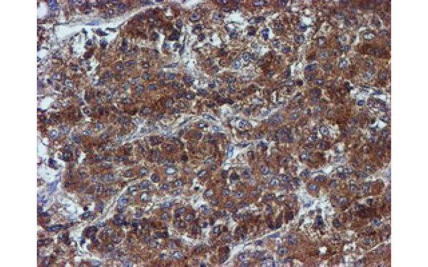
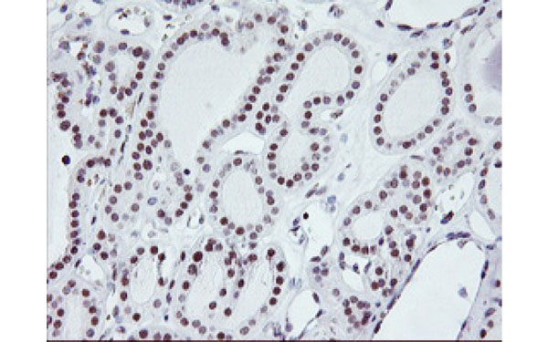
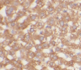
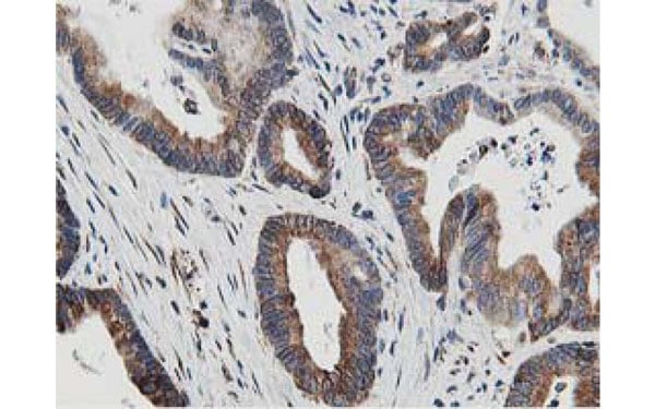
(Immunohistochemical analysis of PTPRE protein in paraffin embedded Adenocarcinoma of Human colon tissue using PTPRE antibody)
IHC (Immunohistochemisry)
(Immunohistochemical analysis of PTPRE protein in paraffin embedded Adenocarcinoma of Human colon tissue using PTPRE antibody)
PTPRE, Monoclonal Antibody (Cat# AAA107235)
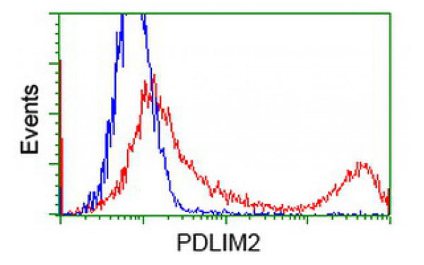
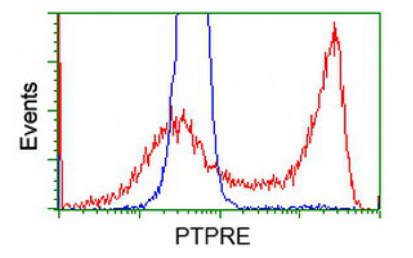
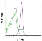 FCM/FACS (Flow Cytometry)
(Flow Cytometric analysis using TdT antibodyStaining of C57BL/6 thymocytes with CD4 antibody (FITC) and CD8a antibody PErCP-Cy5.5 followed by fixation permeabilization with the Foxp3 Stainng Buffers and subsequent staining with 0.25 ug of Anti-Mouse TdT (PE) (purple histogram). CD4 single positives are shown in gray and CD8 single positives in green.)
FCM/FACS (Flow Cytometry)
(Flow Cytometric analysis using TdT antibodyStaining of C57BL/6 thymocytes with CD4 antibody (FITC) and CD8a antibody PErCP-Cy5.5 followed by fixation permeabilization with the Foxp3 Stainng Buffers and subsequent staining with 0.25 ug of Anti-Mouse TdT (PE) (purple histogram). CD4 single positives are shown in gray and CD8 single positives in green.)
TdT, Monoclonal Antibody (Cat# AAA107237)
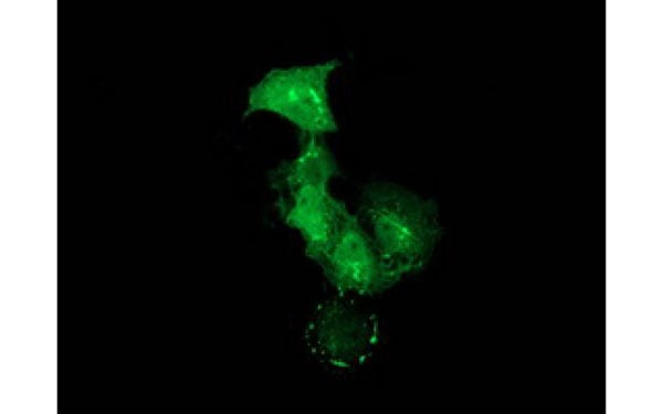 IF (Immunofluorescence)
(Immunofluorescent staining of COS7 cells transiently transfected with recombinant PDLIM2 protein using PDLIM2 antibody)
IF (Immunofluorescence)
(Immunofluorescent staining of COS7 cells transiently transfected with recombinant PDLIM2 protein using PDLIM2 antibody)
PDLIM2, Monoclonal Antibody (Cat# AAA107404)
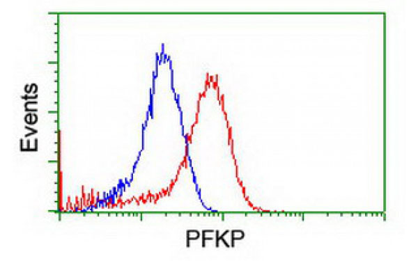
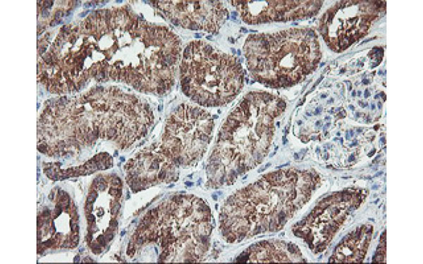
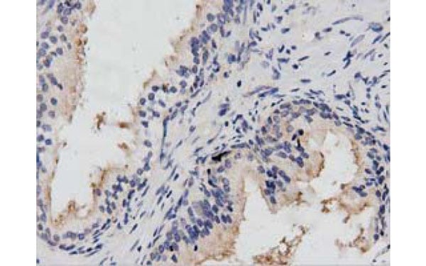
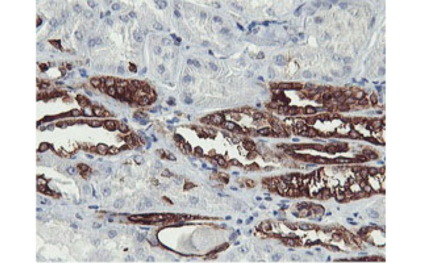 IHC (Immunohistochemisry)
(Immunohistochemical analysis of PFKP protein in paraffin embedded Human Kidney tissue using PFKP antibody)
IHC (Immunohistochemisry)
(Immunohistochemical analysis of PFKP protein in paraffin embedded Human Kidney tissue using PFKP antibody)
PFKP, Monoclonal Antibody (Cat# AAA107419)
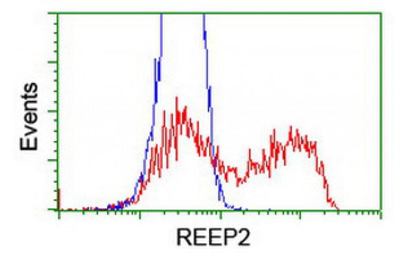
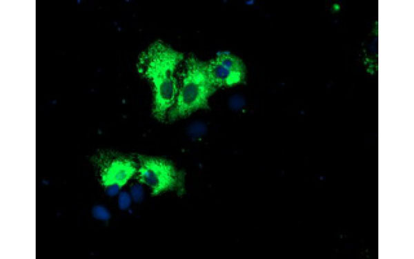 IF (Immunofluorescence)
(Immunofluorescent staining of COS7 cells transiently transfected with recombinant REEP2 protein using REEP2 antibody)
IF (Immunofluorescence)
(Immunofluorescent staining of COS7 cells transiently transfected with recombinant REEP2 protein using REEP2 antibody)
REEP2, Monoclonal Antibody (Cat# AAA107435)
 IF (Immunofluorescence)
(Immunofluorescent staining of COS7 cells transiently transfected with recombinant LIN7B protein using LIN7B antibody)
IF (Immunofluorescence)
(Immunofluorescent staining of COS7 cells transiently transfected with recombinant LIN7B protein using LIN7B antibody)
LIN7B, Monoclonal Antibody (Cat# AAA107460)
 IHC (Immunohistochemisry)
(Immunohistochemical analysis of NEK6 protein in paraffin embedded Carcinoma of Human kidney tissue using NEK6 antibody)
IHC (Immunohistochemisry)
(Immunohistochemical analysis of NEK6 protein in paraffin embedded Carcinoma of Human kidney tissue using NEK6 antibody)
NEK6, Monoclonal Antibody (Cat# AAA106911)
 IF (Immunofluorescence)
(Immunofluorescent staining of COS7 cells transiently transfected with recombinant ITFG2 protein using ITFG2 antibody)
IF (Immunofluorescence)
(Immunofluorescent staining of COS7 cells transiently transfected with recombinant ITFG2 protein using ITFG2 antibody)
ITFG2, Monoclonal Antibody (Cat# AAA106936)
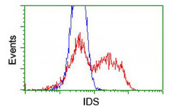
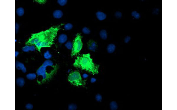 IF (Immunofluorescence)
(Immunofluorescent staining of COS7 cells transiently transfected with recombinant IDS protein using IDS antibody)
IF (Immunofluorescence)
(Immunofluorescent staining of COS7 cells transiently transfected with recombinant IDS protein using IDS antibody)
IDS, Monoclonal Antibody (Cat# AAA106942)
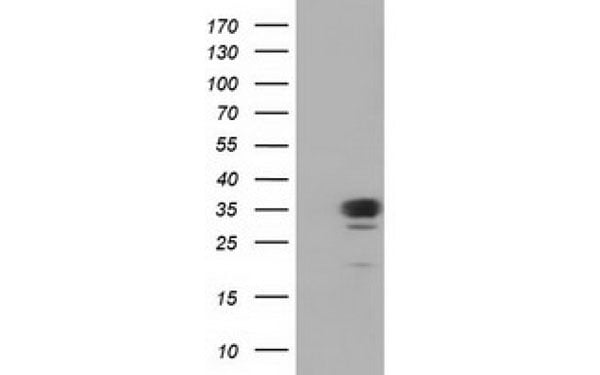
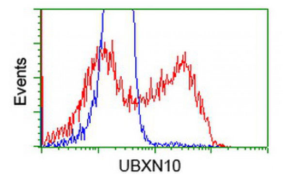
 IF (Immunofluorescence)
(Immunofluorescent staining of COS7 cells transiently transfected with recombinant UBXN10 protein using UBXN10 antibody)
IF (Immunofluorescence)
(Immunofluorescent staining of COS7 cells transiently transfected with recombinant UBXN10 protein using UBXN10 antibody)
UBXN10, Monoclonal Antibody (Cat# AAA106960)
 IF (Immunofluorescence)
(Immunofluorescent staining of COS7 cells transiently transfected with recombinant TMOD1 protein using TMOD1 antibody)
IF (Immunofluorescence)
(Immunofluorescent staining of COS7 cells transiently transfected with recombinant TMOD1 protein using TMOD1 antibody)
TMOD1, Monoclonal Antibody (Cat# AAA106968)
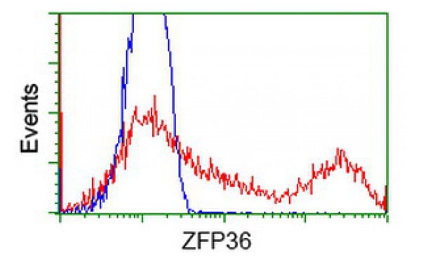
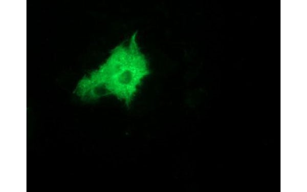 IF (Immunofluorescence)
(Immunofluorescent staining of COS7 cells transiently transfected with recombinant ZFP36 protein using ZFP36 antibody)
IF (Immunofluorescence)
(Immunofluorescent staining of COS7 cells transiently transfected with recombinant ZFP36 protein using ZFP36 antibody)
ZFP36, Monoclonal Antibody (Cat# AAA106971)
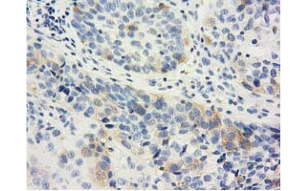
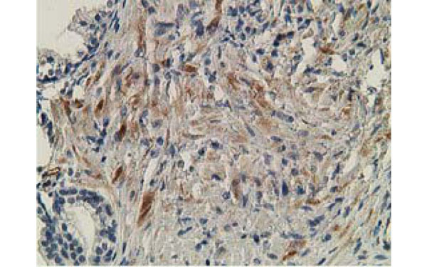
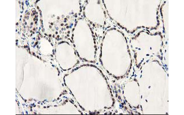
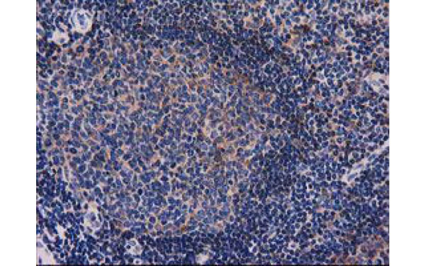
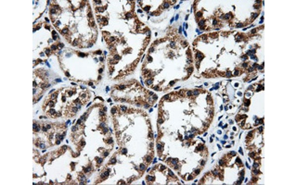
 IHC (Immunohistochemisry)
(Immunohistochemical analysis of RNF113B protein in paraffin embedded Carcinoma of Human thyroid tissue using RNF113B antibody)
IHC (Immunohistochemisry)
(Immunohistochemical analysis of RNF113B protein in paraffin embedded Carcinoma of Human thyroid tissue using RNF113B antibody)
RNF113B, Monoclonal Antibody (Cat# AAA106976)
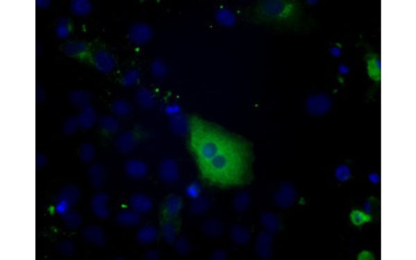
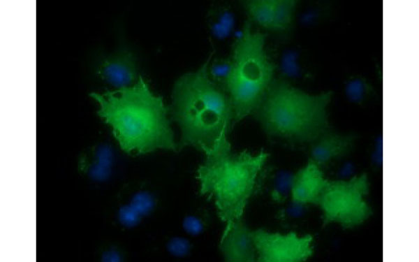
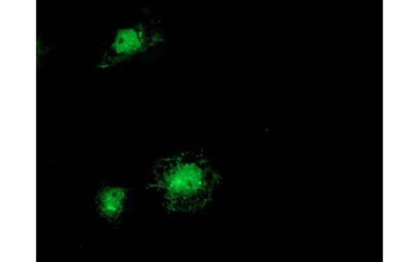
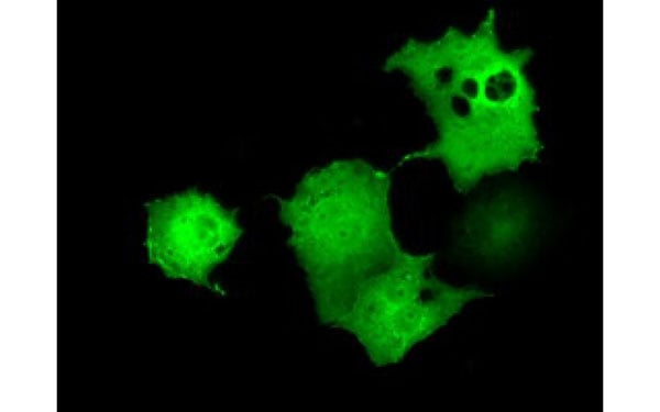
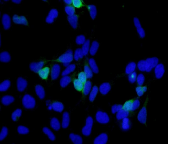
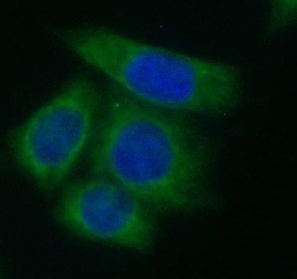
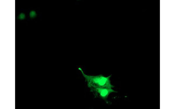
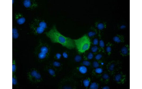
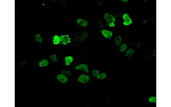
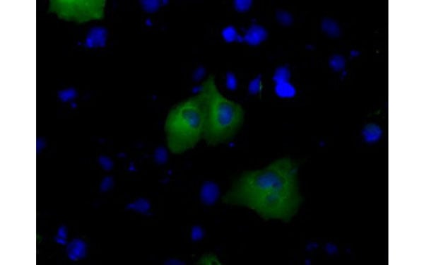
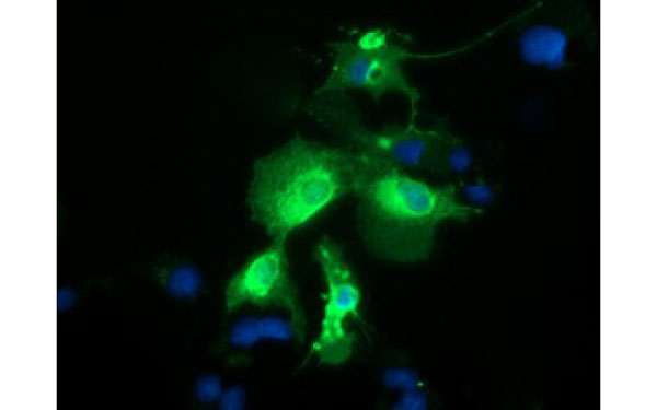 IF (Immunofluorescence)
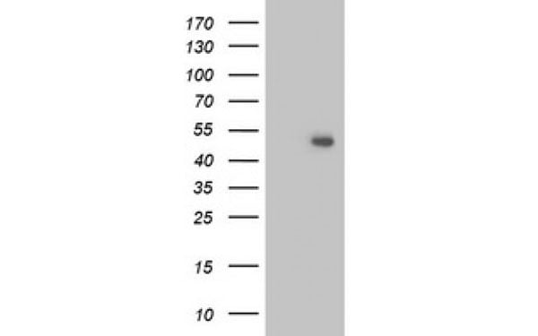
(Immunofluorescent staining of COS7 cells transiently transfected with recombinant PTPRE protein using PTPRE antibody)
IF (Immunofluorescence)
(Immunofluorescent staining of COS7 cells transiently transfected with recombinant PTPRE protein using PTPRE antibody)
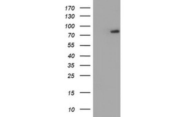
PTPRE, Monoclonal Antibody (Cat# AAA106980)
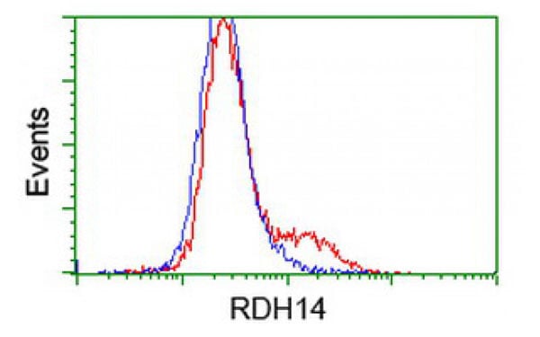
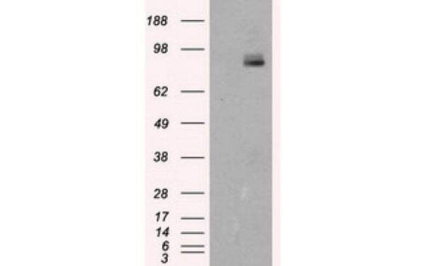
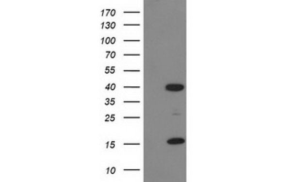
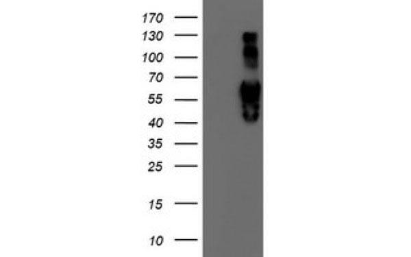
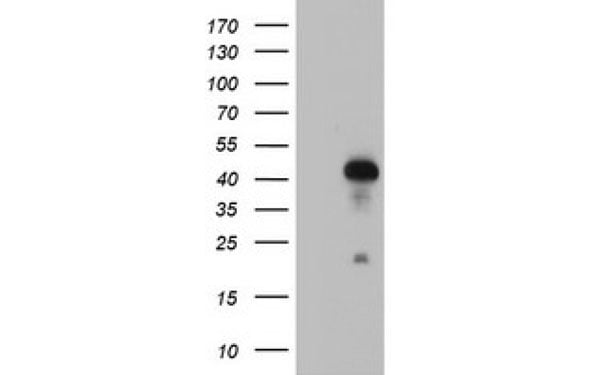
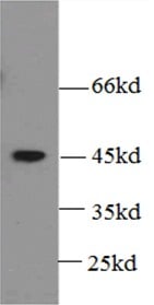
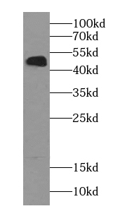
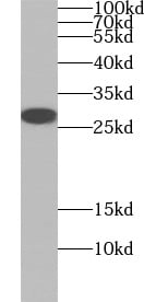
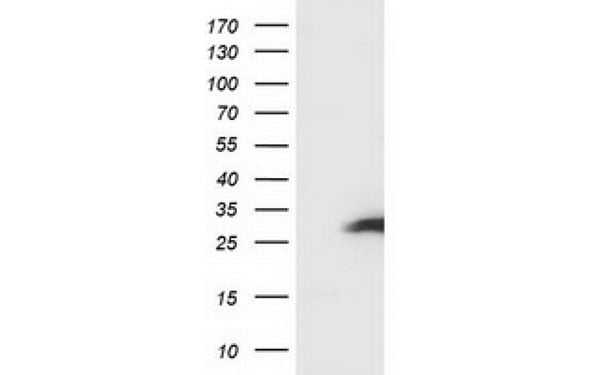
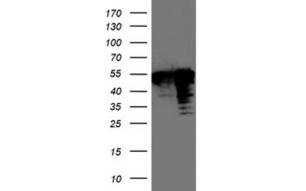
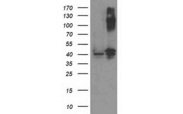
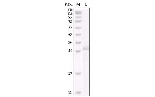
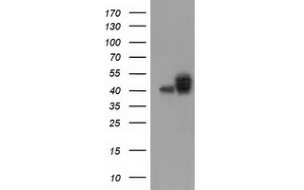
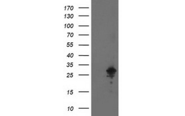
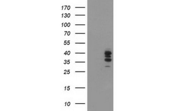
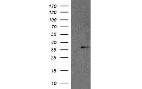 WB (Western Blot)
(Western Blot analysis of HEK293T cell lysates (5 ug) transfected with either recombinant RDH14 protein (Right) or empty vector (Left) detected with RDH14 antibody)
WB (Western Blot)
(Western Blot analysis of HEK293T cell lysates (5 ug) transfected with either recombinant RDH14 protein (Right) or empty vector (Left) detected with RDH14 antibody)
RDH14, Monoclonal Antibody (Cat# AAA106983)
 IF (Immunofluorescence)
(Immunofluorescent staining of COS7 cells transiently transfected with recombinant OGFOD1 protein using OGFOD1 antibody)
IF (Immunofluorescence)
(Immunofluorescent staining of COS7 cells transiently transfected with recombinant OGFOD1 protein using OGFOD1 antibody)
OGFOD1, Monoclonal Antibody (Cat# AAA106985)
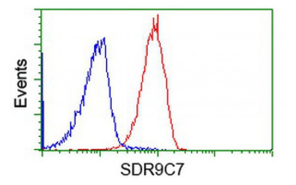
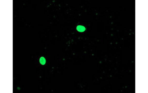
 IF (Immunofluorescence)
(Immunofluorescent staining of COS7 cells transiently transfected with recombinant SDR9C7 protein using SDR9C7 antibody)
IF (Immunofluorescence)
(Immunofluorescent staining of COS7 cells transiently transfected with recombinant SDR9C7 protein using SDR9C7 antibody)
SDR9C7, Monoclonal Antibody (Cat# AAA107323)
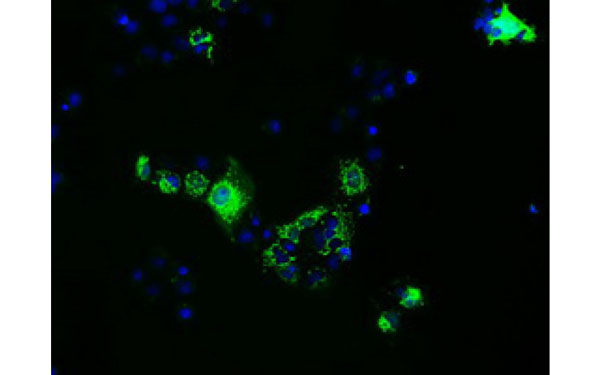
 FCM/FACS (Flow Cytometry)
(Staining of 3-day ConA stimulated (right) or unstimulated (left) BALB/c mouse splenocytes with 0.06 ug of Ly-6A/E antibody (PE). Total viable cells were used for analysis. Markers were set based on the autofluorescence sample.)
FCM/FACS (Flow Cytometry)
(Staining of 3-day ConA stimulated (right) or unstimulated (left) BALB/c mouse splenocytes with 0.06 ug of Ly-6A/E antibody (PE). Total viable cells were used for analysis. Markers were set based on the autofluorescence sample.)
Ly6A/E, Monoclonal Antibody (Cat# AAA107326)
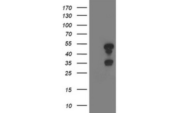
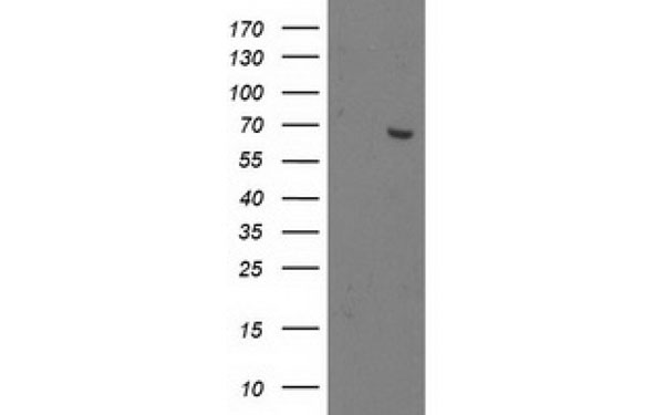
 WB (Western Blot)
(Western Blot analysis of HEK293T cell lysates (5 ug) transfected with either recombinant LXN protein (Right) or empty vector (Left) detected with LXN antibody)
WB (Western Blot)
(Western Blot analysis of HEK293T cell lysates (5 ug) transfected with either recombinant LXN protein (Right) or empty vector (Left) detected with LXN antibody)
LXN, Monoclonal Antibody (Cat# AAA107337)
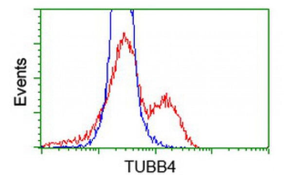
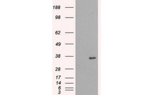
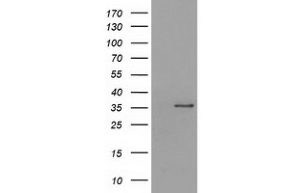
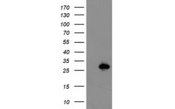
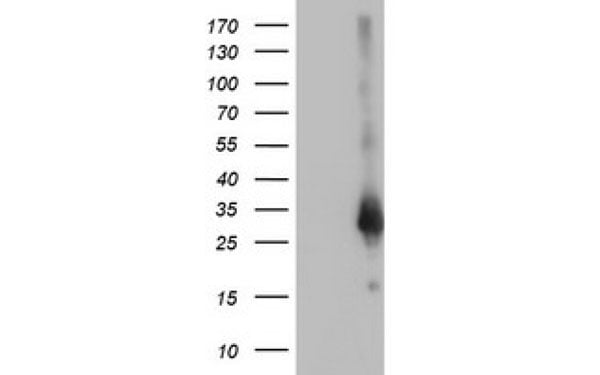
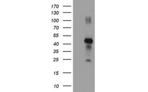
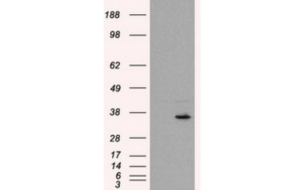
 WB (Western Blot)
(Western Blot analysis of HEK293T cell lysates (5 ug) transfected with either recombinant TUBB4 protein (Right) or empty vector (Left) detected with TUBB4 antibody)
WB (Western Blot)
(Western Blot analysis of HEK293T cell lysates (5 ug) transfected with either recombinant TUBB4 protein (Right) or empty vector (Left) detected with TUBB4 antibody)
TUBB4, Monoclonal Antibody (Cat# AAA107350)
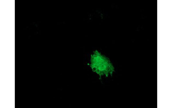 IF (Immunofluorescence)
(Immunofluorescent staining of COS7 cells transiently transfected with recombinant SNX8 protein using SNX8 antibody)
IF (Immunofluorescence)
(Immunofluorescent staining of COS7 cells transiently transfected with recombinant SNX8 protein using SNX8 antibody)
SNX8, Monoclonal Antibody (Cat# AAA107362)
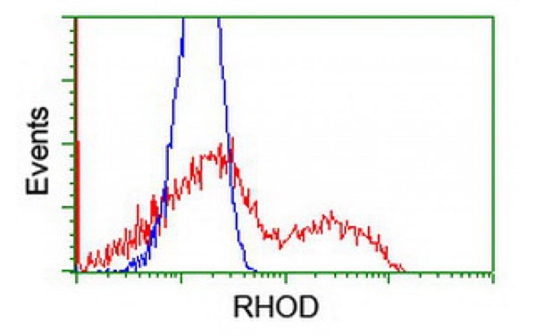
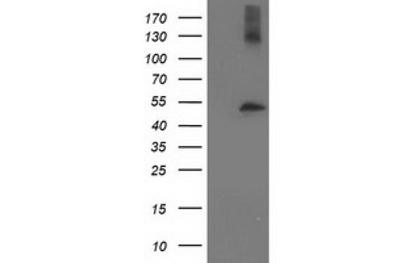
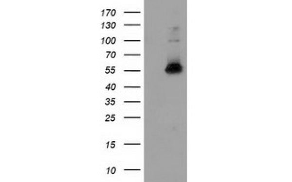
 WB (Western Blot)
(Western Blot analysis of HEK293T cell lysates (5 ug) transfected with either recombinant RHOD protein (Right) or empty vector (Left) detected with RHOD antibody)
WB (Western Blot)
(Western Blot analysis of HEK293T cell lysates (5 ug) transfected with either recombinant RHOD protein (Right) or empty vector (Left) detected with RHOD antibody)
RHOD, Monoclonal Antibody (Cat# AAA107380)
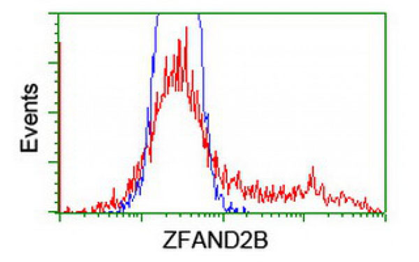
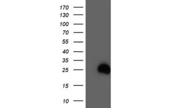
 WB (Western Blot)
(Western Blot analysis of HEK293T cell lysates (5 ug) transfected with either recombinant ZFAND2B protein (Right) or empty vector (Left) detected with ZFAND2B antibody)
WB (Western Blot)
(Western Blot analysis of HEK293T cell lysates (5 ug) transfected with either recombinant ZFAND2B protein (Right) or empty vector (Left) detected with ZFAND2B antibody)
ZFAND2B, Monoclonal Antibody (Cat# AAA107387)
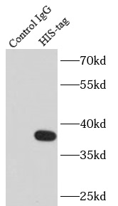
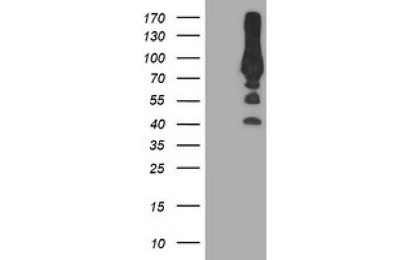
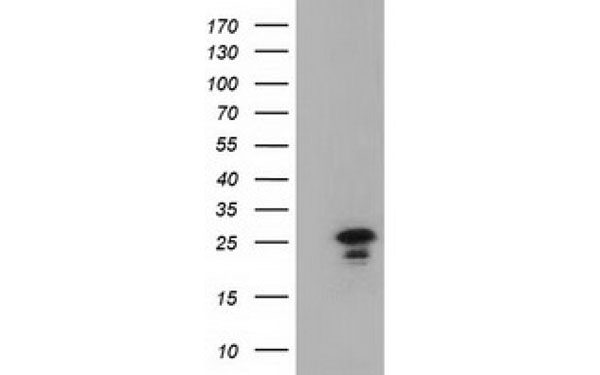
 WB (Western Blot)
(Western blot of 6*His-tagged fusion protein with AAA102666 (anti-6*HIS tag) at dilution of 1:5000.)
WB (Western Blot)
(Western blot of 6*His-tagged fusion protein with AAA102666 (anti-6*HIS tag) at dilution of 1:5000.)
6*His, Monoclonal Antibody (Cat# AAA102666)
Protein A+G purification
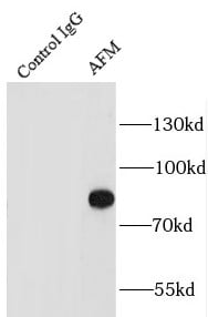
 WB (Western Blot)
(human blood tissue were subjected to SDS PAGE followed by western blot with AAA102671 (AFM antibody) at dilution of 1:1000)
WB (Western Blot)
(human blood tissue were subjected to SDS PAGE followed by western blot with AAA102671 (AFM antibody) at dilution of 1:1000)
AFM, Monoclonal Antibody (Cat# AAA102671)
Protein A+G purification
 WB (Western Blot)
(HeLa cells were subjected to SDS PAGE followed by western blot with AAA102672 (AHCY antibody) at dilution of 1:1000)
WB (Western Blot)
(HeLa cells were subjected to SDS PAGE followed by western blot with AAA102672 (AHCY antibody) at dilution of 1:1000)
AHCY, Monoclonal Antibody (Cat# AAA102672)
Protein A+G purification
 WB (Western Blot)
(HeLa cells were subjected to SDS PAGE followed by western blot with AAA102673 (AK2 antibody) at dilution of 1:4000)
WB (Western Blot)
(HeLa cells were subjected to SDS PAGE followed by western blot with AAA102673 (AK2 antibody) at dilution of 1:4000)
AK2, Monoclonal Antibody (Cat# AAA102673)
Protein A+G purification
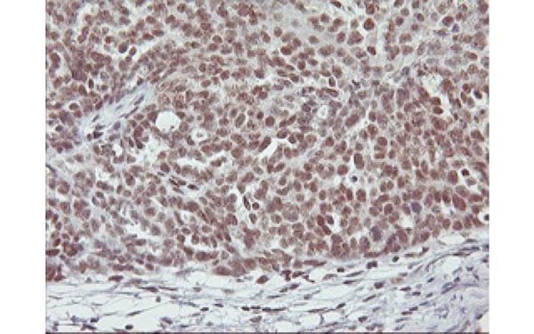
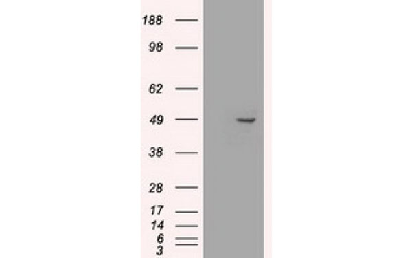
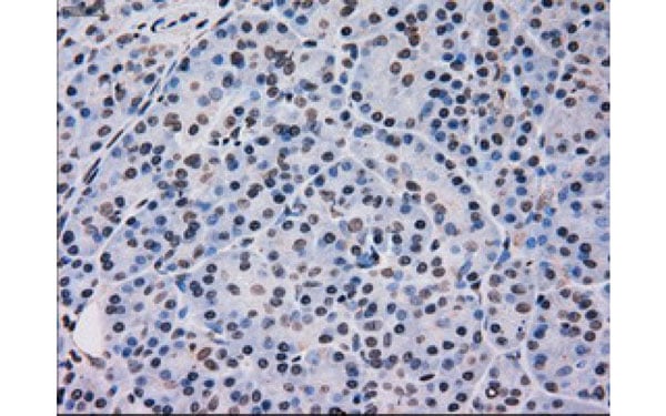 IHC (Immunohiostchemistry)
(Immunohistochemical analysis of MAPK12 protein in paraffin embedded Human pancreas tissue using MAPK12 antibody)
IHC (Immunohiostchemistry)
(Immunohistochemical analysis of MAPK12 protein in paraffin embedded Human pancreas tissue using MAPK12 antibody)
MAPK12, Monoclonal Antibody (Cat# AAA106663)
 WB (Western Blot)
(Western Blot analysis of HEK293T cell lysates (5 ug) transfected with either recombinant RNPEP protein (Right) or empty vector (Left) detected with RNPEP antibody)
WB (Western Blot)
(Western Blot analysis of HEK293T cell lysates (5 ug) transfected with either recombinant RNPEP protein (Right) or empty vector (Left) detected with RNPEP antibody)
RNPEP, Monoclonal Antibody (Cat# AAA106672)
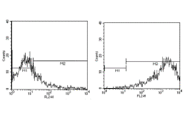
 FCM/FACS (Flow Cytometry)
(Flow Cytometric analysis using IL4 antibodyNormal human peripheral blood cells were stimulated with PMA and Ionomycin in the presence of brefeldin A for 6 hours. The cells were surface stained with CD3 antibody (FITC) and intracellularly stained with IL-4 antibody (PE) (right panel). Left panel demonstrates unlabelled antibody blocking control.)
FCM/FACS (Flow Cytometry)
(Flow Cytometric analysis using IL4 antibodyNormal human peripheral blood cells were stimulated with PMA and Ionomycin in the presence of brefeldin A for 6 hours. The cells were surface stained with CD3 antibody (FITC) and intracellularly stained with IL-4 antibody (PE) (right panel). Left panel demonstrates unlabelled antibody blocking control.)
IL4, Monoclonal Antibody (Cat# AAA106674)
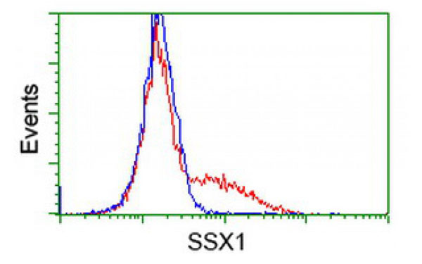
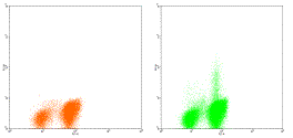
 IF (Immunofluorescence)
(Immunofluorescent staining of COS7 cells transiently transfected with recombinant SSX1 protein using SSX1 antibody)
IF (Immunofluorescence)
(Immunofluorescent staining of COS7 cells transiently transfected with recombinant SSX1 protein using SSX1 antibody)
SSX1, Monoclonal Antibody (Cat# AAA106688)
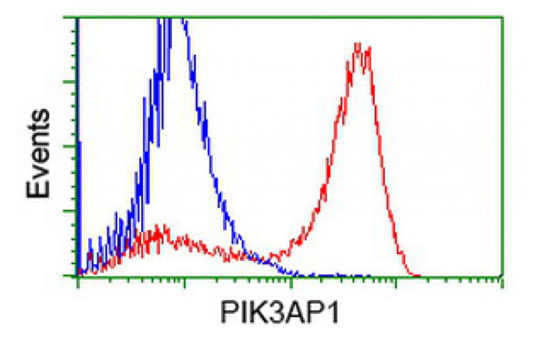
 IF (Immunofluorescence)
(Immunofluorescent staining of COS7 cells transiently transfected with recombinant PIK3AP1 protein using PIK3AP1 antibody)
IF (Immunofluorescence)
(Immunofluorescent staining of COS7 cells transiently transfected with recombinant PIK3AP1 protein using PIK3AP1 antibody)
PIK3AP1, Monoclonal Antibody (Cat# AAA106691)
 IF (Immunofluorescence)
(Immunofluorescent staining of COS7 cells transiently transfected with recombinant NPR3 protein using NPR3 antibody)
IF (Immunofluorescence)
(Immunofluorescent staining of COS7 cells transiently transfected with recombinant NPR3 protein using NPR3 antibody)
NPR3, Monoclonal Antibody (Cat# AAA106693)
HPV16 E7, Monoclonal Antibody (Cat# AAA106694)
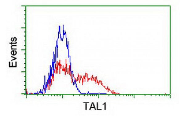
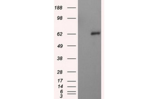
 WB (Western Blot)
(Western Blot analysis of HEK293T cell lysates (5 ug) transfected with either recombinant TAL1 protein (Right) or empty vector (Left) detected with TAL1 antibody)
WB (Western Blot)
(Western Blot analysis of HEK293T cell lysates (5 ug) transfected with either recombinant TAL1 protein (Right) or empty vector (Left) detected with TAL1 antibody)
TAL1, Monoclonal Antibody (Cat# AAA106705)
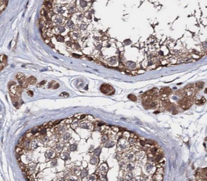
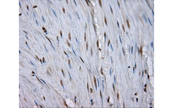
 IHC (Immunohistochemisry)
(Immunohistochemical analysis of RNF113B protein in paraffin embedded Adenocarcinoma of Human ovary tissue using RNF113B antibody)
IHC (Immunohistochemisry)
(Immunohistochemical analysis of RNF113B protein in paraffin embedded Adenocarcinoma of Human ovary tissue using RNF113B antibody)
RNF113B, Monoclonal Antibody (Cat# AAA106710)
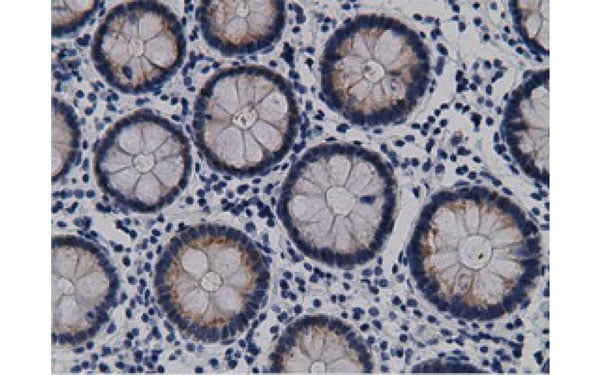
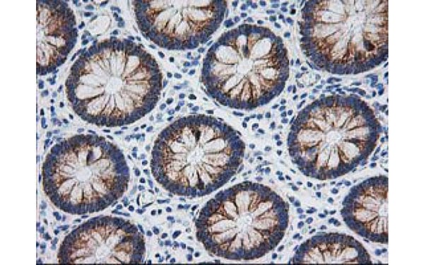
 IHC (Immunohiostchemistry)
(Immunohistochemical analysis of LIPG protein in paraffin embedded Human colon tissue using LIPG antibody)
IHC (Immunohiostchemistry)
(Immunohistochemical analysis of LIPG protein in paraffin embedded Human colon tissue using LIPG antibody)
LIPG, Monoclonal Antibody (Cat# AAA106723)
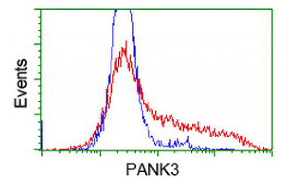
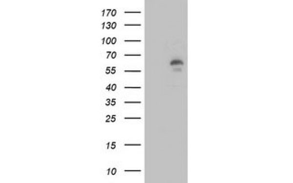
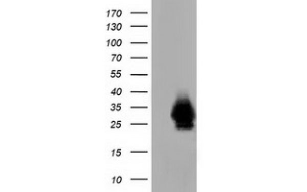
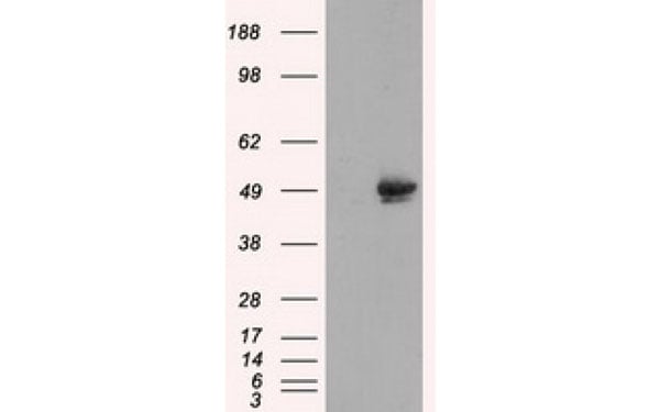
 WB (Western Blot)
(Western Blot analysis of HEK293T cell lysates (5 ug) transfected with either recombinant PANK3 protein (Right) or empty vector (Left) detected with PANK3 antibody)
WB (Western Blot)
(Western Blot analysis of HEK293T cell lysates (5 ug) transfected with either recombinant PANK3 protein (Right) or empty vector (Left) detected with PANK3 antibody)
PANK3, Monoclonal Antibody (Cat# AAA106732)
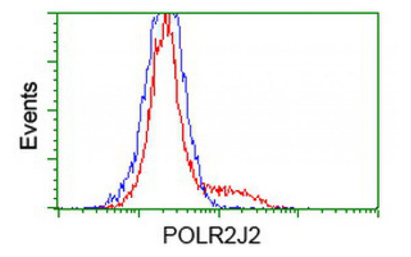
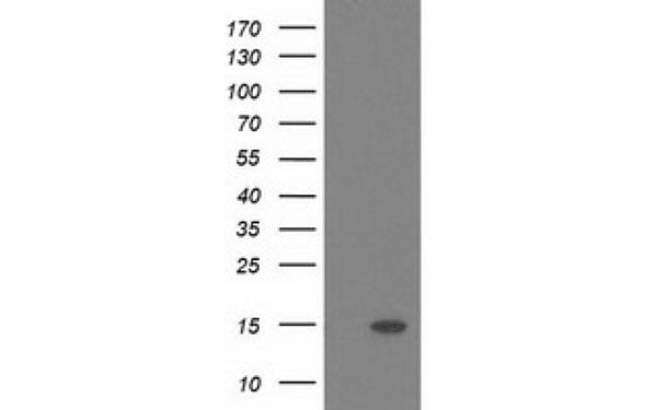 WB (Western Blot)
(Western Blot analysis of HEK293T cell lysates (5 ug) transfected with either recombinant POLR2J2 protein (Right) or empty vector (Left) detected with POLR2J2 antibody)
WB (Western Blot)
(Western Blot analysis of HEK293T cell lysates (5 ug) transfected with either recombinant POLR2J2 protein (Right) or empty vector (Left) detected with POLR2J2 antibody)
POLR2J2, Monoclonal Antibody (Cat# AAA106734)
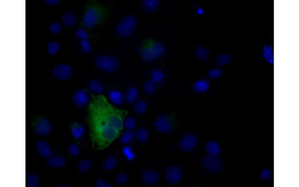
 IF (Immunofluorescence)
(Immunofluorescent staining of COS7 cells transiently transfected with recombinant CRYM protein using CRYM antibody)
IF (Immunofluorescence)
(Immunofluorescent staining of COS7 cells transiently transfected with recombinant CRYM protein using CRYM antibody)
CRYM, Monoclonal Antibody (Cat# AAA106196)
 IF (Immunofluorescence)
(Immunofluorescent staining of COS7 cells transiently transfected with recombinant FAHD2A protein using FAHD2A antibody)
IF (Immunofluorescence)
(Immunofluorescent staining of COS7 cells transiently transfected with recombinant FAHD2A protein using FAHD2A antibody)
FAHD2A, Monoclonal Antibody (Cat# AAA106478)
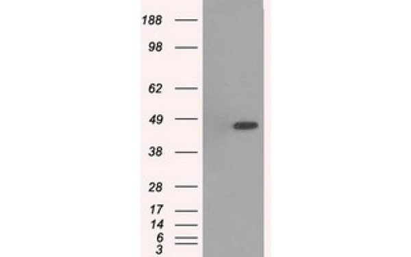
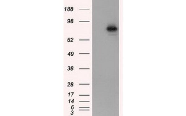
 WB (Western Blot)
(Western Blot showing HER-2 antibody used against truncated HER-2 recombinant protein.)
WB (Western Blot)
(Western Blot showing HER-2 antibody used against truncated HER-2 recombinant protein.)
HER2, Monoclonal Antibody (Cat# AAA106479)
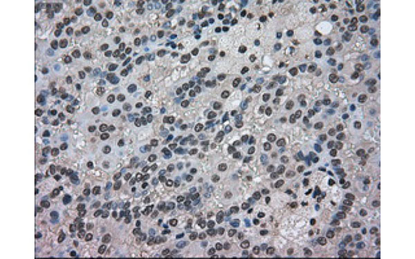
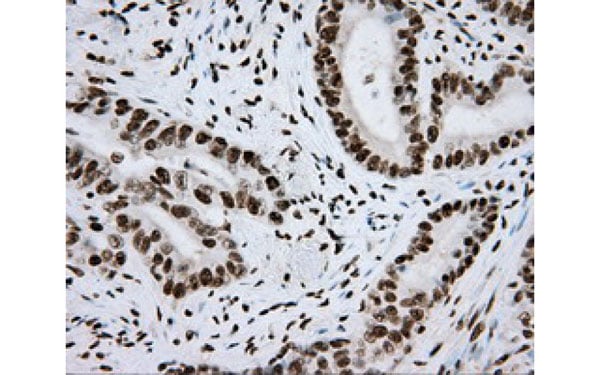
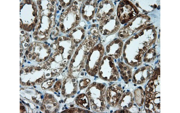 IHC (Immunohiostchemistry)
(Immunohistochemical analysis of CELF1 protein in paraffin embedded Human kidney tissue using CELF1 antibody)
IHC (Immunohiostchemistry)
(Immunohistochemical analysis of CELF1 protein in paraffin embedded Human kidney tissue using CELF1 antibody)
CELF1, Monoclonal Antibody (Cat# AAA106482)
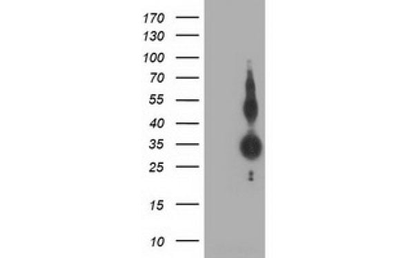
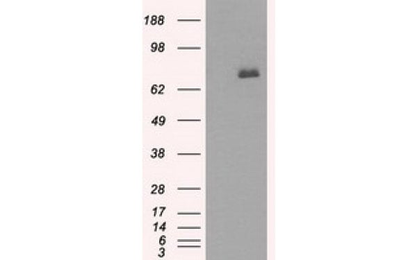
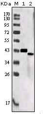 WB (Western Blot)
(Western Blot analysis using SORL1 antibodyWestern Blot showing SORL1 antibody used against truncated SORL1 recombinant protein (1) and SORL1 (aa2159-2214)-hIgGFc transfected CHO-K1 cell lysate (2).)
WB (Western Blot)
(Western Blot analysis using SORL1 antibodyWestern Blot showing SORL1 antibody used against truncated SORL1 recombinant protein (1) and SORL1 (aa2159-2214)-hIgGFc transfected CHO-K1 cell lysate (2).)
SORL1, Monoclonal Antibody (Cat# AAA106485)
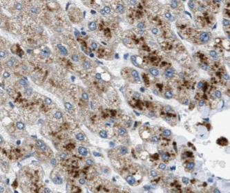
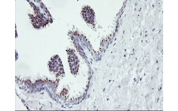
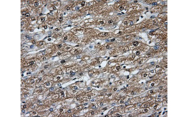
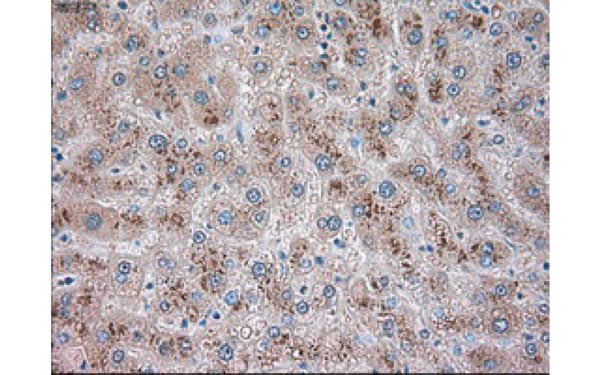 IHC (Immunohiostchemistry)
(Immunohistochemical analysis of GFAP protein in paraffin embedded Human liver tissue using GFAP antibody)
IHC (Immunohiostchemistry)
(Immunohistochemical analysis of GFAP protein in paraffin embedded Human liver tissue using GFAP antibody)
GFAP, Monoclonal Antibody (Cat# AAA106516)
 IF (Immunofluorescence)
(Immunofluorescent staining of COS7 cells transiently transfected with recombinant ARNT protein using ARNT antibody)
IF (Immunofluorescence)
(Immunofluorescent staining of COS7 cells transiently transfected with recombinant ARNT protein using ARNT antibody)
ARNT, Monoclonal Antibody (Cat# AAA106517)
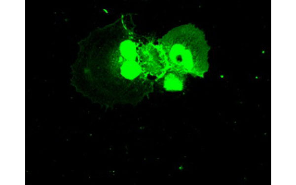
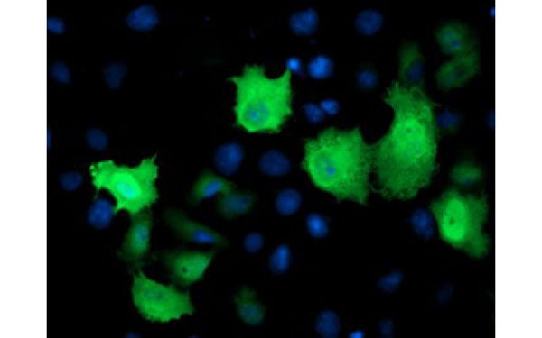
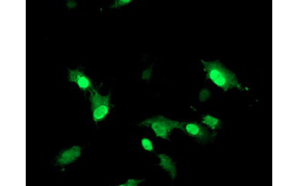
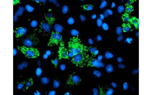
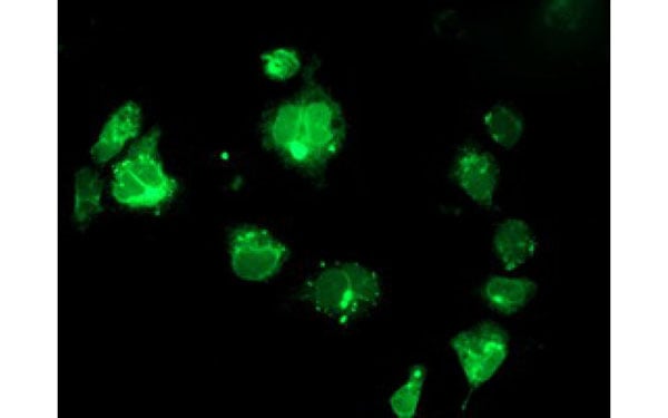
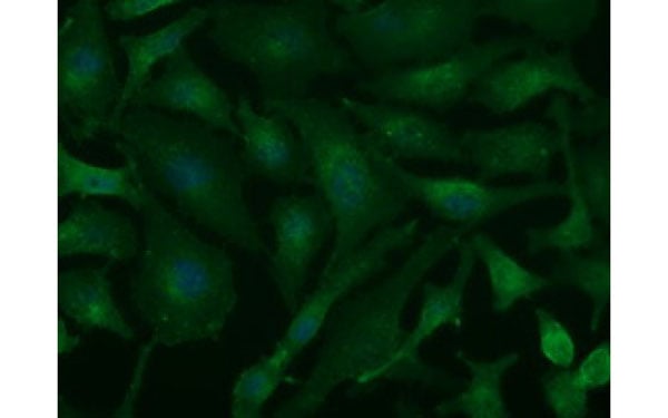 IF (Immunofluorescence)
(Immunofluorescent staining of endogenous BSG protein in Hela cells using BSG antibody)
IF (Immunofluorescence)
(Immunofluorescent staining of endogenous BSG protein in Hela cells using BSG antibody)
BSG, Monoclonal Antibody (Cat# AAA106527)
 IF (Immunofluorescence)
(Immunofluorescent staining of COS7 cells transiently transfected with recombinant AK4 protein using AK4 antibody)
IF (Immunofluorescence)
(Immunofluorescent staining of COS7 cells transiently transfected with recombinant AK4 protein using AK4 antibody)
AK4, Monoclonal Antibody (Cat# AAA106543)
 IF (Immunofluorescence)
(Immunofluorescent staining of COS7 cells transiently transfected with recombinant BTK protein using BTK antibody)
IF (Immunofluorescence)
(Immunofluorescent staining of COS7 cells transiently transfected with recombinant BTK protein using BTK antibody)
BTK, Monoclonal Antibody (Cat# AAA106550)
 IF (Immunofluorescence)
(Immunofluorescent staining of COS7 cells transiently transfected with recombinant SSR1 protein using SSR1 antibody)
IF (Immunofluorescence)
(Immunofluorescent staining of COS7 cells transiently transfected with recombinant SSR1 protein using SSR1 antibody)
SSR1, Monoclonal Antibody (Cat# AAA106583)
What are Monoclonal Antibodies?
Monoclonal antibodies are specialized laboratory-produced proteins developed for binding to specific biological antigens or other molecular targets. Since they come from a single cell (or clone), they are especially consistent and accurate in the data they are involved in producing.
This type of antibody material has been shown to be a powerful tool in finding and subsequently destroying harmful cells in an organism, such as those found in cancers or various autoimmune diseases. This makes them excellent aids in medical testing and research, which is why they are so widely used.
AAA Biotech offers a comprehensive range of high-quality monoclonal antibodies that perform effectively in various laboratory tests, including (amongst others) ELISA, western blotting, immunohistochemistry, and flow cytometry. All of the products in our catalog are thoroughly quality tested to make sure that they are reliable and will consistently perform well in your research.
What Are The Uses of Monoclonal Antibodies
Monoclonal antibodies are used in many lab tests, including (amongst others) ELISA, western blotting, immunohistochemistry, and flow cytometry.
ELISA is a test that helps detect a specific substance/analyte in a sample. It uses antibodies (often monoclonal) bound to a solid surface (such as the well of a microplate) to “capture” the substance/analyte in the sample and immobilize it so that the detection antibody component can then bind to it and produce a signal, which can then be measured.
Western blotting identifies specific proteins in a sample. The sample is first separated on a gel, and then antibodies are applied that will typically bind to the target, which will all be localized to a single band in a lane.
Immunohistochemistry helps locate specific proteins in cells or tissue samples using antibodies.
Flow cytometry looks at and sorts cells. It uses antibodies that are conjugated to reporter molecules called “fluorophores”, which, under special lights, emit light themselves, which can then be measured by a detector instrument.
How Monoclonal Antibodies Are Used as Medicine?
Please note that all of the products listed in AAA Biotech’s also known as AAA Bio or AAABio catalog are strictly for research-use only (RUO).
Monoclonal antibodies can also be used as therapeutic/medical treatments, particularly in the context of cancers. They are designed to find and bind to specific cells or proteins, helping the immune system recognize and attack the cancer. These treatments work in different ways, such as:
- Radioimmunotherapy attaches a small amount of radioactive molecule to the antibody, so it delivers the radiation directly to the cancer cells that the antibody is specifically binding to.
- Antibody-directed enzyme prodrug therapy uses antibodies that are specifically bound to special enzymes. These enzymes activate a harmless drug in the body and turn it into a cancer-killing drug only near the cancer cells—this helps avoid harming healthy cells.
- Immunoliposomes are tiny “bubbles” filled with medicine/drug and coated with antibodies. They carry the drug straight to the cancer cells.
Why Buy Monoclonal Antibodies From Us?
At AAA Biotech, we provide high-performance monoclonal antibodies designed to support a wide range of research needs.
1. Validated for Versatile Applications
The antibodies in our catalog are extensively validated and compatible with multiple techniques, including (but not limited to) ELISA, flow cytometry (FC), immunocytochemistry (ICC), immunofluorescence (IF), immunohistochemistry (IHC), immunoprecipitation (IP), and western blotting (WB).
2. Wide Selection & Specialized Options
We offer antibodies for common and rare species, that are available in various conjugated forms, and also in recombinant formats. Essentially, there is almost anything one might need to meet their experimental model’s requirements.
3. High-Quality Proteins
Our proteins meet high purity standards—90% or more as confirmed by SDS-PAGE. Many are available with tags like His, Flag, GST, or MBP, and we also supply native and biologically active proteins for functional studies.
Frequently Asked Questions
1. Are your monoclonal antibodies validated for specific applications?
Yes, our antibodies are tested and validated for use in methods such as ELISA, western blot, IHC, flow cytometry, and more. Refer to specific product pages or datasheets for individual product information.
2. How do I choose the right monoclonal antibody for my application?
Review the product details directly for application validation, species reactivity, and target information. You may also contact our support team at any time for help.
3. How quickly can I receive my order?
Most orders are processed and shipped within 1–3 business days, depending on product availability and your shipping location.