Get accurate results in your research with our Monoclonal Antibodies, which are specially made to target exactly what you require for your research, and will produce consistent, reliable performance in lab tests.
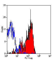
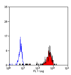
Application Data (Staining of human peripheral blood platelets with Mouse anti Human CD36)
Application Data (Staining of human peripheral blood platelets with Mouse anti Human CD36)
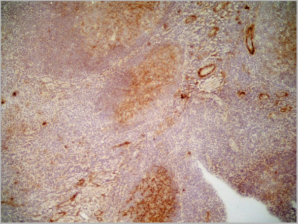
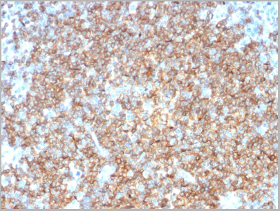
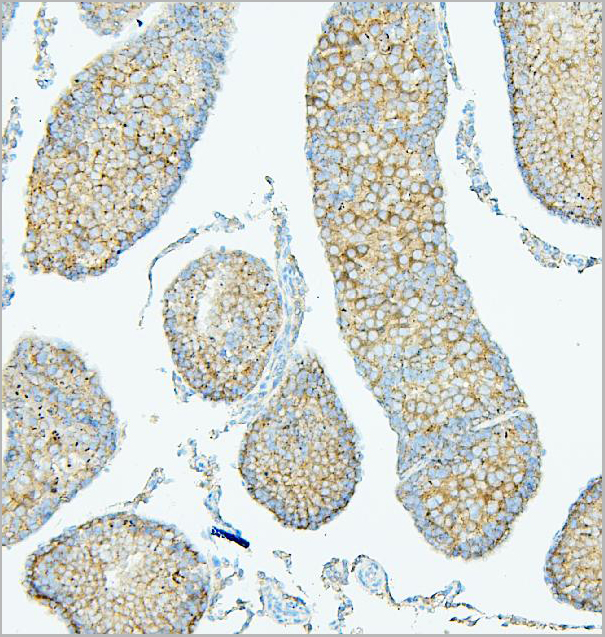
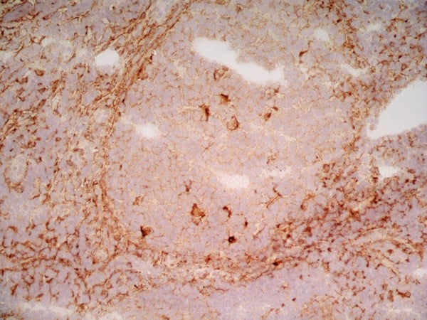
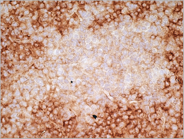
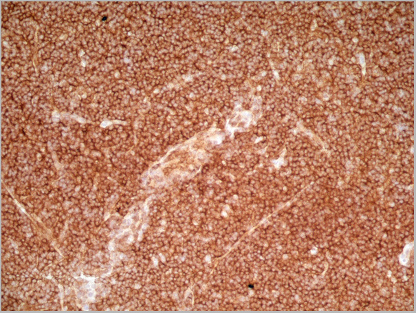
Application Data (Immunoperoxidase staining of human tonsil cryosection with Mouse anti Human CD36 antibody, clone SMø followed by the HISTAR detection system . High power SMθ)
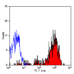
Application Data (Staining of human peripheral blood lymphocytes with Mouse anti Human CD36: FITC)
Application Data (Staining of human peripheral blood monocytes with Mouse anti Human CD36: Azide Free)
Application Data (Staining of human peripheral blood monocytes with Mouse anti Human CD36: Alexa Fluor 647)
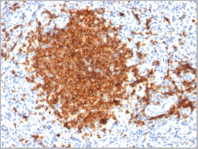
Application Data (Immunoperoxidase staining of human tonsil cryosection with Mouse anti Human CD36 antibody, clone SMø followed by the HISTAR detection system . Low power)
Application Data (Immunoperoxidase staining of human tonsil cryosection with Mouse anti Human CD36 antibody, clone SMø followed by the HISTAR detection system . Medium power)
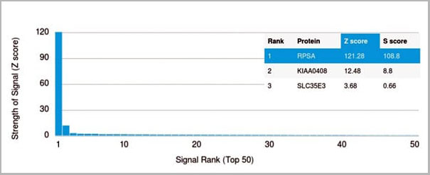
Application Data (Analysis of Protein Array containing more than 19,000 full-length human proteins using Laminin Receptor Monospecific Mouse Monoclonal Antibody (RPSA/2699) Z- and S- Score: The Z-score represents the strength of a signal that a monoclonal antibody (MAb) (in combination with a fluorescently-tagged anti-IgG secondary antibody) produces when binding to a particular protein on the HuProtTM array. Z-scores are described in units of standard deviations (SD's) above the mean value of all signals generated on that array. If targets on HuProtTM are arranged in descending order of the Z-score, the S-score is the difference (also in units of SD's) between the Z-score. S-score therefore represents the relative target specificity of a MAb to its intended target. A MAb is considered to specific to its intended target, if the MAb has an S-score of at least 2.5. For example, if a MAb binds to protein X with a Z-score of 43 and to protein Y with a Z-score of 14, then the S-score for the binding of that MAb to protein X is equal to 29.)
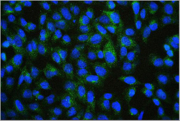
IF (Immunofluorescence) (Immunofluorescent staining of paraformaldehyde-Raji cells using Laminin Receptor Monospecific Mouse Monoclonal Antibody (RPSA/2699) followed by goat anti-Mouse IgG conjugated to CF488 (green). Nuclei are labeled with RedDot (red).)
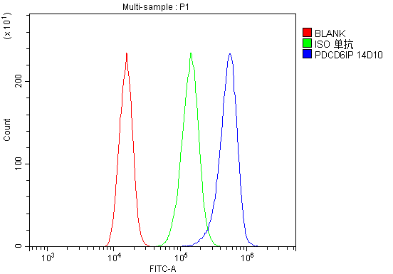
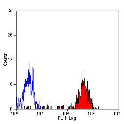
FCM (Flow Cytometry) (Flow Cytometric Analysis of paraformaldehyde-fixed Raji cells. Laminin Receptor Monospecific Mouse Monoclonal Antibody (RPSA/2699) followed by goat anti-Mouse IgG-CF488 (blue) and Isotype Control (Red).)
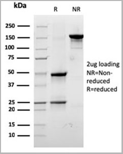
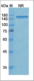
SDS-PAGE (SDS-PAGE Analysis Purified Laminin Receptor Monospecific Mouse Monoclonal Antibody (RPSA/2699). Confirmation of Integrity and Purity of Antibody)
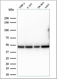
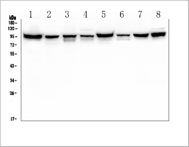
WB (Western Blot) (Western Blot Analysis of human THP-1, U937, SK-BR3, and A431 cell lysates. Laminin Receptor Monospecific Mouse Monoclonal Antibody (RPSA/2699).)
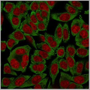
IF (Immunofluorescence) (Immunofluorescent staining of HeLa cells using Laminin Receptor Monospecific Mouse Monoclonal Antibody (RPSA/2699) followed by goat anti-Mouse IgG conjugated to CF488 (green). Nuclei are labeled with RedDot (red).)
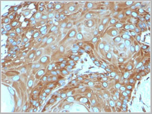
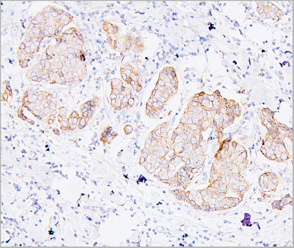
IHC (Immunohistochemistry) (Formalin-fixed, paraffin-embedded human Basal Cell Carcinoma stained with Laminin Receptor Monospecific Mouse Monoclonal Antibody (RPSA/2699).)
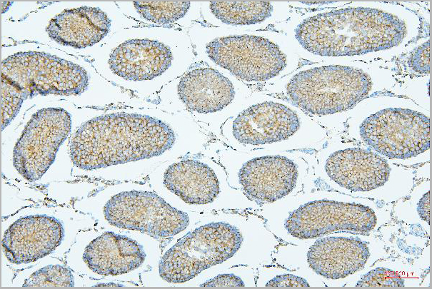
IHC (Immunohistochemistry) (Formalin-fixed, paraffin-embedded human Tonsil stained with Laminin Receptor Monospecific Mouse Monoclonal Antibody (RPSA/2699).)
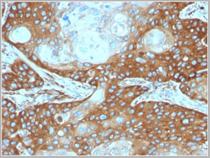
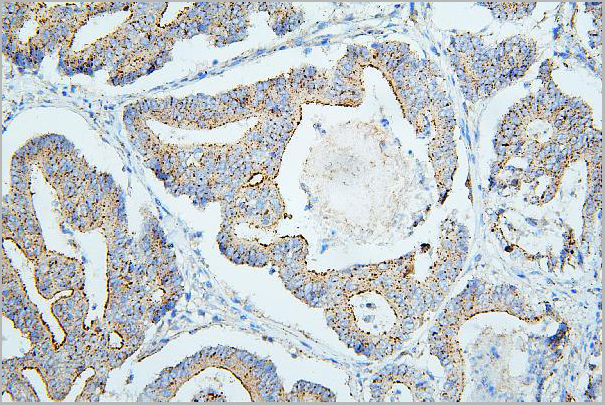
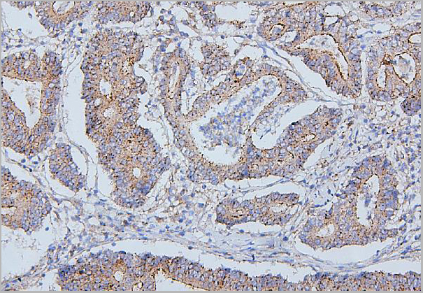
IHC (Immunohistochemistry) (Formalin-fixed, paraffin-embedded human Cervix Carcinoma stained with Laminin Receptor Monospecific Mouse Monoclonal Antibody (RPSA/2699).)
IHC (Immunohistochemistry) (Formalin-fixed, paraffin-embedded human Colon Carcinoma stained with Laminin Receptor Monospecific Mouse Monoclonal Antibody (RPSA/2699).)
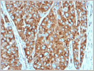
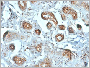
IHC (Immunohistochemistry) (Formalin-fixed, paraffin-embedded human Breast Carcinoma stained with Laminin Receptor Monospecific Mouse Monoclonal Antibody (RPSA/2699).)
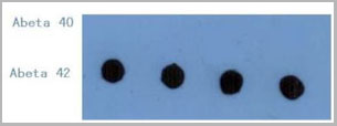
DB (Dot Blot) (Dot Blot: The figure below is the result of using 1ug/mL anti- Aβ42 clone CA9 10C11 to detect 10ng/dot Aβ42 or Aβ40. The antibody showed no cross-reactivity with Aβ40.)
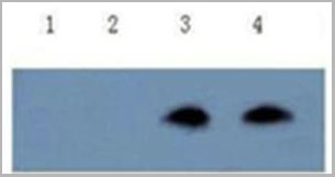
WB (Western Blot) (Sandwich ELISA: In combination with anti-amyloid peptide N-terminus capture antibody (mAb clone NT 5B8, Cat. No.: please inquire), the antibody can detect Aβ42 in sandwich ELISA assayWestern Blot: The figure below is the result of using 2ug/mL anti- Aβ42 clone CA9 10C11 to detect Aβ42 on Western blot following Tris-Tricine separation gel electrophoresis. The antibody showed no cross-reactivity with Aβ40.Lane 1 &2 : Aβ40 Lane 3&4: Aβ42)
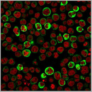
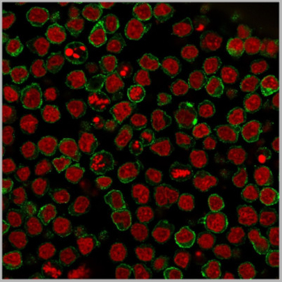
IF (Immunofluorescence) (Immunofluorescent staining of paraformaldehyde-fixed Ramos cells using CD22-Monospecific Mouse Monoclonal Antibody (BLCAM/1795) followed by goat anti-Mouse IgG conjugated to CF488 (green). Nuclei are stained with Reddot.)
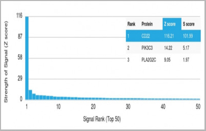
ELISA (Analysis of Protein Array containing more than 19,000 full-length human proteins using CD22-Monospecific Mouse Monoclonal Antibody (BLCAM/1795) Z-and S- Score: The Z-score represents the strength of a signal that a monoclonal antibody (MAb) (in combination with a fluorescently-tagged anti-IgG secondary antibody) produces when binding to a particular protein on the HuProtTM array. Z-scores are described in units of standard deviations (SD's) above the mean value of all signals generated on that array. If targets on HuProtTM are arranged in descendingorder of the Z-score, the S-score is the difference (also in units of SD's) between the Z-score. S-score therefore represents the relative target specificity of a MAb to its intended target. A MAb is considered to specific to its intended target, if the MAb has an S-score of at least 2.5. For example, if a MAb binds to protein X with a Z-score of 43 and to protein Y with a Z-score of 14, then the S-score for the binding of that MAb to protein X is equal to 29.)
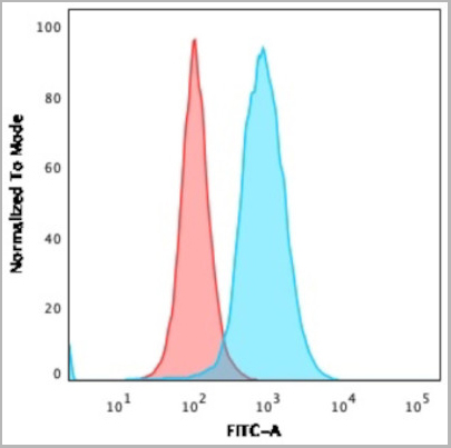
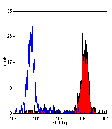
FCM (Flow Cytometry) (Flow Cytometric Analysis of paraformaldehyde-fixed MOLT4 cells. CD22-Monospecific Mouse Monoclonal Antibody (BLCAM/1795) followed by goat anti-Mouse IgG-CF488 (Blue); Isotype Control (Red).)
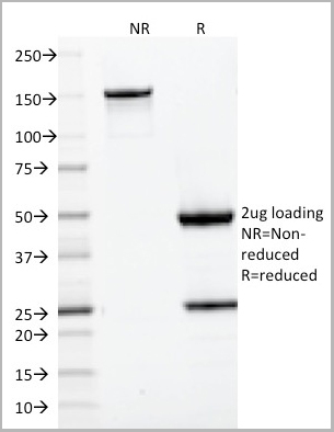
SDS-PAGE (SDS-PAGE Analysis Purified CD22-Monospecific Mouse Monoclonal Antibody (BLCAM/1795). Confirmation of Purity and Integrity of Antibody)
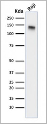
WB (Western Blot) (Western Blot Analysis of Raji cell lysate using CD22-Monospecific Mouse Monoclonal Antibody (BLCAM/1795).)
WB (Western Blot) (Western Blot Analysis of Raji and Ramos Cell Lysates using CD22 Mouse Monoclonal Antibody (BLCAM/1795).)
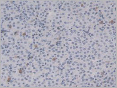
IHC (Immunohistochemistry) (Formalin-fixed, paraffin-embedded human Tonsil stained with CD22 Mouse Monoclonal Antibody (BLCAM/1795).)
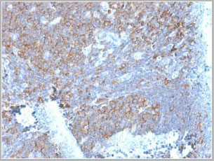
IHC (Immunohistochemistry) (Formalin-fixed, paraffin-embedded human Spleen stained with CD22 Mouse Monoclonal Antibody (BLCAM/1795).)
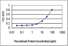
Application Data (Detection limit for recombinant GST tagged TCEA3 is approximately 0.03ng/ml as a capture antibody.)
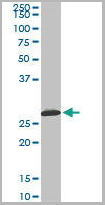
WB (Western Blot) (TCEA3 monoclonal antibody (M08), clone 4E11 Western Blot analysis of TCEA3 expression in HepG2.)
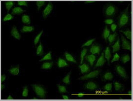
IF (Immunofluorescence) (Immunofluorescence of monoclonal antibody to TCEA3 on HepG2 cell. [antibody concentration 10 ug/ml])
IF (Immunofluorescence) (Immunofluorescence of monoclonal antibody to TCEA3 on HepG2 cell. [antibody concentration 10 ug/ml])
IHC (Immunohistochemistry) (Immunoperoxidase of monoclonal antibody to TCEA3 on formalin-fixed paraffin-embedded human salivary gland. [antibody concentration 3 ug/ml])
Application Data (Immunoperoxidase of monoclonal antibody to TCEA3 on formalin-fixed paraffin-embedded human salivary gland. [antibody concentration 3 ug/ml])
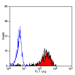
Application Data (Staining of human peripheral blood monocytes probed with Mouse anti Human CD14:FITC)
Application Data (Staining of human peripheral blood monocytes with Mouse anti Human CD14:RPE-Alexa Fluor 750)
Application Data (Staining of human peripheral blood monocytes with Mouse anti Human CD14)
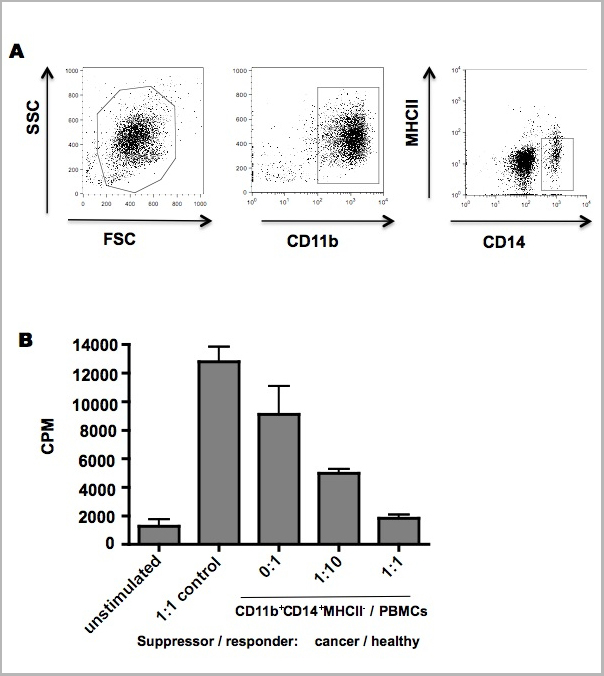
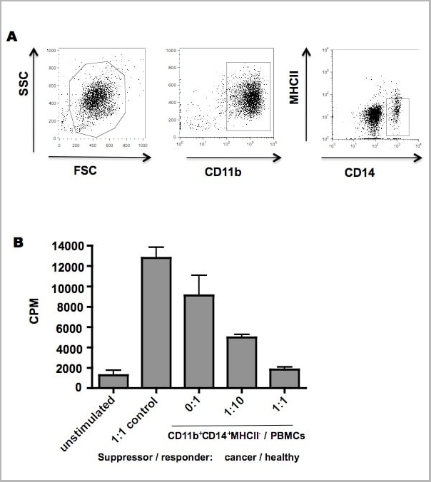
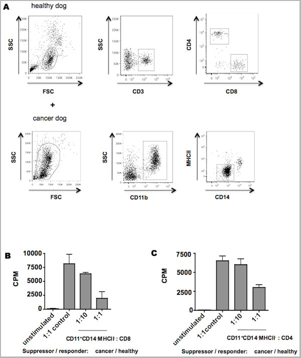
Application Data (Published customer image: CD11b+CD14+MHCII- cells demonstrate ability to suppressive T cell proliferation. (A) CD11b+CD14+MHCII- cells were sorted from peripheral blood sample of an osteosarcoma dog (B) and co-cultured with healthy dog PBMCs in the presence of mitogen for 72 hs. Non-stimulated PBMCs were used as negative control and PBMCs co-cultured with healthy PMNs were used to control for the effect of adding cells to the assay. Proliferative responses were measured by 3H-thymidine incorporation. CPM, counts per minute. The experiment was performed in triplicate. Mean +/- SEM are shown.From: Goulart MR, Pluhar GE, Ohlfest JR (2012) Identification of Myeloid Derived Suppressor Cells in Dogs with Naturally Occurring Cancer. PLoS ONE 7(3): e33274.)
Application Data (Staining of human peripheral blood monocytes with Mouse anti Human CD14)
Application Data (Staining of human peripheral blood monocytes with Mouse anti Human CD14:Biotin)
Application Data (Published customer image: CD11b+CD14-MHCII- cells suppress T cell proliferation. Facs sorted CD11b+CD14-MHCII- cells isolated from a dog with osteosarcoma or healthy PBMCs were co-incubated with mitogen-stimulated CD4+ and CD8+ T cells isolated from a healthy dog for 72 hs. No stimulated cells were used as negative control. Proliferative responses were measured by 3H-thymidine incorporation from experiments performed in triplicate. CPM, counts per minute. Mean +/- SEM are shown.From: Goulart MR, Pluhar GE, Ohlfest JR (2012) Identification of Myeloid Derived Suppressor Cells in Dogs with Naturally Occurring Cancer. PLoS ONE 7(3): e33274.)
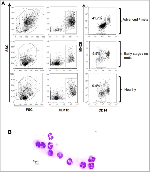
Application Data (Published customer image: Immunophenotyping gating strategy and morphological analysis for MDSC identification in peripheral blood of dogs. PBMCs from healthy dogs and dogs with cancer were stained for the myeloid marker CD11b, monocytic marker CD14 and MHC II. (A) Representative flow cytometric analysis of forward and side scatter and gated CD11b+CD14-MHCII- cells from dogs with advanced or metastatic tumors compared to dogs with early stage non-metastatic tumors and healthy control dogs. Plots are representative of dog with advanced metastatic hemangiosarcoma (top), early stage bladder transitional cell carcinoma (middle) and a healthy dog. (B) FACS sorted CD11b+CD14-MHCII- cells were stained with diff-quick for cell morphology evaluation. A representative example of polymorphonuclear granulocyte morphology of CD11b+CD14-MHCII- cells is shown at 63x magnification.From: Goulart MR, Pluhar GE, Ohlfest JR (2012) Identification of Myeloid Derived Suppressor Cells in Dogs with Naturally Occurring Cancer. PLoS ONE 7(3): e33274.)
Application Data (Staining of human peripheral blood monocytes with Mouse anti Human CD14:RPE-Alexa Fluor 647)
Application Data (Staining of human peripheral blood monocytes with Mouse anti Human CD14:Pacific Blue)
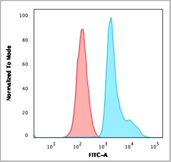
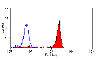
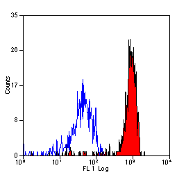
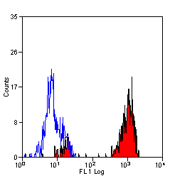
FCM (Flow Cytometry) (Staining of C57BL/6 splenocytes with 0.25 ug of Mouse IgG2a kappa Isotype Control (FITC) (open histogram) or 0.25 ug of Qa-2 antibody (FITC) (filled histogram). Total viable cells were used for analysis.)
Application Data (Detection limit for recombinant GST tagged TCEA3 is approximately 0.03ng/ml as a capture antibody.)
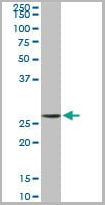
WB (Western Blot) (TCEA3 monoclonal antibody (M08), clone 4E11 Western Blot analysis of TCEA3 expression in HepG2 (Cat # L019V1).)
IF (Immunofluorescence) (Immunofluorescence of monoclonal antibody to TCEA3 on HepG2 cell. [antibody concentration 10 ug/ml])
IF (Immunofluorescence) (Immunofluorescence of monoclonal antibody to TCEA3 on HepG2 cell. [antibody concentration 10 ug/ml])
IHC (Immunohistochemistry) (Immunoperoxidase of monoclonal antibody to TCEA3 on formalin-fixed paraffin-embedded human salivary gland. [antibody concentration 3 ug/ml])
Application Data (Immunoperoxidase of monoclonal antibody to TCEA3 on formalin-fixed paraffin-embedded human salivary gland. [antibody concentration 3 ug/ml])
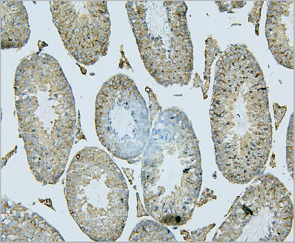
Application Data (Immunoperoxidase staining of a human tonsil cryosection with Mouse anti Human CD91 antibody, clone A2Mr alpha-2 followed by the Histar detection system . Medium power)
Application Data (Immunoperoxidase staining of a human tonsil cryosection with Mouse anti Human CD91 antibody, clone A2Mr alpha-2 followed by the Histar detection system . High power)
Application Data (Immunoperoxidase staining of a human tonsil cryosection with Mouse anti Human CD91 antibody, clone A2Mr alpha-2 followed by the Histar detection system . Low power)
Application Data (Immunoperoxidase staining of a human tonsil cryosection with Mouse anti Human CD91 antibody, clone A2Mr alpha-2 followed by the Histar detection system . Medium power)
Application Data (Immunofluorescence staining of a human tonsil cryosection with Mouse anti Human CD91 antibody, clone A2Mr alpha-2 , red in A and Mouse anti Human CD21, clone LB21 , green in B. C is the merged image with nuclei counterstained blue using DAPI. Medium power)
Application Data (Immunofluorescence staining of a human tonsil cryosection with Mouse anti Human CD91 antibody, clone A2Mr alpha-2 , red in A and Mouse anti Human CD21, clone LB21 , green in B. C is the merged image with nuclei counterstained blue using DAPI. Low power)
Application Data (Immunofluorescence staining of a human tonsil cryosection with Mouse anti Human CD91 antibody, clone A2Mr alpha-2 , red in A and Mouse anti Human CD21, clone LB21 , green in B. C is the merged image with nuclei counterstained blue using DAPI. High power)
Application Data (Staining of human peripheral blood monocytes probed with Mouse anti Human CD14:FITC)
Application Data (Staining of human peripheral blood monocytes with Mouse anti Human CD14:RPE-Alexa Fluor 750)
Application Data (Staining of human peripheral blood monocytes with Mouse anti Human CD14)
Application Data (Published customer image: CD11b+CD14+MHCII- cells demonstrate ability to suppressive T cell proliferation. (A) CD11b+CD14+MHCII- cells were sorted from peripheral blood sample of an osteosarcoma dog (B) and co-cultured with healthy dog PBMCs in the presence of mitogen for 72 hs. Non-stimulated PBMCs were used as negative control and PBMCs co-cultured with healthy PMNs were used to control for the effect of adding cells to the assay. Proliferative responses were measured by 3H-thymidine incorporation. CPM, counts per minute. The experiment was performed in triplicate. Mean +/- SEM are shown.From: Goulart MR, Pluhar GE, Ohlfest JR (2012) Identification of Myeloid Derived Suppressor Cells in Dogs with Naturally Occurring Cancer. PLoS ONE 7(3): e33274.)
Application Data (Staining of human peripheral blood monocytes with Mouse anti Human CD14)
Application Data (Staining of human peripheral blood monocytes with Mouse anti Human CD14:Biotin)
Application Data (Published customer image: CD11b+CD14-MHCII- cells suppress T cell proliferation. Facs sorted CD11b+CD14-MHCII- cells isolated from a dog with osteosarcoma or healthy PBMCs were co-incubated with mitogen-stimulated CD4+ and CD8+ T cells isolated from a healthy dog for 72 hs. No stimulated cells were used as negative control. Proliferative responses were measured by 3H-thymidine incorporation from experiments performed in triplicate. CPM, counts per minute. Mean +/- SEM are shown.From: Goulart MR, Pluhar GE, Ohlfest JR (2012) Identification of Myeloid Derived Suppressor Cells in Dogs with Naturally Occurring Cancer. PLoS ONE 7(3): e33274.)
Application Data (Published customer image: Immunophenotyping gating strategy and morphological analysis for MDSC identification in peripheral blood of dogs. PBMCs from healthy dogs and dogs with cancer were stained for the myeloid marker CD11b, monocytic marker CD14 and MHC II. (A) Representative flow cytometric analysis of forward and side scatter and gated CD11b+CD14-MHCII- cells from dogs with advanced or metastatic tumors compared to dogs with early stage non-metastatic tumors and healthy control dogs. Plots are representative of dog with advanced metastatic hemangiosarcoma (top), early stage bladder transitional cell carcinoma (middle) and a healthy dog. (B) FACS sorted CD11b+CD14-MHCII- cells were stained with diff-quick for cell morphology evaluation. A representative example of polymorphonuclear granulocyte morphology of CD11b+CD14-MHCII- cells is shown at 63x magnification.From: Goulart MR, Pluhar GE, Ohlfest JR (2012) Identification of Myeloid Derived Suppressor Cells in Dogs with Naturally Occurring Cancer. PLoS ONE 7(3): e33274.)
Application Data (Staining of human peripheral blood monocytes with Mouse anti Human CD14:RPE-Alexa Fluor 647)
Application Data (Staining of human peripheral blood monocytes with Mouse anti Human CD14:Pacific Blue)
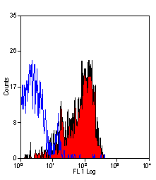
Application Data (Staining of human peripheral blood platelets with Mouse anti Human CD36)
Application Data (Staining of human peripheral blood platelets with Mouse anti Human CD36)
Application Data (Immunoperoxidase staining of human tonsil cryosection with Mouse anti Human CD36 antibody, clone SMø followed by the HISTAR detection system . High power SMθ)
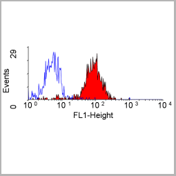
Application Data (Staining of human peripheral blood lymphocytes with Mouse anti Human CD36: FITC)
Application Data (Staining of human peripheral blood monocytes with Mouse anti Human CD36: Azide Free)
Application Data (Staining of human peripheral blood monocytes with Mouse anti Human CD36: Alexa Fluor 647)
Application Data (Immunoperoxidase staining of human tonsil cryosection with Mouse anti Human CD36 antibody, clone SMø followed by the HISTAR detection system . Low power)
Application Data (Immunoperoxidase staining of human tonsil cryosection with Mouse anti Human CD36 antibody, clone SMø followed by the HISTAR detection system . Medium power)
Application Data (Staining of human peripheral blood monocytes probed with Mouse anti Human CD14:FITC)
Application Data (Staining of human peripheral blood monocytes with Mouse anti Human CD14:RPE-Alexa Fluor 750)
Application Data (Staining of human peripheral blood monocytes with Mouse anti Human CD14)
Application Data (Published customer image: CD11b+CD14+MHCII- cells demonstrate ability to suppressive T cell proliferation. (A) CD11b+CD14+MHCII- cells were sorted from peripheral blood sample of an osteosarcoma dog (B) and co-cultured with healthy dog PBMCs in the presence of mitogen for 72 hs. Non-stimulated PBMCs were used as negative control and PBMCs co-cultured with healthy PMNs were used to control for the effect of adding cells to the assay. Proliferative responses were measured by 3H-thymidine incorporation. CPM, counts per minute. The experiment was performed in triplicate. Mean +/- SEM are shown.From: Goulart MR, Pluhar GE, Ohlfest JR (2012) Identification of Myeloid Derived Suppressor Cells in Dogs with Naturally Occurring Cancer. PLoS ONE 7(3): e33274.)
Application Data (Staining of human peripheral blood monocytes with Mouse anti Human CD14)
Application Data (Staining of human peripheral blood monocytes with Mouse anti Human CD14:Biotin)
Application Data (Published customer image: CD11b+CD14-MHCII- cells suppress T cell proliferation. Facs sorted CD11b+CD14-MHCII- cells isolated from a dog with osteosarcoma or healthy PBMCs were co-incubated with mitogen-stimulated CD4+ and CD8+ T cells isolated from a healthy dog for 72 hs. No stimulated cells were used as negative control. Proliferative responses were measured by 3H-thymidine incorporation from experiments performed in triplicate. CPM, counts per minute. Mean +/- SEM are shown.From: Goulart MR, Pluhar GE, Ohlfest JR (2012) Identification of Myeloid Derived Suppressor Cells in Dogs with Naturally Occurring Cancer. PLoS ONE 7(3): e33274.)
Application Data (Published customer image: Immunophenotyping gating strategy and morphological analysis for MDSC identification in peripheral blood of dogs. PBMCs from healthy dogs and dogs with cancer were stained for the myeloid marker CD11b, monocytic marker CD14 and MHC II. (A) Representative flow cytometric analysis of forward and side scatter and gated CD11b+CD14-MHCII- cells from dogs with advanced or metastatic tumors compared to dogs with early stage non-metastatic tumors and healthy control dogs. Plots are representative of dog with advanced metastatic hemangiosarcoma (top), early stage bladder transitional cell carcinoma (middle) and a healthy dog. (B) FACS sorted CD11b+CD14-MHCII- cells were stained with diff-quick for cell morphology evaluation. A representative example of polymorphonuclear granulocyte morphology of CD11b+CD14-MHCII- cells is shown at 63x magnification.From: Goulart MR, Pluhar GE, Ohlfest JR (2012) Identification of Myeloid Derived Suppressor Cells in Dogs with Naturally Occurring Cancer. PLoS ONE 7(3): e33274.)
Application Data (Staining of human peripheral blood monocytes with Mouse anti Human CD14:RPE-Alexa Fluor 647)
Application Data (Staining of human peripheral blood monocytes with Mouse anti Human CD14:Pacific Blue)
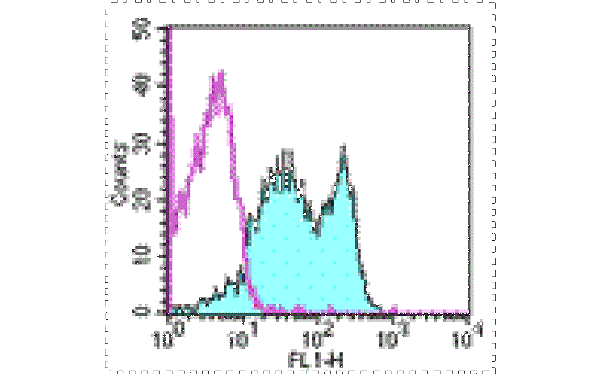
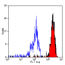
FCM (Flow Cytometry) (FACS analysis of ES-2 cells stained with STIP1 monoclonal antibody clone 2E11 (Green) and non-stained ES-2 cells (Black) as negative control.)
FCM (Flow Cytometry) (FACS analysis of ES-2 cells stained with STIP1 monoclonal antibody clone 2E11 (Green) and non-stained ES-2 cells (Black) as negative control.)
FCM (Flow Cytometry) (FACS analysis of 293 cells stained with STIP1 monoclonal antibody clone 2E11 (Green) and non-stained 293 cells (Black) as negative control.)
FCM (Flow Cytometry) (FACS analysis of 293 cells stained with STIP1 monoclonal antibody clone 2E11 (Green) and non-stained 293 cells (Black) as negative control.)
FCM (Flow Cytometry) (FACS analysis of HeLa cells stained with STIP1 monoclonal antibody clone 2E11 (Green) and non-stained HeLa cells (Black) as negative control.)
FCM (Flow Cytometry) (FACS analysis of HeLa cells stained with STIP1 monoclonal antibody clone 2E11 (Green) and non-stained HeLa cells (Black) as negative control.)
PITPNA (Phosphatidylinositol Transfer Protein alpha Isoform, PI-TP-alpha, PtdIns Transfer Protein alpha, PtdInsTP alpha, PITPN, MGC99649) (AP)
Gene Names
PITPNA; PITPN; VIB1A; HEL-S-36; PI-TPalpha
Reactivity
Human, Mouse, Rat
Applications
EIA, WB
Purity
Purified by Protein A Affinity Chromatography.
Pricing
What are Monoclonal Antibodies?
Monoclonal antibodies are specialized laboratory-produced proteins developed for binding to specific biological antigens or other molecular targets. Since they come from a single cell (or clone), they are especially consistent and accurate in the data they are involved in producing.
This type of antibody material has been shown to be a powerful tool in finding and subsequently destroying harmful cells in an organism, such as those found in cancers or various autoimmune diseases. This makes them excellent aids in medical testing and research, which is why they are so widely used.
AAA Biotech offers a comprehensive range of high-quality monoclonal antibodies that perform effectively in various laboratory tests, including (amongst others) ELISA, western blotting, immunohistochemistry, and flow cytometry. All of the products in our catalog are thoroughly quality tested to make sure that they are reliable and will consistently perform well in your research.
What Are The Uses of Monoclonal Antibodies
Monoclonal antibodies are used in many lab tests, including (amongst others) ELISA, western blotting, immunohistochemistry, and flow cytometry.
ELISA is a test that helps detect a specific substance/analyte in a sample. It uses antibodies (often monoclonal) bound to a solid surface (such as the well of a microplate) to “capture” the substance/analyte in the sample and immobilize it so that the detection antibody component can then bind to it and produce a signal, which can then be measured.
Western blotting identifies specific proteins in a sample. The sample is first separated on a gel, and then antibodies are applied that will typically bind to the target, which will all be localized to a single band in a lane.
Immunohistochemistry helps locate specific proteins in cells or tissue samples using antibodies.
Flow cytometry looks at and sorts cells. It uses antibodies that are conjugated to reporter molecules called “fluorophores”, which, under special lights, emit light themselves, which can then be measured by a detector instrument.
How Monoclonal Antibodies Are Used as Medicine?
Please note that all of the products listed in AAA Biotech’s catalog are strictly for research-use only (RUO).
Monoclonal antibodies can also be used as therapeutic/medical treatments, particularly in the context of cancers. They are designed to find and bind to specific cells or proteins, helping the immune system recognize and attack the cancer. These treatments work in different ways, such as:
Radioimmunotherapy attaches a small amount of radioactive molecule to the antibody, so it delivers the radiation directly to the cancer cells that the antibody is specifically binding to.
Antibody-directed enzyme prodrug therapy uses antibodies that are specifically bound to special enzymes. These enzymes activate a harmless drug in the body and turn it into a cancer-killing drug only near the cancer cells—this helps avoid harming healthy cells.
Immunoliposomes are tiny “bubbles” filled with medicine/drug and coated with antibodies. They carry the drug straight to the cancer cells.
Why Buy Monoclonal Antibodies From Us?
At AAA Biotech, we provide high-performance monoclonal antibodies designed to support a wide range of research needs.
1. Validated for Versatile Applications
The antibodies in our catalog are extensively validated and compatible with multiple techniques, including (but not limited to) ELISA, flow cytometry (FC), immunocytochemistry (ICC), immunofluorescence (IF), immunohistochemistry (IHC), immunoprecipitation (IP), and western blotting (WB).
2. Wide Selection & Specialized Options
We offer antibodies for common and rare species, that are available in various conjugated forms, and also in recombinant formats. Essentially, there is almost anything one might need to meet their experimental model’s requirements.
3. High-Quality Proteins
Our proteins meet high purity standards—90% or more as confirmed by SDS-PAGE. Many are available with tags like His, Flag, GST, or MBP, and we also supply native and biologically active proteins for functional studies.
Frequently Asked Questions
1. Are your monoclonal antibodies validated for specific applications?
Yes, our antibodies are tested and validated for use in methods such as ELISA, western blot, IHC, flow cytometry, and more. Refer to specific product pages or datasheets for individual product information.
2. How do I choose the right monoclonal antibody for my application?
Review the product details directly for application validation, species reactivity, and target information. You may also contact our support team at any time for help.
3. How quickly can I receive my order?
Most orders are processed and shipped within 1–3 business days, depending on product availability and your shipping location.
Submit a Question
Please complete the form below and a representative will contact you as soon as possible.
Request more Information
Please complete the form below and a representative will contact you as soon as possible.
Request a Manual
Please complete the form below and a representative will contact you as soon as possible.
Request a Quote
Please complete the form below and a representative will contact you as soon as possible.


 Application Data
(Staining of human peripheral blood platelets with Mouse anti Human CD36)
Application Data
(Staining of human peripheral blood platelets with Mouse anti Human CD36)
 Application Data
(Detection limit for recombinant GST tagged MTAP is ~0.3ng/ml as a capture antibody.)
Application Data
(Detection limit for recombinant GST tagged MTAP is ~0.3ng/ml as a capture antibody.)


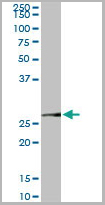
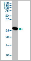
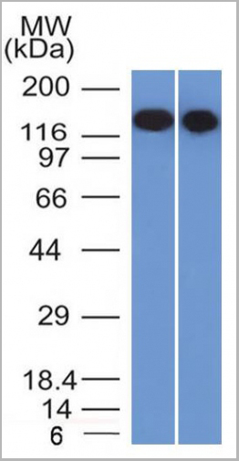
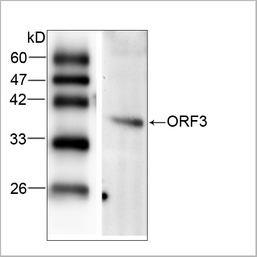
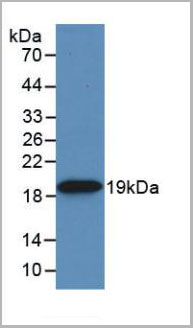
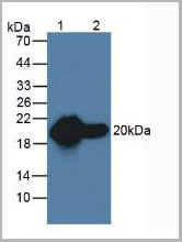
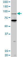
 WB (Western Blot)
(PCAF monoclonal antibody (M04), clone 5E11. Western Blot analysis of PCAF expression in LNCaP.)
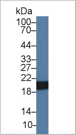
WB (Western Blot)
(PCAF monoclonal antibody (M04), clone 5E11. Western Blot analysis of PCAF expression in LNCaP.)
 Application Data
(Analysis of Protein Array containing more than 19,000 full-length human proteins using Laminin Receptor Monospecific Mouse Monoclonal Antibody (RPSA/2699) Z- and S- Score: The Z-score represents the strength of a signal that a monoclonal antibody (MAb) (in combination with a fluorescently-tagged anti-IgG secondary antibody) produces when binding to a particular protein on the HuProtTM array. Z-scores are described in units of standard deviations (SD's) above the mean value of all signals generated on that array. If targets on HuProtTM are arranged in descending order of the Z-score, the S-score is the difference (also in units of SD's) between the Z-score. S-score therefore represents the relative target specificity of a MAb to its intended target. A MAb is considered to specific to its intended target, if the MAb has an S-score of at least 2.5. For example, if a MAb binds to protein X with a Z-score of 43 and to protein Y with a Z-score of 14, then the S-score for the binding of that MAb to protein X is equal to 29.)
Application Data
(Analysis of Protein Array containing more than 19,000 full-length human proteins using Laminin Receptor Monospecific Mouse Monoclonal Antibody (RPSA/2699) Z- and S- Score: The Z-score represents the strength of a signal that a monoclonal antibody (MAb) (in combination with a fluorescently-tagged anti-IgG secondary antibody) produces when binding to a particular protein on the HuProtTM array. Z-scores are described in units of standard deviations (SD's) above the mean value of all signals generated on that array. If targets on HuProtTM are arranged in descending order of the Z-score, the S-score is the difference (also in units of SD's) between the Z-score. S-score therefore represents the relative target specificity of a MAb to its intended target. A MAb is considered to specific to its intended target, if the MAb has an S-score of at least 2.5. For example, if a MAb binds to protein X with a Z-score of 43 and to protein Y with a Z-score of 14, then the S-score for the binding of that MAb to protein X is equal to 29.)
 DB (Dot Blot)
(Dot Blot: The figure below is the result of using 1ug/mL anti- Aβ42 clone CA9 10C11 to detect 10ng/dot Aβ42 or Aβ40. The antibody showed no cross-reactivity with Aβ40.)
DB (Dot Blot)
(Dot Blot: The figure below is the result of using 1ug/mL anti- Aβ42 clone CA9 10C11 to detect 10ng/dot Aβ42 or Aβ40. The antibody showed no cross-reactivity with Aβ40.)
 IF (Immunofluorescence)
(Immunofluorescent staining of paraformaldehyde-fixed Ramos cells using CD22-Monospecific Mouse Monoclonal Antibody (BLCAM/1795) followed by goat anti-Mouse IgG conjugated to CF488 (green). Nuclei are stained with Reddot.)
IF (Immunofluorescence)
(Immunofluorescent staining of paraformaldehyde-fixed Ramos cells using CD22-Monospecific Mouse Monoclonal Antibody (BLCAM/1795) followed by goat anti-Mouse IgG conjugated to CF488 (green). Nuclei are stained with Reddot.)
 WB (Western Blot)


 Application Data
(Detection limit for recombinant GST tagged TCEA3 is approximately 0.03ng/ml as a capture antibody.)
Application Data
(Detection limit for recombinant GST tagged TCEA3 is approximately 0.03ng/ml as a capture antibody.)


 Application Data
(Staining of human peripheral blood monocytes probed with Mouse anti Human CD14:FITC)
Application Data
(Staining of human peripheral blood monocytes probed with Mouse anti Human CD14:FITC)


 Application Data
(Detection limit for recombinant GST tagged TCEA3 is approximately 0.03ng/ml as a capture antibody.)
Application Data
(Detection limit for recombinant GST tagged TCEA3 is approximately 0.03ng/ml as a capture antibody.)
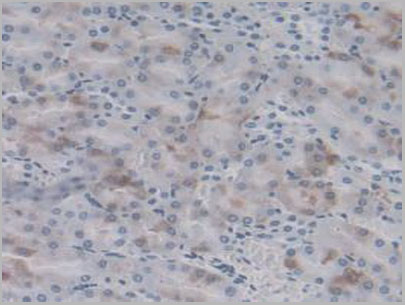
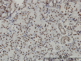
 IHC (Immunohistochemistry)
(DAB staining on IHC-P; Samples: Rat Adrenal Gland Tissue)
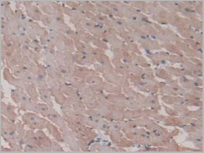
IHC (Immunohistochemistry)
(DAB staining on IHC-P; Samples: Rat Adrenal Gland Tissue)


 Application Data
(Immunoperoxidase staining of a human tonsil cryosection with Mouse anti Human CD91 antibody, clone A2Mr alpha-2 followed by the Histar detection system . Medium power)
Application Data
(Immunoperoxidase staining of a human tonsil cryosection with Mouse anti Human CD91 antibody, clone A2Mr alpha-2 followed by the Histar detection system . Medium power)


 Application Data
(Staining of human peripheral blood monocytes probed with Mouse anti Human CD14:FITC)
Application Data
(Staining of human peripheral blood monocytes probed with Mouse anti Human CD14:FITC)


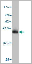
 WB (Western Blot)
(SPP1 monoclonal antibody (M09), clone 3E11. Western Blot analysis of SPP1 expression in NIH/3T3.)
WB (Western Blot)
(SPP1 monoclonal antibody (M09), clone 3E11. Western Blot analysis of SPP1 expression in NIH/3T3.)


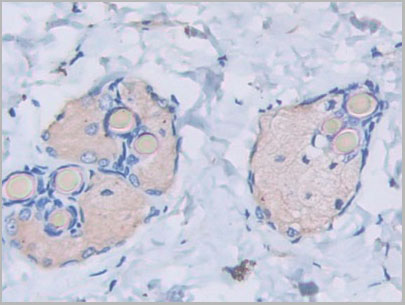
 IHC (Immunohistchemistry)
(Formalin-fixed, paraffin-embedded human Colon Carcinoma stained with AF488 Conjugate of Nucleolin Monoclonal Antibody (364-5 + NCL/902).)
IHC (Immunohistchemistry)
(Formalin-fixed, paraffin-embedded human Colon Carcinoma stained with AF488 Conjugate of Nucleolin Monoclonal Antibody (364-5 + NCL/902).)


 Application Data
(Staining of human peripheral blood monocytes probed with Mouse anti Human CD14:FITC)
Application Data
(Staining of human peripheral blood monocytes probed with Mouse anti Human CD14:FITC)


 Application Data
(Staining of human peripheral blood platelets with Mouse anti Human CD36)
Application Data
(Staining of human peripheral blood platelets with Mouse anti Human CD36)


 Application Data
(Staining of human peripheral blood monocytes probed with Mouse anti Human CD14:FITC)
Application Data
(Staining of human peripheral blood monocytes probed with Mouse anti Human CD14:FITC)


 FCM (Flow Cytometry)
(FACS analysis of ES-2 cells stained with STIP1 monoclonal antibody clone 2E11 (Green) and non-stained ES-2 cells (Black) as negative control.)
FCM (Flow Cytometry)
(FACS analysis of ES-2 cells stained with STIP1 monoclonal antibody clone 2E11 (Green) and non-stained ES-2 cells (Black) as negative control.)


 Application Data
(Staining of mouse peripheral blood lymphocytes with Hamster anti Mouse CD29: Low Endotoxin)
Application Data
(Staining of mouse peripheral blood lymphocytes with Hamster anti Mouse CD29: Low Endotoxin)


 WB (Western Blot)
(PCAF monoclonal antibody (M04), clone 5E11. Western Blot analysis of PCAF expression in LNCaP.)
WB (Western Blot)
(PCAF monoclonal antibody (M04), clone 5E11. Western Blot analysis of PCAF expression in LNCaP.)