Filters
▼Clonality
▼Type
▼Reactivity
▼Gene Name
▼Isotype
▼Host
▼Application
▼Clone
▼Monoclonal Antibodies
Get accurate results in your research with our Monoclonal Antibodies, which are specially made to target exactly what you require for your research, and will produce consistent, reliable performance in lab tests.
Viewing 5900-5950 of 27597 product results

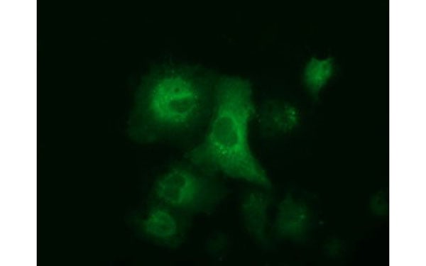
 IF (Immunofluorescence)
(Immunofluorescent staining of COS7 cells transiently transfected with recombinant PDE4A protein using PDE4A antibody)
IF (Immunofluorescence)
(Immunofluorescent staining of COS7 cells transiently transfected with recombinant PDE4A protein using PDE4A antibody)
PDE4A, Monoclonal Antibody (Cat# AAA106878)
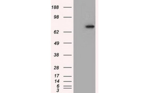
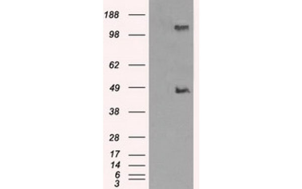
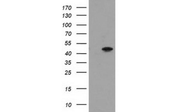
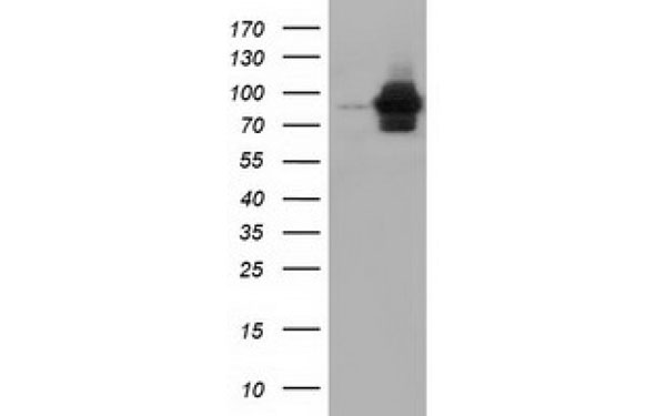
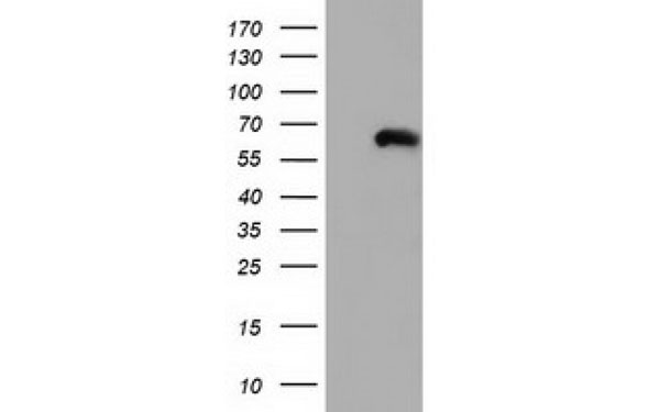
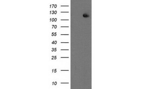
 WB (Western Blot)
(Western Blot analysis of HEK293T cell lysates (5 ug) transfected with either recombinant PVRL1 protein (Right) or empty vector (Left) detected with PVRL1 antibody)
WB (Western Blot)
(Western Blot analysis of HEK293T cell lysates (5 ug) transfected with either recombinant PVRL1 protein (Right) or empty vector (Left) detected with PVRL1 antibody)
PVRL1, Monoclonal Antibody (Cat# AAA106886)
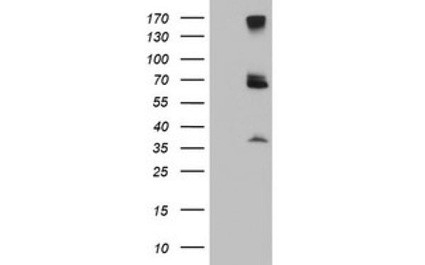
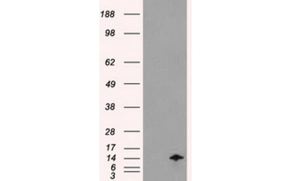
 WB (Western Blot)
(Western Blot analysis of HEK293T cell lysates (5 ug) transfected with either recombinant METTL21A protein (Right) or empty vector (Left) detected with METTL21A antibody)
WB (Western Blot)
(Western Blot analysis of HEK293T cell lysates (5 ug) transfected with either recombinant METTL21A protein (Right) or empty vector (Left) detected with METTL21A antibody)
METTL21A, Monoclonal Antibody (Cat# AAA106888)
 IF (Immunofluorescence)
(Immunofluorescent staining of COS7 cells transiently transfected with recombinant LOX protein using LOX antibody)
IF (Immunofluorescence)
(Immunofluorescent staining of COS7 cells transiently transfected with recombinant LOX protein using LOX antibody)
LOX, Monoclonal Antibody (Cat# AAA106899)
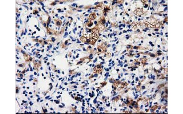
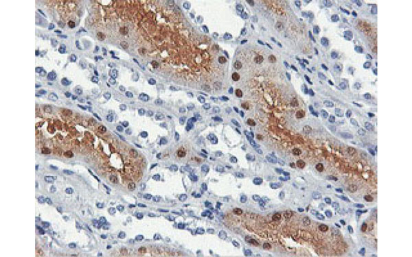
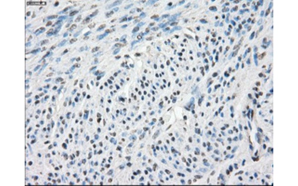
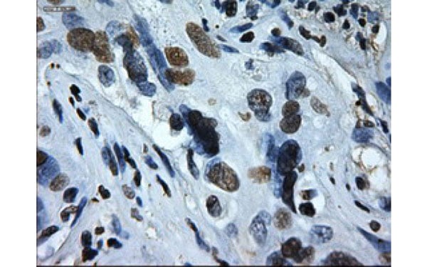
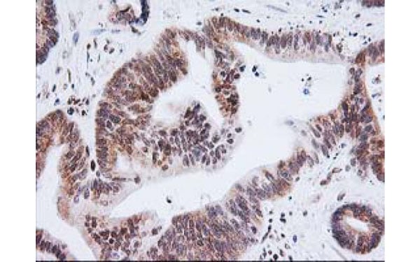
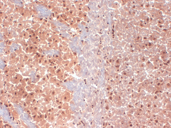
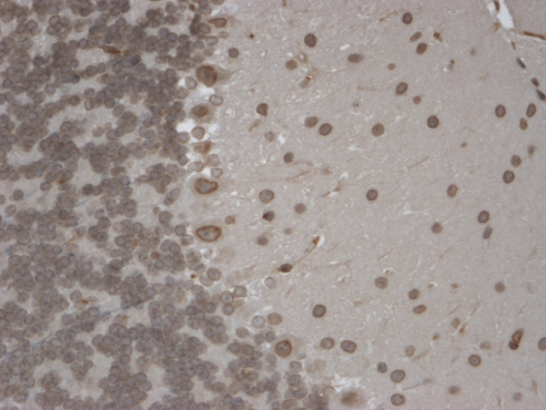
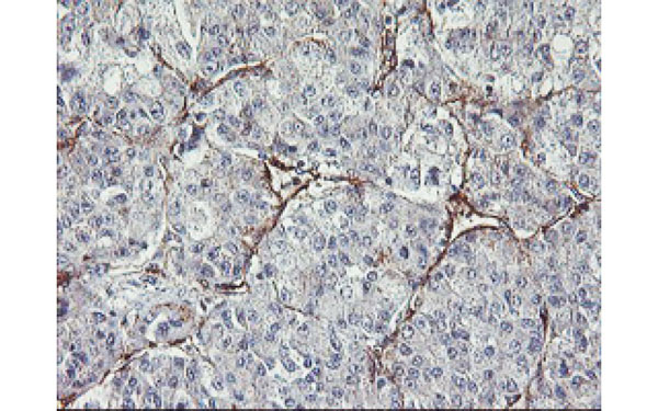 IHC (Immunohistochemisry)
(Immunohistochemical analysis of MIER2 protein in paraffin embedded Carcinoma of Human liver tissue using MIER2 antibody)
IHC (Immunohistochemisry)
(Immunohistochemical analysis of MIER2 protein in paraffin embedded Carcinoma of Human liver tissue using MIER2 antibody)
MIER2, Monoclonal Antibody (Cat# AAA106907)
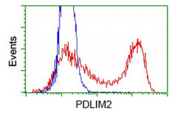
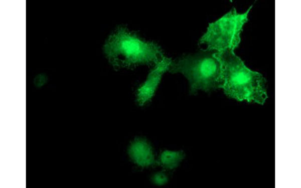
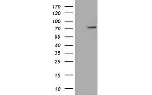
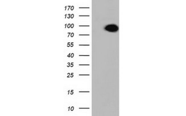
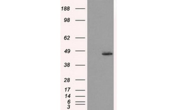
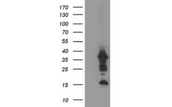 WB (Western Blot)
(Western Blot analysis of HEK293T cell lysates (5 ug) transfected with either recombinant PDLIM2 protein (Right) or empty vector (Left) detected with PDLIM2 antibody)
WB (Western Blot)
(Western Blot analysis of HEK293T cell lysates (5 ug) transfected with either recombinant PDLIM2 protein (Right) or empty vector (Left) detected with PDLIM2 antibody)
PDLIM2, Monoclonal Antibody (Cat# AAA106908)
 IF (Immunofluorescence)
(Immunofluorescent staining of COS7 cells transiently transfected with recombinant PDE4A protein using PDE4A antibody)
IF (Immunofluorescence)
(Immunofluorescent staining of COS7 cells transiently transfected with recombinant PDE4A protein using PDE4A antibody)
PDE4A, Monoclonal Antibody (Cat# AAA106919)

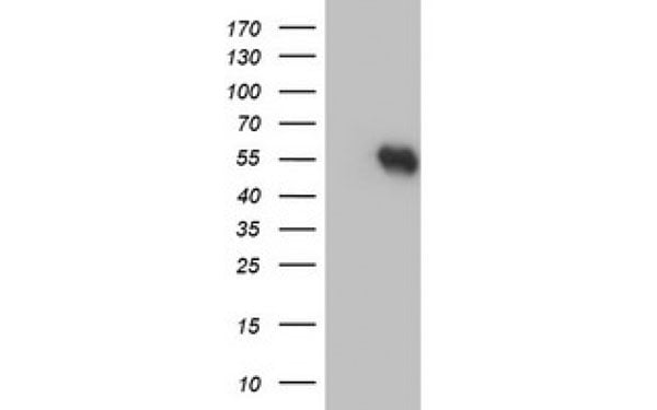
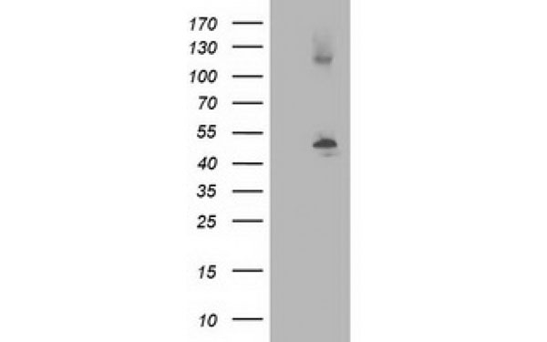
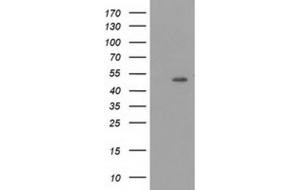
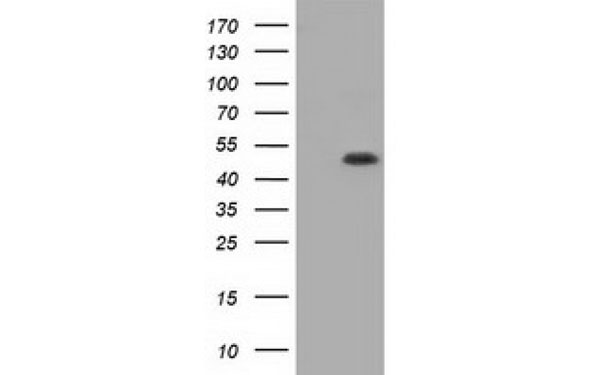
 WB (Western Blot)
(Western Blot analysis of HEK293T cell lysates (5 ug) transfected with either recombinant PSMD3 protein (Right) or empty vector (Left) detected with PSMD3 antibody)
WB (Western Blot)
(Western Blot analysis of HEK293T cell lysates (5 ug) transfected with either recombinant PSMD3 protein (Right) or empty vector (Left) detected with PSMD3 antibody)
PSMD3, Monoclonal Antibody (Cat# AAA106925)
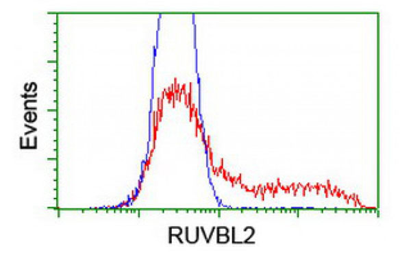
 IF (Immunofluorescence)
(Immunofluorescent staining of COS7 cells transiently transfected with recombinant RUVBL2 protein using RUVBL2 antibody)
IF (Immunofluorescence)
(Immunofluorescent staining of COS7 cells transiently transfected with recombinant RUVBL2 protein using RUVBL2 antibody)
RUVBL2, Monoclonal Antibody (Cat# AAA106927)
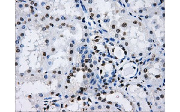
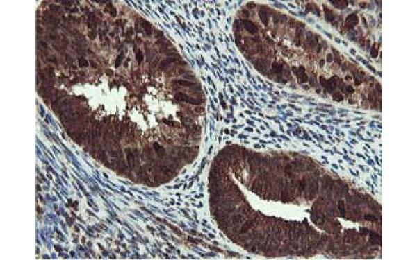
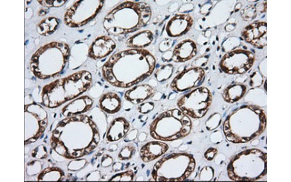
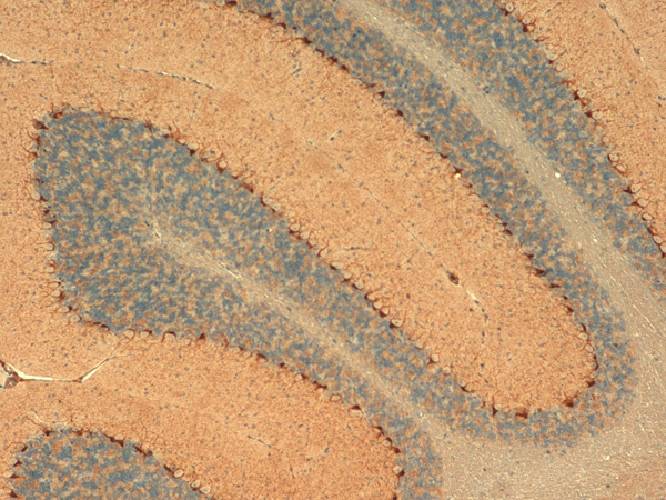
 IHC (Immunohiostchemistry)
(Immunohistochemical analysis of ALDH1L1 protein in paraffin embedded Carcinoma of Human kidney tissue using ALDH1L1 antibody)
IHC (Immunohiostchemistry)
(Immunohistochemical analysis of ALDH1L1 protein in paraffin embedded Carcinoma of Human kidney tissue using ALDH1L1 antibody)
ALDH1L1, Monoclonal Antibody (Cat# AAA106523)
 IF (Immunofluorescence)
(Immunofluorescent staining of COS7 cells transiently transfected with recombinant ALDH3A1 protein using ALDH3A1 antibody)
IF (Immunofluorescence)
(Immunofluorescent staining of COS7 cells transiently transfected with recombinant ALDH3A1 protein using ALDH3A1 antibody)
ALDH3A1, Monoclonal Antibody (Cat# AAA106532)
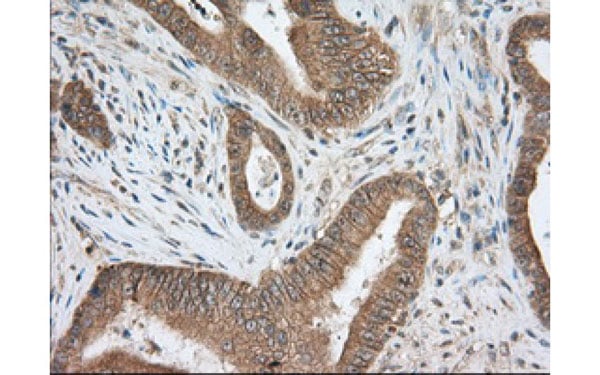
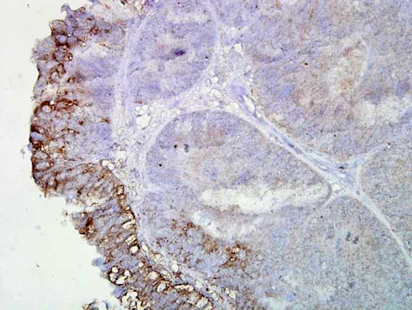
 IHC (Immunohiostchemistry)
(Immunohistochemical analysis of CRYAB protein in paraffin embedded Adenocarcinoma of Human colon tissue using CRYAB antibody)
IHC (Immunohiostchemistry)
(Immunohistochemical analysis of CRYAB protein in paraffin embedded Adenocarcinoma of Human colon tissue using CRYAB antibody)
CRYAB, Monoclonal Antibody (Cat# AAA106538)
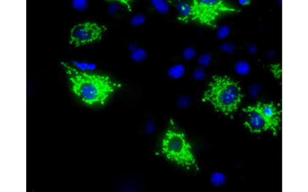
 IF (Immunofluorescence)
(Immunofluorescent staining of COS7 cells transiently transfected with recombinant HDAC6 protein using HDAC6 antibody)
IF (Immunofluorescence)
(Immunofluorescent staining of COS7 cells transiently transfected with recombinant HDAC6 protein using HDAC6 antibody)
HDAC6, Monoclonal Antibody (Cat# AAA106541)
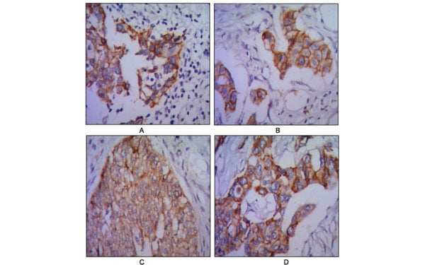
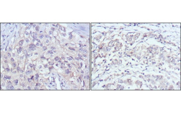 IHC (Immunohiostchemistry)
(Immunohistochemical analysis of paraffin-embedded human lung cancer (left) and gastric cancer (right) using PAK2 antibody with DAB staining.)
IHC (Immunohiostchemistry)
(Immunohistochemical analysis of paraffin-embedded human lung cancer (left) and gastric cancer (right) using PAK2 antibody with DAB staining.)
PAK2, Monoclonal Antibody (Cat# AAA106555)
 IF (Immunofluorescence)
(Immunofluorescent staining of endogenous TUBB4 protein in Hela cells using TUBB4 antibody)
IF (Immunofluorescence)
(Immunofluorescent staining of endogenous TUBB4 protein in Hela cells using TUBB4 antibody)
TUBB4, Monoclonal Antibody (Cat# AAA106576)
 WB (Western Blot)
(Western Blot analysis of HEK293T cell lysates (5 ug) transfected with either recombinant MTRF1L protein (Right) or empty vector (Left) detected with MTRF1L antibody)
WB (Western Blot)
(Western Blot analysis of HEK293T cell lysates (5 ug) transfected with either recombinant MTRF1L protein (Right) or empty vector (Left) detected with MTRF1L antibody)
MTRF1L, Monoclonal Antibody (Cat# AAA106578)
 IF (Immunofluorescence)
(Immunofluorescent staining of COS7 cells transiently transfected with recombinant TTLL12 protein using TTLL12 antibody)
IF (Immunofluorescence)
(Immunofluorescent staining of COS7 cells transiently transfected with recombinant TTLL12 protein using TTLL12 antibody)
TTLL12, Monoclonal Antibody (Cat# AAA106580)
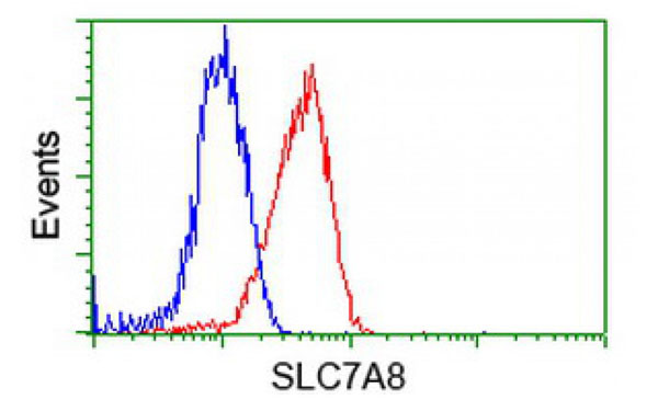
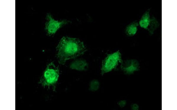
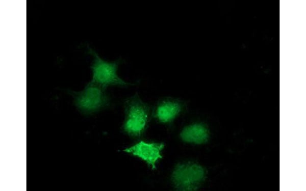
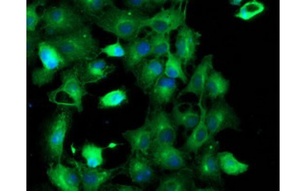
 IF (Immunofluorescence)
(Immunofluorescent staining of COS7 cells transiently transfected with recombinant SLC7A8 protein using SLC7A8 antibody)
IF (Immunofluorescence)
(Immunofluorescent staining of COS7 cells transiently transfected with recombinant SLC7A8 protein using SLC7A8 antibody)
SLC7A8, Monoclonal Antibody (Cat# AAA107031)
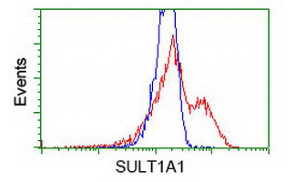
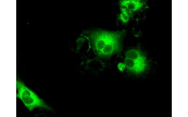
 IF (Immunofluorescence)
(Immunofluorescent staining of COS7 cells transiently transfected with recombinant SulT1A1 protein using SulT1A1 antibody)
IF (Immunofluorescence)
(Immunofluorescent staining of COS7 cells transiently transfected with recombinant SulT1A1 protein using SulT1A1 antibody)
SulT1A1, Monoclonal Antibody (Cat# AAA107036)
 IF (Immunofluorescence)
(Immunofluorescent staining of COS7 cells transiently transfected with recombinant KHK protein using KHK antibody)
IF (Immunofluorescence)
(Immunofluorescent staining of COS7 cells transiently transfected with recombinant KHK protein using KHK antibody)
KHK, Monoclonal Antibody (Cat# AAA107041)
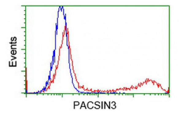
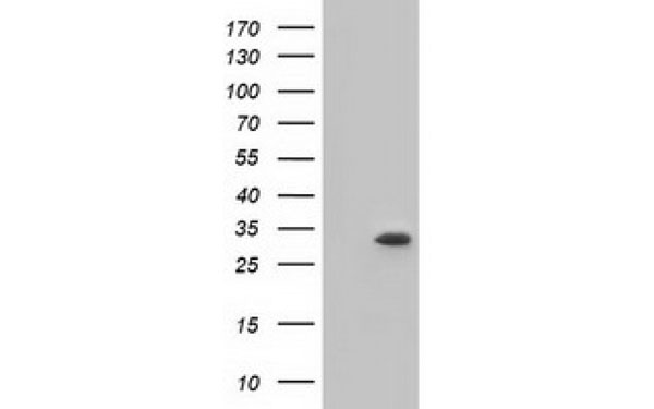
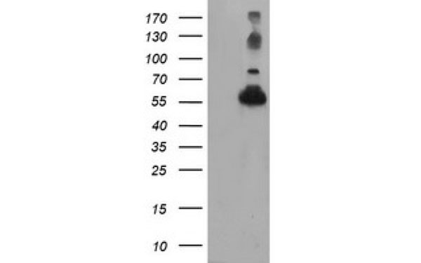
 WB (Western Blot)
(Western Blot analysis of HEK293T cell lysates (5 ug) transfected with either recombinant PACSIN3 protein (Right) or empty vector (Left) detected with PACSIN3 antibody)
WB (Western Blot)
(Western Blot analysis of HEK293T cell lysates (5 ug) transfected with either recombinant PACSIN3 protein (Right) or empty vector (Left) detected with PACSIN3 antibody)
PACSIN3, Monoclonal Antibody (Cat# AAA107048)
 IF (Immunofluorescence)
(Immunofluorescent staining of COS7 cells transiently transfected with recombinant TUBAL3 protein using TUBAL3 antibody)
IF (Immunofluorescence)
(Immunofluorescent staining of COS7 cells transiently transfected with recombinant TUBAL3 protein using TUBAL3 antibody)
TUBAL3, Monoclonal Antibody (Cat# AAA107057)
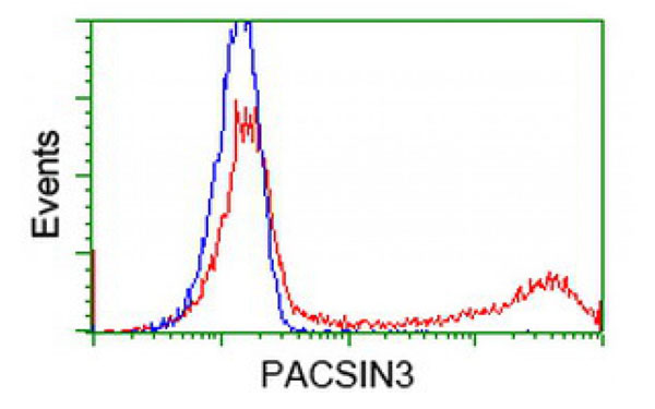
 WB (Western Blot)
(Western Blot analysis of HEK293T cell lysates (5 ug) transfected with either recombinant PACSIN3 protein (Right) or empty vector (Left) detected with PACSIN3 antibody)
WB (Western Blot)
(Western Blot analysis of HEK293T cell lysates (5 ug) transfected with either recombinant PACSIN3 protein (Right) or empty vector (Left) detected with PACSIN3 antibody)
PACSIN3, Monoclonal Antibody (Cat# AAA107068)
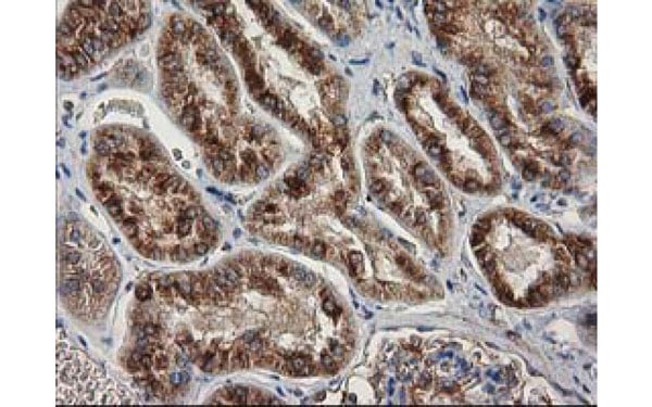
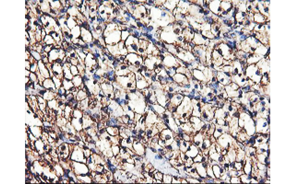
 IHC (Immunohistochemisry)
(Immunohistochemical analysis of SHPK protein in paraffin embedded Human Kidney tissue using SHPK antibody)
IHC (Immunohistochemisry)
(Immunohistochemical analysis of SHPK protein in paraffin embedded Human Kidney tissue using SHPK antibody)
SHPK, Monoclonal Antibody (Cat# AAA107086)
HPV18 E7, Monoclonal Antibody (Cat# AAA107094)
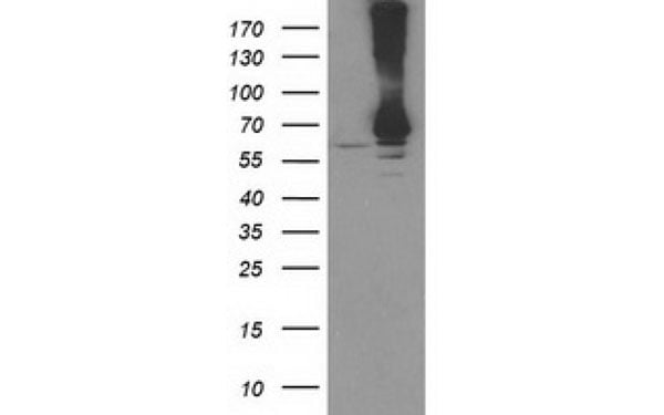
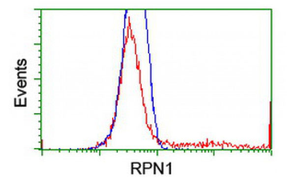
 IF (Immunofluorescence)
(Immunofluorescent staining of COS7 cells transiently transfected with recombinant RPN1 protein using RPN1 antibody)
IF (Immunofluorescence)
(Immunofluorescent staining of COS7 cells transiently transfected with recombinant RPN1 protein using RPN1 antibody)
RPN1, Monoclonal Antibody (Cat# AAA107102)
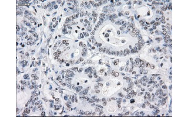
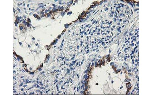
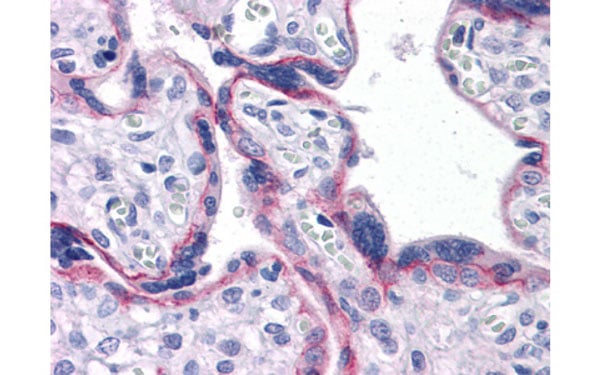
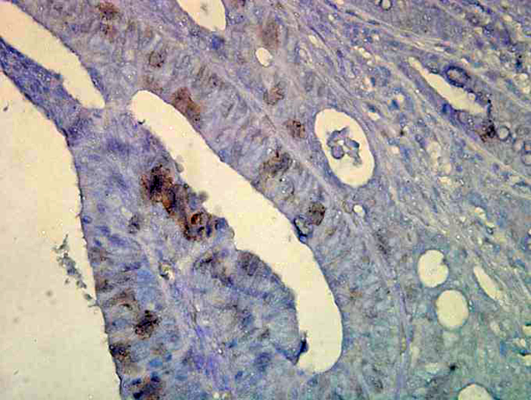
 IHC (Immunohiostchemistry)
(Immunohistochemical analysis of CA9 protein in paraffin embedded Human endometrium tissue using CA9 antibody)
IHC (Immunohiostchemistry)
(Immunohistochemical analysis of CA9 protein in paraffin embedded Human endometrium tissue using CA9 antibody)
CA9, Monoclonal Antibody (Cat# AAA106469)
 IF (Immunofluorescence)
(Immunofluorescent staining of COS7 cells transiently transfected with recombinant ALDH1L1 protein using ALDH1L1 antibody)
IF (Immunofluorescence)
(Immunofluorescent staining of COS7 cells transiently transfected with recombinant ALDH1L1 protein using ALDH1L1 antibody)
ALDH1L1, Monoclonal Antibody (Cat# AAA106471)
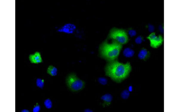
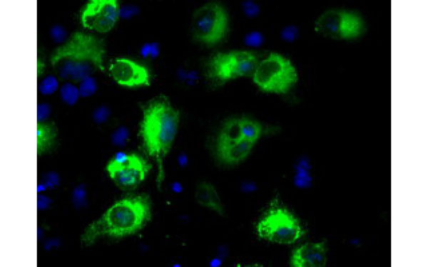
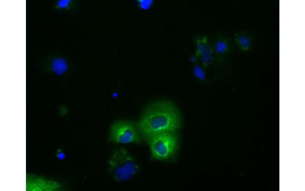
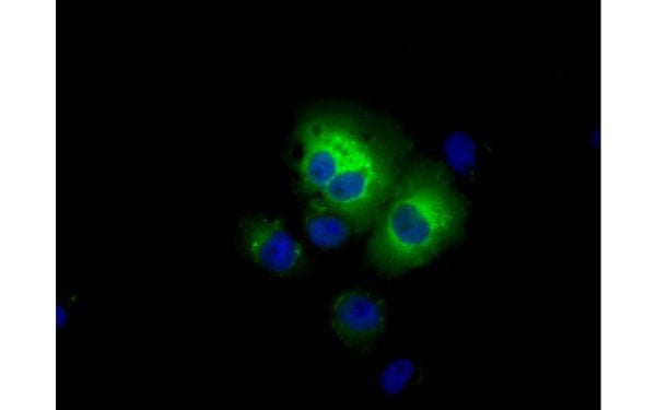
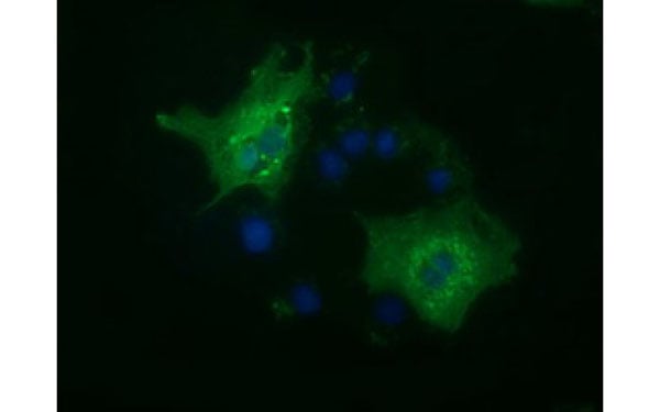
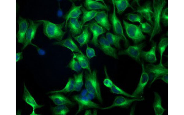
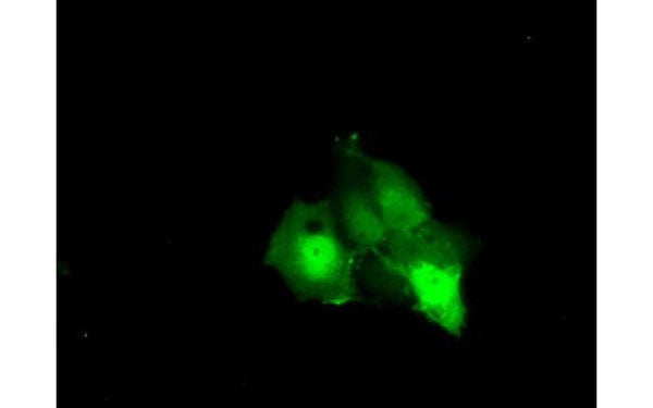
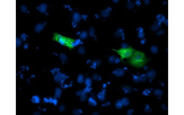
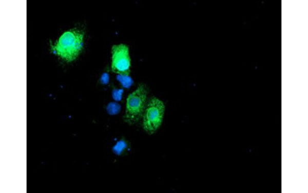
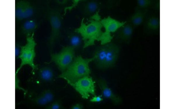
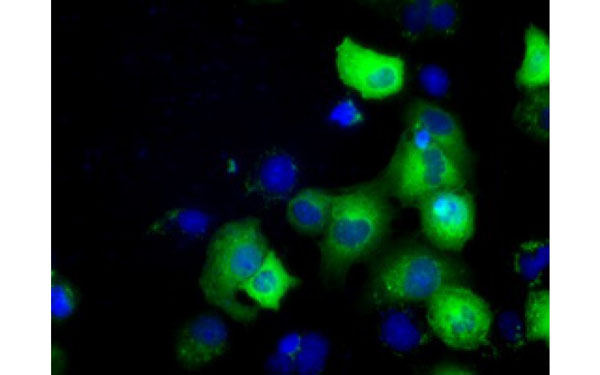
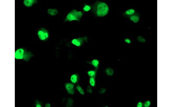
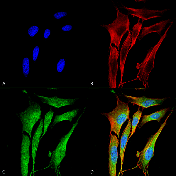
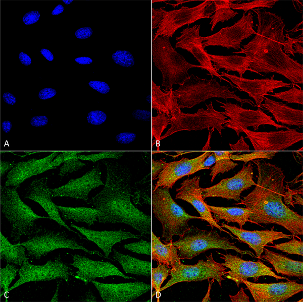
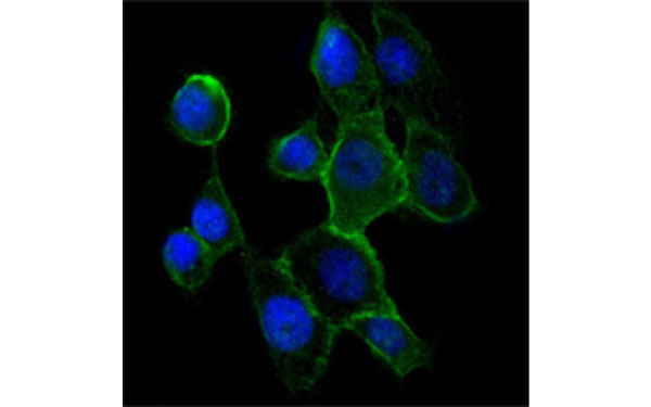 IF (Immunofluorescence)
(Immunofluorescence analysis of A431 cells using CDH2 antibody (green). Blue: DRAQ5 fluorescent DNA dye.)
IF (Immunofluorescence)
(Immunofluorescence analysis of A431 cells using CDH2 antibody (green). Blue: DRAQ5 fluorescent DNA dye.)
CDH2, Monoclonal Antibody (Cat# AAA106475)
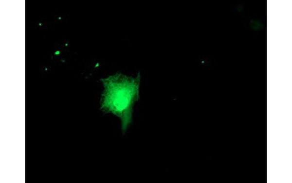
 IF (Immunofluorescence)
(Immunofluorescent staining of COS7 cells transiently transfected with recombinant CORO1B protein using CORO1B antibody)
IF (Immunofluorescence)
(Immunofluorescent staining of COS7 cells transiently transfected with recombinant CORO1B protein using CORO1B antibody)
CORO1B, Monoclonal Antibody (Cat# AAA106480)
 IHC (Immunohiostchemistry)
(Immunohistochemical analysis of ID2 protein in paraffin embedded Carcinoma of Human liver tissue using ID2 antibody)
IHC (Immunohiostchemistry)
(Immunohistochemical analysis of ID2 protein in paraffin embedded Carcinoma of Human liver tissue using ID2 antibody)
ID2, Monoclonal Antibody (Cat# AAA106500)
 IF (Immunofluorescence)
(Immunofluorescent staining of COS7 cells transiently transfected with recombinant GORASP1 protein using GORASP1 antibody)
IF (Immunofluorescence)
(Immunofluorescent staining of COS7 cells transiently transfected with recombinant GORASP1 protein using GORASP1 antibody)
GORASP1, Monoclonal Antibody (Cat# AAA106508)
 IF (Immunofluorescence)
(Immunofluorescent staining of COS7 cells transiently transfected with recombinant ERCC4 protein using ERCC4 antibody)
IF (Immunofluorescence)
(Immunofluorescent staining of COS7 cells transiently transfected with recombinant ERCC4 protein using ERCC4 antibody)
ERCC4, Monoclonal Antibody (Cat# AAA106509)
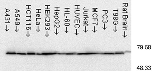
 WB (Western Blot)
(Western Blot analysis of Rat brain membrane lysate showing detection of SHANK3 protein using Mouse Anti-SHANK3 Monoclonal Antibody, Clone S69-46. Load: 15 ug. Block: 1.5% BSA for 30 minutes at RT. Primary Antibody: Mouse Anti-SHANK3 Monoclonal Antibody at 1:1000 for 2 hours at RT. Secondary Antibody: Sheep Anti-Mouse IgG: HRP for 1 hour at RT.)
WB (Western Blot)
(Western Blot analysis of Rat brain membrane lysate showing detection of SHANK3 protein using Mouse Anti-SHANK3 Monoclonal Antibody, Clone S69-46. Load: 15 ug. Block: 1.5% BSA for 30 minutes at RT. Primary Antibody: Mouse Anti-SHANK3 Monoclonal Antibody at 1:1000 for 2 hours at RT. Secondary Antibody: Sheep Anti-Mouse IgG: HRP for 1 hour at RT.)
SHANK3, Monoclonal Antibody (Cat# AAA103171)
 WB (Western Blot)
(Western Blot analysis of Rat brain membrane lysate showing detection of VGLUT1 protein using Mouse Anti-VGLUT1 Monoclonal Antibody, Clone S28-9. Primary Antibody: Mouse Anti-VGLUT1 Monoclonal Antibody at 1:1000.)
WB (Western Blot)
(Western Blot analysis of Rat brain membrane lysate showing detection of VGLUT1 protein using Mouse Anti-VGLUT1 Monoclonal Antibody, Clone S28-9. Primary Antibody: Mouse Anti-VGLUT1 Monoclonal Antibody at 1:1000.)
VGLUT1, Monoclonal Antibody (Cat# AAA103172)

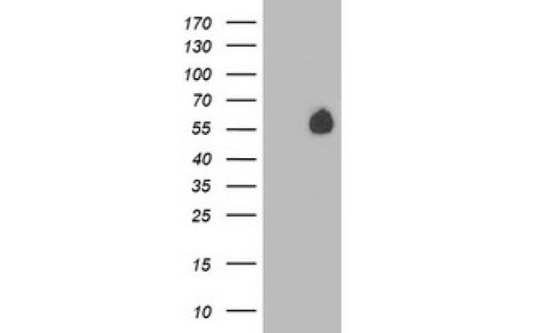
 WB (Western Blot)
(Western Blot analysis of Human Heat Shocked cervical cancer cell line (HeLa) lysate showing detection of Hsp60 protein using Mouse Anti-Hsp60 Monoclonal Antibody, Clone LK-2. Load: 15 ug. Block: 1.5% BSA for 30 minutes at RT. Primary Antibody: Mouse Anti-Hsp60 Monoclonal Antibody at 1:1000 for 2 hours at RT. Secondary Antibody: Sheep Anti-Mouse IgG: HRP for 1 hour at RT.)
WB (Western Blot)
(Western Blot analysis of Human Heat Shocked cervical cancer cell line (HeLa) lysate showing detection of Hsp60 protein using Mouse Anti-Hsp60 Monoclonal Antibody, Clone LK-2. Load: 15 ug. Block: 1.5% BSA for 30 minutes at RT. Primary Antibody: Mouse Anti-Hsp60 Monoclonal Antibody at 1:1000 for 2 hours at RT. Secondary Antibody: Sheep Anti-Mouse IgG: HRP for 1 hour at RT.)
Hsp60, Monoclonal Antibody (Cat# AAA103179)
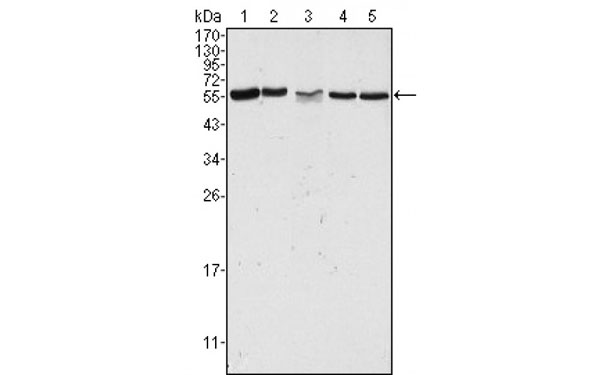
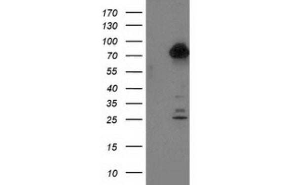
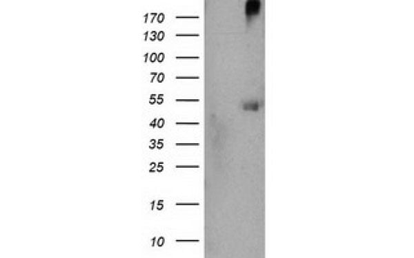
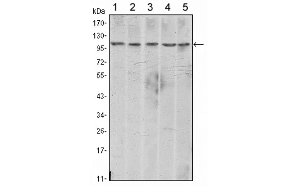
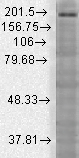
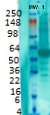
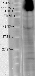
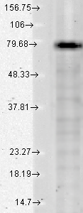
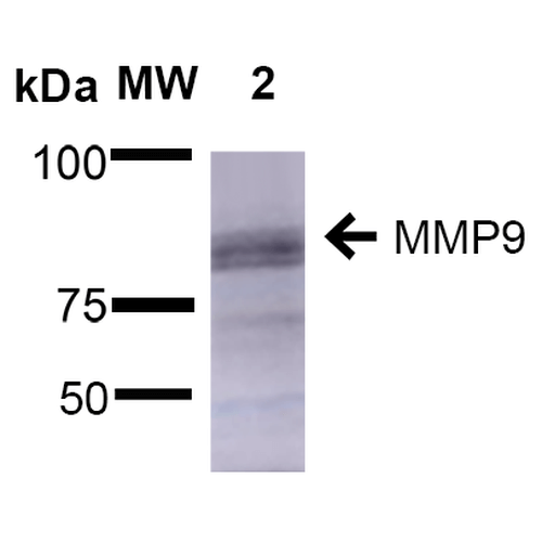 WB (Western Blot)
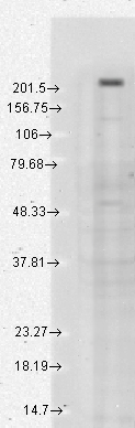
(Western Blot analysis of Rat Brain showing detection of ~92 kDa and ~82 kDa (pro and active) MMP9 protein using Mouse Anti-MMP9 Monoclonal Antibody, Clone S51-82 . Lane 1: Molecular Weight Ladder (MW). Lane 2: Rat Brain. Load: 15 ug. Block: 5% Skim Milk in 1X TBST. Primary Antibody: Mouse Anti-MMP9 Monoclonal Antibody at 1:1000 for 2 hours at RT. Secondary Antibody: Goat Anti-Mouse IgG: HRP at 1:2000 for 60 min at RT. Color Development: ECL solution for 5 min at RT. Predicted/Observed Size: ~92 kDa and ~82 kDa (pro and active).)
WB (Western Blot)
(Western Blot analysis of Rat Brain showing detection of ~92 kDa and ~82 kDa (pro and active) MMP9 protein using Mouse Anti-MMP9 Monoclonal Antibody, Clone S51-82 . Lane 1: Molecular Weight Ladder (MW). Lane 2: Rat Brain. Load: 15 ug. Block: 5% Skim Milk in 1X TBST. Primary Antibody: Mouse Anti-MMP9 Monoclonal Antibody at 1:1000 for 2 hours at RT. Secondary Antibody: Goat Anti-Mouse IgG: HRP at 1:2000 for 60 min at RT. Color Development: ECL solution for 5 min at RT. Predicted/Observed Size: ~92 kDa and ~82 kDa (pro and active).)
MMP9, Monoclonal Antibody (Cat# AAA103204)
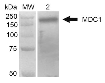
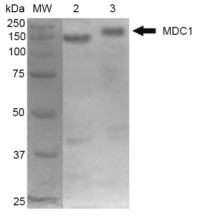 WB (Western Blot)
(Western Blot analysis of Mouse Cortex and Cerebellum showing detection of 184 kDa MDC1 protein using Mouse Anti-MDC1 Monoclonal Antibody, Clone P2B11. Lane 1: MW ladder. Lane 2: Mouse Cortex. Lane 3: Mouse Cerebellum. Load: 10 ug. Block: 5% Skim Milk in 1X TBST. Primary Antibody: Mouse Anti-MDC1 Monoclonal Antibody at 1:1000 for 2 hours RT. Secondary Antibody: Goat Anti-Mouse at 1:2000 for 60 min at RT. Color Development: ECL solution for 5 min in RT. Predicted/Observed Size: 184 kDa.)
WB (Western Blot)
(Western Blot analysis of Mouse Cortex and Cerebellum showing detection of 184 kDa MDC1 protein using Mouse Anti-MDC1 Monoclonal Antibody, Clone P2B11. Lane 1: MW ladder. Lane 2: Mouse Cortex. Lane 3: Mouse Cerebellum. Load: 10 ug. Block: 5% Skim Milk in 1X TBST. Primary Antibody: Mouse Anti-MDC1 Monoclonal Antibody at 1:1000 for 2 hours RT. Secondary Antibody: Goat Anti-Mouse at 1:2000 for 60 min at RT. Color Development: ECL solution for 5 min in RT. Predicted/Observed Size: 184 kDa.)
MDC1, Monoclonal Antibody (Cat# AAA103210)
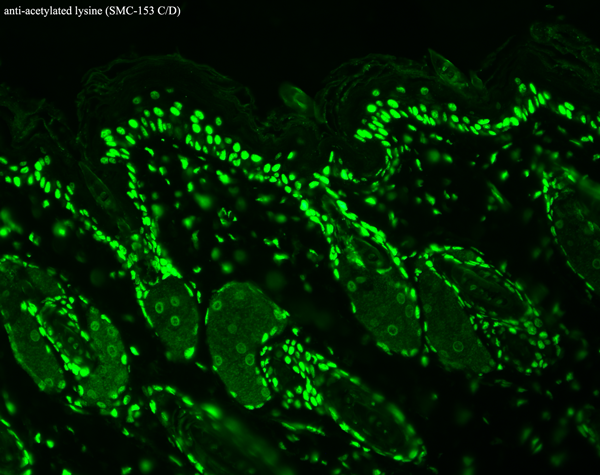
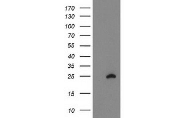
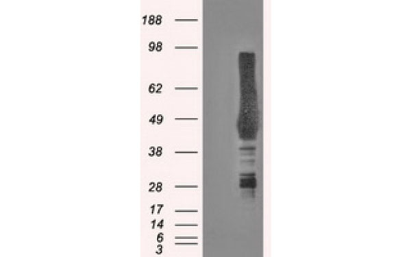
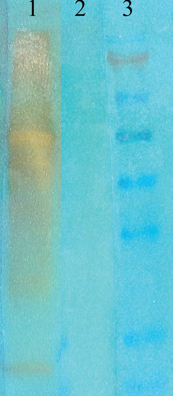 WB (Western Blot)
(Western Blot analysis of acetylated lysine showing detection of Acetylated Lysine protein using Mouse Anti-Acetylated Lysine Monoclonal Antibody, Clone 7F8. Primary Antibody: Mouse Anti-Acetylated Lysine Monoclonal Antibody at 1:1000. (1) acetylated BSA (75ng of protein), (2) non-acetylated BSA, and (3) marker.)
WB (Western Blot)
(Western Blot analysis of acetylated lysine showing detection of Acetylated Lysine protein using Mouse Anti-Acetylated Lysine Monoclonal Antibody, Clone 7F8. Primary Antibody: Mouse Anti-Acetylated Lysine Monoclonal Antibody at 1:1000. (1) acetylated BSA (75ng of protein), (2) non-acetylated BSA, and (3) marker.)
Acetylated Lysine, Monoclonal Antibody (Cat# AAA103218)
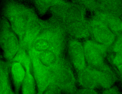
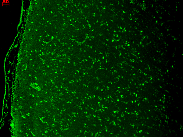
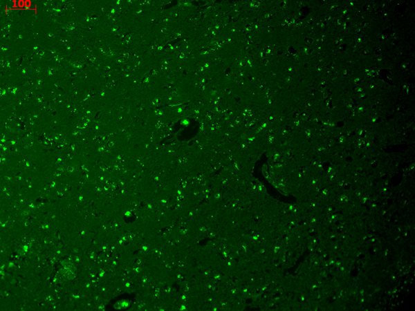
 IHC (Immunohistochemisry)
(Immunohistochemistry analysis using Mouse Anti-HCN4 Monoclonal Antibody, Clone S114-10. Tissue: hippocampus. Species: Human. Fixation: Bouin's Fixative and paraffin-embedded. Primary Antibody: Mouse Anti-HCN4 Monoclonal Antibody at 1:100 for 1 hour at RT. Secondary Antibody: FITC Goat Anti-Mouse (green) at 1:50 for 1 hour at RT.)
IHC (Immunohistochemisry)
(Immunohistochemistry analysis using Mouse Anti-HCN4 Monoclonal Antibody, Clone S114-10. Tissue: hippocampus. Species: Human. Fixation: Bouin's Fixative and paraffin-embedded. Primary Antibody: Mouse Anti-HCN4 Monoclonal Antibody at 1:100 for 1 hour at RT. Secondary Antibody: FITC Goat Anti-Mouse (green) at 1:50 for 1 hour at RT.)
HCN4, Monoclonal Antibody (Cat# AAA103220)
 WB (Western Blot)
(Western Blot analysis of Human Cell lysates showing detection of Rhodopsin protein using Mouse Anti-Rhodopsin Monoclonal Antibody, Clone 1D4. Load: 15 ug. Block: 1.5% BSA for 30 minutes at RT. Primary Antibody: Mouse Anti-Rhodopsin Monoclonal Antibody at 1:1000 for 2 hours at RT. Secondary Antibody: Sheep Anti-Mouse IgG: HRP for 1 hour at RT.)
WB (Western Blot)
(Western Blot analysis of Human Cell lysates showing detection of Rhodopsin protein using Mouse Anti-Rhodopsin Monoclonal Antibody, Clone 1D4. Load: 15 ug. Block: 1.5% BSA for 30 minutes at RT. Primary Antibody: Mouse Anti-Rhodopsin Monoclonal Antibody at 1:1000 for 2 hours at RT. Secondary Antibody: Sheep Anti-Mouse IgG: HRP for 1 hour at RT.)
Rhodopsin, Monoclonal Antibody (Cat# AAA103222)
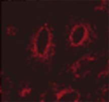
 IHC (Immunohiostchemistry)
(Immunohistochemistry analysis using Mouse Anti-CaV1.3 Calcium Channel Monoclonal Antibody, Clone S48A-9. Tissue: hippocampus. Species: Human. Fixation: Bouin's Fixative and paraffin-embedded. Primary Antibody: Mouse Anti-CaV1.3 Calcium Channel Monoclonal Antibody at 1:1000 for 1 hour at RT. Secondary Antibody: FITC Goat Anti-Mouse (green) at 1:50 for 1 hour at RT.)
IHC (Immunohiostchemistry)
(Immunohistochemistry analysis using Mouse Anti-CaV1.3 Calcium Channel Monoclonal Antibody, Clone S48A-9. Tissue: hippocampus. Species: Human. Fixation: Bouin's Fixative and paraffin-embedded. Primary Antibody: Mouse Anti-CaV1.3 Calcium Channel Monoclonal Antibody at 1:1000 for 1 hour at RT. Secondary Antibody: FITC Goat Anti-Mouse (green) at 1:50 for 1 hour at RT.)
Cav1.3, Monoclonal Antibody (Cat# AAA103233)
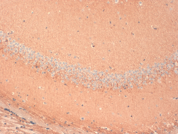
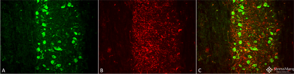
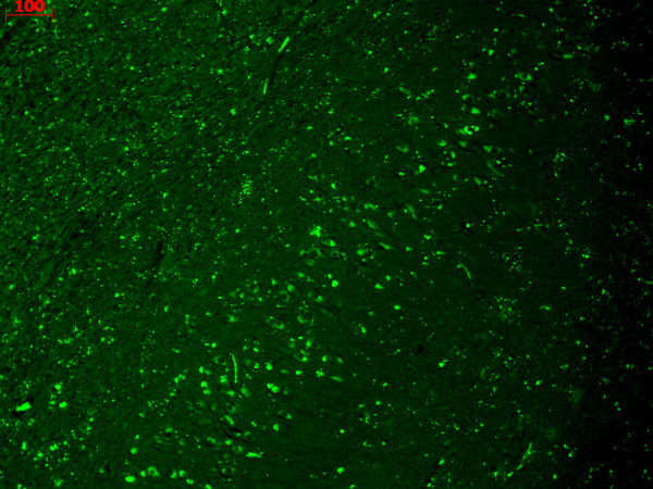
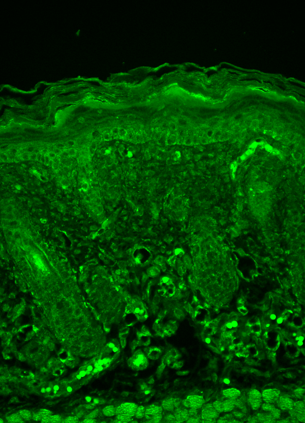
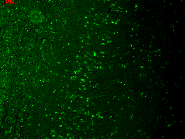
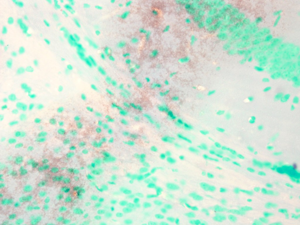
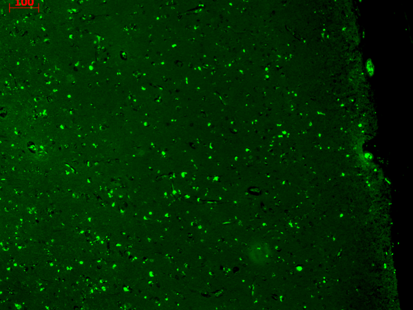
 IHC (Immunohistochemistry)
(Immunohistochemistry analysis using Mouse Anti-HCN1 Monoclonal Antibody, Clone S70-28. Tissue: Frozen brain section. Species: Mouse. Fixation: 10% Formalin Solution for 12-24 hours at RT. Primary Antibody: Mouse Anti-HCN1 Monoclonal Antibody at 1:1000 for 1 hour at RT. Secondary Antibody: HRP/DAB Detection System: Biotinylated Goat Anti-Mouse, Streptavidin Peroxidase, DAB Chromogen (brown) for 30 minutes at RT. Counterstain: Mayer Hematoxylin (purple/blue) nuclear stain at 250-500 ul for 5 minutes at RT.)
IHC (Immunohistochemistry)
(Immunohistochemistry analysis using Mouse Anti-HCN1 Monoclonal Antibody, Clone S70-28. Tissue: Frozen brain section. Species: Mouse. Fixation: 10% Formalin Solution for 12-24 hours at RT. Primary Antibody: Mouse Anti-HCN1 Monoclonal Antibody at 1:1000 for 1 hour at RT. Secondary Antibody: HRP/DAB Detection System: Biotinylated Goat Anti-Mouse, Streptavidin Peroxidase, DAB Chromogen (brown) for 30 minutes at RT. Counterstain: Mayer Hematoxylin (purple/blue) nuclear stain at 250-500 ul for 5 minutes at RT.)
HCN1, Monoclonal Antibody (Cat# AAA103241)
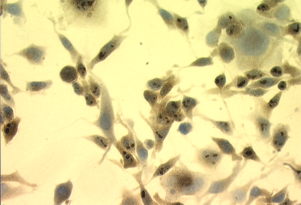
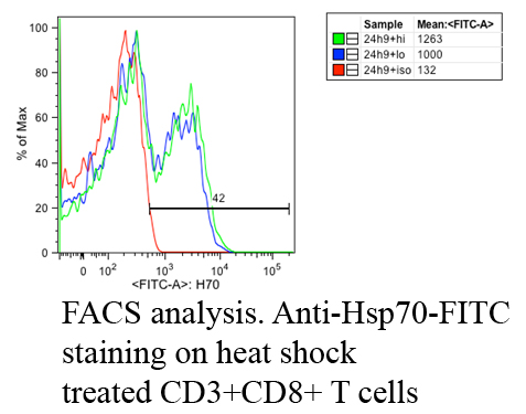 IF (Immunofluorescence)
(Fluorescence Activated Cell Sorting analysis using Mouse Anti-Hsp70: FITC Monoclonal Antibody, Clone C92. Tissue: Heat Shocked CD3+ CD8+ T cells . Species: Mouse. Primary Antibody: Mouse Anti-Hsp70: FITC Monoclonal Antibody at 1:1000. Courtesy of: Cheryl Cameron, Vaccine and Gene Therapy Instit. Florida.)
IF (Immunofluorescence)
(Fluorescence Activated Cell Sorting analysis using Mouse Anti-Hsp70: FITC Monoclonal Antibody, Clone C92. Tissue: Heat Shocked CD3+ CD8+ T cells . Species: Mouse. Primary Antibody: Mouse Anti-Hsp70: FITC Monoclonal Antibody at 1:1000. Courtesy of: Cheryl Cameron, Vaccine and Gene Therapy Instit. Florida.)
Hsp70, Monoclonal Antibody (Cat# AAA103243)
 WB (Western Blot)
(Western Blot analysis of Human Cell lysates showing detection of TrpM7 protein using Mouse Anti-TrpM7 Monoclonal Antibody, Clone S74-25. Load: 15 ug. Block: 1.5% BSA for 30 minutes at RT. Primary Antibody: Mouse Anti-TrpM7 Monoclonal Antibody at 1:1000 for 2 hours at RT. Secondary Antibody: Sheep Anti-Mouse IgG: HRP for 1 hour at RT.)
WB (Western Blot)
(Western Blot analysis of Human Cell lysates showing detection of TrpM7 protein using Mouse Anti-TrpM7 Monoclonal Antibody, Clone S74-25. Load: 15 ug. Block: 1.5% BSA for 30 minutes at RT. Primary Antibody: Mouse Anti-TrpM7 Monoclonal Antibody at 1:1000 for 2 hours at RT. Secondary Antibody: Sheep Anti-Mouse IgG: HRP for 1 hour at RT.)
TrpM7, Monoclonal Antibody (Cat# AAA103244)
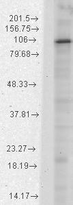
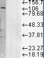
 WB (Western Blot)
(Western Blot analysis of Human SH-SY5Y showing detection of Copper Transporting ATPase 1 protein using Mouse Anti-Copper Transporting ATPase 1 Monoclonal Antibody, Clone S60-4 . Lane 1: MW Ladder. Lane 2: 10 ug SH-SY5Y. Load: 10 ug. Block: 5% Skim Milk powder in TBST. Primary Antibody: Mouse Anti-Copper Transporting ATPase 1 Monoclonal Antibody at 1:500 for 2 hours at RT with shaking. Secondary Antibody: Goat anti-mouse IgG:HRP at 1:4000 for 1 hour at RT with shaking. Color Development: Chemiluminescent for HRP (Moss) for 5 min in RT.)
WB (Western Blot)
(Western Blot analysis of Human SH-SY5Y showing detection of Copper Transporting ATPase 1 protein using Mouse Anti-Copper Transporting ATPase 1 Monoclonal Antibody, Clone S60-4 . Lane 1: MW Ladder. Lane 2: 10 ug SH-SY5Y. Load: 10 ug. Block: 5% Skim Milk powder in TBST. Primary Antibody: Mouse Anti-Copper Transporting ATPase 1 Monoclonal Antibody at 1:500 for 2 hours at RT with shaking. Secondary Antibody: Goat anti-mouse IgG:HRP at 1:4000 for 1 hour at RT with shaking. Color Development: Chemiluminescent for HRP (Moss) for 5 min in RT.)
Copper-Transporting ATPase1, Monoclonal Antibody (Cat# AAA103253)


 WB (Western Blot)
(Western Blot analysis of Human Heat Shocked cervical cancer cell line (HeLa) lysate showing detection of Hsp60 protein using Mouse Anti-Hsp60 Monoclonal Antibody, Clone LK-2. Load: 15 ug. Block: 1.5% BSA for 30 minutes at RT. Primary Antibody: Mouse Anti-Hsp60 Monoclonal Antibody at 1:1000 for 2 hours at RT. Secondary Antibody: Sheep Anti-Mouse IgG: HRP for 1 hour at RT.)
WB (Western Blot)
(Western Blot analysis of Human Heat Shocked cervical cancer cell line (HeLa) lysate showing detection of Hsp60 protein using Mouse Anti-Hsp60 Monoclonal Antibody, Clone LK-2. Load: 15 ug. Block: 1.5% BSA for 30 minutes at RT. Primary Antibody: Mouse Anti-Hsp60 Monoclonal Antibody at 1:1000 for 2 hours at RT. Secondary Antibody: Sheep Anti-Mouse IgG: HRP for 1 hour at RT.)
Hsp60, Monoclonal Antibody (Cat# AAA103272)
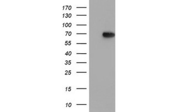
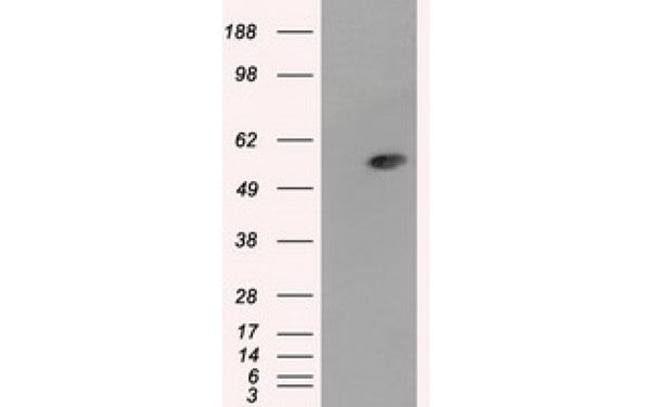
 WB (Western Blot)
(Western Blot analysis of Rat liver microsome lysate showing detection of LAMP1 protein using Mouse Anti-LAMP1 Monoclonal Antibody, Clone Ly1C6. Load: 15 ug. Block: 1.5% BSA for 30 minutes at RT. Primary Antibody: Mouse Anti-LAMP1 Monoclonal Antibody at 1:1000 for 2 hours at RT. Secondary Antibody: Sheep Anti-Mouse IgG: HRP for 1 hour at RT.)
WB (Western Blot)
(Western Blot analysis of Rat liver microsome lysate showing detection of LAMP1 protein using Mouse Anti-LAMP1 Monoclonal Antibody, Clone Ly1C6. Load: 15 ug. Block: 1.5% BSA for 30 minutes at RT. Primary Antibody: Mouse Anti-LAMP1 Monoclonal Antibody at 1:1000 for 2 hours at RT. Secondary Antibody: Sheep Anti-Mouse IgG: HRP for 1 hour at RT.)
LAMP1, Monoclonal Antibody (Cat# AAA103273)
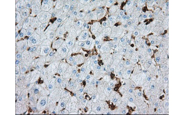
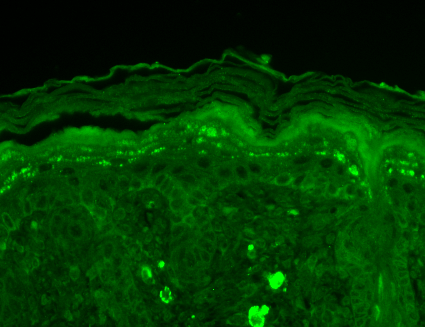
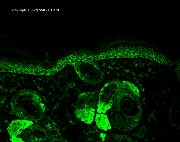
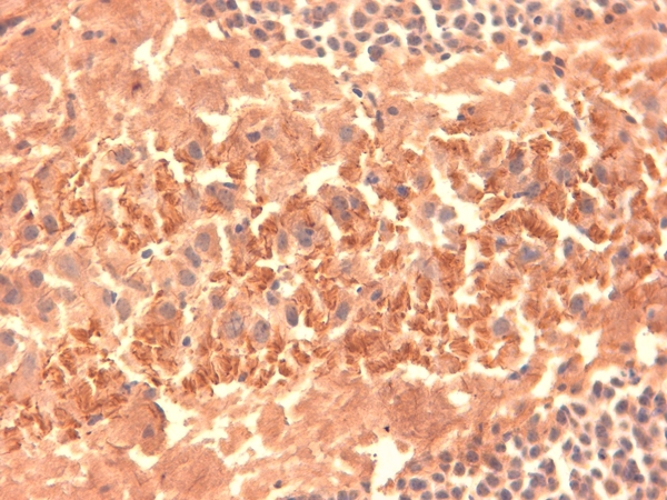
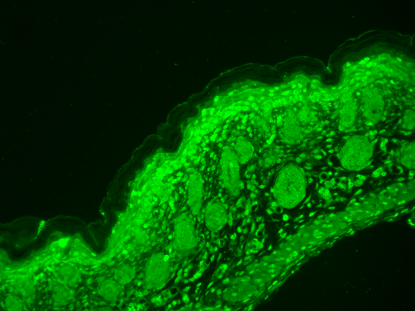
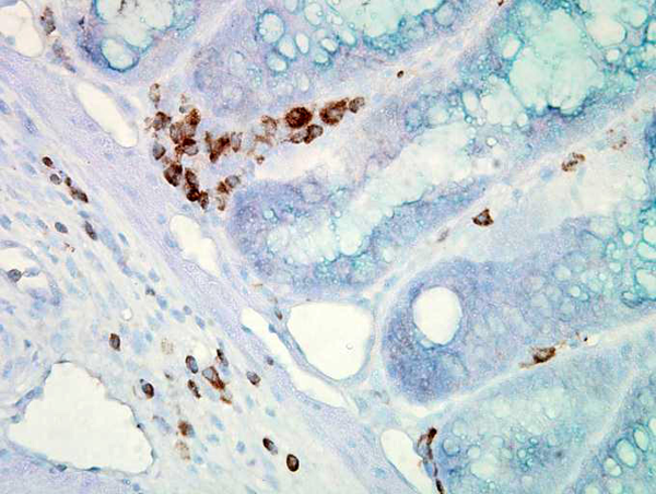
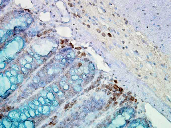 IHC (Immunohiostchemistry)
(Immunohistochemistry analysis using Mouse Anti-Hsp90 Monoclonal Antibody, Clone AC-16. Tissue: inflamed colon. Species: Mouse. Fixation: Formalin. Primary Antibody: Mouse Anti-Hsp90 Monoclonal Antibody at 1:2000 for 12 hours at 4 degree C. Secondary Antibody: Biotin Goat Anti-Mouse at 1:2000 for 1 hour at RT. Counterstain: Mayer Hematoxylin (purple/blue) nuclear stain at 200 ul for 2 minutes at RT. Localization: Inflammatory cells. Magnification: 40x. Mostly inflammatory cells, some mucosa.)
IHC (Immunohiostchemistry)
(Immunohistochemistry analysis using Mouse Anti-Hsp90 Monoclonal Antibody, Clone AC-16. Tissue: inflamed colon. Species: Mouse. Fixation: Formalin. Primary Antibody: Mouse Anti-Hsp90 Monoclonal Antibody at 1:2000 for 12 hours at 4 degree C. Secondary Antibody: Biotin Goat Anti-Mouse at 1:2000 for 1 hour at RT. Counterstain: Mayer Hematoxylin (purple/blue) nuclear stain at 200 ul for 2 minutes at RT. Localization: Inflammatory cells. Magnification: 40x. Mostly inflammatory cells, some mucosa.)
Hsp90, Monoclonal Antibody (Cat# AAA103274)
 WB (Western Blot)
(Western Blot analysis of hamster T-CHO cell lysate showing detection of KCNQ1 protein using Mouse Anti-KCNQ1 Monoclonal Antibody, Clone S37A-10. Load: 15 ug. Block: 1.5% BSA for 30 minutes at RT. Primary Antibody: Mouse Anti-KCNQ1 Monoclonal Antibody at 1:1000 for 2 hours at RT. Secondary Antibody: Sheep Anti-Mouse IgG: HRP for 1 hour at RT.)
WB (Western Blot)
(Western Blot analysis of hamster T-CHO cell lysate showing detection of KCNQ1 protein using Mouse Anti-KCNQ1 Monoclonal Antibody, Clone S37A-10. Load: 15 ug. Block: 1.5% BSA for 30 minutes at RT. Primary Antibody: Mouse Anti-KCNQ1 Monoclonal Antibody at 1:1000 for 2 hours at RT. Secondary Antibody: Sheep Anti-Mouse IgG: HRP for 1 hour at RT.)
KCNQ1, Monoclonal Antibody (Cat# AAA103289)
What are Monoclonal Antibodies?
Monoclonal antibodies are specialized laboratory-produced proteins developed for binding to specific biological antigens or other molecular targets. Since they come from a single cell (or clone), they are especially consistent and accurate in the data they are involved in producing.
This type of antibody material has been shown to be a powerful tool in finding and subsequently destroying harmful cells in an organism, such as those found in cancers or various autoimmune diseases. This makes them excellent aids in medical testing and research, which is why they are so widely used.
AAA Biotech offers a comprehensive range of high-quality monoclonal antibodies that perform effectively in various laboratory tests, including (amongst others) ELISA, western blotting, immunohistochemistry, and flow cytometry. All of the products in our catalog are thoroughly quality tested to make sure that they are reliable and will consistently perform well in your research.
What Are The Uses of Monoclonal Antibodies
Monoclonal antibodies are used in many lab tests, including (amongst others) ELISA, western blotting, immunohistochemistry, and flow cytometry.
ELISA is a test that helps detect a specific substance/analyte in a sample. It uses antibodies (often monoclonal) bound to a solid surface (such as the well of a microplate) to “capture” the substance/analyte in the sample and immobilize it so that the detection antibody component can then bind to it and produce a signal, which can then be measured.
Western blotting identifies specific proteins in a sample. The sample is first separated on a gel, and then antibodies are applied that will typically bind to the target, which will all be localized to a single band in a lane.
Immunohistochemistry helps locate specific proteins in cells or tissue samples using antibodies.
Flow cytometry looks at and sorts cells. It uses antibodies that are conjugated to reporter molecules called “fluorophores”, which, under special lights, emit light themselves, which can then be measured by a detector instrument.
How Monoclonal Antibodies Are Used as Medicine?
Please note that all of the products listed in AAA Biotech’s also known as AAA Bio or AAABio catalog are strictly for research-use only (RUO).
Monoclonal antibodies can also be used as therapeutic/medical treatments, particularly in the context of cancers. They are designed to find and bind to specific cells or proteins, helping the immune system recognize and attack the cancer. These treatments work in different ways, such as:
- Radioimmunotherapy attaches a small amount of radioactive molecule to the antibody, so it delivers the radiation directly to the cancer cells that the antibody is specifically binding to.
- Antibody-directed enzyme prodrug therapy uses antibodies that are specifically bound to special enzymes. These enzymes activate a harmless drug in the body and turn it into a cancer-killing drug only near the cancer cells—this helps avoid harming healthy cells.
- Immunoliposomes are tiny “bubbles” filled with medicine/drug and coated with antibodies. They carry the drug straight to the cancer cells.
Why Buy Monoclonal Antibodies From Us?
At AAA Biotech, we provide high-performance monoclonal antibodies designed to support a wide range of research needs.
1. Validated for Versatile Applications
The antibodies in our catalog are extensively validated and compatible with multiple techniques, including (but not limited to) ELISA, flow cytometry (FC), immunocytochemistry (ICC), immunofluorescence (IF), immunohistochemistry (IHC), immunoprecipitation (IP), and western blotting (WB).
2. Wide Selection & Specialized Options
We offer antibodies for common and rare species, that are available in various conjugated forms, and also in recombinant formats. Essentially, there is almost anything one might need to meet their experimental model’s requirements.
3. High-Quality Proteins
Our proteins meet high purity standards—90% or more as confirmed by SDS-PAGE. Many are available with tags like His, Flag, GST, or MBP, and we also supply native and biologically active proteins for functional studies.
Frequently Asked Questions
1. Are your monoclonal antibodies validated for specific applications?
Yes, our antibodies are tested and validated for use in methods such as ELISA, western blot, IHC, flow cytometry, and more. Refer to specific product pages or datasheets for individual product information.
2. How do I choose the right monoclonal antibody for my application?
Review the product details directly for application validation, species reactivity, and target information. You may also contact our support team at any time for help.
3. How quickly can I receive my order?
Most orders are processed and shipped within 1–3 business days, depending on product availability and your shipping location.