Filters
▼Clonality
▼Type
▼Reactivity
▼Gene Name
▼Isotype
▼Host
▼Application
▼Clone
▼Monoclonal Antibodies
Get accurate results in your research with our Monoclonal Antibodies, which are specially made to target exactly what you require for your research, and will produce consistent, reliable performance in lab tests.
Viewing 6000-6050 of 27597 product results
Phytase, Monoclonal Antibody (Cat# AAA107327)
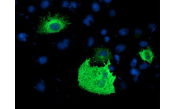 IF (Immunofluorescence)
(Immunofluorescent staining of COS7 cells transiently transfected with recombinant PDE1B protein using PDE1B antibody)
IF (Immunofluorescence)
(Immunofluorescent staining of COS7 cells transiently transfected with recombinant PDE1B protein using PDE1B antibody)
PDE1B, Monoclonal Antibody (Cat# AAA107328)
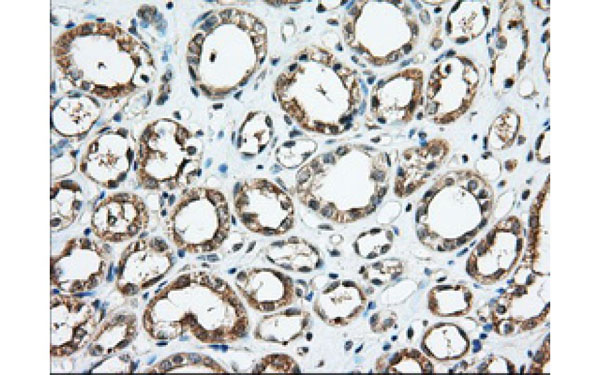
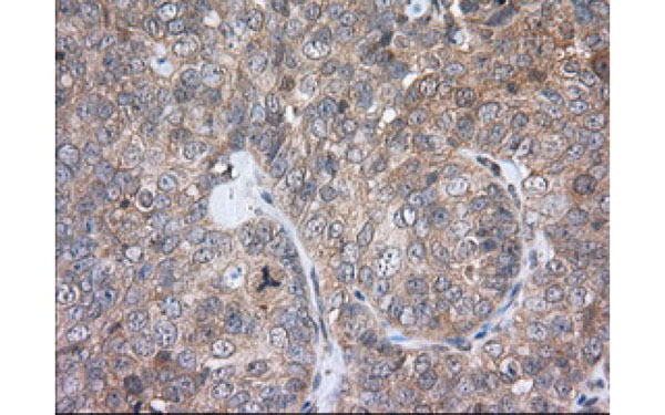
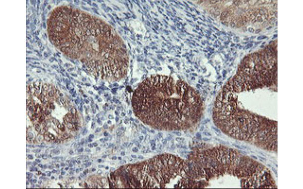 IHC (Immunohistochemisry)
(Immunohistochemical analysis of NLN protein in paraffin embedded Adenocarcinoma of Human endometrium tissue using NLN antibody)
IHC (Immunohistochemisry)
(Immunohistochemical analysis of NLN protein in paraffin embedded Adenocarcinoma of Human endometrium tissue using NLN antibody)
NLN, Monoclonal Antibody (Cat# AAA107347)
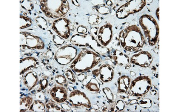
 IHC (Immunohistochemisry)
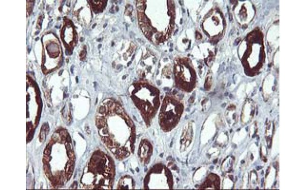
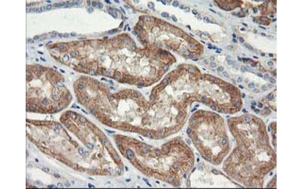
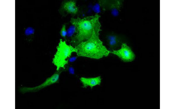
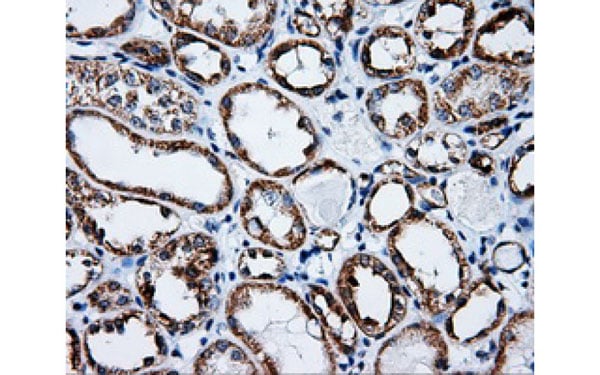
(Immunohistochemical analysis of NAPEPLD protein in paraffin embedded Human Kidney tissue using NAPEPLD antibody)
IHC (Immunohistochemisry)
(Immunohistochemical analysis of NAPEPLD protein in paraffin embedded Human Kidney tissue using NAPEPLD antibody)
NAPEPLD, Monoclonal Antibody (Cat# AAA107348)
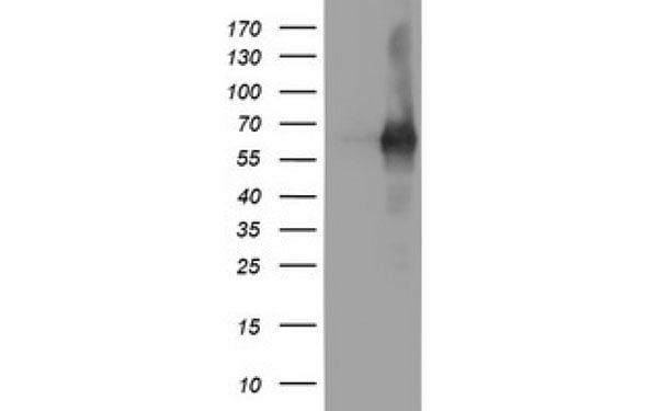
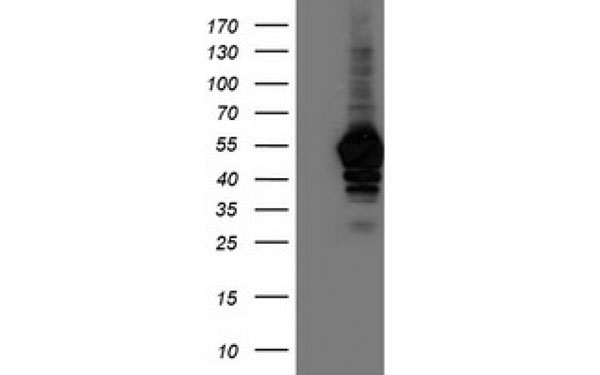
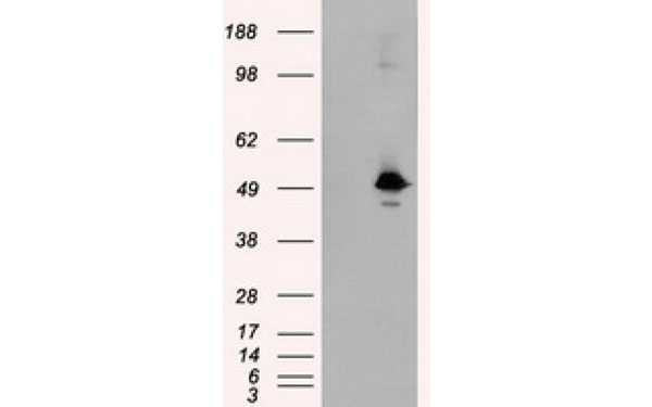
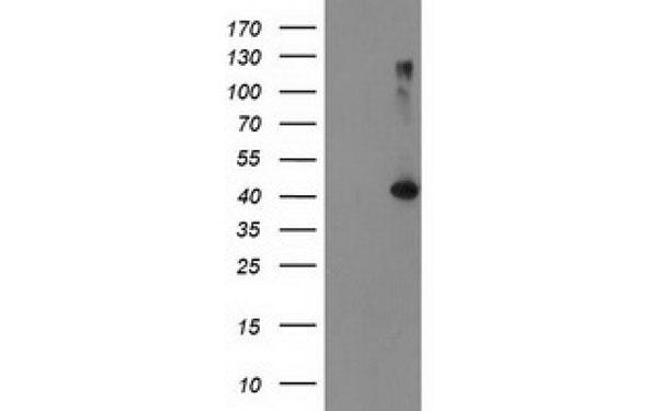 WB (Western Blot)
(Western Blot analysis of HEK293T cell lysates (5 ug) transfected with either recombinant TBC1D21 protein (Right) or empty vector (Left) detected with TBC1D21 antibody)
WB (Western Blot)
(Western Blot analysis of HEK293T cell lysates (5 ug) transfected with either recombinant TBC1D21 protein (Right) or empty vector (Left) detected with TBC1D21 antibody)
TBC1D21, Monoclonal Antibody (Cat# AAA107358)
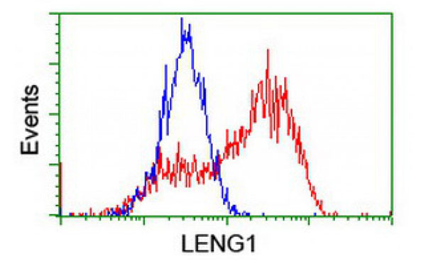
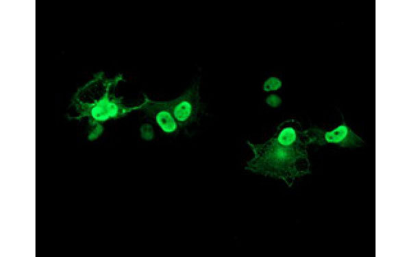 IF (Immunofluorescence)
(Immunofluorescent staining of COS7 cells transiently transfected with recombinant LENG1 protein using LENG1 antibody)
IF (Immunofluorescence)
(Immunofluorescent staining of COS7 cells transiently transfected with recombinant LENG1 protein using LENG1 antibody)
LENG1, Monoclonal Antibody (Cat# AAA107201)
 IF (Immunofluorescence)
(Immunofluorescent staining of COS7 cells transiently transfected with recombinant KLHL2 protein using KLHL2 antibody)
IF (Immunofluorescence)
(Immunofluorescent staining of COS7 cells transiently transfected with recombinant KLHL2 protein using KLHL2 antibody)
KLHL2, Monoclonal Antibody (Cat# AAA107214)
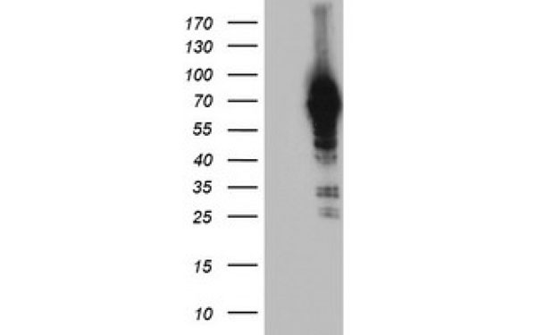
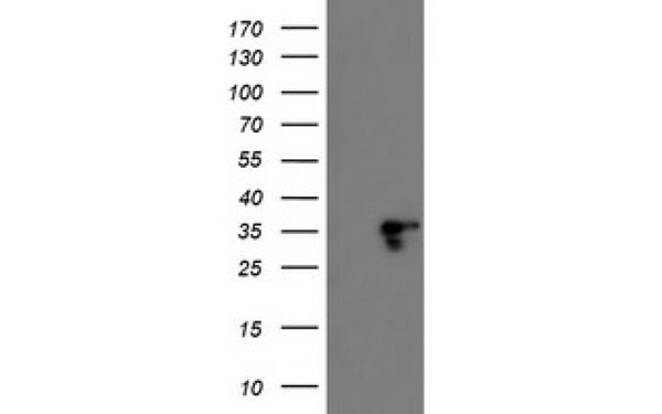
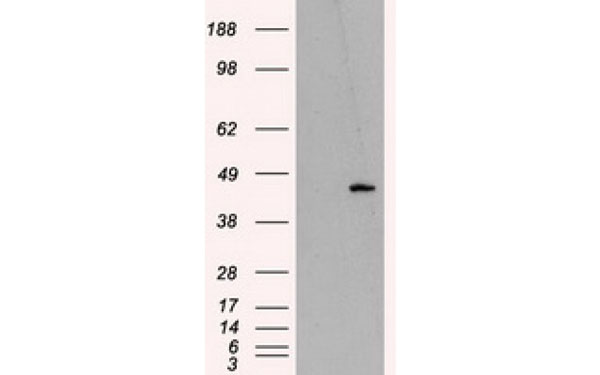
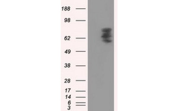
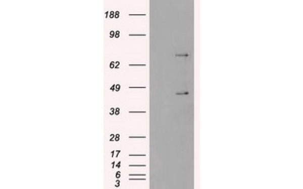
 WB (Western Blot)
(Western Blot analysis of HEK293T cell lysates (5 ug) transfected with either recombinant SulT2A1 protein (Right) or empty vector (Left) detected with SulT2A1 antibody)
WB (Western Blot)
(Western Blot analysis of HEK293T cell lysates (5 ug) transfected with either recombinant SulT2A1 protein (Right) or empty vector (Left) detected with SulT2A1 antibody)
SulT2A1, Monoclonal Antibody (Cat# AAA107216)
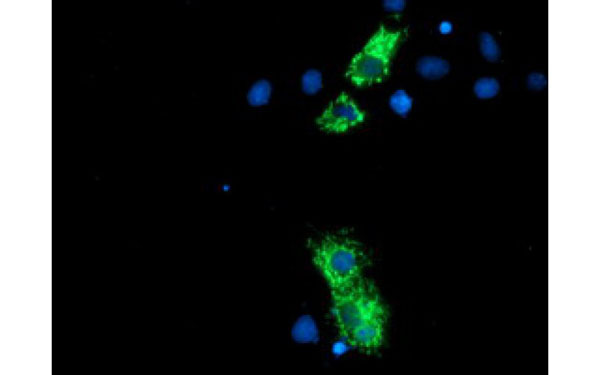
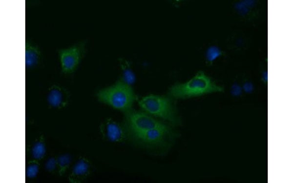
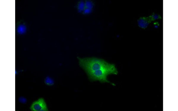
 IF (Immunofluorescence)
(Immunofluorescent staining of COS7 cells transiently transfected with recombinant MTRF1L protein using MTRF1L antibody)
IF (Immunofluorescence)
(Immunofluorescent staining of COS7 cells transiently transfected with recombinant MTRF1L protein using MTRF1L antibody)
MTRF1L, Monoclonal Antibody (Cat# AAA107224)
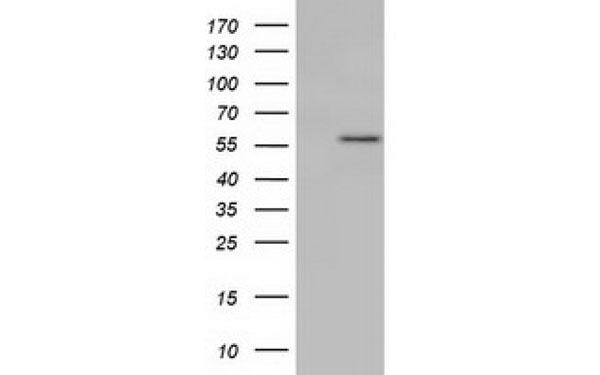
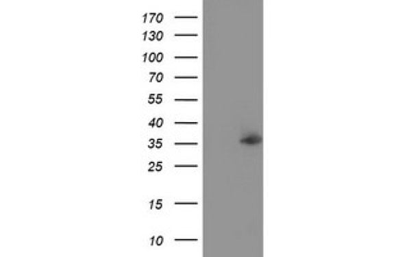
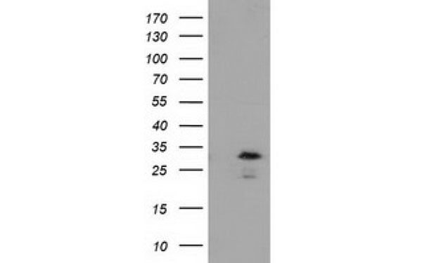
 WB (Western Blot)
(Western Blot analysis of HEK293T cell lysates (5 ug) transfected with either recombinant SERPINA5 protein (Right) or empty vector (Left) detected with SERPINA5 antibody)
WB (Western Blot)
(Western Blot analysis of HEK293T cell lysates (5 ug) transfected with either recombinant SERPINA5 protein (Right) or empty vector (Left) detected with SERPINA5 antibody)
SERPINA5, Monoclonal Antibody (Cat# AAA107230)
 WB (Western Blot)
(Western Blot analysis of HEK293T cell lysates (5 ug) transfected with either recombinant PSMA7 protein (Right) or empty vector (Left) detected with PSMA7 antibody)
WB (Western Blot)
(Western Blot analysis of HEK293T cell lysates (5 ug) transfected with either recombinant PSMA7 protein (Right) or empty vector (Left) detected with PSMA7 antibody)
PSMA7, Monoclonal Antibody (Cat# AAA107233)
 WB (Western Blot)
(Western Blot analysis of HEK293T cell lysates (5 ug) transfected with either recombinant MOBKL2B protein (Right) or empty vector (Left) detected with MOBKL2B antibody)
WB (Western Blot)
(Western Blot analysis of HEK293T cell lysates (5 ug) transfected with either recombinant MOBKL2B protein (Right) or empty vector (Left) detected with MOBKL2B antibody)
MOBKL2B, Monoclonal Antibody (Cat# AAA107253)
 IHC (Immunohiostchemistry)
(Immunohistochemical analysis of KIAA1609 protein in paraffin embedded Human kidney tissue using KIAA1609 antibody)
IHC (Immunohiostchemistry)
(Immunohistochemical analysis of KIAA1609 protein in paraffin embedded Human kidney tissue using KIAA1609 antibody)
KIAA1609, Monoclonal Antibody (Cat# AAA107264)

 IF (Immunofluorescence)
(Immunofluorescent staining of COS7 cells transiently transfected with recombinant KHK protein using KHK antibody)
IF (Immunofluorescence)
(Immunofluorescent staining of COS7 cells transiently transfected with recombinant KHK protein using KHK antibody)
KHK, Monoclonal Antibody (Cat# AAA107269)
 WB (Western Blot)
(Western Blot analysis of HEK293T cell lysates (5 ug) transfected with either recombinant WWTR1 protein (Right) or empty vector (Left) detected with WWTR1 antibody)
WB (Western Blot)
(Western Blot analysis of HEK293T cell lysates (5 ug) transfected with either recombinant WWTR1 protein (Right) or empty vector (Left) detected with WWTR1 antibody)
WWTR1, Monoclonal Antibody (Cat# AAA107542)
 WB (Western Blot)
(Western Blot analysis of HEK293T cell lysates (5 ug) transfected with either recombinant NUDT6 protein (Right) or empty vector (Left) detected with NUDT6 antibody)
WB (Western Blot)
(Western Blot analysis of HEK293T cell lysates (5 ug) transfected with either recombinant NUDT6 protein (Right) or empty vector (Left) detected with NUDT6 antibody)
NUDT6, Monoclonal Antibody (Cat# AAA107546)
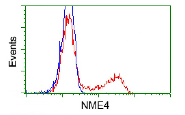
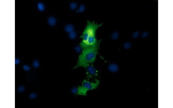
 IF (Immunofluorescence)
(Immunofluorescent staining of COS7 cells transiently transfected with recombinant NME4 protein using NME4 antibody)
IF (Immunofluorescence)
(Immunofluorescent staining of COS7 cells transiently transfected with recombinant NME4 protein using NME4 antibody)
NME4, Monoclonal Antibody (Cat# AAA107370)
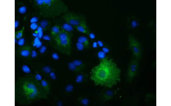
 IF (Immunofluorescence)
(Immunofluorescent staining of COS7 cells transiently transfected with recombinant NUDT6 protein using NUDT6 antibody)
IF (Immunofluorescence)
(Immunofluorescent staining of COS7 cells transiently transfected with recombinant NUDT6 protein using NUDT6 antibody)
NUDT6, Monoclonal Antibody (Cat# AAA107393)
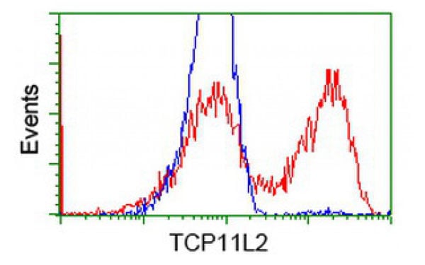
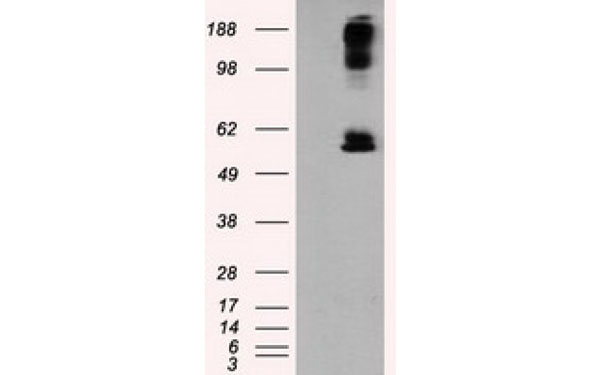
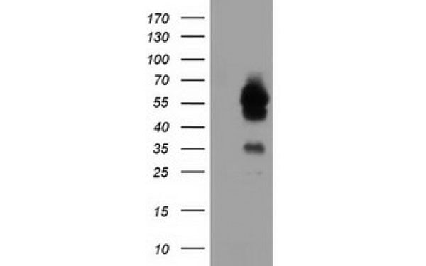 WB (Western Blot)
(Western Blot analysis of HEK293T cell lysates (5 ug) transfected with either recombinant TCP11L2 protein (Right) or empty vector (Left) detected with TCP11L2 antibody)
WB (Western Blot)
(Western Blot analysis of HEK293T cell lysates (5 ug) transfected with either recombinant TCP11L2 protein (Right) or empty vector (Left) detected with TCP11L2 antibody)
TCP11L2, Monoclonal Antibody (Cat# AAA107395)
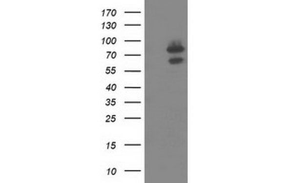 WB (Western Blot)
(Western Blot analysis of HEK293T cell lysates (5 ug) transfected with either recombinant OSBPL11 protein (Right) or empty vector (Left) detected with OSBPL11 antibody)
WB (Western Blot)
(Western Blot analysis of HEK293T cell lysates (5 ug) transfected with either recombinant OSBPL11 protein (Right) or empty vector (Left) detected with OSBPL11 antibody)
OSBPL11, Monoclonal Antibody (Cat# AAA107400)
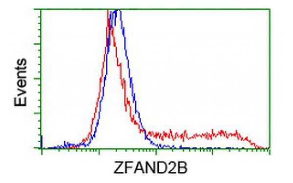
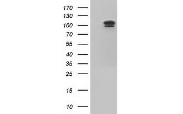
 WB (Western Blot)
(Western Blot analysis of HEK293T cell lysates (5 ug) transfected with either recombinant ZFAND2B protein (Right) or empty vector (Left) detected with ZFAND2B antibody)
WB (Western Blot)
(Western Blot analysis of HEK293T cell lysates (5 ug) transfected with either recombinant ZFAND2B protein (Right) or empty vector (Left) detected with ZFAND2B antibody)
ZFAND2B, Monoclonal Antibody (Cat# AAA107405)
 WB (Western Blot)
(Western Blot analysis of HEK293T cell lysates (5 ug) transfected with either recombinant USP5 protein (Right) or empty vector (Left) detected with USP5 antibody)
WB (Western Blot)
(Western Blot analysis of HEK293T cell lysates (5 ug) transfected with either recombinant USP5 protein (Right) or empty vector (Left) detected with USP5 antibody)
USP5, Monoclonal Antibody (Cat# AAA107411)

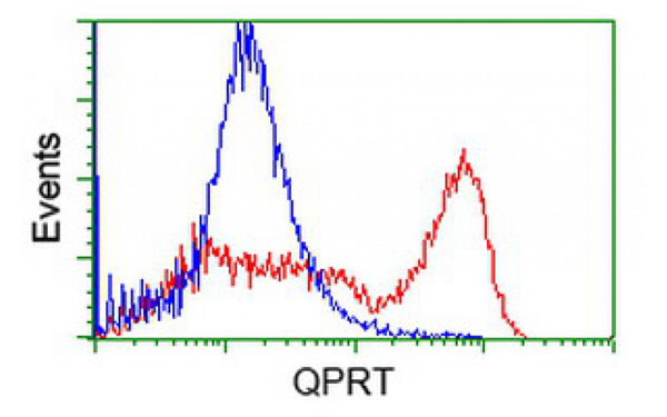
 IF (Immunofluorescence)
(Immunofluorescent staining of COS7 cells transiently transfected with recombinant QPRT protein using QPRT antibody)
IF (Immunofluorescence)
(Immunofluorescent staining of COS7 cells transiently transfected with recombinant QPRT protein using QPRT antibody)
QPRT, Monoclonal Antibody (Cat# AAA107422)
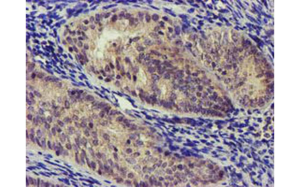
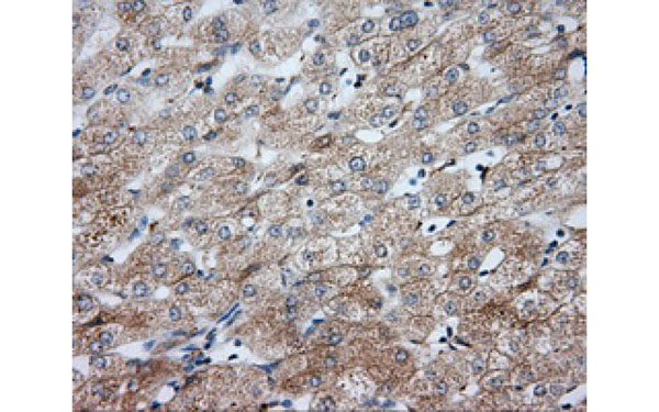
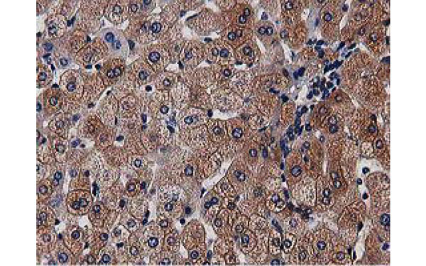
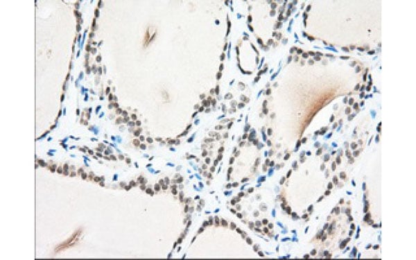
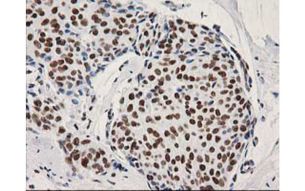
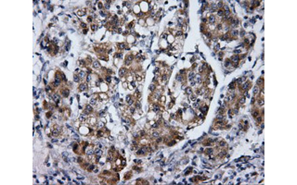 IHC (Immunohiostchemistry)
(Immunohistochemical analysis of LIPG protein in paraffin embedded Carcinoma of Human liver tissue using LIPG antibody)
IHC (Immunohiostchemistry)
(Immunohistochemical analysis of LIPG protein in paraffin embedded Carcinoma of Human liver tissue using LIPG antibody)
LIPG, Monoclonal Antibody (Cat# AAA107438)
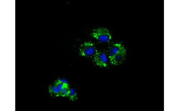
 IF (Immunofluorescence)
(Immunofluorescent staining of COS7 cells transiently transfected with recombinant SDS protein using SDS antibody)
IF (Immunofluorescence)
(Immunofluorescent staining of COS7 cells transiently transfected with recombinant SDS protein using SDS antibody)
SDS, Monoclonal Antibody (Cat# AAA107442)
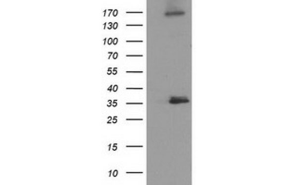
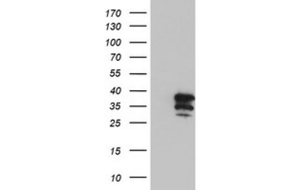
 WB (Western Blot)
(Western Blot analysis of HEK293T cell lysates (5 ug) transfected with either recombinant PUS7 protein (Right) or empty vector (Left) detected with PUS7 antibody)
WB (Western Blot)
(Western Blot analysis of HEK293T cell lysates (5 ug) transfected with either recombinant PUS7 protein (Right) or empty vector (Left) detected with PUS7 antibody)
PUS7, Monoclonal Antibody (Cat# AAA107443)
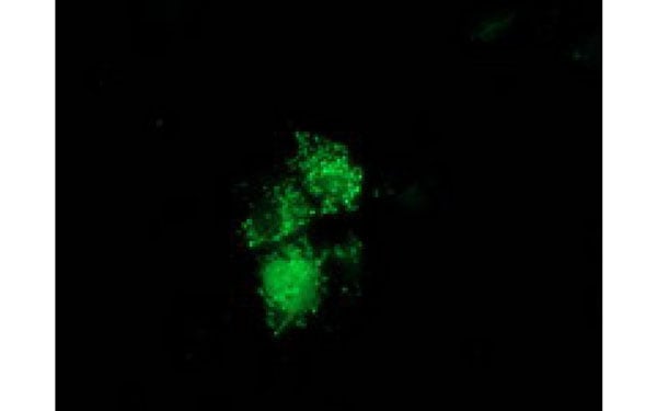
 IF (Immunofluorescence)
(Immunofluorescent staining of COS7 cells transiently transfected with recombinant PDE4A protein using PDE4A antibody)
IF (Immunofluorescence)
(Immunofluorescent staining of COS7 cells transiently transfected with recombinant PDE4A protein using PDE4A antibody)
PDE4A, Monoclonal Antibody (Cat# AAA107466)
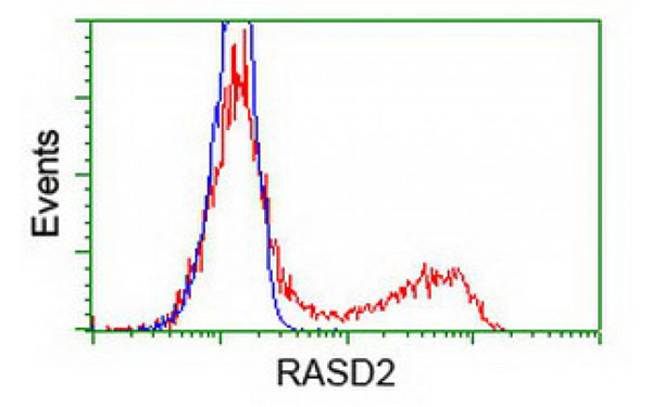
 IF (Immunofluorescence)
(Immunofluorescent staining of COS7 cells transiently transfected with recombinant RASD2 protein using RASD2 antibody)
IF (Immunofluorescence)
(Immunofluorescent staining of COS7 cells transiently transfected with recombinant RASD2 protein using RASD2 antibody)
RASD2, Monoclonal Antibody (Cat# AAA107470)
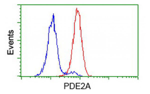
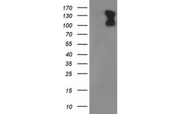 WB (Western Blot)
(Western Blot analysis of HEK293T cell lysates (5 ug) transfected with either recombinant PDE2A protein (Right) or empty vector (Left) detected with PDE2A antibody)
WB (Western Blot)
(Western Blot analysis of HEK293T cell lysates (5 ug) transfected with either recombinant PDE2A protein (Right) or empty vector (Left) detected with PDE2A antibody)
PDE2A, Monoclonal Antibody (Cat# AAA107496)
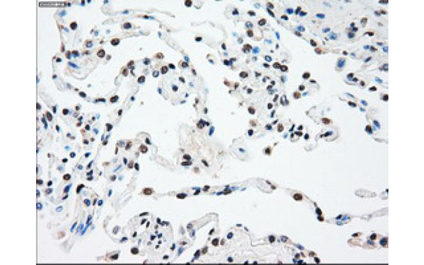
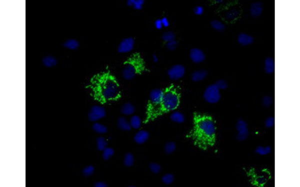
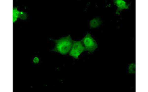
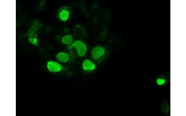
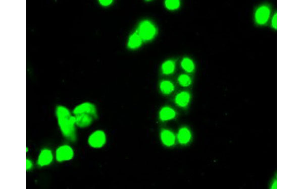 IF (Immunofluorescence)
(Immunofluorescent staining of endogenous SSB protein in HT29 cells using SSB antibody)
IF (Immunofluorescence)
(Immunofluorescent staining of endogenous SSB protein in HT29 cells using SSB antibody)
SSB, Monoclonal Antibody (Cat# AAA107501)
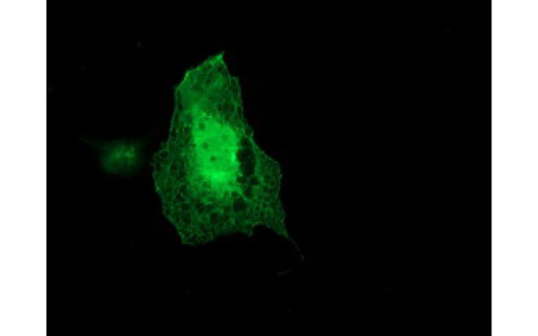 IF (Immunofluorescence)
(Immunofluorescent staining of COS7 cells transiently transfected with recombinant ARCN1 protein using ARCN1 antibody)
IF (Immunofluorescence)
(Immunofluorescent staining of COS7 cells transiently transfected with recombinant ARCN1 protein using ARCN1 antibody)
ARCN1, Monoclonal Antibody (Cat# AAA107509)
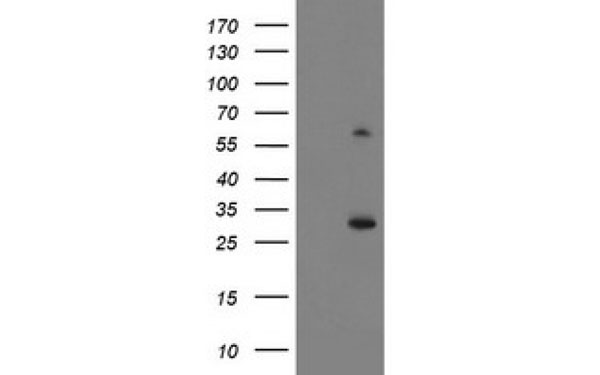 WB (Western Blot)
(Western Blot analysis of HEK293T cell lysates (5 ug) transfected with either recombinant KCTD14 protein (Right) or empty vector (Left) detected with KCTD14 antibody)
WB (Western Blot)
(Western Blot analysis of HEK293T cell lysates (5 ug) transfected with either recombinant KCTD14 protein (Right) or empty vector (Left) detected with KCTD14 antibody)
KCTD14, Monoclonal Antibody (Cat# AAA107447)
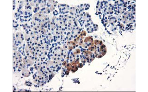
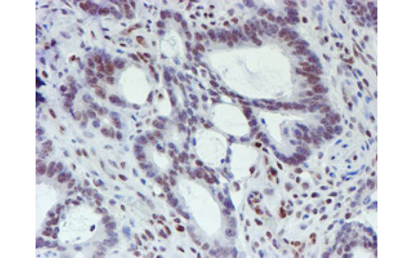
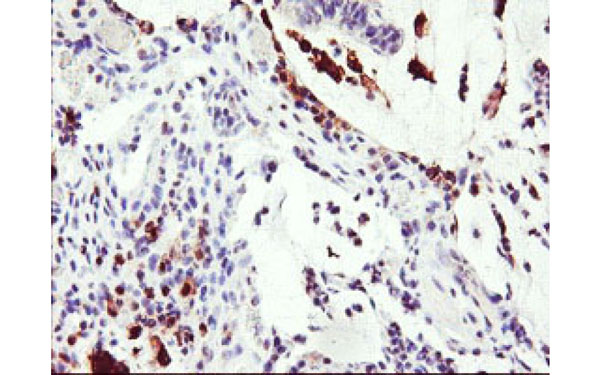
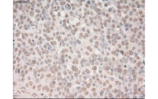 IHC (Immunohistochemisry)
(Immunohistochemical analysis of SOX17 protein in paraffin embedded Adenocarcinoma of Human ovary tissue using SOX17 antibody)
IHC (Immunohistochemisry)
(Immunohistochemical analysis of SOX17 protein in paraffin embedded Adenocarcinoma of Human ovary tissue using SOX17 antibody)
SOX17, Monoclonal Antibody (Cat# AAA107451)
 IF (Immunofluorescence)
(Immunofluorescent staining of COS7 cells transiently transfected with recombinant LENG1 protein using LENG1 antibody)
IF (Immunofluorescence)
(Immunofluorescent staining of COS7 cells transiently transfected with recombinant LENG1 protein using LENG1 antibody)
LENG1, Monoclonal Antibody (Cat# AAA107458)
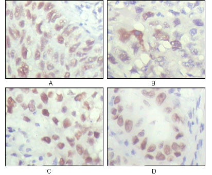
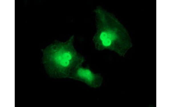
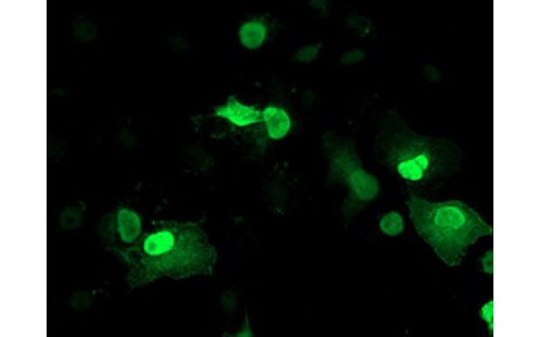
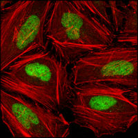 IF (Immunofluorescence)
(Figure 3. Confocal immunofluorescence analysis of Hela cells using CHK2 mouse mAb (green), showing nuclear localization. Red. Actin filaments have been labeled with DY-554 phalloidin.)
IF (Immunofluorescence)
(Figure 3. Confocal immunofluorescence analysis of Hela cells using CHK2 mouse mAb (green), showing nuclear localization. Red. Actin filaments have been labeled with DY-554 phalloidin.)
CHK2, Monoclonal Antibody (Cat# AAA108638)
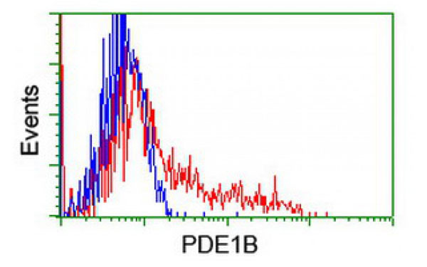
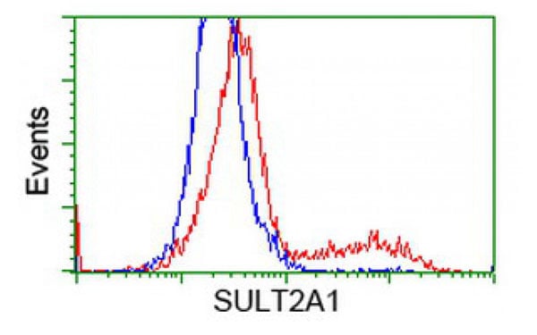
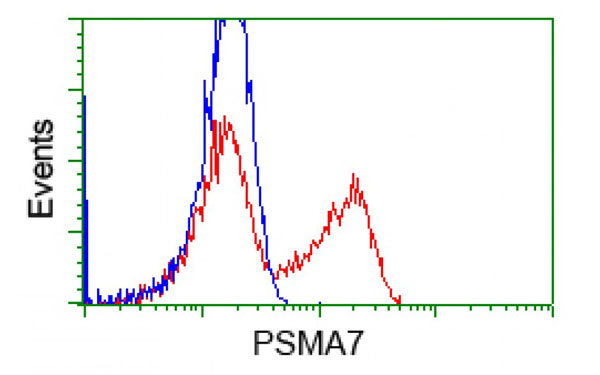
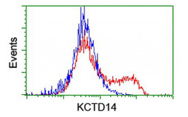
 FCM/FACS (Flow Cytometry)
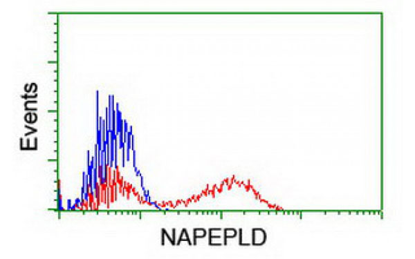
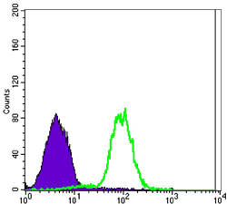
(Figure 3: Flow cytometric analysis of NTERA-2 cells using CA9 mouse mAb (green) and negative control (purple).)
FCM/FACS (Flow Cytometry)
(Figure 3: Flow cytometric analysis of NTERA-2 cells using CA9 mouse mAb (green) and negative control (purple).)
CA9, Monoclonal Antibody (Cat# AAA108668)
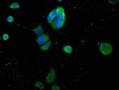
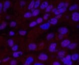 IF (Immunofluorescence)
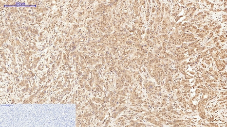
(Immunofluorescence analysis of Human liver cancer tissue using XRCC4 Monoclonal Antibody at dilution of 1:200.)
IF (Immunofluorescence)
(Immunofluorescence analysis of Human liver cancer tissue using XRCC4 Monoclonal Antibody at dilution of 1:200.)
XRCC4, Monoclonal Antibody (Cat# AAA171612)
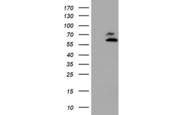
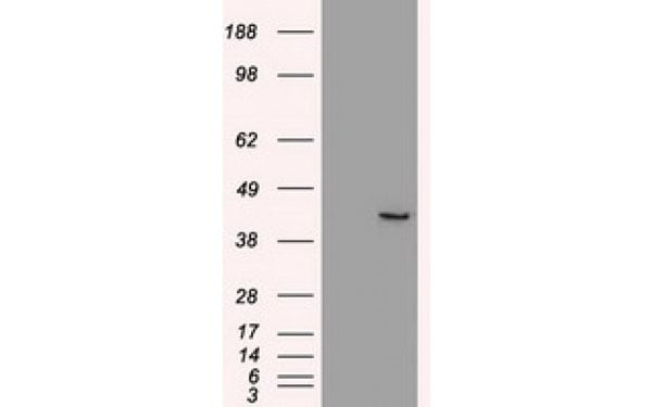
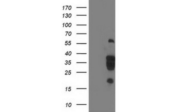
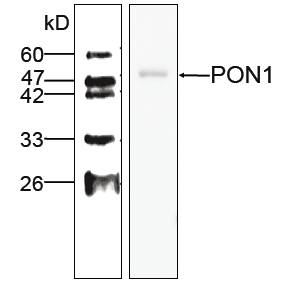 WB (Western Blot)
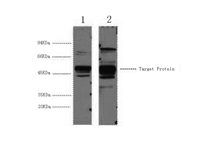
(WB (1:1000) analysis of PON 1 expression in Hela whole cell lysate with Anti-PON 1 (AAA109354))
WB (Western Blot)
(WB (1:1000) analysis of PON 1 expression in Hela whole cell lysate with Anti-PON 1 (AAA109354))
PON 1, Monoclonal Antibody (Cat# AAA109354)

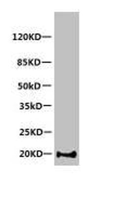
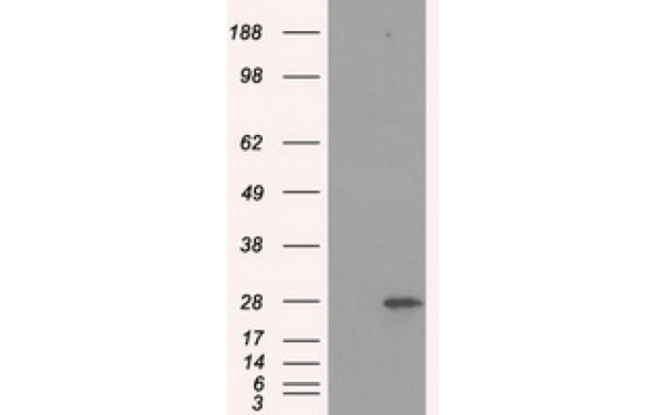
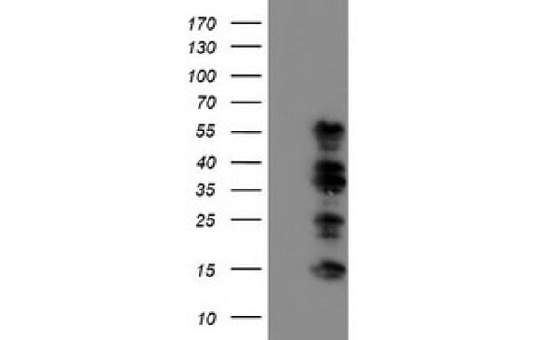
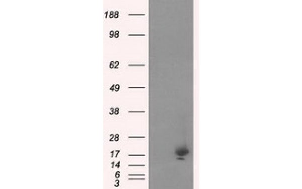
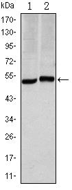
 WB (Western Blot)
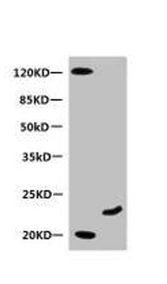
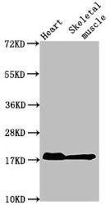
(Positive WB detected in: Mouse heart tissue, Mouse skeletal muscle tissueAll lanes: MB antibody at 1:2000SecondaryGoat polyclonal to Mouse IgG at 1/50000 dilutionPredicted band size: 18 kDaObserved band size: 18 kDa)
WB (Western Blot)
(Positive WB detected in: Mouse heart tissue, Mouse skeletal muscle tissueAll lanes: MB antibody at 1:2000SecondaryGoat polyclonal to Mouse IgG at 1/50000 dilutionPredicted band size: 18 kDaObserved band size: 18 kDa)
Myoglobin, Monoclonal Antibody (Cat# AAA114272)
CD11a (activitor), Monoclonal Antibody (Cat# AAA77516)
Mouse anti Human IgG3 (G3m(U) allotype specific), Monoclonal Secondary Antibody (Cat# AAA77528)
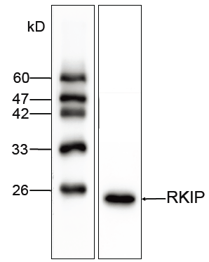 Application Data
RKIP, Monoclonal Antibody (Cat# AAA109271)
 Application Data
DYKDDDDK-Tag, Monoclonal Antibody (Cat# AAA108501)
 Application Data
amyloid beta peptide N-terminal, Monoclonal Antibody (Cat# AAA77929)
RSV, Monoclonal Antibody (Cat# AAA78047)
Influenza Virus B Group, Monoclonal Antibody (Cat# AAA78072)
Chromatography on protein A Sepharose for MAbs InB12, InB27, InB36, InB64, InB114, InB204, InB210, InB213, 8-5, 13-9
C-reactive Protein, Monoclonal Antibody (Cat# AAA78078)
Hepatitis B Virus Surface Antigen, Monoclonal Antibody (Cat# AAA78087)
Purification: Protein G chromatography
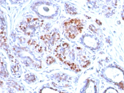
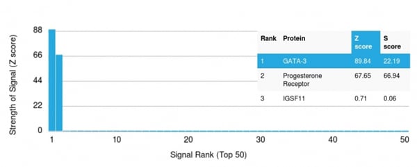 Application Data
(Analysis of Protein Array containing more than 19,000 full-length human proteins using GATA-3 Mouse Monoclonal Antibody (GATA3/2442). Z- and S- Score: The Z-score represents the strength of a signal that a monoclonal antibody (MAb) (in combination with a fluorescently-tagged anti-IgG secondary antibody) produces when binding to a particular protein on the HuProtTM array. Z-scores are described in units of standard deviations (SD's) above the mean value of all signals generated on that array. If targets on HuProtTM are arranged in descending order of the Z-score, the S-score is the difference (also in units of SD's) between the Z-score. S-score therefore represents the relative target specificity of a MAb to its intended target. A MAb is considered to specific to its intended target, if the MAb has an S-score of at least 2.5. For example, if a MAb binds to protein X with a Z-score of 43 and to protein Y with a Z-score of 14, then the S-score for the binding of that MAb to protein X is equal to 29.)
Application Data
(Analysis of Protein Array containing more than 19,000 full-length human proteins using GATA-3 Mouse Monoclonal Antibody (GATA3/2442). Z- and S- Score: The Z-score represents the strength of a signal that a monoclonal antibody (MAb) (in combination with a fluorescently-tagged anti-IgG secondary antibody) produces when binding to a particular protein on the HuProtTM array. Z-scores are described in units of standard deviations (SD's) above the mean value of all signals generated on that array. If targets on HuProtTM are arranged in descending order of the Z-score, the S-score is the difference (also in units of SD's) between the Z-score. S-score therefore represents the relative target specificity of a MAb to its intended target. A MAb is considered to specific to its intended target, if the MAb has an S-score of at least 2.5. For example, if a MAb binds to protein X with a Z-score of 43 and to protein Y with a Z-score of 14, then the S-score for the binding of that MAb to protein X is equal to 29.)
GATA-3, Monoclonal Antibody (Cat# AAA214745)
What are Monoclonal Antibodies?
Monoclonal antibodies are specialized laboratory-produced proteins developed for binding to specific biological antigens or other molecular targets. Since they come from a single cell (or clone), they are especially consistent and accurate in the data they are involved in producing.
This type of antibody material has been shown to be a powerful tool in finding and subsequently destroying harmful cells in an organism, such as those found in cancers or various autoimmune diseases. This makes them excellent aids in medical testing and research, which is why they are so widely used.
AAA Biotech offers a comprehensive range of high-quality monoclonal antibodies that perform effectively in various laboratory tests, including (amongst others) ELISA, western blotting, immunohistochemistry, and flow cytometry. All of the products in our catalog are thoroughly quality tested to make sure that they are reliable and will consistently perform well in your research.
What Are The Uses of Monoclonal Antibodies
Monoclonal antibodies are used in many lab tests, including (amongst others) ELISA, western blotting, immunohistochemistry, and flow cytometry.
ELISA is a test that helps detect a specific substance/analyte in a sample. It uses antibodies (often monoclonal) bound to a solid surface (such as the well of a microplate) to “capture” the substance/analyte in the sample and immobilize it so that the detection antibody component can then bind to it and produce a signal, which can then be measured.
Western blotting identifies specific proteins in a sample. The sample is first separated on a gel, and then antibodies are applied that will typically bind to the target, which will all be localized to a single band in a lane.
Immunohistochemistry helps locate specific proteins in cells or tissue samples using antibodies.
Flow cytometry looks at and sorts cells. It uses antibodies that are conjugated to reporter molecules called “fluorophores”, which, under special lights, emit light themselves, which can then be measured by a detector instrument.
How Monoclonal Antibodies Are Used as Medicine?
Please note that all of the products listed in AAA Biotech’s also known as AAA Bio or AAABio catalog are strictly for research-use only (RUO).
Monoclonal antibodies can also be used as therapeutic/medical treatments, particularly in the context of cancers. They are designed to find and bind to specific cells or proteins, helping the immune system recognize and attack the cancer. These treatments work in different ways, such as:
- Radioimmunotherapy attaches a small amount of radioactive molecule to the antibody, so it delivers the radiation directly to the cancer cells that the antibody is specifically binding to.
- Antibody-directed enzyme prodrug therapy uses antibodies that are specifically bound to special enzymes. These enzymes activate a harmless drug in the body and turn it into a cancer-killing drug only near the cancer cells—this helps avoid harming healthy cells.
- Immunoliposomes are tiny “bubbles” filled with medicine/drug and coated with antibodies. They carry the drug straight to the cancer cells.
Why Buy Monoclonal Antibodies From Us?
At AAA Biotech, we provide high-performance monoclonal antibodies designed to support a wide range of research needs.
1. Validated for Versatile Applications
The antibodies in our catalog are extensively validated and compatible with multiple techniques, including (but not limited to) ELISA, flow cytometry (FC), immunocytochemistry (ICC), immunofluorescence (IF), immunohistochemistry (IHC), immunoprecipitation (IP), and western blotting (WB).
2. Wide Selection & Specialized Options
We offer antibodies for common and rare species, that are available in various conjugated forms, and also in recombinant formats. Essentially, there is almost anything one might need to meet their experimental model’s requirements.
3. High-Quality Proteins
Our proteins meet high purity standards—90% or more as confirmed by SDS-PAGE. Many are available with tags like His, Flag, GST, or MBP, and we also supply native and biologically active proteins for functional studies.
Frequently Asked Questions
1. Are your monoclonal antibodies validated for specific applications?
Yes, our antibodies are tested and validated for use in methods such as ELISA, western blot, IHC, flow cytometry, and more. Refer to specific product pages or datasheets for individual product information.
2. How do I choose the right monoclonal antibody for my application?
Review the product details directly for application validation, species reactivity, and target information. You may also contact our support team at any time for help.
3. How quickly can I receive my order?
Most orders are processed and shipped within 1–3 business days, depending on product availability and your shipping location.