Filters
▼Clonality
▼Type
▼Reactivity
▼Gene Name
▼Isotype
▼Host
▼Application
▼Clone
▼Active Proteins
AAA Biotech also known as AAA Bio or AAABio provides a variety of high-quality recombinant and natural/native proteins that are proven to work in a wide range of experiments. Explore our products to find the active protein that best fits your needs or experimental model.
Viewing 1800-1850 of 2567 product results
KBP-Type Peptidyl-Prolyl Cis-Trans Isomerase, Active Protein (Cat# AAA38285)
Follicle Stimulating Hormone, Active Protein (Cat# AAA38350)
Mitogen-Activated Protein Kinase 1, Active Protein (Cat# AAA38388)
Vascular Endothelial Growth Factor receptor-1, Active Protein (Cat# AAA38398)
Superoxide Dismutase-1, Active Protein (Cat# AAA38724)
IL-32 alpha, Active Protein (Cat# AAA38596)
Glutathione S-Transferase, Active Protein (Cat# AAA38680)
Thioredoxin Reductase 1, Active Protein (Cat# AAA38824)
Lactate Dehydrogenase 2, Active Protein (Cat# AAA44810)
 Application Data
(Measured by its binding ability in a functional ELISA. Immobilized Mouse ACVR2B at 2ug/mL (100 ul/well) can bind biotinylated mouse INHBA-His, The EC50 of biotinylated mouse INHBA-His is 40-95 ng/mL.)
Application Data
(Measured by its binding ability in a functional ELISA. Immobilized Mouse ACVR2B at 2ug/mL (100 ul/well) can bind biotinylated mouse INHBA-His, The EC50 of biotinylated mouse INHBA-His is 40-95 ng/mL.)
ACVR2B, Active Protein (Cat# AAA258137)
 Application Data
(Measured in a cell proliferation assay using BALB/c 3T3 mouse fibroblasts. The ED50 for this effect is typically 0.8-3.3 ug/mL.)
Application Data
(Measured in a cell proliferation assay using BALB/c 3T3 mouse fibroblasts. The ED50 for this effect is typically 0.8-3.3 ug/mL.)
FGF8, Active Protein (Cat# AAA258049)
 Application Data
(Immobilized human ACE2 protein (mFc tag) at 2 ug/mL (100 uL/well) can bind SARS-CoV-2 Spike S1+S2 (D80A, LAL242-244 deletion, R246I, K417N, E484K, N501Y, D614G, A701V) Protein (ECD, His Tag),the EC50 of SARS-CoV-2 Spike S1+S2 (D80A, LAL242-244 deletion, R246I, K417N, E484K, N501Y, D614G, A701V) Protein (ECD, His Tag) is 100-600 ng/mL.)
Application Data
(Immobilized human ACE2 protein (mFc tag) at 2 ug/mL (100 uL/well) can bind SARS-CoV-2 Spike S1+S2 (D80A, LAL242-244 deletion, R246I, K417N, E484K, N501Y, D614G, A701V) Protein (ECD, His Tag),the EC50 of SARS-CoV-2 Spike S1+S2 (D80A, LAL242-244 deletion, R246I, K417N, E484K, N501Y, D614G, A701V) Protein (ECD, His Tag) is 100-600 ng/mL.)
COVID 19 Spike S1+S2 (D80A, LAL242-244 deletion, R246I, K417N, E484K, N501Y, D614G, A701V) Protein (ECD, His Tag) Coronavirus, Active Protein (Cat# AAA258077)
 Application Data
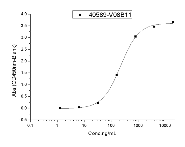
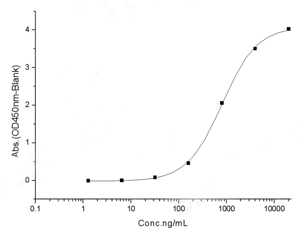
(Measured by its binding ability in a functional ELISA.Immobilized human ACE2 protein (Fc tag)(10108-H05H) at 2 ug/mL (100 uL/well) can bind SARS-CoV-2 (2019-nCoV) Spike S1+S2 ECD (D614G)-His(40589-V08B4), the EC50 of SARS-CoV-2 (2019-nCoV) Spike S1+S2 ECD (D614G)-His(40589-V08B4) is 0.5-1.2 ug/mL.)
Application Data
(Measured by its binding ability in a functional ELISA.Immobilized human ACE2 protein (Fc tag)(10108-H05H) at 2 ug/mL (100 uL/well) can bind SARS-CoV-2 (2019-nCoV) Spike S1+S2 ECD (D614G)-His(40589-V08B4), the EC50 of SARS-CoV-2 (2019-nCoV) Spike S1+S2 ECD (D614G)-His(40589-V08B4) is 0.5-1.2 ug/mL.)
COVID 19 Spike S1+S2 ECD (D614G)-His Coronavirus, Active Protein (Cat# AAA258079)
 Bioactivity
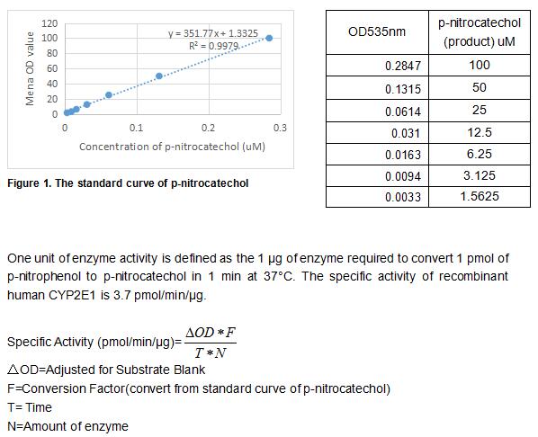
(The cytochrome P450 enzyme CYP2E1 catalyzes the oxidative metabolism of many solvents and other small organic molecules. CYP2E1 is expressed in adult and fetal human liver in addition to extrahepatic tissues such as lung and placenta. Treatment of primary cultures of human hepatocytes with ethanol induces CYP2E1 protein, and this is consistent with the finding that hepatic CYP2E1 protein and mRNA levels are increased in individuals with alcoholism. Although only a few drugs (e.g., acetaminophen have been identified as substrates for CYP2E1, many low molecular weight procarcinogens are activated by this cytochrome P450 (P450). Chlorzoxazone 6-hydroxylation, N-nitrosodimethylamine N-demethylation and p-nitrophenol hydroxylation can be used to measure the catalytic activity of CYP2E1. Thus, the recombinant human CYP2E1 activity was measured by its ability to hydroxylate p-nitrophenol to p-nitrocatechol. The reaction was performed in 50 mM potassium phosphate, pH 7.4 (Assay Buffer), initiated by addition 20 uL of 500 ug/ml CYP2E1 to 10 uL of 5 mM substrate p-nitrophenol and 30 ul of 26 mM NADPH in a total volume of 500 ul. Incubated at 37 degree C for 30min, then read at a wavelength of 535 nm after acidification of the reaction mixture with trichloroacetic acid followed by neutralization using 2 M NaOH.)
Bioactivity
(The cytochrome P450 enzyme CYP2E1 catalyzes the oxidative metabolism of many solvents and other small organic molecules. CYP2E1 is expressed in adult and fetal human liver in addition to extrahepatic tissues such as lung and placenta. Treatment of primary cultures of human hepatocytes with ethanol induces CYP2E1 protein, and this is consistent with the finding that hepatic CYP2E1 protein and mRNA levels are increased in individuals with alcoholism. Although only a few drugs (e.g., acetaminophen have been identified as substrates for CYP2E1, many low molecular weight procarcinogens are activated by this cytochrome P450 (P450). Chlorzoxazone 6-hydroxylation, N-nitrosodimethylamine N-demethylation and p-nitrophenol hydroxylation can be used to measure the catalytic activity of CYP2E1. Thus, the recombinant human CYP2E1 activity was measured by its ability to hydroxylate p-nitrophenol to p-nitrocatechol. The reaction was performed in 50 mM potassium phosphate, pH 7.4 (Assay Buffer), initiated by addition 20 uL of 500 ug/ml CYP2E1 to 10 uL of 5 mM substrate p-nitrophenol and 30 ul of 26 mM NADPH in a total volume of 500 ul. Incubated at 37 degree C for 30min, then read at a wavelength of 535 nm after acidification of the reaction mixture with trichloroacetic acid followed by neutralization using 2 M NaOH.)
Cytochrome P450 2E1 (CYP2E1), Active Protein (Cat# AAA161816)
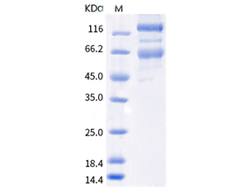
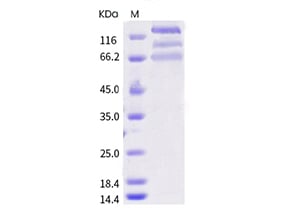
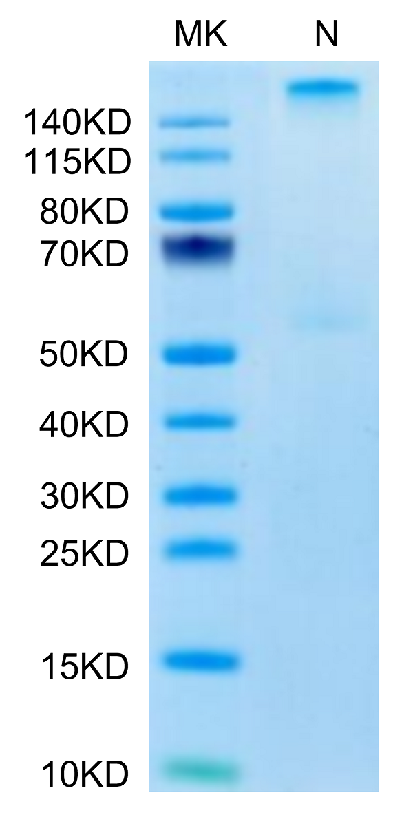
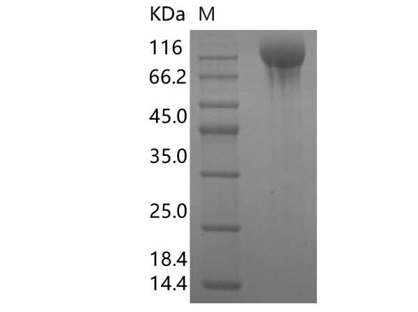 SDS-PAGE
(Recombinant SARS-CoV-2 S1+S2 trimer Protein (C-His Tag)(Omicron))
SDS-PAGE
(Recombinant SARS-CoV-2 S1+S2 trimer Protein (C-His Tag)(Omicron))
COVID 19 Spike S1+S2 trimer Coronavirus, Active Protein (Cat# AAA177998)
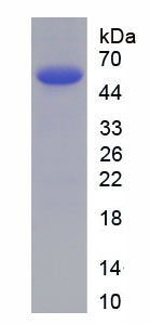
 SDS-PAGE
COVID 19 Spike RBD Coronavirus, Active Protein (Cat# AAA177012)
Interleukin 4, Active Protein (Cat# AAA62204)
BETA GALACTOSIDASE, Active Protein (Cat# AAA50519)
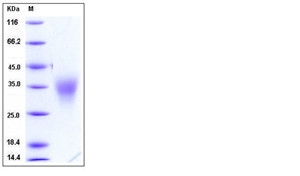
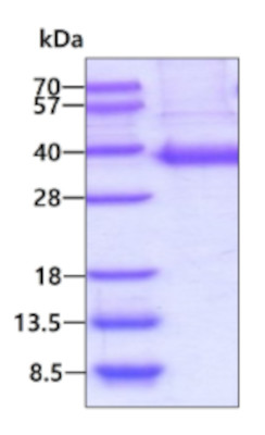
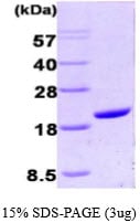
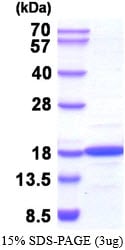
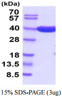 SDS-PAGE
(3 ug by SDS-PAGE under reducing condition and visualized by coomassie blue stain.)
SDS-PAGE
(3 ug by SDS-PAGE under reducing condition and visualized by coomassie blue stain.)
Beta-lactaminase, Active Protein (Cat# AAA48963)
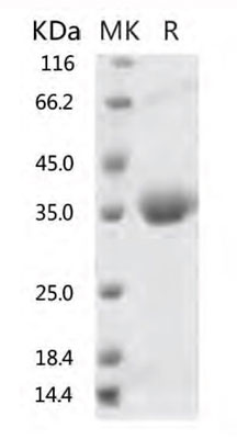
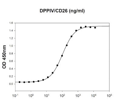 Bioactivity
(MERS-CoV Spike RBD is coated at 2 ug/ml (100 ul/well) can bind human DPPIV/CD26 in a functional ELISA assay.)
Bioactivity
(MERS-CoV Spike RBD is coated at 2 ug/ml (100 ul/well) can bind human DPPIV/CD26 in a functional ELISA assay.)
MERS-CoV Spike, Active Protein (Cat# AAA48981)
MERS-CoV Spike, Active Protein (Cat# AAA48982)
ICAM-1/CD54, Active Protein (Cat# AAA48983)
Fc gamma RIIA/CD32a (R167H), Active Protein (Cat# AAA48984)
EphA3, Active Protein (Cat# AAA48999)
Osteocrin, Active Protein (Cat# AAA49001)
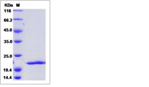
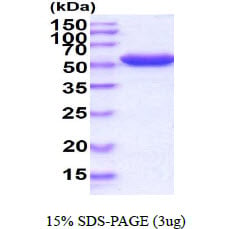
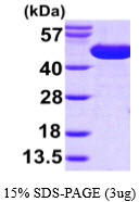
 SDS-PAGE
(3ug by SDS-PAGE under reducing condition and visualized by coomassie blue stain)
SDS-PAGE
(3ug by SDS-PAGE under reducing condition and visualized by coomassie blue stain)
Cyclophilin A, Active Protein (Cat# AAA48477)
 SDS-PAGE
G6PD, Active Protein (Cat# AAA48520)
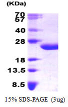
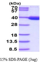
 SDS-PAGE
(3ug by SDS-PAGE under reducing condition and visualized by coomassie blue stain.)
SDS-PAGE
(3ug by SDS-PAGE under reducing condition and visualized by coomassie blue stain.)
Carbonic anhydrase 2, Active Protein (Cat# AAA48538)
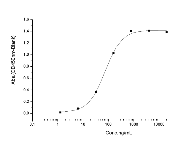
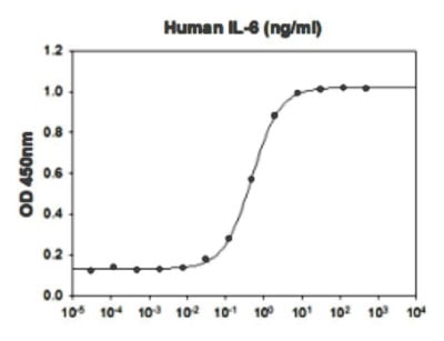 Application Data
(Biological Acitivity: Human IL-6 stimulates cell proliferation of the TF-1 human erythroleukemic cells. The ED50 range )
Application Data
(Biological Acitivity: Human IL-6 stimulates cell proliferation of the TF-1 human erythroleukemic cells. The ED50 range )
Interleukin 6, Active Protein (Cat# AAA48546)
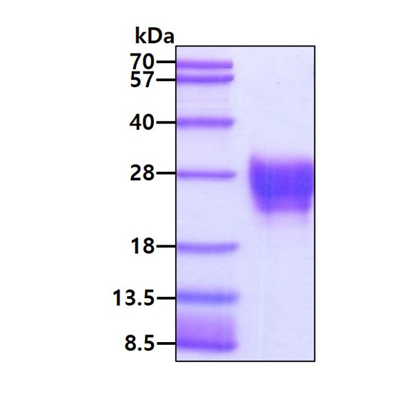
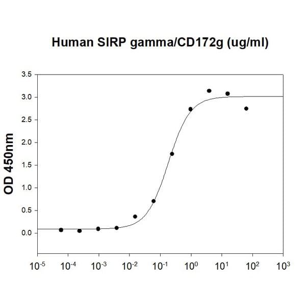 Bioactivity
(Human CD47 is coated at 1 ug/ml (100 ul/well) can bind Human SIRP gamma/CD172g (CAT# ATGP4030) in a Functional ELISA assay.)
Bioactivity
(Human CD47 is coated at 1 ug/ml (100 ul/well) can bind Human SIRP gamma/CD172g (CAT# ATGP4030) in a Functional ELISA assay.)
CD47, Active Protein (Cat# AAA48392)
 Bioactivity
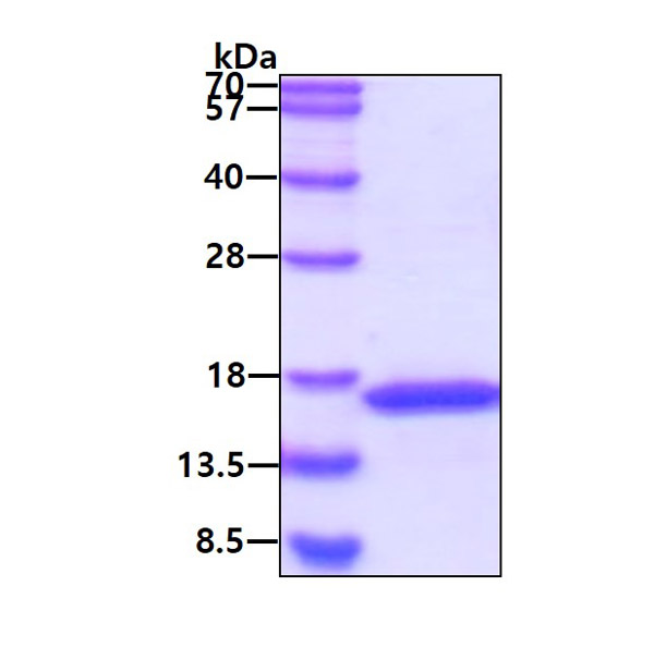
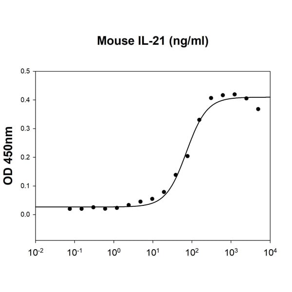
(Mouse IL-21 stimulates IFN-g secretion of the NK-92 human natural killer cells. The ED50 range )
Bioactivity
(Mouse IL-21 stimulates IFN-g secretion of the NK-92 human natural killer cells. The ED50 range )
IL-21, Active Protein (Cat# AAA48409)
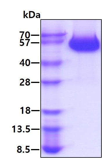
 SDS-PAGE
NME2, Active Protein (Cat# AAA48916)
 SDS-PAGE
SurA, Active Protein (Cat# AAA48603)
 SDS-PAGE
(3ug by SDS-PAGE under reducing condition and visualized by coomassie blue stain)
SDS-PAGE
(3ug by SDS-PAGE under reducing condition and visualized by coomassie blue stain)
LDHB, Active Protein (Cat# AAA48607)
 Bioactivity
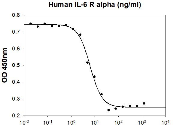
(Human IL-6 R alpha inhibits Human IL-6 induced cell proliferation in the M1 mouse myeloid leukemia cells. The ED50 range 20 ng/ml.)
Bioactivity
(Human IL-6 R alpha inhibits Human IL-6 induced cell proliferation in the M1 mouse myeloid leukemia cells. The ED50 range 20 ng/ml.)
IL-6R alpha, Active Protein (Cat# AAA48327)
 Bioactivity
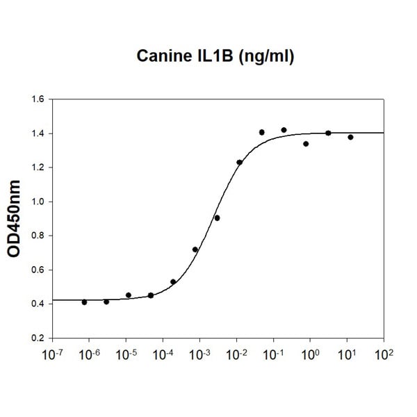
(Canine IL-1B stimulates cell proliferation of the D10.G4.1 mouse helper T cells. The ED50 range range )
Bioactivity
(Canine IL-1B stimulates cell proliferation of the D10.G4.1 mouse helper T cells. The ED50 range range )
IL-1 beta/IL-1F2, Active Protein (Cat# AAA48366)
Sonic Hedgehog, Active Protein (Cat# AAA75584)
BD4, Active Protein (Cat# AAA75586)
TLR3, Active Protein (Cat# AAA75594)
ANGPTL-7, Active Protein (Cat# AAA76054)
IL2RA (CD25), Active Protein (Cat# AAA76070)
CD32b (FcgR2b), Active Protein (Cat# AAA76086)
EGFL7, Active Protein (Cat# AAA76105)
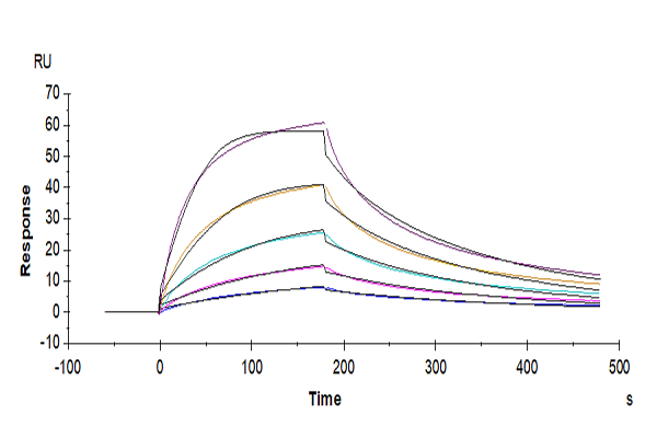 Application Data
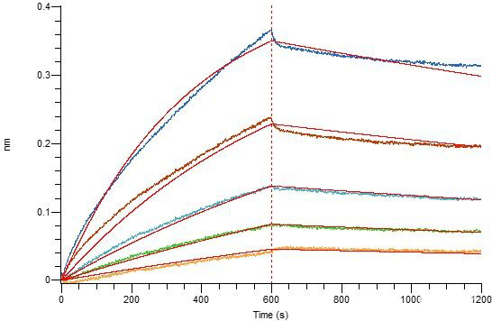
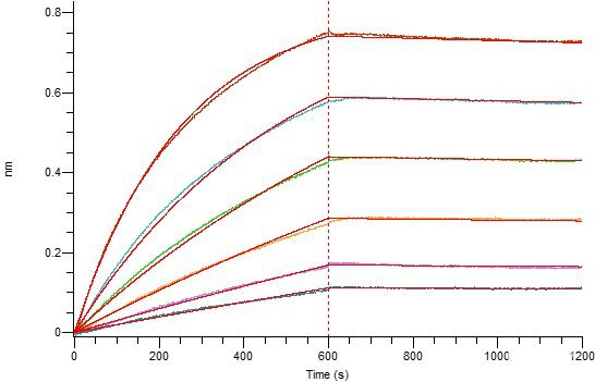
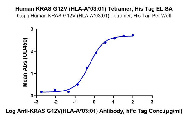
(KRAS G12V (HLA-A*03:01) Tetramer SPRSPR image shows curve for KRAS G12V(HLA-A*03:01) and immobilized Antibody binding, with a calculated affinity constant of 42nM)
Application Data
(KRAS G12V (HLA-A*03:01) Tetramer SPRSPR image shows curve for KRAS G12V(HLA-A*03:01) and immobilized Antibody binding, with a calculated affinity constant of 42nM)
HLA-A*03:01 KRAS G12V, Active Protein (Cat# AAA76371)

 Application Data
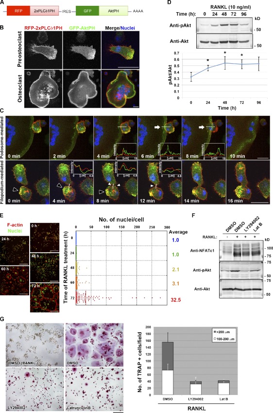
(Polarized membrane extensions mediate osteoclast fusion.(D, top) Immunoblot analysis of lysates of RAW264.7 macrophages stimulated with RANKL for the indicated times with antibodies to Ser473-phosphorylated (p) or total forms of Akt.)
Application Data
(Polarized membrane extensions mediate osteoclast fusion.(D, top) Immunoblot analysis of lysates of RAW264.7 macrophages stimulated with RANKL for the indicated times with antibodies to Ser473-phosphorylated (p) or total forms of Akt.)
Soluble RANK ligand, Active Protein (Cat# AAA76384)
 Application Data
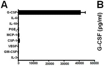
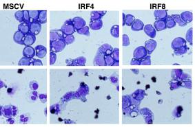
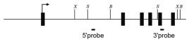
(Generation of conditional TAK1-deficient mice.Colony formation by BM cells from Map3k7)
Application Data
(Generation of conditional TAK1-deficient mice.Colony formation by BM cells from Map3k7)
Granulocyte Colony Stimulating Factor, Active Protein (Cat# AAA76390)

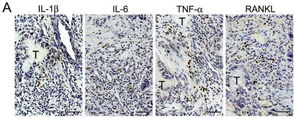
 Application Data
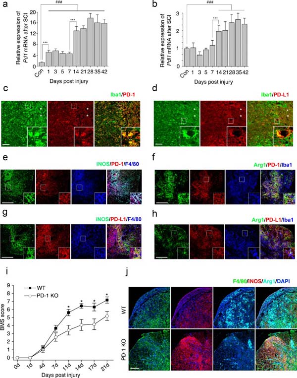
(Expression of programmed death-1 (PD-1) in macrophages/microglia after spinal cord injury (SCI) in mice.Immunohistochemistry in macrophages/microglia 14 days postinjury (dpi) in wild-type mice for (c) PD-1, (d) it’s ligand, PD-L1, and (e) PD-1 in M1 cells [inducible nitric oxide synthase (iNOS)+]; (f) PD-1 in M2 cells [arginase 1 (Arg1+)]; (g) PD-L1 in M1 cells (iNOS+); and (h) PD-L1 in M2 cells (Arg1+).)
Application Data
(Expression of programmed death-1 (PD-1) in macrophages/microglia after spinal cord injury (SCI) in mice.Immunohistochemistry in macrophages/microglia 14 days postinjury (dpi) in wild-type mice for (c) PD-1, (d) it’s ligand, PD-L1, and (e) PD-1 in M1 cells [inducible nitric oxide synthase (iNOS)+]; (f) PD-1 in M2 cells [arginase 1 (Arg1+)]; (g) PD-L1 in M1 cells (iNOS+); and (h) PD-L1 in M2 cells (Arg1+).)
PD-L1 (B7-H1), Active Protein (Cat# AAA76402)


 Application Data
(Galectin 3 (GAL3) is a member of the lectin family, of which 14 mammalian galectins have been identified. It is also a member of the beta-galactoside-binding protein family that plays an important role in cell-cell adhesion, cell-matrix interactions, macrophage activation, angiogenesis, metastasis, apoptosis. The protein also has been demonstrated to be involved in cancer, inflammation and fibrosis, heart disease, and stroke. GAL3 is expressed in the nucleus, cytoplasm, mitochondrion, cell surface, and extracellular space. It also can agglutinate red blood. In this case, we choose rabbit erythrocyte (RaE) to assay its ability of agglutination. A general procedure for hemagglutination assay (HA) is as follows, two-fold dilute the recombinant rat GAL3 with 0.01M PBS (pH7.4), add 50L a serial dilution of GAL3 to each well of a U or V-bottom shaped 96-well microtiter plate. The final well serves as a negative control with no GAL3, replace with 50L 0.01M PBS. Then add 50L 1% RaE to each well and mixed gently. The plate is incubated for 1-2 hours at room temperature. The results are shown in Figure 1. The minimal effective concentration of GAL3 is 2.5g/mL. (A) 1% RaE treated with 2.5g/mL GAL3 for 2h; (B) Negative control without GAL3.Figure. The hemagglutination activity of recombinant rat GAL3.)
Application Data
(Galectin 3 (GAL3) is a member of the lectin family, of which 14 mammalian galectins have been identified. It is also a member of the beta-galactoside-binding protein family that plays an important role in cell-cell adhesion, cell-matrix interactions, macrophage activation, angiogenesis, metastasis, apoptosis. The protein also has been demonstrated to be involved in cancer, inflammation and fibrosis, heart disease, and stroke. GAL3 is expressed in the nucleus, cytoplasm, mitochondrion, cell surface, and extracellular space. It also can agglutinate red blood. In this case, we choose rabbit erythrocyte (RaE) to assay its ability of agglutination. A general procedure for hemagglutination assay (HA) is as follows, two-fold dilute the recombinant rat GAL3 with 0.01M PBS (pH7.4), add 50L a serial dilution of GAL3 to each well of a U or V-bottom shaped 96-well microtiter plate. The final well serves as a negative control with no GAL3, replace with 50L 0.01M PBS. Then add 50L 1% RaE to each well and mixed gently. The plate is incubated for 1-2 hours at room temperature. The results are shown in Figure 1. The minimal effective concentration of GAL3 is 2.5g/mL. (A) 1% RaE treated with 2.5g/mL GAL3 for 2h; (B) Negative control without GAL3.Figure. The hemagglutination activity of recombinant rat GAL3.)
Galectin 3, Active Protein (Cat# AAA150087)
 Bioactivity
(Tumor protein p53 (TP53), also known as p53, cellular tumor antigen p53 has many mechanisms of anticancer function and plays a role in apoptosis, genomic stability, and inhibition of angiogenesis. TP53 act as a tumor suppressor in many tumor types; induces growth arrest or apoptosis depending on the physiological circumstances and cell type. It also Involved in cell cycle regulation as a trans-activator that acts to negatively regulate cell division by controlling a set of genes required for this process. Besides, CREB Binding Protein (CREBBP) has been identified as an interactor of TP53, thus a binding ELISA assay was conducted to detect the interaction of recombinant rat TP53 and recombinant rat CREBBP. Briefly, TP53 were diluted serially in PBS, with 0.01% BSA (pH 7.4). Duplicate samples of 100L were then transferred to CREBBP-coated microtiter wells and incubated for 2h at 37. Wells were washed with PBST and incubated for 1h with anti-TP53 pAb, then aspirated and washed 3 times. After incubation with HRP labelled secondary antibody, wells were aspirated and washed 3 times. With the addition of substrate solution, wells were incubated 15-25 minutes at 37. Finally, add 50uL stop solution to the wells and read at 450nm immediately. The binding activity of TP53 and CREBBP was shown in Figure 1, and this effect was in a dose dependent manner.Figure. The binding activity of TP53 with CREBBP.)
Bioactivity
(Tumor protein p53 (TP53), also known as p53, cellular tumor antigen p53 has many mechanisms of anticancer function and plays a role in apoptosis, genomic stability, and inhibition of angiogenesis. TP53 act as a tumor suppressor in many tumor types; induces growth arrest or apoptosis depending on the physiological circumstances and cell type. It also Involved in cell cycle regulation as a trans-activator that acts to negatively regulate cell division by controlling a set of genes required for this process. Besides, CREB Binding Protein (CREBBP) has been identified as an interactor of TP53, thus a binding ELISA assay was conducted to detect the interaction of recombinant rat TP53 and recombinant rat CREBBP. Briefly, TP53 were diluted serially in PBS, with 0.01% BSA (pH 7.4). Duplicate samples of 100L were then transferred to CREBBP-coated microtiter wells and incubated for 2h at 37. Wells were washed with PBST and incubated for 1h with anti-TP53 pAb, then aspirated and washed 3 times. After incubation with HRP labelled secondary antibody, wells were aspirated and washed 3 times. With the addition of substrate solution, wells were incubated 15-25 minutes at 37. Finally, add 50uL stop solution to the wells and read at 450nm immediately. The binding activity of TP53 and CREBBP was shown in Figure 1, and this effect was in a dose dependent manner.Figure. The binding activity of TP53 with CREBBP.)
Tumor Protein p53, Active Protein (Cat# AAA150105)
What Are Active Proteins?
Proteins are large molecules made up of long chains of amino acids.
They will typically fold into a very particular 3-dimensional shape/conformation, that is sometimes referred to as their “native” form, which allows them to work properly in the body. For the purposes of product categorization, AAA Biotech will typically refer to proteins purified from their original animal host as being “native” proteins (this is to signify their difference compared to their “recombinant” or “synthetic” protein counterparts).
If a protein successfully folds into the correct shape, it is will typically display high fidelity characteristics to its original protein in its original animal host, and be classified as an active protein, as it will be able to function “normally” in most enzymatic or binding capacities. If it loses this shape, due to factors such as heat or strong chemicals (such as detergents), it becomes inactive and is no longer able to perform its basic functions. All of the proteins in this category are made under strict quality control, and they are active, pure, low in contaminants, and stable.
Most are stored as freeze-dried powders and come without extra tags, so they’re very close to the actual natural/native form.
Key Applications of Active Proteins
1. Scientific Research
- Aid in the study of how proteins function in the body
- Aid in understanding various disease processes
2. Drug Development
- Powerful tools to investigate how potential drugs interact with specific proteins
- Ideal for identifying drug targets
3. Cell Culture
- Are routinely utilized to support cell growth and function (e.g., using exogenous growth factors)
- Can be used to promote cellular development into specific types (differentiation)
4. Diagnostics
- Regularly utilized in tests to detect diseases or infections (e.g., COVID-19, cancer)
- Note: All products are strictly for research-use only (RUO).
5. Therapeutics
- Some active proteins are used directly as treatments (e.g., insulin, enzymes)
- Note: All products are strictly for research-use only (RUO).
6. Vaccine Development
- Used to create or test vaccines by mimicking parts of viruses or bacteria
7. Biochemical Assays
- They can facilitate the characterization of enzyme activity, binding strength, or protein interactions in lab tests
Why Buy Active Proteins from AAA Biotech?
- High biological activity – Verified to perform as expected or indicated on datasheet
- Strict quality control – We are confident in our active proteins’ reliability and consistency
- High purity & low endotoxin – Ideal for applications involving sensitive or precious samples/components
- Freeze-dried for stability – Long shelf life and straightforward storage
- Mostly tag-free – Closer to natural/native protein form
FAQ
1. What are active proteins used for in research?
Active proteins are used primarily in the study of how proteins function, in characterizing/discovering drug interactions, supporting cell growth, running biochemical assays, and in development of diagnostics or therapeutics.
2. How are AAA Biotech's active proteins validated?
AAA Biotech’s active proteins are validated through strict quality control and functional assays to ensure they are properly folded and active. “Active”, though, can be an ambiguous term, so if a specific “activity” or “binding” capability of a protein is of crucial interest to you, please inquire with us prior to purchase, and we will provide further details on how the “Active” modifier was determined to be applicable.
3. Are these proteins tested for biological activity?
Yes, all active proteins from AAA Biotech are tested to confirm they have the expected biological activity before being offered for use. Though, said “biological activity” can be either “enzymatic”, “binding”, or both.