Filters
▼Clonality
▼Type
▼Reactivity
▼Gene Name
▼Isotype
▼Host
▼Application
▼Clone
▼Active Proteins
AAA Biotech also known as AAA Bio or AAABio provides a variety of high-quality recombinant and natural/native proteins that are proven to work in a wide range of experiments. Explore our products to find the active protein that best fits your needs or experimental model.
Viewing 1900-1950 of 2567 product results
Monocyte Chemotactic Protein-1/MCAF, Active Protein (Cat# AAA38081)
Macrophage Inflammatory protein-1 beta, Active Protein (Cat# AAA38083)
Monocyte Chemotactic Protein-3, Active Protein (Cat# AAA38087)
(a) Analysis by RP-HPLC.
(b) Analysis by SDS-PAGE.
GRO-alpha, Active Protein (Cat# AAA38089)
IL-6 Receptor (IL-6R), Active Protein (Cat# AAA76077)
IL-12 receptor beta 1, Active Protein (Cat# AAA76114)
 Application Data
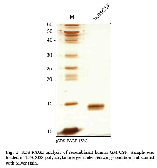
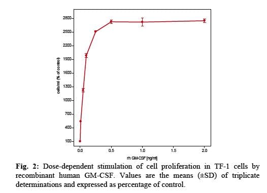
GM-CSF, Active Protein (Cat# AAA79117)
r-3alpha-Hydroxysteroid Dehydrogenase, Active Protein (Cat# AAA78965)
 Bioactivity
(Human Proinsulin in a cell proliferation assay using MCF7 human breast cancer cell. The ED50 range )
Bioactivity
(Human Proinsulin in a cell proliferation assay using MCF7 human breast cancer cell. The ED50 range )
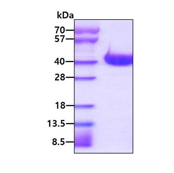
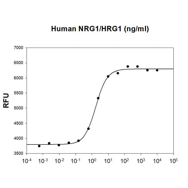
NRG1/HRG1, Active Protein (Cat# AAA48387)
 Bioactivity
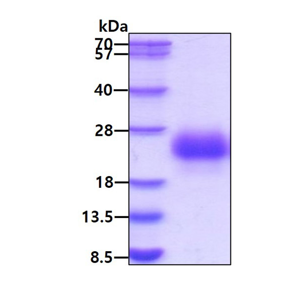
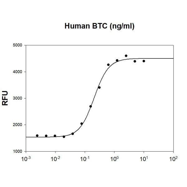
(Human BTC stimulates cell proliferation of the Balb/3T3 mouse embryonic fibroblast cells. The ED50 range )
Bioactivity
(Human BTC stimulates cell proliferation of the Balb/3T3 mouse embryonic fibroblast cells. The ED50 range )
Betacellulin/BTC, Active Protein (Cat# AAA48393)
 Bioactivity
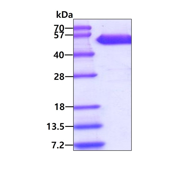
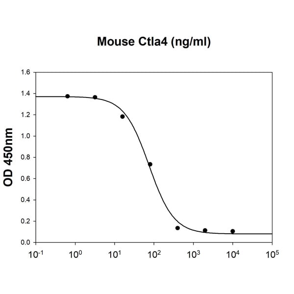
(Mouse Ctla4 inhibits IL-2 secretion of the stimulated Jurkat human acute T cell leukemia cells with Human B7-1/CD80. The ED50 range )
Bioactivity
(Mouse Ctla4 inhibits IL-2 secretion of the stimulated Jurkat human acute T cell leukemia cells with Human B7-1/CD80. The ED50 range )
Ctla-4, Active Protein (Cat# AAA48408)
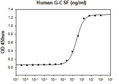 Application Data
(Human G-CSF stimulates cell proliferation of the M-NFS-60 mouse myelogenous leukemia lymphoblast cells. The ED50 range )
Application Data
(Human G-CSF stimulates cell proliferation of the M-NFS-60 mouse myelogenous leukemia lymphoblast cells. The ED50 range )
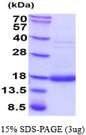
G-CSF, Active Protein (Cat# AAA48456)
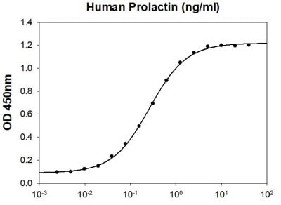 Bioactivity
(Human Prolactin in a cell proliferation assay using Nb2-11 Rat lymphoma cells. The ED50 range is less than or equal to 0.5ng/ml.)
Bioactivity
(Human Prolactin in a cell proliferation assay using Nb2-11 Rat lymphoma cells. The ED50 range is less than or equal to 0.5ng/ml.)
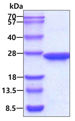
Prolactin, Active Protein (Cat# AAA48461)
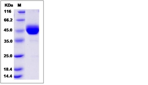
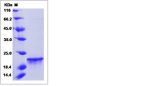
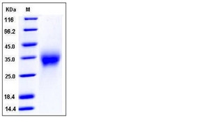
 SDS-PAGE
(3ug by SDS-PAGE under reducing condition and visualized by coomassie blue stain)
SDS-PAGE
(3ug by SDS-PAGE under reducing condition and visualized by coomassie blue stain)
Caspase-3, Active Protein (Cat# AAA48316)
 Bioactivity
(SARS-CoV Spike RBD is coated at 5ug/ml (100 ul/well) can bind ACE-2 (CAT# ATGP3963) in a Functional ELISA assay.)
Bioactivity
(SARS-CoV Spike RBD is coated at 5ug/ml (100 ul/well) can bind ACE-2 (CAT# ATGP3963) in a Functional ELISA assay.)
COVID 19 Spike RBD Coronavirus, Active Protein (Cat# AAA48369)
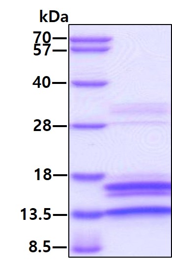
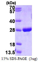
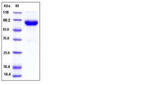
 SDS-PAGE
Cyclophilin F, Active Protein (Cat# AAA48516)
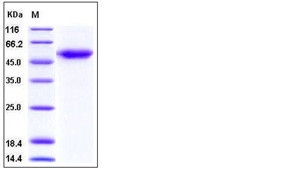
 SDS-PAGE
GSTP1, Active Protein (Cat# AAA48543)
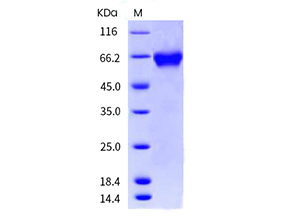
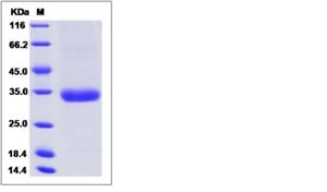
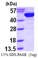 SDS-PAGE
BLMH, Active Protein (Cat# AAA48677)
Lactate Dehydrogenase 4, Active Protein (Cat# AAA44812)
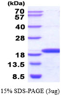 SDS-PAGE
NME3, Active Protein (Cat# AAA48925)
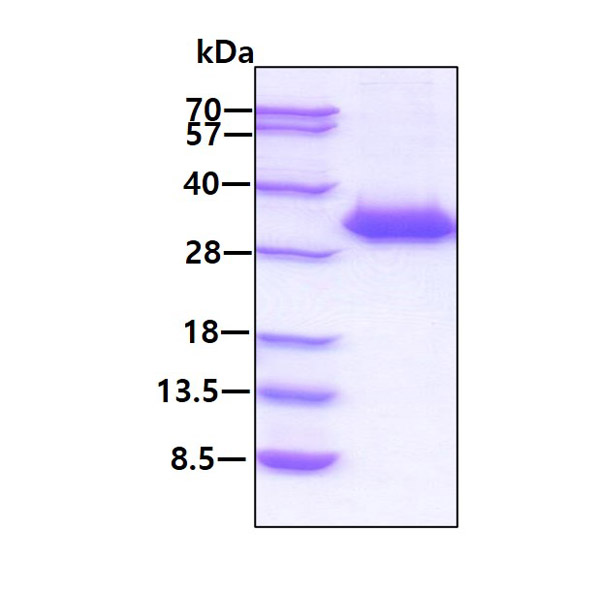
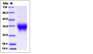
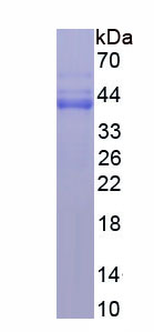
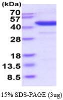 SDS-PAGE
(3ug by SDS-PAGE under reducing condition and visualized by coomassie blue stain.)
SDS-PAGE
(3ug by SDS-PAGE under reducing condition and visualized by coomassie blue stain.)
IDO1, Active Protein (Cat# AAA48930)
IFN-omega, Active Protein (Cat# AAA48989)
PlGF-1/PGF, Active Protein (Cat# AAA48993)
TRANCE/RANK L/TNFSF11, Active Protein (Cat# AAA49005)
IL-7, Active Protein (Cat# AAA49006)
VEGFR1/Flt-1, Active Protein (Cat# AAA49007)
Granulocyte Macrophage Colony Stimulating Factor (rHuGM-CSF), Active Protein (Cat# AAA47951)
CCL19, Active Protein (Cat# AAA47963)
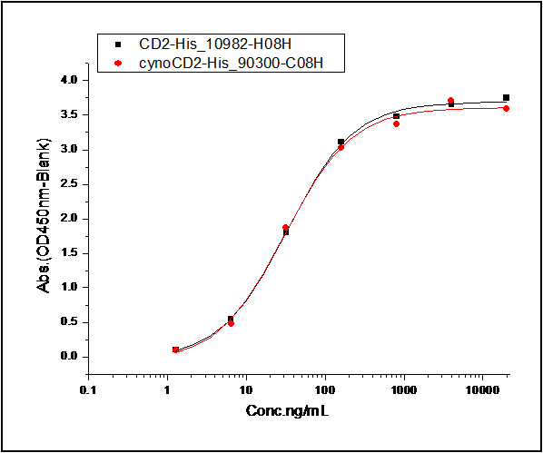 Application Data
(1. Measured by its binding ability in a functional ELISA. Immobilized human CD2-His at 10 ug/ml (100 ul/well) can bind human CD58-Fc, The EC50 of human CD58-Fc is 0.04-0.1 ug/ml. 2. Measured by its binding ability in a functional ELISA. Immobilized Cynomolgus CD2-His at 10 ug/ml (100 ul/well) can bind human CD58-Fc, The EC50 of human CD58-Fc is 0.04-0.10 ug/ml.)
Application Data
(1. Measured by its binding ability in a functional ELISA. Immobilized human CD2-His at 10 ug/ml (100 ul/well) can bind human CD58-Fc, The EC50 of human CD58-Fc is 0.04-0.1 ug/ml. 2. Measured by its binding ability in a functional ELISA. Immobilized Cynomolgus CD2-His at 10 ug/ml (100 ul/well) can bind human CD58-Fc, The EC50 of human CD58-Fc is 0.04-0.10 ug/ml.)
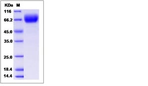
CD58, Active Protein (Cat# AAA258011)


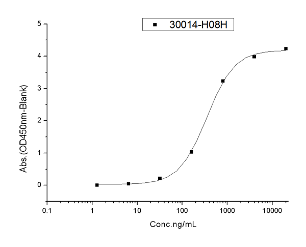
 Application Data
(Measured in a cell proliferation assay using human umbilical vein endothelial cells (HUVEC). The ED50 for this effect is typically 1-5 ng/mL.)
Application Data
(Measured in a cell proliferation assay using human umbilical vein endothelial cells (HUVEC). The ED50 for this effect is typically 1-5 ng/mL.)
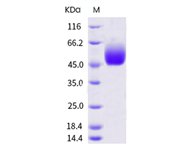
VEGF121b, Active Protein (Cat# AAA258033)
 Application Data
(Measured by its binding ability in a functional ELISA. Immobilized human CD47 at 2 ug/ml (100 ul/well) can bind human SIRPa(V2)-His, the EC50 of human SIRPa(V2)-His is 200-800 ng/mL.)
Application Data
(Measured by its binding ability in a functional ELISA. Immobilized human CD47 at 2 ug/ml (100 ul/well) can bind human SIRPa(V2)-His, the EC50 of human SIRPa(V2)-His is 200-800 ng/mL.)
SIRP alpha, Active Protein (Cat# AAA258062)
 Application Data
(Measured by its ability to inhibit BMP9-induced alkaline phosphatase production by MC3T3-E1 cells. The ED50 for this effect is typically 5-15 ng/mL in the presence of 2 ng/mL of recombinant human BMP-9.)
Application Data
(Measured by its ability to inhibit BMP9-induced alkaline phosphatase production by MC3T3-E1 cells. The ED50 for this effect is typically 5-15 ng/mL in the presence of 2 ng/mL of recombinant human BMP-9.)
ALK-1, Active Protein (Cat# AAA258252)
 Application Data
(Measured by its ability to neutralize Activin-mediated inhibition on MPC11 cell proliferation. The ED50 for this effect is typically 10-50 ng/mL in the presence of 10 ng/mL recombinant Activin A.)
Application Data
(Measured by its ability to neutralize Activin-mediated inhibition on MPC11 cell proliferation. The ED50 for this effect is typically 10-50 ng/mL in the presence of 10 ng/mL recombinant Activin A.)
ACVR2B, Active Protein (Cat# AAA258136)
 Application Data
(Measured by its ability to inhibit BMP9 induced alkaline phosphatase production by MC3T3E1 mouse chondrogenic cells. David, L. et al. (2007) Blood 109:1953. The ED50 for this effect is typically 5-15 ng/mL in the presence of 2 ng/mL of recombiant human BMP9.)
Application Data
(Measured by its ability to inhibit BMP9 induced alkaline phosphatase production by MC3T3E1 mouse chondrogenic cells. David, L. et al. (2007) Blood 109:1953. The ED50 for this effect is typically 5-15 ng/mL in the presence of 2 ng/mL of recombiant human BMP9.)
ALK-1, Active Protein (Cat# AAA257825)
 Application Data
(Measured by its binding ability in a functional ELISA. Immobilized Inhibin Human, Mouse, Rat, Cynomolgus, Rhesus Inhibin beta A/Activin A at 2 ug/ml (100 ul/well) can bind Human ACVR2B hFc, the EC50 of Human ACVR2B hFc is 12-60 ng/mL.)
Application Data
(Measured by its binding ability in a functional ELISA. Immobilized Inhibin Human, Mouse, Rat, Cynomolgus, Rhesus Inhibin beta A/Activin A at 2 ug/ml (100 ul/well) can bind Human ACVR2B hFc, the EC50 of Human ACVR2B hFc is 12-60 ng/mL.)
ACVR2B, Active Protein (Cat# AAA257849)
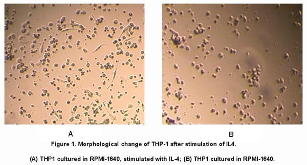

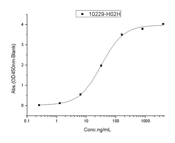
 Application Data
Interleukin 4 (IL4), Active Protein (Cat# AAA146607)
 Bioactivity
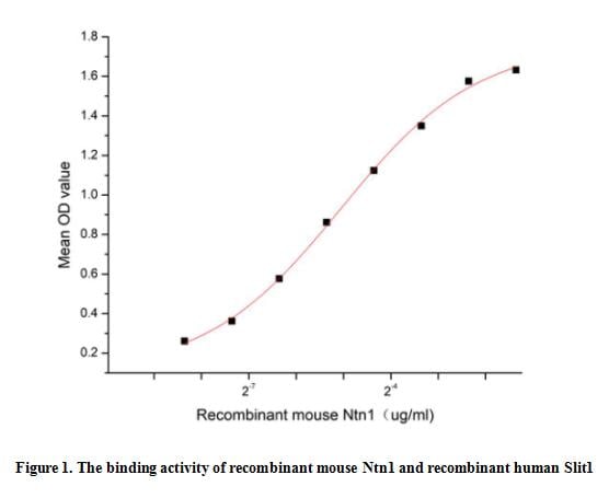
(Netrin 1 (Ntn1) is included in a family of laminin-related secreted proteins. Mouse Ntn1 is a 66-76 kDa glycoprotein that is well known for its involvement in axonal guidance during embryonic development and as an enhancer of cancer cell metastasis. Ntn1 is also involved in outgrowth and migration orientation in the developing CNS and plays a significant role in the morphogenesis of endothelial cells and vascular smooth-muscle cells. It is also involved in the processes of cytoskeleton reorganization, angiogenesis, epithelial cell adhesion, and cell migration in the lungs, mammary gland, and pancreas. Slit Homolog 1 (Slit1) can acts as a potent promoter of both Netrin-1 attractive and repulsive activities on distinct neuronal cell types, thereby opening novel perspectives on the role of combinations of cues in brain wiring. Thus a functional binding ELISA assay was conducted to detect the interaction of recombinant mouse Ntn1 and recombinant human Slit1. Briefly, Ntn1 was diluted serially in PBS with 0.01% BSA (pH 7.4). Duplicate samples of 100 ul were then transferred to Slit1-coated microtiter wells and incubated for 1h at 37 degree C. Wells were washed with PBST and incubated for 1h with anti-Ntn1 pAb, then aspirated and washed 3 times. After incubation with HRP labelled secondary antibody for 1h at 37 degree C, wells were aspirated and washed 5 times. With the addition of substrate solution, wells were incubated 15-25 minutes at 37 degree C. Finally, add 50 uL stop solution to the wells and read at 450/630 nm immediately. The binding activity of recombinant mouse Ntn1 and recombinant human Slit1 was shown in Figure 1, the EC50 for this effect is 0.03 ug/mL.)
Bioactivity
(Netrin 1 (Ntn1) is included in a family of laminin-related secreted proteins. Mouse Ntn1 is a 66-76 kDa glycoprotein that is well known for its involvement in axonal guidance during embryonic development and as an enhancer of cancer cell metastasis. Ntn1 is also involved in outgrowth and migration orientation in the developing CNS and plays a significant role in the morphogenesis of endothelial cells and vascular smooth-muscle cells. It is also involved in the processes of cytoskeleton reorganization, angiogenesis, epithelial cell adhesion, and cell migration in the lungs, mammary gland, and pancreas. Slit Homolog 1 (Slit1) can acts as a potent promoter of both Netrin-1 attractive and repulsive activities on distinct neuronal cell types, thereby opening novel perspectives on the role of combinations of cues in brain wiring. Thus a functional binding ELISA assay was conducted to detect the interaction of recombinant mouse Ntn1 and recombinant human Slit1. Briefly, Ntn1 was diluted serially in PBS with 0.01% BSA (pH 7.4). Duplicate samples of 100 ul were then transferred to Slit1-coated microtiter wells and incubated for 1h at 37 degree C. Wells were washed with PBST and incubated for 1h with anti-Ntn1 pAb, then aspirated and washed 3 times. After incubation with HRP labelled secondary antibody for 1h at 37 degree C, wells were aspirated and washed 5 times. With the addition of substrate solution, wells were incubated 15-25 minutes at 37 degree C. Finally, add 50 uL stop solution to the wells and read at 450/630 nm immediately. The binding activity of recombinant mouse Ntn1 and recombinant human Slit1 was shown in Figure 1, the EC50 for this effect is 0.03 ug/mL.)
Netrin 1 (Ntn1), Active Protein (Cat# AAA161863)
CMV pp65, Active Protein (Cat# AAA224571)
 Bioactivity
Membrane Cofactor (CD46), Active Protein (Cat# AAA243727)
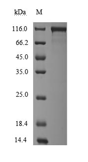
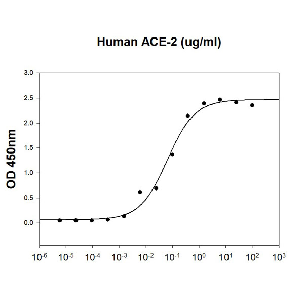
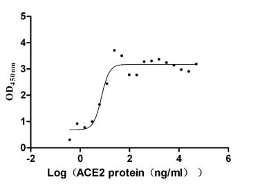 Bioactivity
Angiotensin-converting enzyme (ACE2), Active Protein (Cat# AAA244034)
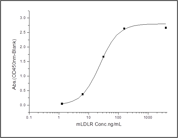 Application Data
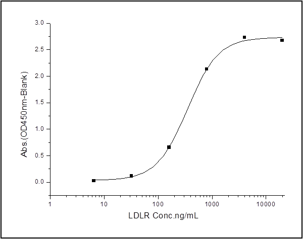
(Measured by its binding ability in a functional ELISA. Immobilized human APOH-his at 2 ug/mL (100 ul/well) can bind biotinylated mouse LDLR-his, The EC50 of biotinylated mouse LDLR-his is 26 ng/mL.)
Application Data
(Measured by its binding ability in a functional ELISA. Immobilized human APOH-his at 2 ug/mL (100 ul/well) can bind biotinylated mouse LDLR-his, The EC50 of biotinylated mouse LDLR-his is 26 ng/mL.)
Apolipoprotein H, Active Protein (Cat# AAA257970)
 Application Data
(Measured by its binding ability in a functional ELISA. Immobilized human TDGF1 at 2 ug/ml (100 ul/well) can bind human ALK-4 with a linear range of 0.032-4 ug/ml.)
Application Data
(Measured by its binding ability in a functional ELISA. Immobilized human TDGF1 at 2 ug/ml (100 ul/well) can bind human ALK-4 with a linear range of 0.032-4 ug/ml.)
ALK4/ACVR1B, Active Protein (Cat# AAA257899)
 Application Data
(Measured by its ability to inhibit BMP9 induced alkaline phosphatase production by MC3T3E1 mouse chondrogenic cells. David, L. et al. (2007) Blood 109:1953. The ED50 for this effect is typically 50-200 ng/mL in the presence of 2 ng/mL of recombiant human BMP9.)
Application Data
(Measured by its ability to inhibit BMP9 induced alkaline phosphatase production by MC3T3E1 mouse chondrogenic cells. David, L. et al. (2007) Blood 109:1953. The ED50 for this effect is typically 50-200 ng/mL in the presence of 2 ng/mL of recombiant human BMP9.)
ALK-1, Active Protein (Cat# AAA257826)
 Application Data
(Measured by its ability to neutralize Activin-mediated inhibition on MPC11 cell proliferation. The ED50 for this effect is typically 0.3-2 ug/mL in the presence of 10 ng/mL recombinant Activin A.)
Application Data
(Measured by its ability to neutralize Activin-mediated inhibition on MPC11 cell proliferation. The ED50 for this effect is typically 0.3-2 ug/mL in the presence of 10 ng/mL recombinant Activin A.)
ACVR2B, Active Protein (Cat# AAA257850)
 Application Data
(Measured in a cell proliferation assay using BALB/c 3T3 mouse fibroblasts. The ED50 for this effect is typically 0.4-1.6 ug/mL.)
Application Data
(Measured in a cell proliferation assay using BALB/c 3T3 mouse fibroblasts. The ED50 for this effect is typically 0.4-1.6 ug/mL.)
FGF8, Active Protein (Cat# AAA258053)
 Application Data
(Measured by its ability to neutralize Activin-mediated inhibition on MPC11 cell proliferation. The ED50 for this effect is typically 0.2-1 ug/mL in the presence of 10 ng/mL recombinant Activin A.)
Application Data
(Measured by its ability to neutralize Activin-mediated inhibition on MPC11 cell proliferation. The ED50 for this effect is typically 0.2-1 ug/mL in the presence of 10 ng/mL recombinant Activin A.)
ACVR2B, Active Protein (Cat# AAA258265)
 Application Data
(Measured by its binding ability in a functional ELISA. Immobilized Mouse Angiopoietin-2-His at 2 ug/ml (100 ul/well) can bind mouse TEK-Fc, the EC50 of mouse TEK-Fc is 350-900 ng/mL.)
Application Data
(Measured by its binding ability in a functional ELISA. Immobilized Mouse Angiopoietin-2-His at 2 ug/ml (100 ul/well) can bind mouse TEK-Fc, the EC50 of mouse TEK-Fc is 350-900 ng/mL.)
Angiopoietin-2, Active Protein (Cat# AAA258152)
 Application Data
(Measured by its ability to neutralize Activin-mediated inhibition on MPC11 cell proliferation. The ED50 for this effect is typically 0.6-3 ug/mL in the presence of 10 ng/ml Recombinant Human Activin A.)
Application Data
(Measured by its ability to neutralize Activin-mediated inhibition on MPC11 cell proliferation. The ED50 for this effect is typically 0.6-3 ug/mL in the presence of 10 ng/ml Recombinant Human Activin A.)
ACVR2A, Active Protein (Cat# AAA258185)
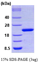
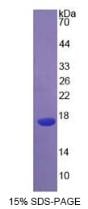
> 95% as determined by SEC-HPLC
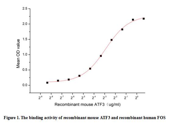 Bioactivity
(Activating transcription factor 3 (ATF3) is a stress-induced transcription factor that plays vital roles in modulating metabolism, immunity and oncogenesis. ATF3 acts as a hub of the cellular adaptive-response network. Multiple extracellular signals, such as endoplasmic reticulum (ER) stress, cytokines, chemokines, and LPS, are connected to ATF3 induction. Proto-oncogene c-Fos can bind to ATF3 to regulate signal transduction, cell proliferation and differentiation. Thus a functional ELISA assay was conducted to detect the interaction of recombinant mouse ATF3 and recombinant human FOS. Briefly, ATF3 was diluted serially in PBS with 0.01% BSA (pH 7.4). Duplicate samples of 100 ul were then transferred to FOS-coated microtiter wells and incubated for 1h at 37 degree C. Wells were washed with PBST and incubated for 1h with anti-ATF3 pAb, then aspirated and washed 3 times. After incubation with HRP labelled secondary antibody for 1h at 37 degree C, wells were aspirated and washed 5 times. With the addition of substrate solution, wells were incubated 15-25 minutes at 37 degree C. Finally, add 50 uL stop solution to the wells and read at 450/630nm immediately. The binding activity of recombinant mouse ATF3 and recombinant human FOS was shown in Figure 1, the EC50 for this effect is 0.13 ug/mL.)
Bioactivity
(Activating transcription factor 3 (ATF3) is a stress-induced transcription factor that plays vital roles in modulating metabolism, immunity and oncogenesis. ATF3 acts as a hub of the cellular adaptive-response network. Multiple extracellular signals, such as endoplasmic reticulum (ER) stress, cytokines, chemokines, and LPS, are connected to ATF3 induction. Proto-oncogene c-Fos can bind to ATF3 to regulate signal transduction, cell proliferation and differentiation. Thus a functional ELISA assay was conducted to detect the interaction of recombinant mouse ATF3 and recombinant human FOS. Briefly, ATF3 was diluted serially in PBS with 0.01% BSA (pH 7.4). Duplicate samples of 100 ul were then transferred to FOS-coated microtiter wells and incubated for 1h at 37 degree C. Wells were washed with PBST and incubated for 1h with anti-ATF3 pAb, then aspirated and washed 3 times. After incubation with HRP labelled secondary antibody for 1h at 37 degree C, wells were aspirated and washed 5 times. With the addition of substrate solution, wells were incubated 15-25 minutes at 37 degree C. Finally, add 50 uL stop solution to the wells and read at 450/630nm immediately. The binding activity of recombinant mouse ATF3 and recombinant human FOS was shown in Figure 1, the EC50 for this effect is 0.13 ug/mL.)
Activating Transcription Factor 3 (ATF3), Active Protein (Cat# AAA161885)

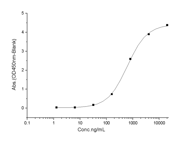
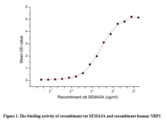 Bioactivity
(The Semaphorin 3A(SEMA3A) which belongs to the semaphorin family can function as either a chemorepulsive agent, inhibiting axonal outgrowth, or as a chemoattractive agent, stimulating the growth of apical dendrites. In both cases, the protein is vital for normal neuronal pattern development. Semaphorin 3A is secreted protein containing a Sema domain, an immunoglobulin C2-like domain and a basic domain near the carboxyl tail. It can be secreted by neurons and surrounding tissue to guide migrating cells and axons in the developing nervous system. Besides, Neuropilin 1 (NRP1) has been identified as an interactor of SEMA3A, thus a functional binding ELISA assay was conducted to detect the interaction of recombinant rat SEMA3A and recombinant human NRP1. Briefly, SEMA3A were diluted serially in PBS, with 0.01% BSA (pH 7.4). Duplicate samples of 100 ul were then transferred to NRP1-coated microtiter wells and incubated for 2h at 37 degree C. Wells were washed with PBST and incubated for 1h with anti-SEMA3A pAb, then aspirated and washed 3 times. After incubation with HRP labelled secondary antibody, wells were aspirated and washed 3 times. With the addition of substrate solution, wells were incubated 15-25 minutes at 37 degree C. Finally, add 50 ul stop solution to the wells and read at 450 nm immediately. The binding activity of recombinant rat SEMA3A and recombinant human NRP1 was shown in Figure 1, the EC50 for this effect is 1.22 ug/mL.)
Bioactivity
(The Semaphorin 3A(SEMA3A) which belongs to the semaphorin family can function as either a chemorepulsive agent, inhibiting axonal outgrowth, or as a chemoattractive agent, stimulating the growth of apical dendrites. In both cases, the protein is vital for normal neuronal pattern development. Semaphorin 3A is secreted protein containing a Sema domain, an immunoglobulin C2-like domain and a basic domain near the carboxyl tail. It can be secreted by neurons and surrounding tissue to guide migrating cells and axons in the developing nervous system. Besides, Neuropilin 1 (NRP1) has been identified as an interactor of SEMA3A, thus a functional binding ELISA assay was conducted to detect the interaction of recombinant rat SEMA3A and recombinant human NRP1. Briefly, SEMA3A were diluted serially in PBS, with 0.01% BSA (pH 7.4). Duplicate samples of 100 ul were then transferred to NRP1-coated microtiter wells and incubated for 2h at 37 degree C. Wells were washed with PBST and incubated for 1h with anti-SEMA3A pAb, then aspirated and washed 3 times. After incubation with HRP labelled secondary antibody, wells were aspirated and washed 3 times. With the addition of substrate solution, wells were incubated 15-25 minutes at 37 degree C. Finally, add 50 ul stop solution to the wells and read at 450 nm immediately. The binding activity of recombinant rat SEMA3A and recombinant human NRP1 was shown in Figure 1, the EC50 for this effect is 1.22 ug/mL.)
Semaphorin 3A (SEMA3A), Active Protein (Cat# AAA161937)
What Are Active Proteins?
Proteins are large molecules made up of long chains of amino acids.
They will typically fold into a very particular 3-dimensional shape/conformation, that is sometimes referred to as their “native” form, which allows them to work properly in the body. For the purposes of product categorization, AAA Biotech will typically refer to proteins purified from their original animal host as being “native” proteins (this is to signify their difference compared to their “recombinant” or “synthetic” protein counterparts).
If a protein successfully folds into the correct shape, it is will typically display high fidelity characteristics to its original protein in its original animal host, and be classified as an active protein, as it will be able to function “normally” in most enzymatic or binding capacities. If it loses this shape, due to factors such as heat or strong chemicals (such as detergents), it becomes inactive and is no longer able to perform its basic functions. All of the proteins in this category are made under strict quality control, and they are active, pure, low in contaminants, and stable.
Most are stored as freeze-dried powders and come without extra tags, so they’re very close to the actual natural/native form.
Key Applications of Active Proteins
1. Scientific Research
- Aid in the study of how proteins function in the body
- Aid in understanding various disease processes
2. Drug Development
- Powerful tools to investigate how potential drugs interact with specific proteins
- Ideal for identifying drug targets
3. Cell Culture
- Are routinely utilized to support cell growth and function (e.g., using exogenous growth factors)
- Can be used to promote cellular development into specific types (differentiation)
4. Diagnostics
- Regularly utilized in tests to detect diseases or infections (e.g., COVID-19, cancer)
- Note: All products are strictly for research-use only (RUO).
5. Therapeutics
- Some active proteins are used directly as treatments (e.g., insulin, enzymes)
- Note: All products are strictly for research-use only (RUO).
6. Vaccine Development
- Used to create or test vaccines by mimicking parts of viruses or bacteria
7. Biochemical Assays
- They can facilitate the characterization of enzyme activity, binding strength, or protein interactions in lab tests
Why Buy Active Proteins from AAA Biotech?
- High biological activity – Verified to perform as expected or indicated on datasheet
- Strict quality control – We are confident in our active proteins’ reliability and consistency
- High purity & low endotoxin – Ideal for applications involving sensitive or precious samples/components
- Freeze-dried for stability – Long shelf life and straightforward storage
- Mostly tag-free – Closer to natural/native protein form
FAQ
1. What are active proteins used for in research?
Active proteins are used primarily in the study of how proteins function, in characterizing/discovering drug interactions, supporting cell growth, running biochemical assays, and in development of diagnostics or therapeutics.
2. How are AAA Biotech's active proteins validated?
AAA Biotech’s active proteins are validated through strict quality control and functional assays to ensure they are properly folded and active. “Active”, though, can be an ambiguous term, so if a specific “activity” or “binding” capability of a protein is of crucial interest to you, please inquire with us prior to purchase, and we will provide further details on how the “Active” modifier was determined to be applicable.
3. Are these proteins tested for biological activity?
Yes, all active proteins from AAA Biotech are tested to confirm they have the expected biological activity before being offered for use. Though, said “biological activity” can be either “enzymatic”, “binding”, or both.