Filters
▼Clonality
▼Type
▼Reactivity
▼Gene Name
▼Isotype
▼Host
▼Application
▼Clone
▼Active Proteins
AAA Biotech also known as AAA Bio or AAABio provides a variety of high-quality recombinant and natural/native proteins that are proven to work in a wide range of experiments. Explore our products to find the active protein that best fits your needs or experimental model.
Viewing 2000-2050 of 2567 product results
Zinc-Alpha 2 Glycoprotein, Active Protein (Cat# AAA38476)
Myostatin, Active Protein (Cat# AAA38198)
4-1BB Receptor, Active Protein (Cat# AAA38219)
Urokinase, Active Protein (Cat# AAA38242)
Lactate Dehydrogenase B, Active Protein (Cat# AAA38250)
Peroxidase, Active Protein (Cat# AAA38278)
Vascular Endothelial Growth Factor C, Active Protein (Cat# AAA38552)
Beta Defensin -2, Active Protein (Cat# AAA38587)
Beta Defensin -4, Active Protein (Cat# AAA38588)
Glucagon, Active Protein (Cat# AAA38343)
(a) Analysis by RP-HPLC.
(b) Analysis by SDS-PAGE.
Myostatin, Active Protein (Cat# AAA76059)
 ELISA
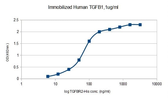
(Biotinylated TGFB-1 ELISAImmobilized Biotinylated Human TGF Beta1 at 1ug/ml (100ul/Well). Response curve for TGFR2 with alinear range of 2-100 ng/ml was determined by ELISA)
ELISA
(Biotinylated TGFB-1 ELISAImmobilized Biotinylated Human TGF Beta1 at 1ug/ml (100ul/Well). Response curve for TGFR2 with alinear range of 2-100 ng/ml was determined by ELISA)
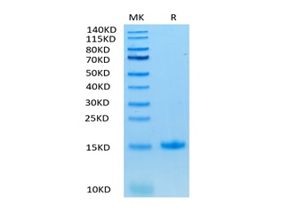
TGF Beta-1, Active Protein (Cat# AAA76386)
 ELISA
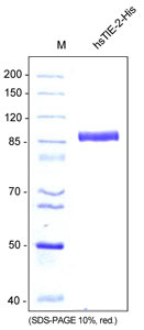
(Functional ELISA to test the binding of sTIE-2 to its ligand Ang-2. Ang-2 was coated on the plate and increasing amounts of recombinant human soluble sTIE-2 was added)
ELISA
(Functional ELISA to test the binding of sTIE-2 to its ligand Ang-2. Ang-2 was coated on the plate and increasing amounts of recombinant human soluble sTIE-2 was added)
TIE-2, soluble, Active Protein (Cat# AAA79307)
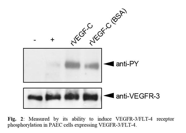
 Application Data
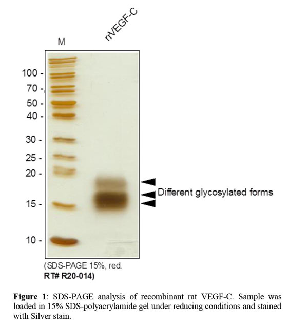
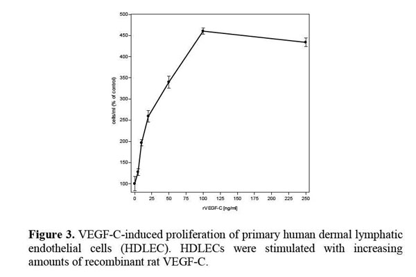
VEGF-C, Active Protein (Cat# AAA79180)
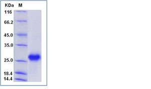
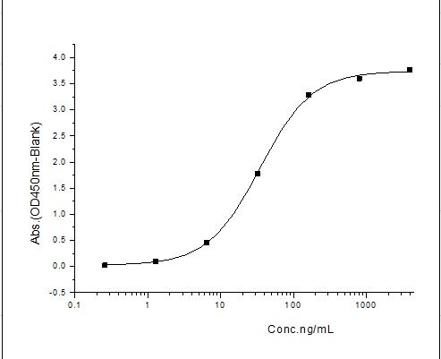
 Application Data
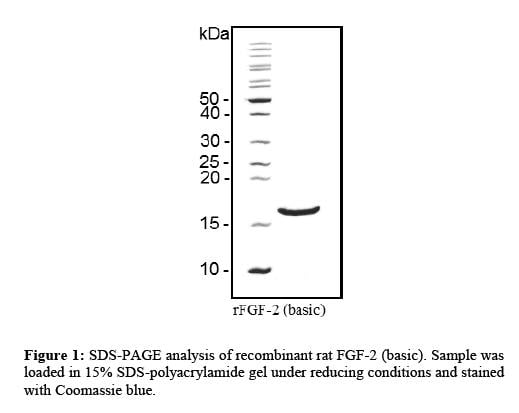
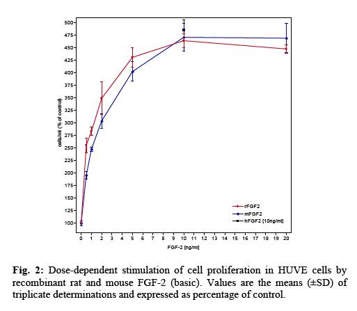
FGF-2 (basic), Active Protein (Cat# AAA79225)
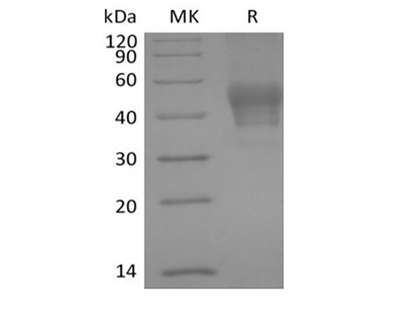
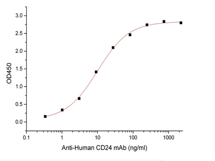 Bioactivity
Signal Transducer CD24/CD24, Active Protein (Cat# AAA177953)
Vascular Endothelial Growth Factor-Sf9, Active Protein (Cat# AAA38118)
Leptin, Active Protein (Cat# AAA38120)
Interferon-beta 1b, Active Protein (Cat# AAA38124)
Insulin Like Growth Factor-1, Active Protein (Cat# AAA38145)
Soluble RANK Ligand, Active Protein (Cat# AAA38163)
Fibroblast Growth Factor-basic, Active Protein (Cat# AAA38177)
Granulocyte Macrophage-Colony Stimulating Factor, Active Protein (Cat# AAA38190)
Platelet Derived Growth Factor-BB, Active Protein (Cat# AAA38195)
TNF-Related Apoptosis Inducing Ligand/Apo2L, Active Protein (Cat# AAA38208)
Resistin, Active Protein (Cat# AAA38215)
Leptin, Active Protein (Cat# AAA38234)
Urate Oxidase, Active Protein (Cat# AAA38272)
Deoxyribonuclease I, Active Protein (Cat# AAA38277)
Bone Morphogenetic protein-13, Active Protein (Cat# AAA38030)
Interferon-alpha 2c, Active Protein (Cat# AAA38061)
(a) Analysis by RP-HPLC.
(b) Analysis by SDS-PAGE.
Epithelial Neutrophil-Activating Protein 78, Active Protein (Cat# AAA38090)
Creatine Kinase Brain, Active Protein (Cat# AAA38098)
Salmonella Typhimurium Uridine phosphorylase, Active Protein (Cat# AAA38288)
CD14 CHO, Active Protein (Cat# AAA38511)
Peroxiredoxin-3, Active Protein (Cat# AAA38770)
Beta Defensin -1, Active Protein (Cat# AAA38586)
Growth Hormone, Active Protein (Cat# AAA38592)
Trefoil Factor-1, Active Protein (Cat# AAA38601)
Mitogen Activated Kinase Kinase 1, Active Protein (Cat# AAA38618)
Bleomycin Hydrolase, Active Protein (Cat# AAA38866)
LR3 Insulin Like Growth Factor-1, Active Protein (Cat# AAA38973)
(a) Analysis by RP-HPLC
(b) Analysis by SDS-PAGE
Thyroid Stimulating Hormone, Active Protein (Cat# AAA39027)
Luteinizing Hormone, Active Protein (Cat# AAA44840)
Creatine Kinase, Active Protein (Cat# AAA44789)
Kallikrein, Active Protein (Cat# AAA44797)
Lactate Dehydrogenase, Active Protein (Cat# AAA44807)
Pyruvate Kinase, Active Protein (Cat# AAA44819)
 Application Data
(Measured by its binding ability in a functional ELISA. Immobilized TNFRSF18-His at 10 ug/mL (100 uL/well) can bind TNFSF18-mFc, the EC50 of TNFSF18-mFc is 20-60 ng/mL.)
Application Data
(Measured by its binding ability in a functional ELISA. Immobilized TNFRSF18-His at 10 ug/mL (100 uL/well) can bind TNFSF18-mFc, the EC50 of TNFSF18-mFc is 20-60 ng/mL.)
GITR, Active Protein (Cat# AAA258024)
 Application Data
(Measured in a cell proliferation assay using TF-1 human erythroleukemic cells. The ED50 for this effect is typically 0.015-0.06 ug/mL.)
Application Data
(Measured in a cell proliferation assay using TF-1 human erythroleukemic cells. The ED50 for this effect is typically 0.015-0.06 ug/mL.)
Cardiotrophin 1, Active Protein (Cat# AAA258040)
What Are Active Proteins?
Proteins are large molecules made up of long chains of amino acids.
They will typically fold into a very particular 3-dimensional shape/conformation, that is sometimes referred to as their “native” form, which allows them to work properly in the body. For the purposes of product categorization, AAA Biotech will typically refer to proteins purified from their original animal host as being “native” proteins (this is to signify their difference compared to their “recombinant” or “synthetic” protein counterparts).
If a protein successfully folds into the correct shape, it is will typically display high fidelity characteristics to its original protein in its original animal host, and be classified as an active protein, as it will be able to function “normally” in most enzymatic or binding capacities. If it loses this shape, due to factors such as heat or strong chemicals (such as detergents), it becomes inactive and is no longer able to perform its basic functions. All of the proteins in this category are made under strict quality control, and they are active, pure, low in contaminants, and stable.
Most are stored as freeze-dried powders and come without extra tags, so they’re very close to the actual natural/native form.
Key Applications of Active Proteins
1. Scientific Research
- Aid in the study of how proteins function in the body
- Aid in understanding various disease processes
2. Drug Development
- Powerful tools to investigate how potential drugs interact with specific proteins
- Ideal for identifying drug targets
3. Cell Culture
- Are routinely utilized to support cell growth and function (e.g., using exogenous growth factors)
- Can be used to promote cellular development into specific types (differentiation)
4. Diagnostics
- Regularly utilized in tests to detect diseases or infections (e.g., COVID-19, cancer)
- Note: All products are strictly for research-use only (RUO).
5. Therapeutics
- Some active proteins are used directly as treatments (e.g., insulin, enzymes)
- Note: All products are strictly for research-use only (RUO).
6. Vaccine Development
- Used to create or test vaccines by mimicking parts of viruses or bacteria
7. Biochemical Assays
- They can facilitate the characterization of enzyme activity, binding strength, or protein interactions in lab tests
Why Buy Active Proteins from AAA Biotech?
- High biological activity – Verified to perform as expected or indicated on datasheet
- Strict quality control – We are confident in our active proteins’ reliability and consistency
- High purity & low endotoxin – Ideal for applications involving sensitive or precious samples/components
- Freeze-dried for stability – Long shelf life and straightforward storage
- Mostly tag-free – Closer to natural/native protein form
FAQ
1. What are active proteins used for in research?
Active proteins are used primarily in the study of how proteins function, in characterizing/discovering drug interactions, supporting cell growth, running biochemical assays, and in development of diagnostics or therapeutics.
2. How are AAA Biotech's active proteins validated?
AAA Biotech’s active proteins are validated through strict quality control and functional assays to ensure they are properly folded and active. “Active”, though, can be an ambiguous term, so if a specific “activity” or “binding” capability of a protein is of crucial interest to you, please inquire with us prior to purchase, and we will provide further details on how the “Active” modifier was determined to be applicable.
3. Are these proteins tested for biological activity?
Yes, all active proteins from AAA Biotech are tested to confirm they have the expected biological activity before being offered for use. Though, said “biological activity” can be either “enzymatic”, “binding”, or both.