Filters
▼Clonality
▼Type
▼Reactivity
▼Gene Name
▼Isotype
▼Host
▼Application
▼Clone
▼Active Proteins
AAA Biotech also known as AAA Bio or AAABio provides a variety of high-quality recombinant and natural/native proteins that are proven to work in a wide range of experiments. Explore our products to find the active protein that best fits your needs or experimental model.
Viewing 2200-2250 of 2567 product results
Creatine Kinase MB, Active Protein (Cat# AAA44790)
Isocitrate Dehydrogenase, Active Protein (Cat# AAA44806)
Lactate Dehydrogenase, Active Protein (Cat# AAA44808)
Lactate Dehydrogenase 1, Active Protein (Cat# AAA44809)
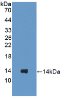
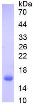
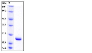 SDS-PAGE
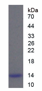
SUMO1, Active Protein (Cat# AAA258013)
 Application Data
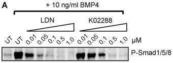
(Measured by its ability to inhibit BMP9-induced alkaline phosphatase production by MC3T3E1 mouse chondrogenic cells. David, L. et al. (2007) Blood 109:1953. The ED50 for this effect is typically 60-300 ng/mL in the presence of 2 ng/mL of recombiant human BMP9.)
Application Data
(Measured by its ability to inhibit BMP9-induced alkaline phosphatase production by MC3T3E1 mouse chondrogenic cells. David, L. et al. (2007) Blood 109:1953. The ED50 for this effect is typically 60-300 ng/mL in the presence of 2 ng/mL of recombiant human BMP9.)
ALK-1, Active Protein (Cat# AAA258143)
 Application Data
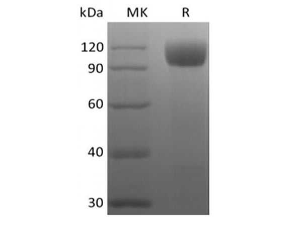
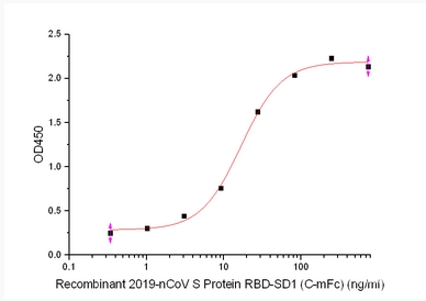
(Measured by its binding ability in a functional ELISA. Immobilized Human ACE2 (mFc tag) at 2 ug/mL (100 uL/well) can bind SARS-CoV-2 (2019-nCoV) Spike S1+S2 ECD (HV69-70 deletion, Y144 deletion, N501Y, A570D, D614G, P681H, T716I, S982A, D1118H)-His, the EC50 of SARS-CoV-2 (2019-nCoV) Spike S1+S2 ECD (HV69-70 deletion, Y144 deletion, N501Y, A570D, D614G, P681H, T716I, S982A, D1118H)-His is 200-800 ng/mL.)
Application Data
(Measured by its binding ability in a functional ELISA. Immobilized Human ACE2 (mFc tag) at 2 ug/mL (100 uL/well) can bind SARS-CoV-2 (2019-nCoV) Spike S1+S2 ECD (HV69-70 deletion, Y144 deletion, N501Y, A570D, D614G, P681H, T716I, S982A, D1118H)-His, the EC50 of SARS-CoV-2 (2019-nCoV) Spike S1+S2 ECD (HV69-70 deletion, Y144 deletion, N501Y, A570D, D614G, P681H, T716I, S982A, D1118H)-His is 200-800 ng/mL.)
COVID 19 Spike S1+S2 ECD (HV69-70 deletion, Y144 deletion, N501Y, A570D, D614G, P681H, T716I, S982A, D1118H)-His Coronavirus, Active Protein (Cat# AAA258080)
ERYTHROPOIETIN ALPHA, Active Protein (Cat# AAA50528)
IFN GAMMA, Active Protein (Cat# AAA50533)
Fc gamma RIIB/CD32b, Active Protein (Cat# AAA49000)
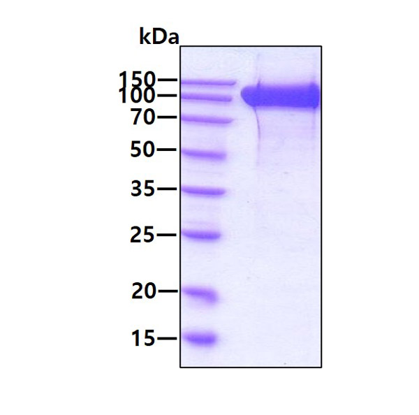
GFR alpha-1, Active Protein (Cat# AAA49003)
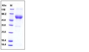
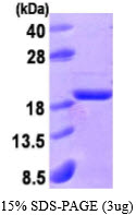
 SDS-PAGE
(3ug by SDS-PAGE under reducing condition and visualized fby coomassie blue stain.)
SDS-PAGE
(3ug by SDS-PAGE under reducing condition and visualized fby coomassie blue stain.)
Peroxiredoxin 6, Active Protein (Cat# AAA48548)
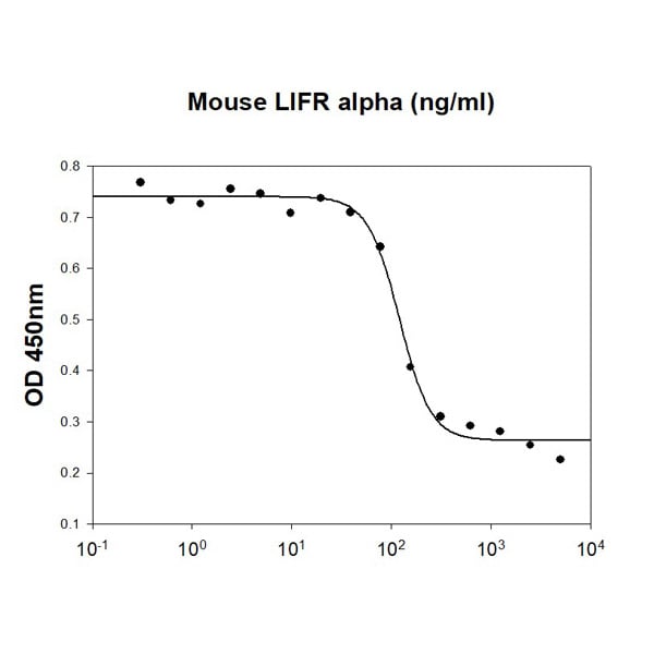 Bioactivity
(Mouse LIFR alpha inhibits human LIF (Cat# ATGP3533) induced cell proliferation in the TF-1 human erythroleukemic cells. The ED50 range is )
Bioactivity
(Mouse LIFR alpha inhibits human LIF (Cat# ATGP3533) induced cell proliferation in the TF-1 human erythroleukemic cells. The ED50 range is )
LIFR alpha, Active Protein (Cat# AAA48367)
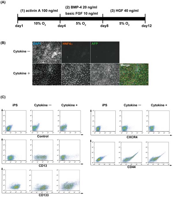
 Application Data
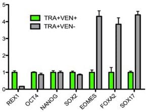
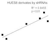
(WNT3 Is a Functional Biomarker for Predicting the DE Differentiation Potential (A) WNT3 levels in control and various knockdown HUES8 sublines correlate with their DE differentiation efficiency.The four protocols included the following treatments in a serum-free medium: (1) A - > A (Activin A only for 4 days); (2) AW - > A (Activin A and Wnt3a for 2 days followed by Activin A only for another 2 days); (3) A - > AX (Activin A only for 2 days followed by Activin A and XAV 939 for another 2 days); and (4) AW - > AX (Activin A and Wnt3a for 2 days followed by Activin A and XAV 939 for another 2 days).)
Application Data
(WNT3 Is a Functional Biomarker for Predicting the DE Differentiation Potential (A) WNT3 levels in control and various knockdown HUES8 sublines correlate with their DE differentiation efficiency.The four protocols included the following treatments in a serum-free medium: (1) A - > A (Activin A only for 4 days); (2) AW - > A (Activin A and Wnt3a for 2 days followed by Activin A only for another 2 days); (3) A - > AX (Activin A only for 2 days followed by Activin A and XAV 939 for another 2 days); and (4) AW - > AX (Activin A and Wnt3a for 2 days followed by Activin A and XAV 939 for another 2 days).)
Activin, Active Protein (Cat# AAA76389)
 Bioactivity
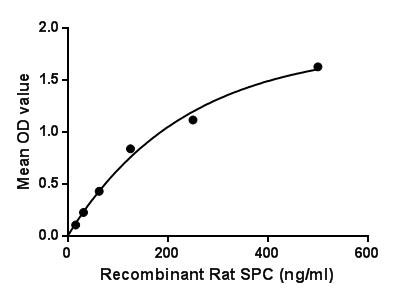
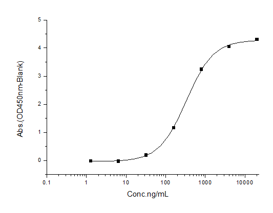
(Surfactant Associated Protein C (SPC) is one of the pulmonary surfactant proteins. It is a membrane protein which manufactures surfactant. The propeptide of pulmonary surfactant C has an N-terminal alpha-helical segment whose suggested function was stabilization of the protein structure, since the latter can irreversibly transform from its native alpha-helical structure to beta-sheet aggregates and form amyloid fibrils. Besides, Monokine Induced By Interferon Gamma (MIg) has been identified as an interactor of SPC, thus a binding ELISA assay was conducted to detect the interaction of recombinant rat SPC and recombinant rat MIg. Briefly, SPC were diluted serially in PBS, with 0.01% BSA (pH 7.4). Duplicate samples of 100L were then transferred to MIg-coated microtiter wells and incubated for 2h at 37. Wells were washed with PBST and incubated for 1h with anti-SPC pAb, then aspirated and washed 3 times. After incubation with HRP labelled secondary antibody, wells were aspirated and washed 3 times. With the addition of substrate solution, wells were incubated 15-25 minutes at 37. Finally, add 50uL stop solution to the wells and read at 450nm immediately. The binding activity of SPC and MIg was shown in Figure 1, and this effect was in a dose dependent manner.Figure. The binding activity of SPC with MIg.)
Bioactivity
(Surfactant Associated Protein C (SPC) is one of the pulmonary surfactant proteins. It is a membrane protein which manufactures surfactant. The propeptide of pulmonary surfactant C has an N-terminal alpha-helical segment whose suggested function was stabilization of the protein structure, since the latter can irreversibly transform from its native alpha-helical structure to beta-sheet aggregates and form amyloid fibrils. Besides, Monokine Induced By Interferon Gamma (MIg) has been identified as an interactor of SPC, thus a binding ELISA assay was conducted to detect the interaction of recombinant rat SPC and recombinant rat MIg. Briefly, SPC were diluted serially in PBS, with 0.01% BSA (pH 7.4). Duplicate samples of 100L were then transferred to MIg-coated microtiter wells and incubated for 2h at 37. Wells were washed with PBST and incubated for 1h with anti-SPC pAb, then aspirated and washed 3 times. After incubation with HRP labelled secondary antibody, wells were aspirated and washed 3 times. With the addition of substrate solution, wells were incubated 15-25 minutes at 37. Finally, add 50uL stop solution to the wells and read at 450nm immediately. The binding activity of SPC and MIg was shown in Figure 1, and this effect was in a dose dependent manner.Figure. The binding activity of SPC with MIg.)
Surfactant Protein C, Active Protein (Cat# AAA150111)
Arginine Deiminase (ADI), Active Protein (Cat# AAA79254)


 Application Data
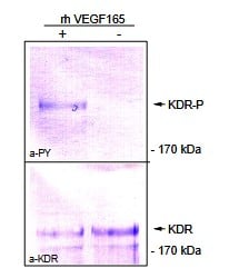
(Fig. 3: Confluent PAE/KDR cells were stimulated with 50ng/ml human VEGF165 for 10 min at 37°C. Cells were lysed and an IP was performed using a mouse anti-human KDR antibody (Cat# WB was performed with a mouse anti-human KDR antibody (Cat# and an anti- Phosphotyrosine antibody.)
Application Data
(Fig. 3: Confluent PAE/KDR cells were stimulated with 50ng/ml human VEGF165 for 10 min at 37°C. Cells were lysed and an IP was performed using a mouse anti-human KDR antibody (Cat# WB was performed with a mouse anti-human KDR antibody (Cat# and an anti- Phosphotyrosine antibody.)
VEGF165, Active Protein (Cat# AAA79288)
alpha-Glucosidase, Active Protein (Cat# AAA78980)
D-Dimer, Active Protein (Cat# AAA224711)
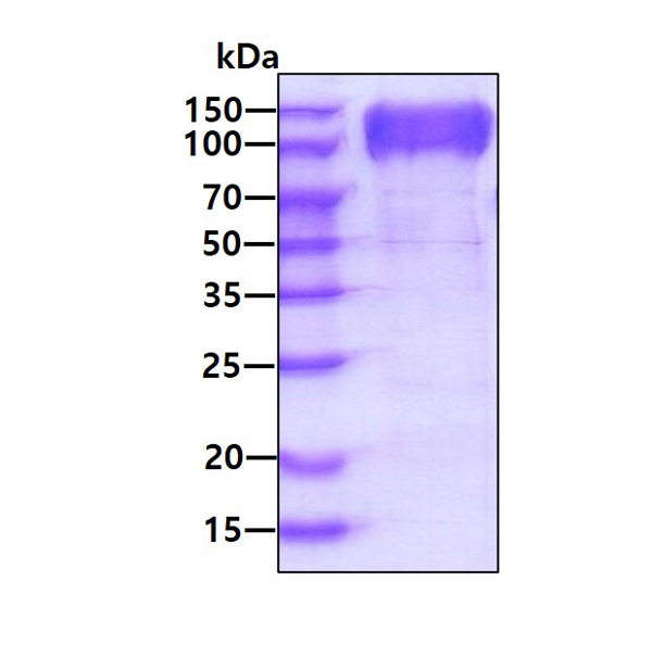
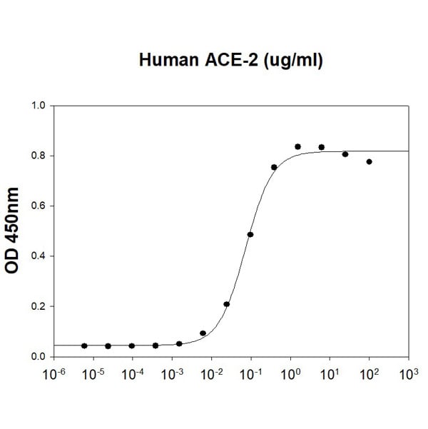
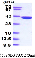
 Bioactivity
Angiotensin-Converting Enzyme 2/ACE-2, Active Protein (Cat# AAA177978)
ALKALINE PHOSPHATASE, Active Protein (Cat# AAA50488)
Matrix Metalloproteinase-7, Active Protein (Cat# AAA38246)
Interleukin-4, Active Protein (Cat# AAA38110)
Insulin Like Growth Factor-1, Active Protein (Cat# AAA38121)
Interleukin-17 A/F, Active Protein (Cat# AAA38661)
Peptidyl-Prolyl Cis/Trans Isomerase NIMA-Interacting 1, Active Protein (Cat# AAA38283)
Carbonic Anhydrase XII, Active Protein (Cat# AAA37995)
Creatine Kinase MB Isoenzyme Type-1, Active Protein (Cat# AAA38099)
Adiponectin, Active Protein (Cat# AAA78131)
ApoE2, Active Protein (Cat# AAA79218)
Adiponectin (Acrp30), Active Protein (Cat# AAA79239)
ApoE3, Active Protein (Cat# AAA79280)
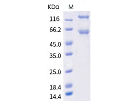
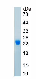
 SDS-PAGE
AKR7A2, Active Protein (Cat# AAA48568)
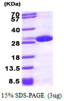
 SDS-PAGE
(3ug by SDS-PAGE under reducing condition and visualized by coomassie blue stain)
SDS-PAGE
(3ug by SDS-PAGE under reducing condition and visualized by coomassie blue stain)
Acid phosphatase 1, Active Protein (Cat# AAA48522)
Thrombin, Active Protein (Cat# AAA44822)
 Bioactivity
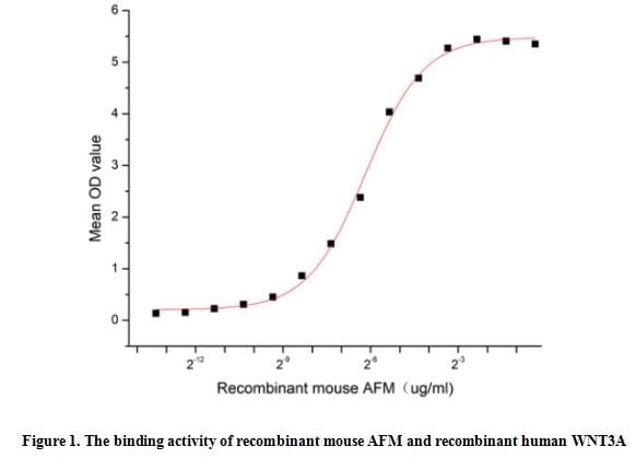
(Afamin (AFM, also known as Alpha -Albumin), a bioactive protein, is expressed in the liver and secreted into the bloodstream. Human afamin is a 87 kDa protein which is encoded by the fourth member of the human albumin gene family. Mouse AFM shares 66% aa sequence identity with Full-length human AFM. This protein can bind and transport vitamin E across the blood-brain barrier in body fluids under conditions where the lipoprotein system is not sufficient, it also can act as a carrier for hydrophobic molecules in body fluids. Essential for the solubility and activity of lipidated Wnt family members. Thus a functional binding ELISA assay was conducted to detect the interaction of recombinant mouse AFM and recombinant human WNT3A. Briefly, AFM was diluted serially in PBS with 0.01% BSA (pH 7.4). Duplicate samples of 100 ul were then transferred to WNT3A-coated microtiter wells and incubated for 1h at 37 degree C. Wells were washed with PBST and incubated for 1h with anti-AFM pAb, then aspirated and washed 3 times. After incubation with HRP labelled secondary antibody for 1h at 37 degree C, wells were aspirated and washed 5 times. With the addition of substrate solution, wells were incubated 15-25 minutes at 37 degree C. Finally, add 50 uL stop solution to the wells and read at 450/630 nm immediately. The binding activity of recombinant mouse AFM and recombinant human WNT3A was shown in Figure 1, the EC50 for this effect is 0.01 ug/mL.)
Bioactivity
(Afamin (AFM, also known as Alpha -Albumin), a bioactive protein, is expressed in the liver and secreted into the bloodstream. Human afamin is a 87 kDa protein which is encoded by the fourth member of the human albumin gene family. Mouse AFM shares 66% aa sequence identity with Full-length human AFM. This protein can bind and transport vitamin E across the blood-brain barrier in body fluids under conditions where the lipoprotein system is not sufficient, it also can act as a carrier for hydrophobic molecules in body fluids. Essential for the solubility and activity of lipidated Wnt family members. Thus a functional binding ELISA assay was conducted to detect the interaction of recombinant mouse AFM and recombinant human WNT3A. Briefly, AFM was diluted serially in PBS with 0.01% BSA (pH 7.4). Duplicate samples of 100 ul were then transferred to WNT3A-coated microtiter wells and incubated for 1h at 37 degree C. Wells were washed with PBST and incubated for 1h with anti-AFM pAb, then aspirated and washed 3 times. After incubation with HRP labelled secondary antibody for 1h at 37 degree C, wells were aspirated and washed 5 times. With the addition of substrate solution, wells were incubated 15-25 minutes at 37 degree C. Finally, add 50 uL stop solution to the wells and read at 450/630 nm immediately. The binding activity of recombinant mouse AFM and recombinant human WNT3A was shown in Figure 1, the EC50 for this effect is 0.01 ug/mL.)
Afamin (AFM), Active Protein (Cat# AAA161881)
 Application Data
COVID 19 Spike S1+S2 Coronavirus, Active Protein (Cat# AAA268887)
 Application Data
(Measured by its binding ability in a functional ELISA. Immobilized MICAh at 10 ug/mL (100 uL/well) can bind S4-Fc3L3-NKG2D, the EC50 of S4-Fc3L3-NKG2D is 200-450 ng/mL.)
Application Data
(Measured by its binding ability in a functional ELISA. Immobilized MICAh at 10 ug/mL (100 uL/well) can bind S4-Fc3L3-NKG2D, the EC50 of S4-Fc3L3-NKG2D is 200-450 ng/mL.)
MICA, Active Protein (Cat# AAA258009)
 WB (Western Blot)
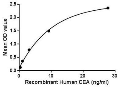
(Figure 4. Western BlotSample: Active recombinant CEA, HumanAntibody: Rabbit Anti-Human CEA Ab)
WB (Western Blot)
(Figure 4. Western BlotSample: Active recombinant CEA, HumanAntibody: Rabbit Anti-Human CEA Ab)
Carcinoembryonic Antigen, Active Protein (Cat# AAA150079)
 Bioactivity
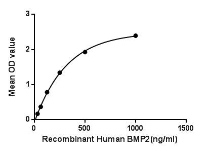
(Figure 1. The binding activity of BMP2 with NOG.)
Bioactivity
(Figure 1. The binding activity of BMP2 with NOG.)
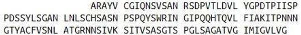
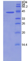
Bone Morphogenetic Protein 2, Active Protein (Cat# AAA150048)
 Bioactivity
(SARS-CoV S1 Subunit is coated at 5 ug/ml (100 ul/well) can bind Human ACE-2 (CAT# ATGP3963) in a Functional ELISA assay.)
Bioactivity
(SARS-CoV S1 Subunit is coated at 5 ug/ml (100 ul/well) can bind Human ACE-2 (CAT# ATGP3963) in a Functional ELISA assay.)
COVID 19 Spike S1 Subunit Coronavirus, Active Protein (Cat# AAA48390)
 SDS-PAGE
(#ug by SDS-PAGE under reducing condition and visualized by coomassie blue stain)
SDS-PAGE
(#ug by SDS-PAGE under reducing condition and visualized by coomassie blue stain)
AKR1B10, Active Protein (Cat# AAA48534)
Follicle Stimulating Hormone, Active Protein (Cat# AAA44845)
Immunogen affinity purified
Cancer Antigen 242, Active Protein (Cat# AAA44859)
gamma-Glutamyl Transferase, Active Protein (Cat# AAA44875)
Procalcitonin (PCT), Active Protein (Cat# AAA44759)
L-Glutamate Dehydrogenase, Active Protein (Cat# AAA44793)
Lactate Dehydrogenase 5, Active Protein (Cat# AAA44813)
acidic FGF (FGF-1), Active Protein (Cat# AAA47950)
Purification: Purified by heparin affinity chromatography and HPLC; single band at 1 ug on silver-stained SDS-PAGE.
What Are Active Proteins?
Proteins are large molecules made up of long chains of amino acids.
They will typically fold into a very particular 3-dimensional shape/conformation, that is sometimes referred to as their “native” form, which allows them to work properly in the body. For the purposes of product categorization, AAA Biotech will typically refer to proteins purified from their original animal host as being “native” proteins (this is to signify their difference compared to their “recombinant” or “synthetic” protein counterparts).
If a protein successfully folds into the correct shape, it is will typically display high fidelity characteristics to its original protein in its original animal host, and be classified as an active protein, as it will be able to function “normally” in most enzymatic or binding capacities. If it loses this shape, due to factors such as heat or strong chemicals (such as detergents), it becomes inactive and is no longer able to perform its basic functions. All of the proteins in this category are made under strict quality control, and they are active, pure, low in contaminants, and stable.
Most are stored as freeze-dried powders and come without extra tags, so they’re very close to the actual natural/native form.
Key Applications of Active Proteins
1. Scientific Research
- Aid in the study of how proteins function in the body
- Aid in understanding various disease processes
2. Drug Development
- Powerful tools to investigate how potential drugs interact with specific proteins
- Ideal for identifying drug targets
3. Cell Culture
- Are routinely utilized to support cell growth and function (e.g., using exogenous growth factors)
- Can be used to promote cellular development into specific types (differentiation)
4. Diagnostics
- Regularly utilized in tests to detect diseases or infections (e.g., COVID-19, cancer)
- Note: All products are strictly for research-use only (RUO).
5. Therapeutics
- Some active proteins are used directly as treatments (e.g., insulin, enzymes)
- Note: All products are strictly for research-use only (RUO).
6. Vaccine Development
- Used to create or test vaccines by mimicking parts of viruses or bacteria
7. Biochemical Assays
- They can facilitate the characterization of enzyme activity, binding strength, or protein interactions in lab tests
Why Buy Active Proteins from AAA Biotech?
- High biological activity – Verified to perform as expected or indicated on datasheet
- Strict quality control – We are confident in our active proteins’ reliability and consistency
- High purity & low endotoxin – Ideal for applications involving sensitive or precious samples/components
- Freeze-dried for stability – Long shelf life and straightforward storage
- Mostly tag-free – Closer to natural/native protein form
FAQ
1. What are active proteins used for in research?
Active proteins are used primarily in the study of how proteins function, in characterizing/discovering drug interactions, supporting cell growth, running biochemical assays, and in development of diagnostics or therapeutics.
2. How are AAA Biotech's active proteins validated?
AAA Biotech’s active proteins are validated through strict quality control and functional assays to ensure they are properly folded and active. “Active”, though, can be an ambiguous term, so if a specific “activity” or “binding” capability of a protein is of crucial interest to you, please inquire with us prior to purchase, and we will provide further details on how the “Active” modifier was determined to be applicable.
3. Are these proteins tested for biological activity?
Yes, all active proteins from AAA Biotech are tested to confirm they have the expected biological activity before being offered for use. Though, said “biological activity” can be either “enzymatic”, “binding”, or both.