Filters
▼Clonality
▼Type
▼Reactivity
▼Gene Name
▼Isotype
▼Host
▼Application
▼Clone
▼Active Proteins
AAA Biotech also known as AAA Bio or AAABio provides a variety of high-quality recombinant and natural/native proteins that are proven to work in a wide range of experiments. Explore our products to find the active protein that best fits your needs or experimental model.
Viewing 2300-2350 of 2567 product results
Aldo-Keto Reductase Family 1 Member C1, Active Protein (Cat# AAA38788)
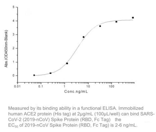
 Application Data
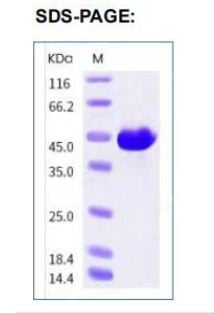
COVID 19 Spike RBD Coronavirus, Active Protein (Cat# AAA268888)
P450 Oxidoreductase, Active Protein (Cat# AAA37991)
Aldo-Keto Reductase Family 1 Member C3, Active Protein (Cat# AAA38665)
Cytidine Deaminase, Active Protein (Cat# AAA38875)
 Application Data
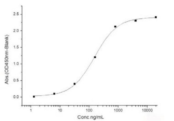
(Measured by its binding ability in a functional ELISA. Immobilized human B7-H6-His at 10 ug/mL (100 uL/well) can bind human NCR3-Fch, the EC50 of human NCR3-Fch is 6-200 ng/mL.)
Application Data
(Measured by its binding ability in a functional ELISA. Immobilized human B7-H6-His at 10 ug/mL (100 uL/well) can bind human NCR3-Fch, the EC50 of human NCR3-Fch is 6-200 ng/mL.)
B7-H6, Active Protein (Cat# AAA258051)
 Application Data
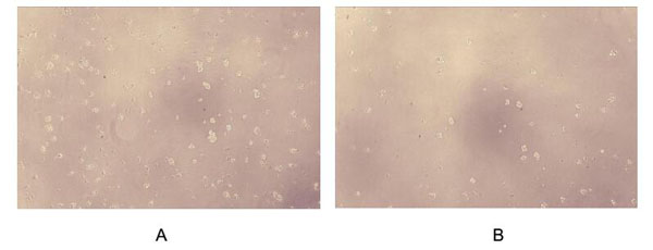
(Figure. Cell proliferation of Raji cells after stimulated with IL6.Interleukin 6 (IL-6) is an interleukin that acts as both a pro-inflammatory cytokine and an anti-inflammatory myokine. Interleukin 6 is secreted by T cells and macrophages to stimulate immune response and also plays a role in fighting infection. It supports the growth of B cells and is antagonistic to regulatory T cells. To test the effect of IL6 on cell proliferation, Raji cells were seeded into triplicate wells of 96-well plates at a density of 5,000 cells/well with 1% serum standard 1640 which contains various concentrations of recombinant dog IL6. After incubated for 5 days, cells were observed by inverted microscope and cell proliferation was measured by Cell Counting Kit-8 (CCK-8). Briefly, 10uL of CCK-8 solution was added to each well of the plate, then the absorbance at 450nm was measured using a microplate reader after incubating the plate for 1-4 hours at 37 degree C. Proliferation of Raji cells after incubation with IL6 for 5 days observed by inverted microscope was shown in Figure 1. Cell viability was assessed by CCK-8 (Cell Counting Kit-8) assay after incubation with recombinant IL6 for 5 days. The result was shown in Figure 2. It was obvious that IL6 significantly increased cell viability of Raji cells.(A) Raji cells cultured in 1640, stimulated with 1ng/mL IL6 for 5 days; (B) Unstimulated Raji cells cultured in 1640 for 5 days.)
Application Data
(Figure. Cell proliferation of Raji cells after stimulated with IL6.Interleukin 6 (IL-6) is an interleukin that acts as both a pro-inflammatory cytokine and an anti-inflammatory myokine. Interleukin 6 is secreted by T cells and macrophages to stimulate immune response and also plays a role in fighting infection. It supports the growth of B cells and is antagonistic to regulatory T cells. To test the effect of IL6 on cell proliferation, Raji cells were seeded into triplicate wells of 96-well plates at a density of 5,000 cells/well with 1% serum standard 1640 which contains various concentrations of recombinant dog IL6. After incubated for 5 days, cells were observed by inverted microscope and cell proliferation was measured by Cell Counting Kit-8 (CCK-8). Briefly, 10uL of CCK-8 solution was added to each well of the plate, then the absorbance at 450nm was measured using a microplate reader after incubating the plate for 1-4 hours at 37 degree C. Proliferation of Raji cells after incubation with IL6 for 5 days observed by inverted microscope was shown in Figure 1. Cell viability was assessed by CCK-8 (Cell Counting Kit-8) assay after incubation with recombinant IL6 for 5 days. The result was shown in Figure 2. It was obvious that IL6 significantly increased cell viability of Raji cells.(A) Raji cells cultured in 1640, stimulated with 1ng/mL IL6 for 5 days; (B) Unstimulated Raji cells cultured in 1640 for 5 days.)
Interleukin 6, Active Protein (Cat# AAA150062)
Adiponectin Trimeric Form, Active Protein (Cat# AAA38123)
Alkaline Phosphatase Placental, Active Protein (Cat# AAA38066)
Alpha-1 Antitrypsin, Active Protein (Cat# AAA38953)
Activin-A, Active Protein (Cat# AAA38992)
Aldehyde Dehydrogenase 2, Active Protein (Cat# AAA38769)
Guanine Deaminase, Active Protein (Cat# AAA39144)
 Application Data
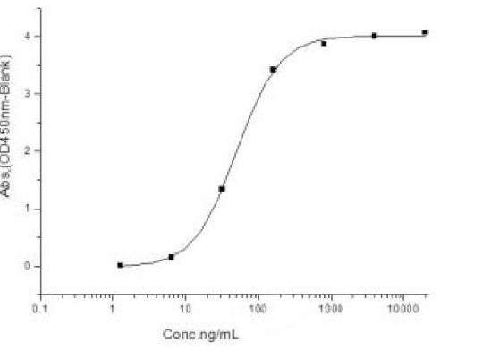
(Measured by its ability to bind human CD32a-His in a functional ELISA.)
Application Data
(Measured by its ability to bind human CD32a-His in a functional ELISA.)
IgG2, Active Protein (Cat# AAA258022)
 Application Data
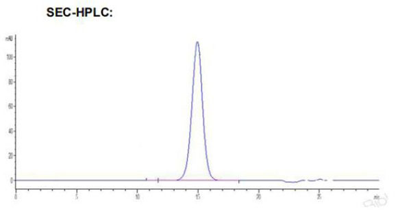
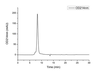
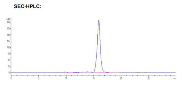
(The purity of SARS-COV-2 Spike S1 Protein with His tag (Cat.AAA281532) was greater than 90% as determined by SEC-HPLC.)
Application Data
(The purity of SARS-COV-2 Spike S1 Protein with His tag (Cat.AAA281532) was greater than 90% as determined by SEC-HPLC.)
COVID 19 Spike S1 Coronavirus, Active Protein (Cat# AAA281532)
BAFF/TNFSF13B, Active Protein (Cat# AAA48992)
Alpha-2 Antiplasmin, Active Protein (Cat# AAA44856)
Adenylate Kinase 2, Active Protein (Cat# AAA38616)
Alpha-Amylase, Active Protein (Cat# AAA44778)
 Application Data
COVID 19 Spike S1 Coronavirus, Active Protein (Cat# AAA268891)
 Application Data
COVID 19 Spike RBD Coronavirus, Active Protein (Cat# AAA268894)
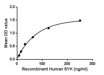 Bioactivity
(Figure. The binding activity of SYK with SIGLEC2.Spleen Tyrosine Kinase (SYK) is a member of the SYK family of tyrosine kinases. These non-receptor cytoplasmic tyrosine kinases share a characteristic dual SH2 domain separated by a linker domain. Within B and T cells respectively, SYK and Zap-70 transmit signals from the B-Cell receptor and T-Cell receptor. SYK plays a similar role in transmitting signals from a variety of cell surface receptors including CD74, Fc Receptor, and integrins. Besides, Sialic Acid Binding Ig Like Lectin 1 (SIGLEC2) has been identified as an interactor of SYK, thus a binding ELISA assay was conducted to detect the interaction of recombinant human SYK and recombinant human SIGLEC2. Briefly, SYK were diluted serially in PBS, with 0.01% BSA (pH 7.4). Duplicate samples of 100uL were then transferred to SIGLEC2-coated microtiter wells and incubated for 2h at 37. Wells were washed with PBST and incubated for 1h with anti-SYK pAb, then aspirated and washed 3 times. After incubation with HRP labelled secondary antibody, wells were aspirated and washed 3 times. With the addition of substrate solution, wells were incubated 15-25 minutes at 37. Finally, add 50uL stop solution to the wells and read at 450nm immediately. The binding activity of SYK and SIGLEC2 was shown in Figure 1, and this effect was in a dose dependent manner.)
Bioactivity
(Figure. The binding activity of SYK with SIGLEC2.Spleen Tyrosine Kinase (SYK) is a member of the SYK family of tyrosine kinases. These non-receptor cytoplasmic tyrosine kinases share a characteristic dual SH2 domain separated by a linker domain. Within B and T cells respectively, SYK and Zap-70 transmit signals from the B-Cell receptor and T-Cell receptor. SYK plays a similar role in transmitting signals from a variety of cell surface receptors including CD74, Fc Receptor, and integrins. Besides, Sialic Acid Binding Ig Like Lectin 1 (SIGLEC2) has been identified as an interactor of SYK, thus a binding ELISA assay was conducted to detect the interaction of recombinant human SYK and recombinant human SIGLEC2. Briefly, SYK were diluted serially in PBS, with 0.01% BSA (pH 7.4). Duplicate samples of 100uL were then transferred to SIGLEC2-coated microtiter wells and incubated for 2h at 37. Wells were washed with PBST and incubated for 1h with anti-SYK pAb, then aspirated and washed 3 times. After incubation with HRP labelled secondary antibody, wells were aspirated and washed 3 times. With the addition of substrate solution, wells were incubated 15-25 minutes at 37. Finally, add 50uL stop solution to the wells and read at 450nm immediately. The binding activity of SYK and SIGLEC2 was shown in Figure 1, and this effect was in a dose dependent manner.)
Spleen Tyrosine Kinase, Active Protein (Cat# AAA150134)
 Application Data
COVID 19 Spike RBD Coronavirus, Active Protein (Cat# AAA268889)
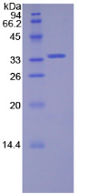
95% as determined by SEC-HPLC
 Bioactivity
(A hallmark of most cancer cells is an altered metabolism involving a shift to aerobic glycolysis with lactate production coupled with a higher uptake of glucose as the main source of energy. Lactate dehydrogenase (LDH) is key to this shift by catalyzing the formation of lactate by reducing pyruvate with NADH, which also generates NAD() that is essential for the continuity of glycolysis. Inhibiting LDH activity has an anti-proliferative effect on cancer cells. It may reverse the resistance of tumor cells to conventional chemo- and radiotherapy. Recent research has renewed interest in LDH as an anticancer drug target. LDH enzymes have three homologous subunits LDHA, LDHB and LDHC. The activity of recombinant rat LDHC was measured by its ability to reduce pyruvate to lactate. The reaction was performed in 25 mM Tris, 100 mM NaCl, pH 7.5 (assay buffer), initiated by addition 50 uL of various concentrations of rrLDHC (diluted by assay buffer) to 50 uL of substrate mixture 1.6 mM beta-NADH and 4 mM sodium pyruvate. The final well serves as a negative control with no rrLDHC, replaced with 50 ul assay buffer. Read at a wavelength of 340 nm in kinetic mode for 5 minutes. The specific activity of recombinant rat LDHC is > 880 pmol/min/ug.)
Bioactivity
(A hallmark of most cancer cells is an altered metabolism involving a shift to aerobic glycolysis with lactate production coupled with a higher uptake of glucose as the main source of energy. Lactate dehydrogenase (LDH) is key to this shift by catalyzing the formation of lactate by reducing pyruvate with NADH, which also generates NAD() that is essential for the continuity of glycolysis. Inhibiting LDH activity has an anti-proliferative effect on cancer cells. It may reverse the resistance of tumor cells to conventional chemo- and radiotherapy. Recent research has renewed interest in LDH as an anticancer drug target. LDH enzymes have three homologous subunits LDHA, LDHB and LDHC. The activity of recombinant rat LDHC was measured by its ability to reduce pyruvate to lactate. The reaction was performed in 25 mM Tris, 100 mM NaCl, pH 7.5 (assay buffer), initiated by addition 50 uL of various concentrations of rrLDHC (diluted by assay buffer) to 50 uL of substrate mixture 1.6 mM beta-NADH and 4 mM sodium pyruvate. The final well serves as a negative control with no rrLDHC, replaced with 50 ul assay buffer. Read at a wavelength of 340 nm in kinetic mode for 5 minutes. The specific activity of recombinant rat LDHC is > 880 pmol/min/ug.)
Lactate Dehydrogenase C (LDHC), Active Protein (Cat# AAA161921)
Apolipoprotein E3, Active Protein (Cat# AAA37992)
Antistreptolysin O, Active Protein (Cat# AAA44827)
Glutamine Synthetase, Active Protein (Cat# AAA39145)
Neutrophil Elastase, Active Protein (Cat# AAA224704)
 Application Data
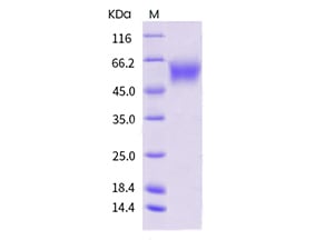
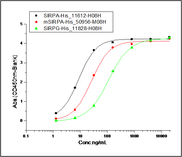
(1. Measured by its binding ability in a functional ELISA. Immobilized human SIRPA-His at 10 ug/ml (100 ul/well) can bind human CD47-Fc, The EC50 of human CD47-Fc is 10.1-23.5 ng/ml. 2. Measured by its binding ability in a functional ELISA. Immobilized mouse SIRPA-His at 10 ug/ml (100 ul/well) can bind human CD47-Fc, The EC50 of human CD47-Fc is 0.05-0.13 ug/ml. 3. Measured by its binding ability in a functional ELISA. Immobilized human SIRPG-His at 10 ug/ml (100 ul/well) can bind human CD47-Fc, The EC50 of human CD47-Fc is 0.58-1.34 ug/ml.)
Application Data
(1. Measured by its binding ability in a functional ELISA. Immobilized human SIRPA-His at 10 ug/ml (100 ul/well) can bind human CD47-Fc, The EC50 of human CD47-Fc is 10.1-23.5 ng/ml. 2. Measured by its binding ability in a functional ELISA. Immobilized mouse SIRPA-His at 10 ug/ml (100 ul/well) can bind human CD47-Fc, The EC50 of human CD47-Fc is 0.05-0.13 ug/ml. 3. Measured by its binding ability in a functional ELISA. Immobilized human SIRPG-His at 10 ug/ml (100 ul/well) can bind human CD47-Fc, The EC50 of human CD47-Fc is 0.58-1.34 ug/ml.)
CD47, Active Protein (Cat# AAA258008)

 Bioactivity
(Annexin V (ANXA5) is a multifunctional protein that is highly expressed on the apical surfaces of syncytiotrophoblasts, and plays an important role in haemostatic regulations, maintaining blood fluidity of the placenta. Lower ANXA5 levels have been observed in M2/ANXA5 haplotype carrying chorion. The association found between the maternal carriage of the M2/ANXA5 haplotype and an elevated risk of IUGR and/or PE supports the hypothesis that carrier status of this haplotype and the consequently reduced placental ANXA5 expression might be responsible, at least partially, for the onset of these gestational vascular complications. Annexin V is a calcium-dependent phospholipid binding protein that can be used to bind Phosphatidylserine (PS) during an early apoptosis event where the PS becomes exposed at the cell surface. Jurkat cells were treated with 10 uM camptothecin for 4h, 2*105 cells which were resuspended in binding buffer were stained with 10 ug recombinant human Annexin V-FITC and 10 ul Propidium iodide (PI) for 20min in dark room temperature. The flow cytometry was used to detect the early apoptotic and late apoptotic of camptothecin-treated Jurkat cells (Figure 1), the combination of Annexin V-FITC and propidium iodide allows for the distinction between early apoptotic cells (Annexin V-FITC positive and propidium iodide negative), late apoptotic and/or necrotic cells (Annexin V-FITC and propidium iodide positive), and viable cells (unstained). Thus, the recombinant human Annexin V-FITC can bind Phosphatidylserine (PS) at early apoptosis of Jurkat.)
Bioactivity
(Annexin V (ANXA5) is a multifunctional protein that is highly expressed on the apical surfaces of syncytiotrophoblasts, and plays an important role in haemostatic regulations, maintaining blood fluidity of the placenta. Lower ANXA5 levels have been observed in M2/ANXA5 haplotype carrying chorion. The association found between the maternal carriage of the M2/ANXA5 haplotype and an elevated risk of IUGR and/or PE supports the hypothesis that carrier status of this haplotype and the consequently reduced placental ANXA5 expression might be responsible, at least partially, for the onset of these gestational vascular complications. Annexin V is a calcium-dependent phospholipid binding protein that can be used to bind Phosphatidylserine (PS) during an early apoptosis event where the PS becomes exposed at the cell surface. Jurkat cells were treated with 10 uM camptothecin for 4h, 2*105 cells which were resuspended in binding buffer were stained with 10 ug recombinant human Annexin V-FITC and 10 ul Propidium iodide (PI) for 20min in dark room temperature. The flow cytometry was used to detect the early apoptotic and late apoptotic of camptothecin-treated Jurkat cells (Figure 1), the combination of Annexin V-FITC and propidium iodide allows for the distinction between early apoptotic cells (Annexin V-FITC positive and propidium iodide negative), late apoptotic and/or necrotic cells (Annexin V-FITC and propidium iodide positive), and viable cells (unstained). Thus, the recombinant human Annexin V-FITC can bind Phosphatidylserine (PS) at early apoptosis of Jurkat.)
Annexin V (ANXA5), Active Protein (Cat# AAA161723)
Jun Proto-Oncogene, Active Protein (Cat# AAA38414)
 ELISA
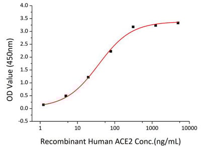
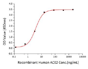
(Immobilized SARS-CoV-2 S1+S2 ECD(S-ECD) at 2ug/mL (100 uL/well) can bind recombinant Human ACE2 with a linear range of 0.15-3.72 ng/mL.)
ELISA
(Immobilized SARS-CoV-2 S1+S2 ECD(S-ECD) at 2ug/mL (100 uL/well) can bind recombinant Human ACE2 with a linear range of 0.15-3.72 ng/mL.)
COVID 19 Spike S1+S2 ECD (S-ECD) His tag (Wild type, pre-fusion state) Coronavirus, Active Protein (Cat# AAA281564)

 Application Data
COVID 19 Spike S1 Coronavirus, Active Protein (Cat# AAA268890)
Alpha-Amylase, Active Protein (Cat# AAA44779)
 Bioactivity
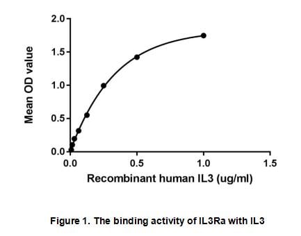
(Interleukin 3 receptor alpha (IL3RA), also known as CD123 (Cluster of Differentiation 123), is a subunit of the functional high-affinity human IL-3 receptor which is a heterodimer. The alpha subunit alone binds IL-3 with low affinity. The beta subunit doe)
Bioactivity
(Interleukin 3 receptor alpha (IL3RA), also known as CD123 (Cluster of Differentiation 123), is a subunit of the functional high-affinity human IL-3 receptor which is a heterodimer. The alpha subunit alone binds IL-3 with low affinity. The beta subunit doe)
Interleukin 3 Receptor Alpha (IL3Ra), Active Protein (Cat# AAA153089)
 Bioactivity
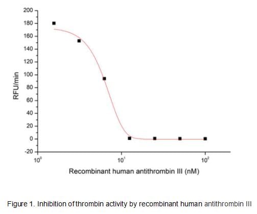
(Antithrombin III, also known as Serpin C1, is a member of the Serpin superfamily of the serine protease inhibitors. It is the principal plasma Serpin of blood clotting proteases and inhibits thrombin as well as several factors such as Xa. Similar to Serpins A5 and D1, its thrombin inhibitory activity is enhanced by heparin. Hereditary and acquired Serpin C1 deficiency is the cause of an increased thrombotic tendency in many cases. For example, acquired Serpin C1 deficiency is a common condition in sepsis, after major trauma or surgery. The activity of recombinant human antithrombin III was measured by its ability to inhibit thrombin cleavage of a fluorogenic peptide substrate Boc-VPR-AMC in the assay buffer 50 mM Tris, 10 mM CaCl2, 150 mM NaCl, 0.05% (w/v) Brij-35, pH 7.5. Thrombin was diluted to 0.5 U with heparin at 50 ug/ml in the assay buffer and 10 ul different concentrations of recombinant human antithrombin III (MW: 50 KD) was incubated with 10 ul diluted thrombin at 37 degree C for 30 minutes. Loading 50 uL of the incubated mixtures which were diluted five-fold in assay buffer into empty wells of a plate, and start the reaction by adding 50 uL of 200 uM substrate. Include a substrate blank containing 50 uL of assay buffer and 50 uL of 200 uM substrate. Then read at excitiation and emission wavelengths of 380 nm and 460 nm, respectively, in kinetic mode for 5 minutes. The result was shown in Figure 1 and it was obvious that recombinant human antithrombin III significantly decreased thrombin activity. The inhibition IC50 was )
Bioactivity
(Antithrombin III, also known as Serpin C1, is a member of the Serpin superfamily of the serine protease inhibitors. It is the principal plasma Serpin of blood clotting proteases and inhibits thrombin as well as several factors such as Xa. Similar to Serpins A5 and D1, its thrombin inhibitory activity is enhanced by heparin. Hereditary and acquired Serpin C1 deficiency is the cause of an increased thrombotic tendency in many cases. For example, acquired Serpin C1 deficiency is a common condition in sepsis, after major trauma or surgery. The activity of recombinant human antithrombin III was measured by its ability to inhibit thrombin cleavage of a fluorogenic peptide substrate Boc-VPR-AMC in the assay buffer 50 mM Tris, 10 mM CaCl2, 150 mM NaCl, 0.05% (w/v) Brij-35, pH 7.5. Thrombin was diluted to 0.5 U with heparin at 50 ug/ml in the assay buffer and 10 ul different concentrations of recombinant human antithrombin III (MW: 50 KD) was incubated with 10 ul diluted thrombin at 37 degree C for 30 minutes. Loading 50 uL of the incubated mixtures which were diluted five-fold in assay buffer into empty wells of a plate, and start the reaction by adding 50 uL of 200 uM substrate. Include a substrate blank containing 50 uL of assay buffer and 50 uL of 200 uM substrate. Then read at excitiation and emission wavelengths of 380 nm and 460 nm, respectively, in kinetic mode for 5 minutes. The result was shown in Figure 1 and it was obvious that recombinant human antithrombin III significantly decreased thrombin activity. The inhibition IC50 was )
Antithrombin (AT), Active Protein (Cat# AAA161886)
 Application Data
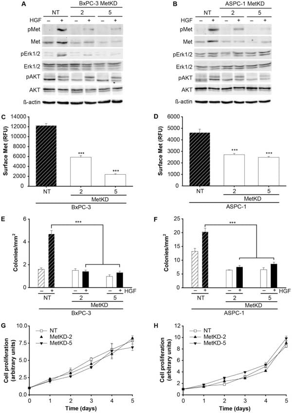
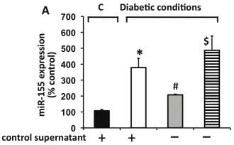
(Conditioned media from BMPC regulates miR-155 expression and fibrogenic response in mouse cardiac fibroblasts in vitro.Neutralizing antibodies against HGF (HGF-Ab) reversed this effect as compared to IgG treated cells.)
Application Data
(Conditioned media from BMPC regulates miR-155 expression and fibrogenic response in mouse cardiac fibroblasts in vitro.Neutralizing antibodies against HGF (HGF-Ab) reversed this effect as compared to IgG treated cells.)
Hepatocyte Growth Factor, Active Protein (Cat# AAA76400)
 Application Data
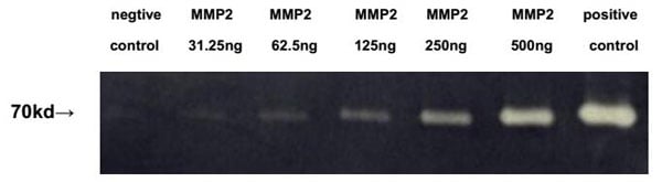
(Mechanism: MMP2 is a zinc-dependent enzymes capable of cleaving components of the extracellular matrix, which belongs to the matrix metalloproteinase (MMP) family.It is a gelatinase A, 72kDa type IV collagenase which can hydrolyze gelatin under certain conditions. Gelatin zymography is mainly used for the detection of the gelatinases, MMP-2 and MMP-9 and It is extremely sensitive because levels of 10pg of MMP-2 can already be detected. Briefly, various concentrations of MMP2 were denatured by SDS loading buffer, electrophoresed through sodium dodecylsulphate–polyacrylamide gel (SDS–PAGE; 10% gels) containing gelatin (1 mg/mL) with nonreducing conditions. After renaturation, incubation and CCB-stained, active MMP2 would hydrolyze gelatin nearby, which was indicated by the white binds on the gel. In this experiment we use heat-denatured MMP2 protein as negative control, and blood sample as positive control.Result: Gelatin hydrolysis by recombinant mouse MMP2 was shown in figure 1.Figure 1. Hydrolysis of gelatin by recombinant mouse MMP2.)
Application Data
(Mechanism: MMP2 is a zinc-dependent enzymes capable of cleaving components of the extracellular matrix, which belongs to the matrix metalloproteinase (MMP) family.It is a gelatinase A, 72kDa type IV collagenase which can hydrolyze gelatin under certain conditions. Gelatin zymography is mainly used for the detection of the gelatinases, MMP-2 and MMP-9 and It is extremely sensitive because levels of 10pg of MMP-2 can already be detected. Briefly, various concentrations of MMP2 were denatured by SDS loading buffer, electrophoresed through sodium dodecylsulphate–polyacrylamide gel (SDS–PAGE; 10% gels) containing gelatin (1 mg/mL) with nonreducing conditions. After renaturation, incubation and CCB-stained, active MMP2 would hydrolyze gelatin nearby, which was indicated by the white binds on the gel. In this experiment we use heat-denatured MMP2 protein as negative control, and blood sample as positive control.Result: Gelatin hydrolysis by recombinant mouse MMP2 was shown in figure 1.Figure 1. Hydrolysis of gelatin by recombinant mouse MMP2.)
Matrix Metalloproteinase 2, Active Protein (Cat# AAA150070)
 Bioactivity
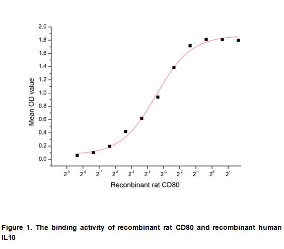
(B-Lymphocyte Activation Antigen B7-1 (CD80) is a membrane receptor that is activated by the binding of CD28 or CTLA-4. The activated protein induces T-cell proliferation and cytokine production. This protein can act as a receptor for adenovirus subgroup B)
Bioactivity
(B-Lymphocyte Activation Antigen B7-1 (CD80) is a membrane receptor that is activated by the binding of CD28 or CTLA-4. The activated protein induces T-cell proliferation and cytokine production. This protein can act as a receptor for adenovirus subgroup B)
B-Lymphocyte Activation Antigen B7-1 (LAB7-1), Active Protein (Cat# AAA153085)
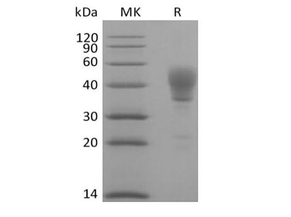
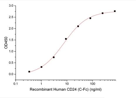 Bioactivity
Signal Transducer CD24/CD24, Active Protein (Cat# AAA177955)
 Application Data
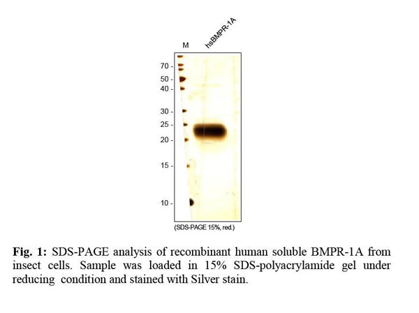
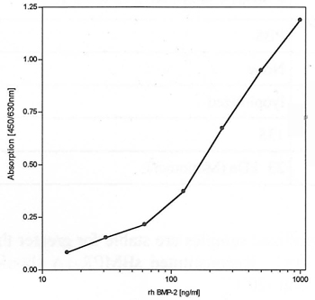
(BMP-2 BioLISA using recombinant human soluble BMPR-IA for capturing and recombinant human BMP-2 as standard. A rabbit anti-human BMP-2 antibody in combination with an goat anti-rabbit Biotin antibody was used for detection.)
Application Data
(BMP-2 BioLISA using recombinant human soluble BMPR-IA for capturing and recombinant human BMP-2 as standard. A rabbit anti-human BMP-2 antibody in combination with an goat anti-rabbit Biotin antibody was used for detection.)
BMP receptor-1A, soluble, Active Protein (Cat# AAA79169)
 Bioactivity
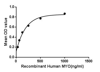
(Myoglobin (symbol Mb or MB or MYO) is an iron- and oxygen-binding protein found in the muscle tissue of vertebrates in general and in almost all mammals. It is related to hemoglobin, which is the iron- and oxygen-binding protein in blood, specifically in the red blood cells. Myoglobin is found in Type I muscle, Type II A and Type II B, but most texts consider myoglobin not to be found in smooth muscle. Besides, Proteasome 26S Subunit, Non ATPase 4 (PSMD4) has been identified as an interactor of MYO, thus a binding ELISA assay was conducted to detect the interaction of recombinant human MYO and recombinant human PSMD4 Briefly, MYO were diluted serially in PBS, with 0.01% BSA (pH 7.4). Duplicate samples of 100L were then transferred to PSMD4-coated microtiter wells and incubated for 2h at 37. Wells were washed with PBST and incubated for 1h with anti-MYO pAb, then aspirated and washed 3 times. After incubation with HRP labelled secondary antibody, wells were aspirated and washed 3 times. With the addition of substrate solution, wells were incubated 15-25 minutes at 37. Finally, add 50uL stop solution to the wells and read at 450nm immediately. The binding activity of MYO and PSMD4 was shown in Figure 1, and this effect was in a dose dependent manner.Figure. The binding activity of MYO with PSMD4.)
Bioactivity
(Myoglobin (symbol Mb or MB or MYO) is an iron- and oxygen-binding protein found in the muscle tissue of vertebrates in general and in almost all mammals. It is related to hemoglobin, which is the iron- and oxygen-binding protein in blood, specifically in the red blood cells. Myoglobin is found in Type I muscle, Type II A and Type II B, but most texts consider myoglobin not to be found in smooth muscle. Besides, Proteasome 26S Subunit, Non ATPase 4 (PSMD4) has been identified as an interactor of MYO, thus a binding ELISA assay was conducted to detect the interaction of recombinant human MYO and recombinant human PSMD4 Briefly, MYO were diluted serially in PBS, with 0.01% BSA (pH 7.4). Duplicate samples of 100L were then transferred to PSMD4-coated microtiter wells and incubated for 2h at 37. Wells were washed with PBST and incubated for 1h with anti-MYO pAb, then aspirated and washed 3 times. After incubation with HRP labelled secondary antibody, wells were aspirated and washed 3 times. With the addition of substrate solution, wells were incubated 15-25 minutes at 37. Finally, add 50uL stop solution to the wells and read at 450nm immediately. The binding activity of MYO and PSMD4 was shown in Figure 1, and this effect was in a dose dependent manner.Figure. The binding activity of MYO with PSMD4.)
Myoglobin, Active Protein (Cat# AAA150089)
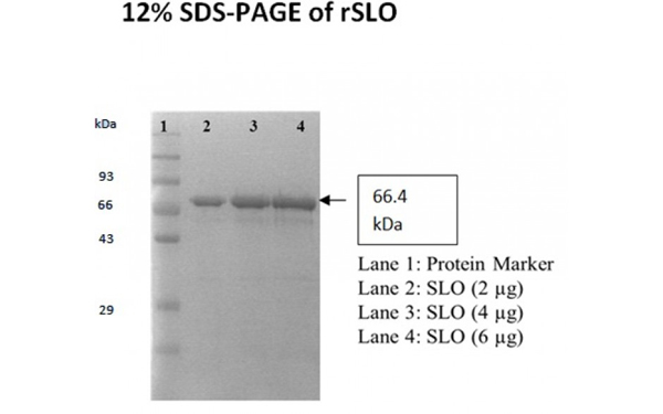
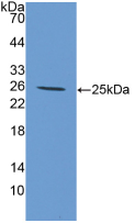
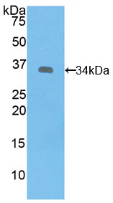
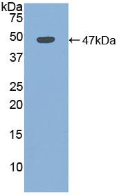
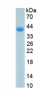
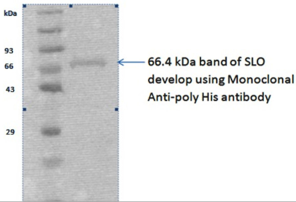 WB (Western Blot)
(Load: 3 ug)
WB (Western Blot)
(Load: 3 ug)
Streptolysin O, Active Protein (Cat# AAA224745)
 Application Data
COVID 19 Papain-Like Protease Coronavirus, Active Protein (Cat# AAA268910)
 Bioactivity
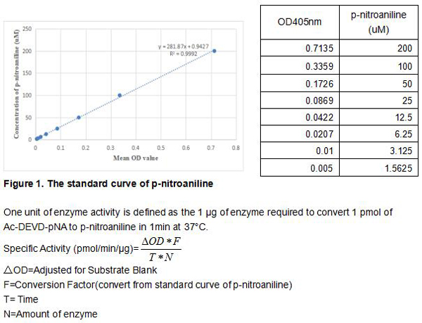
(Caspase 3 is a member of the cysteine-aspartic acid protease (caspase) family. Sequential activation of caspases plays a central role in the execution-phase of cell apoptosis. Caspases exist as inactive proenzymes that undergo proteolytic processing at conserved aspartic residues to produce two subunits, large and small, that dimerize to form the active enzyme. This protein cleaves and activates caspase 6 and 7; and the protein itself is processed and activated by caspases 8, 9, and 10. Caspase 3 can hydrolyze the peptide substrate acetyl-Asp-Glu-Val-Asp-p-nitroanilide (Ac-DEVD-pNA) resulting in the release of the p-nitroaniline (pNA) moiety. p-Nitroaniline has a high absorbance at 405 nm. Thus the activity of recombinant mouse caspase 3 can be measuered by calculating the concentration of the pNA released from the substrate. The reaction was performed in adding 50 ul 2×buffer (50mM HEPES,100mM NaCl,10mM DTT, 2mM EDTA, 10% glycerol) to 96 well plates, then add 50 ul various concentrations of caspase 3 (diluted by 1×buffer, 25mM HEPES, 50mM NaCl, 5mM DTT, 1mM EDTA, 5% glycerol) to each well, finally, add 5 ul 4mM Ac-DEVD-pNA to each well. Cover the 96 well plates and incubate at 37 degree C for 1h. p-Nitroaniline (pNA) Standard curve prepare by double dilute 200 uM pNA with 1×buffer and record the OD value at 405 nm. The specific activity of recombinant mouse caspase3 is >3000 pmol/min/ug.)
Bioactivity
(Caspase 3 is a member of the cysteine-aspartic acid protease (caspase) family. Sequential activation of caspases plays a central role in the execution-phase of cell apoptosis. Caspases exist as inactive proenzymes that undergo proteolytic processing at conserved aspartic residues to produce two subunits, large and small, that dimerize to form the active enzyme. This protein cleaves and activates caspase 6 and 7; and the protein itself is processed and activated by caspases 8, 9, and 10. Caspase 3 can hydrolyze the peptide substrate acetyl-Asp-Glu-Val-Asp-p-nitroanilide (Ac-DEVD-pNA) resulting in the release of the p-nitroaniline (pNA) moiety. p-Nitroaniline has a high absorbance at 405 nm. Thus the activity of recombinant mouse caspase 3 can be measuered by calculating the concentration of the pNA released from the substrate. The reaction was performed in adding 50 ul 2×buffer (50mM HEPES,100mM NaCl,10mM DTT, 2mM EDTA, 10% glycerol) to 96 well plates, then add 50 ul various concentrations of caspase 3 (diluted by 1×buffer, 25mM HEPES, 50mM NaCl, 5mM DTT, 1mM EDTA, 5% glycerol) to each well, finally, add 5 ul 4mM Ac-DEVD-pNA to each well. Cover the 96 well plates and incubate at 37 degree C for 1h. p-Nitroaniline (pNA) Standard curve prepare by double dilute 200 uM pNA with 1×buffer and record the OD value at 405 nm. The specific activity of recombinant mouse caspase3 is >3000 pmol/min/ug.)
Caspase 3 (CASP3), Active Protein (Cat# AAA161756)

 Bioactivity
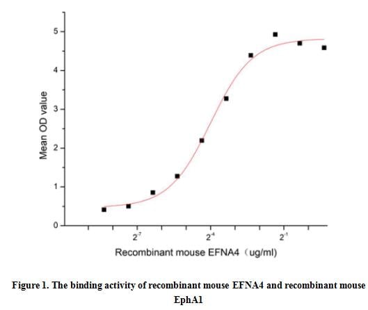
(Ephrin-A4, also known as EFNA4 and EFL-4, is a member of the ligand of the EPH family. It is mainly expressed in the spleen, lymph nodes, ovary, small intestine, and colon of adults, as well as in the heart, lungs, liver, and kidneys of the fetus. It is involved in the development of neurons, blood vessels, and epithelium by regulating cell migration, rejection, and adhesion. Ephrin-A4 has been shown to bind EphA1,EphA2, EphA3, EphA4, EphA5, EphA6, EphA7 and EphB1. Thus a functional binding ELISA assay was conducted to detect the interaction of recombinant mouse EFNA4 and recombinant mouse EPHA1. Briefly, EFNA4 was diluted serially in PBS with 0.01% BSA (pH 7.4). Duplicate samples of 100 ul were then transferred to EphA1-coated microtiter wells and incubated for 1h at 37 degree C. Wells were washed with PBST and incubated for 1h with anti-EFNA4 pAb, then aspirated and washed 3 times. After incubation with HRP labelled secondary antibody for 1h at 37 degree C, wells were aspirated and washed 5 times. With the addition of substrate solution, wells were incubated 15-25 minutes at 37 degree C. Finally, add 50 uL stop solution to the wells and read at 450/630 nm immediately. The binding activity of recombinant mouse EFNA4 and recombinant mouse EphA1 was shown in Figure 1, the EC50 for this effect is 0.06 ug/mL.)
Bioactivity
(Ephrin-A4, also known as EFNA4 and EFL-4, is a member of the ligand of the EPH family. It is mainly expressed in the spleen, lymph nodes, ovary, small intestine, and colon of adults, as well as in the heart, lungs, liver, and kidneys of the fetus. It is involved in the development of neurons, blood vessels, and epithelium by regulating cell migration, rejection, and adhesion. Ephrin-A4 has been shown to bind EphA1,EphA2, EphA3, EphA4, EphA5, EphA6, EphA7 and EphB1. Thus a functional binding ELISA assay was conducted to detect the interaction of recombinant mouse EFNA4 and recombinant mouse EPHA1. Briefly, EFNA4 was diluted serially in PBS with 0.01% BSA (pH 7.4). Duplicate samples of 100 ul were then transferred to EphA1-coated microtiter wells and incubated for 1h at 37 degree C. Wells were washed with PBST and incubated for 1h with anti-EFNA4 pAb, then aspirated and washed 3 times. After incubation with HRP labelled secondary antibody for 1h at 37 degree C, wells were aspirated and washed 5 times. With the addition of substrate solution, wells were incubated 15-25 minutes at 37 degree C. Finally, add 50 uL stop solution to the wells and read at 450/630 nm immediately. The binding activity of recombinant mouse EFNA4 and recombinant mouse EphA1 was shown in Figure 1, the EC50 for this effect is 0.06 ug/mL.)
Ephrin A4 (EFNA4), Active Protein (Cat# AAA161917)
 Bioactivity
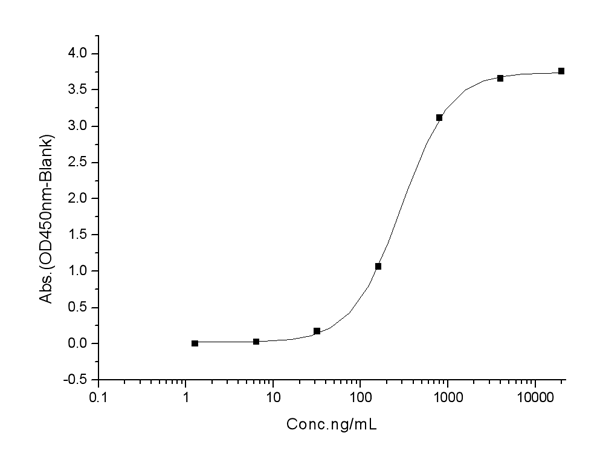
(Neutrophil elastase (NE), also known as polymorphonuclear leukocyte elastase, is a major protease in the primary granules of neutrophils, is involved in microbicidal activity. It is located primarily in the azurophil granules of polymorphonuclear leukocytes. NE is an important factor promoting inflammation, has bactericidal effects, and shortens the inflammatory process. NE also regulates tumor growth by promoting metastasis and tumor microenvironment remodeling. However, NE plays a role in killing tumors under certain conditions and promotes other diseases such as pulmonary ventilation dysfunction. Additionally, it plays a complex role in various physiological processes and mediates several diseases. Myeloperoxidase (MPO) has been identified as an interactor of NE, thus a functional binding ELISA assay was conducted to detect the interaction of recombinant human NE and recombinant rat MPO. Briefly, biotin-linked NE were diluted serially in PBS, with 0.01% BSA (pH 7.4). Duplicate samples of 100 ul were then transferred to MPO-coated microtiter wells and incubated for 1h at 37 degree C. Wells were washed with PBST 3 times and incubation with Streptavidin-HRP for 30min, then wells were aspirated and washed 5 times. With the addition of substrate solution, wells were incubated 15-25 minutes at 37 degree C. Finally, add 50 ul stop solution to the wells and read at 450 nm immediately. The binding activity of recombinant human NE and recombinant rat MPO was shown in Figure 1, the EC50 for this effect is 0.66 ug/mL.)
Bioactivity
(Neutrophil elastase (NE), also known as polymorphonuclear leukocyte elastase, is a major protease in the primary granules of neutrophils, is involved in microbicidal activity. It is located primarily in the azurophil granules of polymorphonuclear leukocytes. NE is an important factor promoting inflammation, has bactericidal effects, and shortens the inflammatory process. NE also regulates tumor growth by promoting metastasis and tumor microenvironment remodeling. However, NE plays a role in killing tumors under certain conditions and promotes other diseases such as pulmonary ventilation dysfunction. Additionally, it plays a complex role in various physiological processes and mediates several diseases. Myeloperoxidase (MPO) has been identified as an interactor of NE, thus a functional binding ELISA assay was conducted to detect the interaction of recombinant human NE and recombinant rat MPO. Briefly, biotin-linked NE were diluted serially in PBS, with 0.01% BSA (pH 7.4). Duplicate samples of 100 ul were then transferred to MPO-coated microtiter wells and incubated for 1h at 37 degree C. Wells were washed with PBST 3 times and incubation with Streptavidin-HRP for 30min, then wells were aspirated and washed 5 times. With the addition of substrate solution, wells were incubated 15-25 minutes at 37 degree C. Finally, add 50 ul stop solution to the wells and read at 450 nm immediately. The binding activity of recombinant human NE and recombinant rat MPO was shown in Figure 1, the EC50 for this effect is 0.66 ug/mL.)
Neutrophil Elastase (NE), Active Protein (Cat# AAA161712)
 Application Data
(Immobilized ACE2 Protein, Human, Recombinant (mFc Tag) at 2 ug/mL (100 uL/well) can bind SARS-CoV-2 Spike S1+S2 (YH145-146 deletion, E484K, D614G) Protein (ECD, His Tag), the EC50 of SARS-CoV-2 Spike S1+S2 (YH145-146 deletion, E484K, D614G) Protein (ECD, His Tag) is 100-700ng/mL.)
Application Data
(Immobilized ACE2 Protein, Human, Recombinant (mFc Tag) at 2 ug/mL (100 uL/well) can bind SARS-CoV-2 Spike S1+S2 (YH145-146 deletion, E484K, D614G) Protein (ECD, His Tag), the EC50 of SARS-CoV-2 Spike S1+S2 (YH145-146 deletion, E484K, D614G) Protein (ECD, His Tag) is 100-700ng/mL.)
COVID 19 Spike S1+S2 (YH145-146 deletion, E484K, D614G) Protein (ECD, His Tag) Coronavirus, Active Protein (Cat# AAA258078)

 Bioactivity
(One unit of enzyme activity is defined as the 1 ug of enzyme required to convert 1 pmol of 4-Nitrophenyl acetate to 4-Nitrophenol in 1min at 37 degree C. The specific activity of recombinant mouse CA4 is > 50 pmol/min/ug.)
Bioactivity
(One unit of enzyme activity is defined as the 1 ug of enzyme required to convert 1 pmol of 4-Nitrophenyl acetate to 4-Nitrophenol in 1min at 37 degree C. The specific activity of recombinant mouse CA4 is > 50 pmol/min/ug.)
Carbonic Anhydrase IV (CA4), Active Protein (Cat# AAA161901)
 Bioactivity
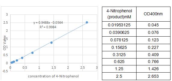
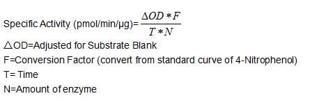
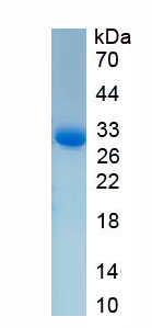
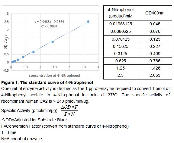
(Carbonic Anhydrase (CA) catalyzes the reversible reaction of CO2 H2O = HCO3- H, which is fundamental to many processes such as respiration, renal tubular acidification and bone resorption. CA2 is a cytosolic enzyme with the highest activity among all known CAs. Mutations in the CA2 gene result in the CA II deficiency syndrome, an autosomal recessive disorder that produces osteopetrosis, renal tubular acidosis and cerebral calcification. The activity of recombinant human CA2 was measured by its ability to hydrolyze 4-Nitrophenyl acetate (4-NPA) to 4-Nitrophenol. The reaction was performed in 12.5 mM Tris, 75 mM NaCl, pH 7.5 (assay buffer), initiated by addition 50 uL of various concentrations of CA2 (diluted by assay buffer) to 50 uL of 2 mM substrate 4-NPA (100 mM stock in Acetone, diluted by assay buffer). Incubated at 37 degree C for 5min, then read at a wavelength of 400 nm.)
Bioactivity
(Carbonic Anhydrase (CA) catalyzes the reversible reaction of CO2 H2O = HCO3- H, which is fundamental to many processes such as respiration, renal tubular acidification and bone resorption. CA2 is a cytosolic enzyme with the highest activity among all known CAs. Mutations in the CA2 gene result in the CA II deficiency syndrome, an autosomal recessive disorder that produces osteopetrosis, renal tubular acidosis and cerebral calcification. The activity of recombinant human CA2 was measured by its ability to hydrolyze 4-Nitrophenyl acetate (4-NPA) to 4-Nitrophenol. The reaction was performed in 12.5 mM Tris, 75 mM NaCl, pH 7.5 (assay buffer), initiated by addition 50 uL of various concentrations of CA2 (diluted by assay buffer) to 50 uL of 2 mM substrate 4-NPA (100 mM stock in Acetone, diluted by assay buffer). Incubated at 37 degree C for 5min, then read at a wavelength of 400 nm.)
Carbonic Anhydrase II (CA2), Active Protein (Cat# AAA161776)
What Are Active Proteins?
Proteins are large molecules made up of long chains of amino acids.
They will typically fold into a very particular 3-dimensional shape/conformation, that is sometimes referred to as their “native” form, which allows them to work properly in the body. For the purposes of product categorization, AAA Biotech will typically refer to proteins purified from their original animal host as being “native” proteins (this is to signify their difference compared to their “recombinant” or “synthetic” protein counterparts).
If a protein successfully folds into the correct shape, it is will typically display high fidelity characteristics to its original protein in its original animal host, and be classified as an active protein, as it will be able to function “normally” in most enzymatic or binding capacities. If it loses this shape, due to factors such as heat or strong chemicals (such as detergents), it becomes inactive and is no longer able to perform its basic functions. All of the proteins in this category are made under strict quality control, and they are active, pure, low in contaminants, and stable.
Most are stored as freeze-dried powders and come without extra tags, so they’re very close to the actual natural/native form.
Key Applications of Active Proteins
1. Scientific Research
- Aid in the study of how proteins function in the body
- Aid in understanding various disease processes
2. Drug Development
- Powerful tools to investigate how potential drugs interact with specific proteins
- Ideal for identifying drug targets
3. Cell Culture
- Are routinely utilized to support cell growth and function (e.g., using exogenous growth factors)
- Can be used to promote cellular development into specific types (differentiation)
4. Diagnostics
- Regularly utilized in tests to detect diseases or infections (e.g., COVID-19, cancer)
- Note: All products are strictly for research-use only (RUO).
5. Therapeutics
- Some active proteins are used directly as treatments (e.g., insulin, enzymes)
- Note: All products are strictly for research-use only (RUO).
6. Vaccine Development
- Used to create or test vaccines by mimicking parts of viruses or bacteria
7. Biochemical Assays
- They can facilitate the characterization of enzyme activity, binding strength, or protein interactions in lab tests
Why Buy Active Proteins from AAA Biotech?
- High biological activity – Verified to perform as expected or indicated on datasheet
- Strict quality control – We are confident in our active proteins’ reliability and consistency
- High purity & low endotoxin – Ideal for applications involving sensitive or precious samples/components
- Freeze-dried for stability – Long shelf life and straightforward storage
- Mostly tag-free – Closer to natural/native protein form
FAQ
1. What are active proteins used for in research?
Active proteins are used primarily in the study of how proteins function, in characterizing/discovering drug interactions, supporting cell growth, running biochemical assays, and in development of diagnostics or therapeutics.
2. How are AAA Biotech's active proteins validated?
AAA Biotech’s active proteins are validated through strict quality control and functional assays to ensure they are properly folded and active. “Active”, though, can be an ambiguous term, so if a specific “activity” or “binding” capability of a protein is of crucial interest to you, please inquire with us prior to purchase, and we will provide further details on how the “Active” modifier was determined to be applicable.
3. Are these proteins tested for biological activity?
Yes, all active proteins from AAA Biotech are tested to confirm they have the expected biological activity before being offered for use. Though, said “biological activity” can be either “enzymatic”, “binding”, or both.