Filters
▼Clonality
▼Type
▼Reactivity
▼Gene Name
▼Isotype
▼Host
▼Application
▼Clone
▼Active Proteins
AAA Biotech also known as AAA Bio or AAABio provides a variety of high-quality recombinant and natural/native proteins that are proven to work in a wide range of experiments. Explore our products to find the active protein that best fits your needs or experimental model.
Viewing 2500-2550 of 2567 product results
Alkaline Phosphatase, Active Protein (Cat# AAA11488)
 SDS-PAGE
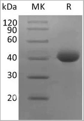
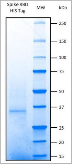
COVID 19 Spike Protein, Biotinylated (RBD-SD1, Avi) Coronavirus, Active Protein (Cat# AAA22155)
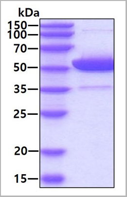
Mucin 16, Active Protein (Cat# AAA14758)
90% as determined by SDS-PAGE under reducing conditions and visualized by silver stain. Endotoxin: <1EU/ug protein (LAL)
 SDS-PAGE
(3ug by SDS-PAGE under reducing condition and visualized by coomassie blue stain.)
SDS-PAGE
(3ug by SDS-PAGE under reducing condition and visualized by coomassie blue stain.)
ALDH2, Active Protein (Cat# AAA11764)
Alkaline Phosphatase, Active Protein (Cat# AAA11487)
Human Alanine Aminotransferase, Active Protein (Cat# AAA14927)
Matrix Metalloproteinase-3, Active Protein (Cat# AAA10817)
Streptokinase, Active Protein (Cat# AAA10820)
(a)Analysis by RP-HPLC.
(b)Analysis by SDS-PAGE
Trefoil Factor-3, Active Protein (Cat# AAA10881)
Sonic HedgeHog, Active Protein (Cat# AAA10853)
Actin, beta Human, Active Protein (Cat# AAA14745)
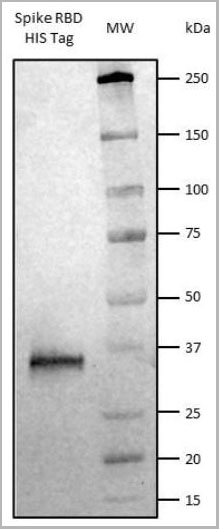 WB (Western Blot)
(Western Blot: Spike RBD HIS Tag detected with Anti-HIS-Tag antibody HRP conjugate showing a band at approx. 30 kDa)
WB (Western Blot)
(Western Blot: Spike RBD HIS Tag detected with Anti-HIS-Tag antibody HRP conjugate showing a band at approx. 30 kDa)
COVID 19 Spike Glycoprotein S1 RBD Coronavirus, Active Protein (Cat# AAA13473)
MHC class I chain-related gene B, Active Protein (Cat# AAA10837)
 Purity
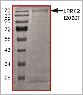
(The purity of LRRK2 (I2020T) was determined to be >70% by densitometry, approx. MW 210 kDa.)
Purity
(The purity of LRRK2 (I2020T) was determined to be >70% by densitometry, approx. MW 210 kDa.)
LRRK2, Active Protein (Cat# AAA14255)
 SDS-PAGE
(3ug by SDS-PAGE under reducing condition and visualized by coomassie blue stain.)
SDS-PAGE
(3ug by SDS-PAGE under reducing condition and visualized by coomassie blue stain.)
Alpha-enolase, Active Protein (Cat# AAA11794)
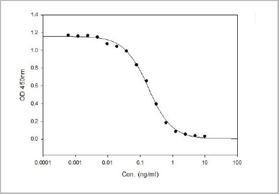
 Application Data
(Immobilized Recombinant SARS-CoV-2 Envelope at 2ug/mL (100 uL/well) can bind Recombinant SARS-CoV-2 Nucleocapsid with a linear range of 1.2-41.1 ng/mL.)
Application Data
(Immobilized Recombinant SARS-CoV-2 Envelope at 2ug/mL (100 uL/well) can bind Recombinant SARS-CoV-2 Nucleocapsid with a linear range of 1.2-41.1 ng/mL.)
COVID 19 Envelope Coronavirus, Active Protein (Cat# AAA28309)
 Application Data
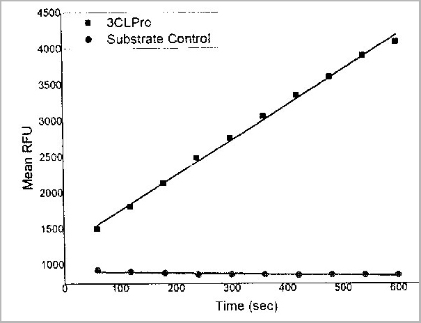
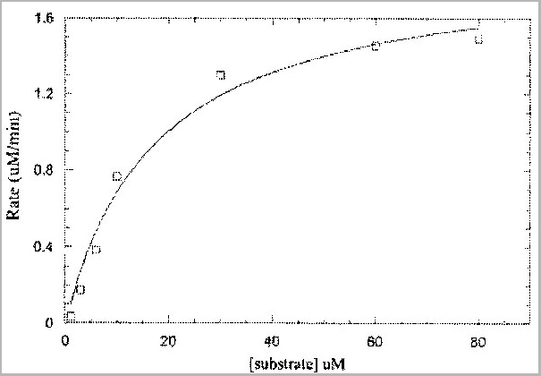
(Measurements of kinetic parameters of Recombinant 2019-nCoV 3C-like Proteinase (CAT# AAA27964) (For reference only).)
Application Data
(Measurements of kinetic parameters of Recombinant 2019-nCoV 3C-like Proteinase (CAT# AAA27964) (For reference only).)
COVID 19 3C-like Proteinase Coronavirus, Active Protein (Cat# AAA27964)
HIV-1 env gp120, Strain YU2, Active Protein (Cat# AAA14753)
 SDS-PAGE
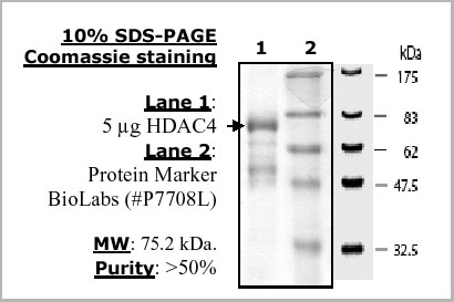
(10% SDS-PAGE Coomassie staining. Lane 1: 3ug F4600-21. Lane 2: Protein Marker.)
SDS-PAGE
(10% SDS-PAGE Coomassie staining. Lane 1: 3ug F4600-21. Lane 2: Protein Marker.)
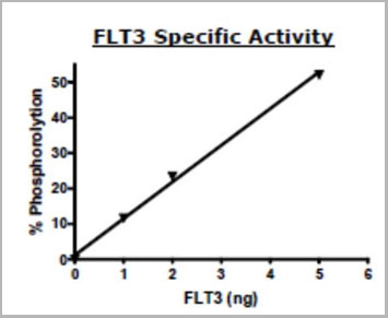
Flt3, active, N-terminal His tag Receptor Tyrosine Kinase, Active Protein (Cat# AAA14760)
 Application Data
(3CL Proteinase inhibitor screening using 2019-nCoV 3CL MPro Recombinant Protein Provided by customer.)
Application Data
(3CL Proteinase inhibitor screening using 2019-nCoV 3CL MPro Recombinant Protein Provided by customer.)
COVID 19 3CL MPro Coronavirus, Active Protein (Cat# AAA27953)
 SDS-PAGE
COVID 19 Spike S1 Protein Coronavirus, Active Protein (Cat# AAA22156)
Hypoxanthine-Guanine Phosphoribosyltransferase, Active Protein (Cat# AAA10904)
5-Lipoxygenase, Active Protein (Cat# AAA14373)
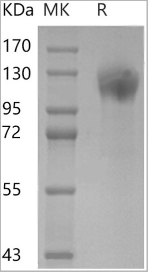
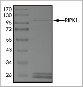 Purity
(The purity of RIPK1 was determined to be >70% by densitometry, approx. MW 108 kDa.)
Purity
(The purity of RIPK1 was determined to be >70% by densitometry, approx. MW 108 kDa.)
HIV1 tat, Active Protein (Cat# AAA14384)
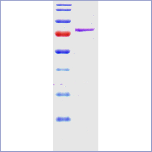 SDS-PAGE
(SDS-PAGE of 78 kDa Grp78 (BiP) protein (AAA17807))
SDS-PAGE
(SDS-PAGE of 78 kDa Grp78 (BiP) protein (AAA17807))
Grp78 (Bip), Active Protein (Cat# AAA17807)
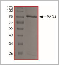 SDS-PAGE
(The purity of PAD4 was determined to be >95% by densitometry. Approx. MW 96 kDa.)
SDS-PAGE
(The purity of PAD4 was determined to be >95% by densitometry. Approx. MW 96 kDa.)
 Sequence
Carbonic Anhydrase IV (CA4), Active Protein (Cat# AAA161902)
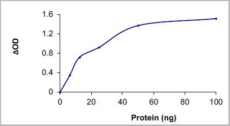
 Application Data
HDAC4, Active Protein (Cat# AAA14763)
 Application Data
(Thioflavin T is a fluorescent dye that binds to beta sheet-rich structures such as those in tau fibrils. Upon binding, the emission spectrum of the dye experiences a red-shift, and increased fluorescence intensity. Thioflavin T emission curves show increased fluorescence (correlated to tau aggregation) in tau K18 P301L monomers over time. Thioflavin T ex = 450 nm, em = 485 nm.)
Application Data
(Thioflavin T is a fluorescent dye that binds to beta sheet-rich structures such as those in tau fibrils. Upon binding, the emission spectrum of the dye experiences a red-shift, and increased fluorescence intensity. Thioflavin T emission curves show increased fluorescence (correlated to tau aggregation) in tau K18 P301L monomers over time. Thioflavin T ex = 450 nm, em = 485 nm.)
Tau, Active Protein (Cat# AAA27662)
Purification: Ion-exchange Purified
 Application Data
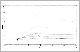
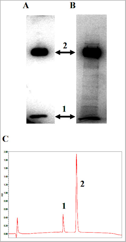
(Fig. 1. Purified CHO derived rhHPA1 was analyzed by:A. Western blot analysis using Polyclonal rabbit anti-HPA1 antibodiesB. SDS-PAGE/GelCode Blue stainingC. RPHPLC; 1, 8-kDa subunit; 2, 50-kDa subunit.)
Application Data
(Fig. 1. Purified CHO derived rhHPA1 was analyzed by:A. Western blot analysis using Polyclonal rabbit anti-HPA1 antibodiesB. SDS-PAGE/GelCode Blue stainingC. RPHPLC; 1, 8-kDa subunit; 2, 50-kDa subunit.)
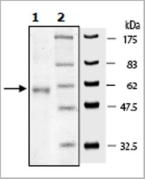
Heparanase-1, Active Protein (Cat# AAA10906)
> 95% on RP-HPLC (Fig. 1, C)
SCF, Active Protein (Cat# AAA14382)

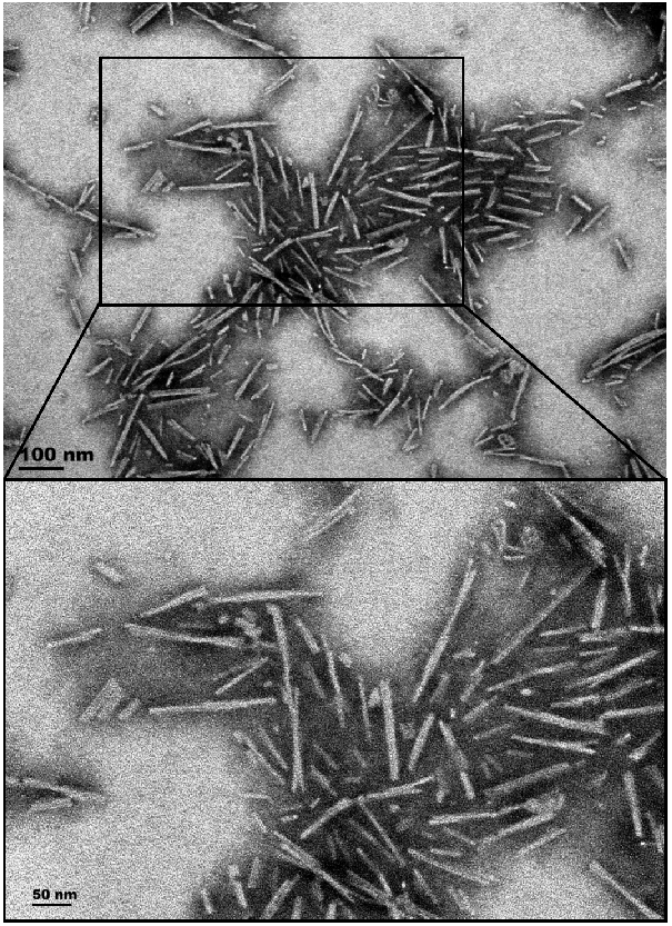
 SDS-PAGE
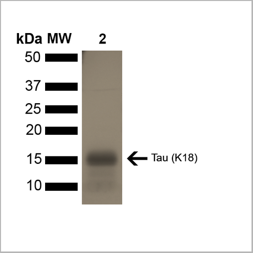
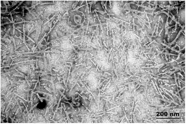
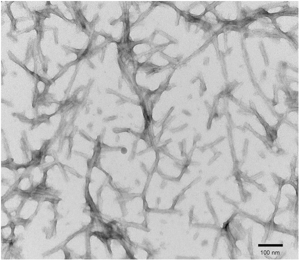
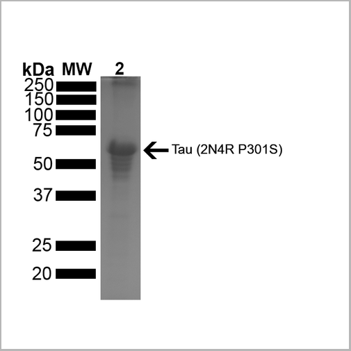
(SDS-PAGE of ~67 kDa Human Tau Protein 2N4R P301S Preformed Fibrils. Lane 1: MW Ladder. Lane 2: Tau Protein Preformed Fibrils)
SDS-PAGE
(SDS-PAGE of ~67 kDa Human Tau Protein 2N4R P301S Preformed Fibrils. Lane 1: MW Ladder. Lane 2: Tau Protein Preformed Fibrils)
Tau441, Active Protein (Cat# AAA27663)
Ion-Exchange Purified
 Biological Activity
(Human TNF-alpha induces cell cytotoxicity in the L-929 mouse fibroblast cells in the presence of the metabolic inhibitor actinomycin D.)
Biological Activity
(Human TNF-alpha induces cell cytotoxicity in the L-929 mouse fibroblast cells in the presence of the metabolic inhibitor actinomycin D.)
Tumor Necrosis Factor-alpha, Active Protein (Cat# AAA11742)
BAFF, Active Protein (Cat# AAA14391)
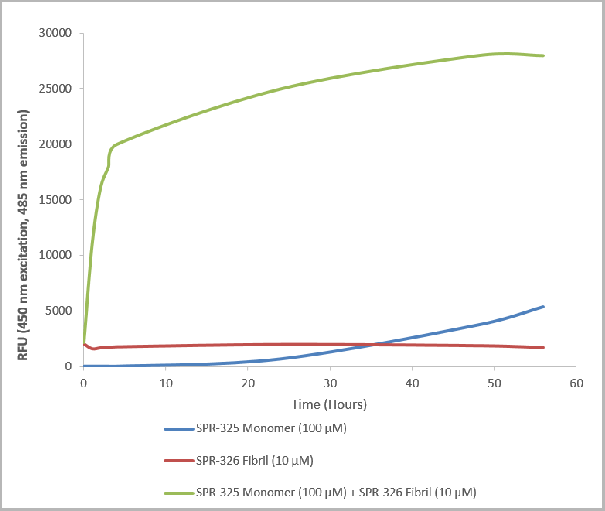 Application Data
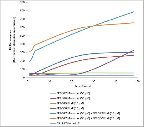
(Thioflavin T is a fluorescent dye that binds to beta sheet-rich structures such as those in alpha synuclein fibrils. Upon binding, the emission spectrum of the dye experiences a red-shift and increased fluorescence intensity. Thioflavin T emission curves show a limited increase in fluorescence (correlated to alpha synuclein aggregation) over time in A53T alpha synuclein monomers . A much greater increase in fluorescence is seen when 100 uM monomer is combined with 10 nM of fibrils (AAA27661) as the fibrils seed the formation of new fibrils from the pool of active monomers. Thioflavin T ex = 450 nm, em = 485 nm.)
Application Data
(Thioflavin T is a fluorescent dye that binds to beta sheet-rich structures such as those in alpha synuclein fibrils. Upon binding, the emission spectrum of the dye experiences a red-shift and increased fluorescence intensity. Thioflavin T emission curves show a limited increase in fluorescence (correlated to alpha synuclein aggregation) over time in A53T alpha synuclein monomers . A much greater increase in fluorescence is seen when 100 uM monomer is combined with 10 nM of fibrils (AAA27661) as the fibrils seed the formation of new fibrils from the pool of active monomers. Thioflavin T ex = 450 nm, em = 485 nm.)
A53T Mutant Alpha Synuclein, Active Protein (Cat# AAA27661)
Ion-Exchange Purified
 SDS-PAGE
KLK4, Active Protein (Cat# AAA27762)
CD137L, Active Protein (Cat# AAA14390)
Cancer Antigen 15-3, Active Protein (Cat# AAA11498)
 SDS-PAGE
C-type lectin domain family 4 member C, Active Protein (Cat# AAA18755)


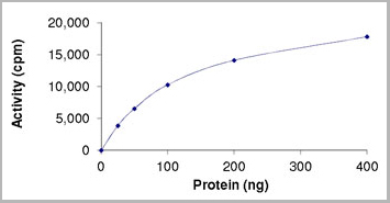
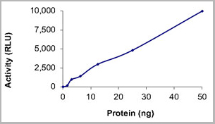
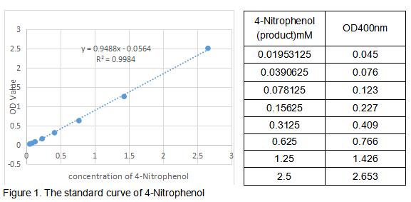
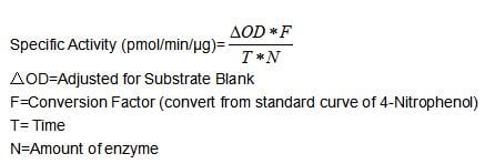
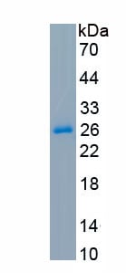
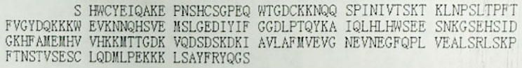
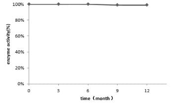 Activity
Proteinase K (PROK), Active Protein (Cat# AAA27052)


 WB (Western Blot)
(Positive WB detected in: His tag-tagged SARS-CoV-2 nucleocapsid recombinant protein from E. Coli SARS-CoV-2 nucleocapsid antibody at 1:1000; Secondary; Peroxidase-Affinipure Goat Anti-Human IgG, Fc? Fragment Specific at 1/20000 dilution; Predicted band size: 48 kDa; Observed band size: 55 kDa)
WB (Western Blot)
(Positive WB detected in: His tag-tagged SARS-CoV-2 nucleocapsid recombinant protein from E. Coli SARS-CoV-2 nucleocapsid antibody at 1:1000; Secondary; Peroxidase-Affinipure Goat Anti-Human IgG, Fc? Fragment Specific at 1/20000 dilution; Predicted band size: 48 kDa; Observed band size: 55 kDa)
COVID 19 Nucleocapsid (NP) Coronavirus, Active Protein (Cat# AAA27032)

 Application Data
(Proliferation assay with TF-I cells.)
Application Data
(Proliferation assay with TF-I cells.)
SCF, Active Protein (Cat# AAA14944)
 SDS-PAGE
(SDS-PAGE analysis of PTP1B (3ug protein; 12% gel) using P9109-14.)
SDS-PAGE
(SDS-PAGE analysis of PTP1B (3ug protein; 12% gel) using P9109-14.)
Protein Tyrosine Phosphatase 1B, Active Protein (Cat# AAA14754)
>95% by SDS PAGE; Purified by using conventional chromatography techniques.
 SDS-PAGE
Transmembrane protease serine 2 (TMPRSS2), Active Protein (Cat# AAA18665)
 SDS-PAGE
uPAR / CD87, Active Protein (Cat# AAA22094)
 SDS-PAGE
Lymphocyte antigen 6 complex locus protein G6d (LY6G6D), Active Protein (Cat# AAA18703)

 Application Data
TNFRSF1B, Active Protein (Cat# AAA23888)
Complement C9, Active Protein (Cat# AAA14378)
What Are Active Proteins?
Proteins are large molecules made up of long chains of amino acids.
They will typically fold into a very particular 3-dimensional shape/conformation, that is sometimes referred to as their “native” form, which allows them to work properly in the body. For the purposes of product categorization, AAA Biotech will typically refer to proteins purified from their original animal host as being “native” proteins (this is to signify their difference compared to their “recombinant” or “synthetic” protein counterparts).
If a protein successfully folds into the correct shape, it is will typically display high fidelity characteristics to its original protein in its original animal host, and be classified as an active protein, as it will be able to function “normally” in most enzymatic or binding capacities. If it loses this shape, due to factors such as heat or strong chemicals (such as detergents), it becomes inactive and is no longer able to perform its basic functions. All of the proteins in this category are made under strict quality control, and they are active, pure, low in contaminants, and stable.
Most are stored as freeze-dried powders and come without extra tags, so they’re very close to the actual natural/native form.
Key Applications of Active Proteins
1. Scientific Research
- Aid in the study of how proteins function in the body
- Aid in understanding various disease processes
2. Drug Development
- Powerful tools to investigate how potential drugs interact with specific proteins
- Ideal for identifying drug targets
3. Cell Culture
- Are routinely utilized to support cell growth and function (e.g., using exogenous growth factors)
- Can be used to promote cellular development into specific types (differentiation)
4. Diagnostics
- Regularly utilized in tests to detect diseases or infections (e.g., COVID-19, cancer)
- Note: All products are strictly for research-use only (RUO).
5. Therapeutics
- Some active proteins are used directly as treatments (e.g., insulin, enzymes)
- Note: All products are strictly for research-use only (RUO).
6. Vaccine Development
- Used to create or test vaccines by mimicking parts of viruses or bacteria
7. Biochemical Assays
- They can facilitate the characterization of enzyme activity, binding strength, or protein interactions in lab tests
Why Buy Active Proteins from AAA Biotech?
- High biological activity – Verified to perform as expected or indicated on datasheet
- Strict quality control – We are confident in our active proteins’ reliability and consistency
- High purity & low endotoxin – Ideal for applications involving sensitive or precious samples/components
- Freeze-dried for stability – Long shelf life and straightforward storage
- Mostly tag-free – Closer to natural/native protein form
FAQ
1. What are active proteins used for in research?
Active proteins are used primarily in the study of how proteins function, in characterizing/discovering drug interactions, supporting cell growth, running biochemical assays, and in development of diagnostics or therapeutics.
2. How are AAA Biotech's active proteins validated?
AAA Biotech’s active proteins are validated through strict quality control and functional assays to ensure they are properly folded and active. “Active”, though, can be an ambiguous term, so if a specific “activity” or “binding” capability of a protein is of crucial interest to you, please inquire with us prior to purchase, and we will provide further details on how the “Active” modifier was determined to be applicable.
3. Are these proteins tested for biological activity?
Yes, all active proteins from AAA Biotech are tested to confirm they have the expected biological activity before being offered for use. Though, said “biological activity” can be either “enzymatic”, “binding”, or both.