Filters
▼Clonality
▼Type
▼Reactivity
▼Gene Name
▼Isotype
▼Host
▼Application
▼Clone
▼Active Proteins
AAA Biotech also known as AAA Bio or AAABio provides a variety of high-quality recombinant and natural/native proteins that are proven to work in a wide range of experiments. Explore our products to find the active protein that best fits your needs or experimental model.
Viewing 2450-2500 of 2567 product results
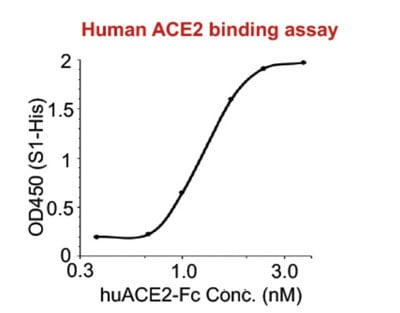
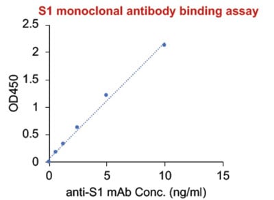
 Bioactivity
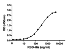
(Bioactivity & antigenicity: Strong binding ability with human ACE2 protein and binding capacity to a human anti-S1 monoclonal antibody (determined by ELISA).)
Bioactivity
(Bioactivity & antigenicity: Strong binding ability with human ACE2 protein and binding capacity to a human anti-S1 monoclonal antibody (determined by ELISA).)
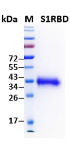
COVID 19 Spike 1 Receptor-Binding Domain (S1RBD) Coronavirus, Active Protein (Cat# AAA60538)
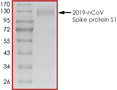
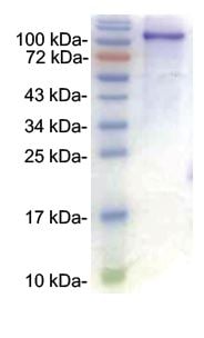
 Purity Info
(The purity was determined to be >80% by densitometry. Approx. MW 64kDa.)
Purity Info
(The purity was determined to be >80% by densitometry. Approx. MW 64kDa.)
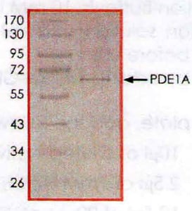
PDE1A, Active Protein (Cat# AAA73096)
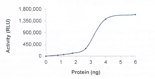
 Bioactivity
(HTRF assay for RPS6KA1/RSK1 activity 1 uM STK S1 substrate was incubated with different concentrations of RPS6KA1/RSK1 protein in 10 ul reaction system containing 1×Enzymatic Buffer, 5 mM MgCl2, 1 mM DTT, and 100 uM ATP for 1 hour. The detection reagents were added and incubated with the reactions for 30 min. All operations and reactions were performed at room temperature, and HTRF assay was used to detect the enzymatic activity.)
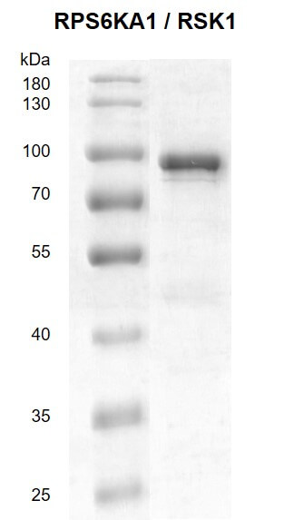
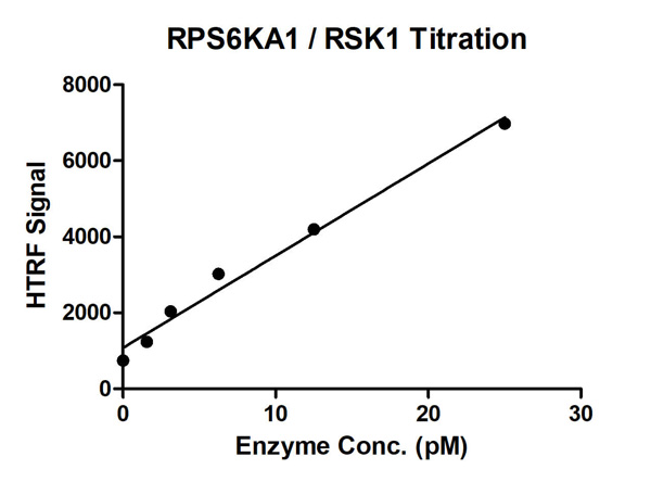
Bioactivity
(HTRF assay for RPS6KA1/RSK1 activity 1 uM STK S1 substrate was incubated with different concentrations of RPS6KA1/RSK1 protein in 10 ul reaction system containing 1×Enzymatic Buffer, 5 mM MgCl2, 1 mM DTT, and 100 uM ATP for 1 hour. The detection reagents were added and incubated with the reactions for 30 min. All operations and reactions were performed at room temperature, and HTRF assay was used to detect the enzymatic activity.)
RPS6KA1/RSK1, Active Protein (Cat# AAA60316)
 Application Data
(The purity of PDE1C was determined to be >90% by densitometry. Approx MW 99kDa)
Application Data
(The purity of PDE1C was determined to be >90% by densitometry. Approx MW 99kDa)
PDE1C, Active Protein (Cat# AAA73095)
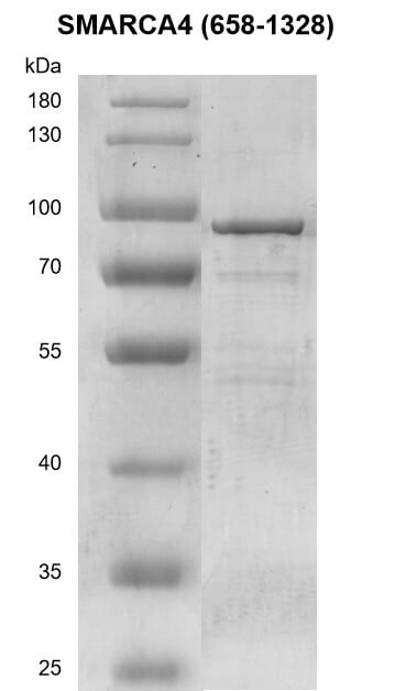
 SDS-PAGE
(SDS-PAGE Analysis - Lane 1: MW Marker, Lane 2: 1 ug Caspase-6.)
SDS-PAGE
(SDS-PAGE Analysis - Lane 1: MW Marker, Lane 2: 1 ug Caspase-6.)
Caspase-6, Active Protein (Cat# AAA77044)
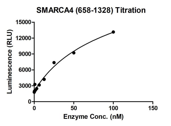 Bioactivity
(ADP-Glo assay for SMARCA4 (658-1328) activity (Data from ICE Bioscience Inc.) 100 uM ATP and 10 nM DNA was incubated with different concentrations of SMARCA4 (658-1328) protein in a 10 ul reaction system containing 20 mM HEPES pH 7.5,10 mM MgCl2, 50 mM NaCl, 0.1%Tween-20, 1 mM DTT for 1 hour, 10 ul ADP-Glo Reagent was added to the products and incubated for 1 hour. Then 20 ul Kinase Detection Reagent incubated for 1 hour. All the operations and reactions were performed at RT. Luminescence measurement is collected by BMG.)
Bioactivity
(ADP-Glo assay for SMARCA4 (658-1328) activity (Data from ICE Bioscience Inc.) 100 uM ATP and 10 nM DNA was incubated with different concentrations of SMARCA4 (658-1328) protein in a 10 ul reaction system containing 20 mM HEPES pH 7.5,10 mM MgCl2, 50 mM NaCl, 0.1%Tween-20, 1 mM DTT for 1 hour, 10 ul ADP-Glo Reagent was added to the products and incubated for 1 hour. Then 20 ul Kinase Detection Reagent incubated for 1 hour. All the operations and reactions were performed at RT. Luminescence measurement is collected by BMG.)
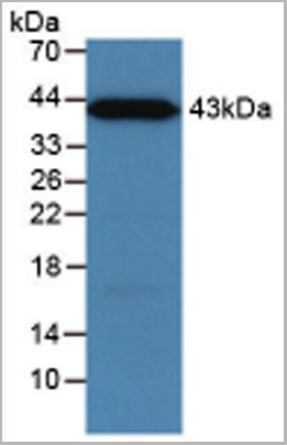
 SDS-PAGE
(3ug by SDS-PAGE under reducing condition and visualized by coomassie blue stain)
SDS-PAGE
(3ug by SDS-PAGE under reducing condition and visualized by coomassie blue stain)
BACE-1, Active Protein (Cat# AAA48325)
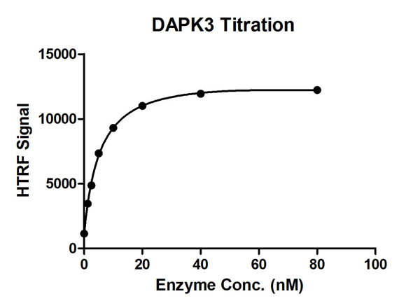
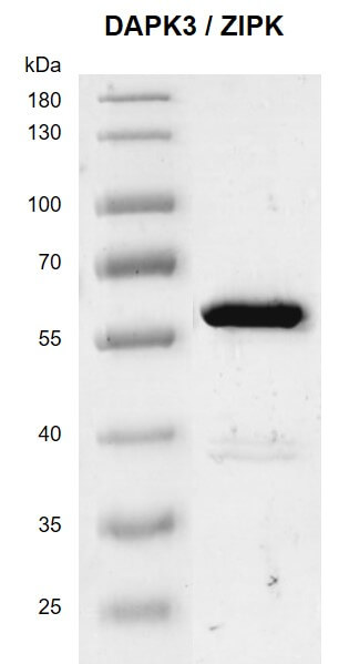
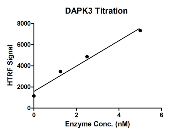 Bioactivity
(HTRF assay for DAPK3/ZIPK; activity 1 uM STK S1 substrate was incubated with different concentrations of DAPK3/ZIPK; protein in a 10 ul reaction system containing 1×Enzymatic Buffer, 10 mM MgCl2, 1 mM DTT and 100 uM ATP for 1 hour. The detection reagents were added and incubated with the reactions for 30 min. All the operations and reactions were performed at room temperature.)
Bioactivity
(HTRF assay for DAPK3/ZIPK; activity 1 uM STK S1 substrate was incubated with different concentrations of DAPK3/ZIPK; protein in a 10 ul reaction system containing 1×Enzymatic Buffer, 10 mM MgCl2, 1 mM DTT and 100 uM ATP for 1 hour. The detection reagents were added and incubated with the reactions for 30 min. All the operations and reactions were performed at room temperature.)
DAPK3/ZIPK, Active Protein (Cat# AAA60341)
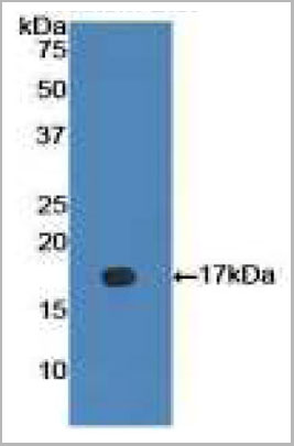
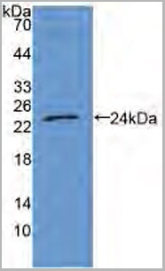
 WB (Western Blot)
(Western BlotSample: Recombinant IL1b, HumanAntibody: Rabbit Anti-Human IL1b Ab)
WB (Western Blot)
(Western BlotSample: Recombinant IL1b, HumanAntibody: Rabbit Anti-Human IL1b Ab)
Interleukin 1 Beta (IL1b), Active Protein (Cat# AAA21056)
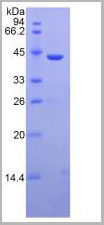
 WB (Western Blot)
(Figure 4. Western BlotSample: Recombinant MBL, Human;Antibody: Rabbit Anti-Human MBL Ab)
WB (Western Blot)
(Figure 4. Western BlotSample: Recombinant MBL, Human;Antibody: Rabbit Anti-Human MBL Ab)
Mannose Binding Lectin (MBL), Active Protein (Cat# AAA21059)
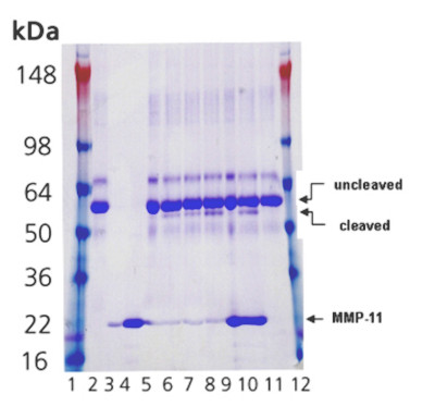 SDS-PAGE
(Coomassie-stained SDS-PAGE showing a timecourse of α1-antitrypsin (3ug) cleavage after incubation at 37°C with or without MMP-11 catalytic domain. Lanes are as follows: 1. Marker; 2. 1.5 ug α1-antitrypsin (0 hours); 3. 50 ng MMP-11, (0 hours); 4. 1 ug MMP-11, (0 hours); 5. 1.5 ug α1-antitrypsin incubated with 50 ng MMP-11 (0 hours); 6. 1.5 ug α1-antitrypsin incubated with 50 ng MMP-11 (3 hours); 7. 1.5 ug α1-antitrypsin incubated with 50 ng MMP-11 (8 hours); 8. 1.5 ug α1-antitrypsin incubated with 50 ng MMP-11 (24 hours); 9. 1.5 ug α1-antitrypsin incubated with 1 ug MMP-11 (0 hours); 10. 1.5 yg α1-antitrypsin incubated with 1 ug MMP-11 (24 hours); 11. 1.5 ug α1-antitrypsin (24 hours); 12. Marker.)
SDS-PAGE
(Coomassie-stained SDS-PAGE showing a timecourse of α1-antitrypsin (3ug) cleavage after incubation at 37°C with or without MMP-11 catalytic domain. Lanes are as follows: 1. Marker; 2. 1.5 ug α1-antitrypsin (0 hours); 3. 50 ng MMP-11, (0 hours); 4. 1 ug MMP-11, (0 hours); 5. 1.5 ug α1-antitrypsin incubated with 50 ng MMP-11 (0 hours); 6. 1.5 ug α1-antitrypsin incubated with 50 ng MMP-11 (3 hours); 7. 1.5 ug α1-antitrypsin incubated with 50 ng MMP-11 (8 hours); 8. 1.5 ug α1-antitrypsin incubated with 50 ng MMP-11 (24 hours); 9. 1.5 ug α1-antitrypsin incubated with 1 ug MMP-11 (0 hours); 10. 1.5 yg α1-antitrypsin incubated with 1 ug MMP-11 (24 hours); 11. 1.5 ug α1-antitrypsin (24 hours); 12. Marker.)
MMP-11, Active Protein (Cat# AAA77048)
Purified by multi-step chromatography
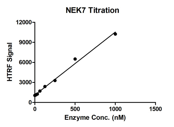 Bioactivity
(HTRF assay for NEK7 activity 1 uM STK S3 substrate was incubated with different concentrations of NEK7 protein in a 10 ul reaction system containing 1×Enzymatic Buffer, 5 mM MgCl2, 1 mM DTT, 100 uM ATP for 1 hour. The 10 ul detection reagents containing anti-STK antibody (1:2) and SA-XL665 (1:100) diluted with 1× Detection Buffer were added and incubated with the reactions for 30 min. All the operations and reactions were performed at room temperature. HTRF assay was used for detection.)
Bioactivity
(HTRF assay for NEK7 activity 1 uM STK S3 substrate was incubated with different concentrations of NEK7 protein in a 10 ul reaction system containing 1×Enzymatic Buffer, 5 mM MgCl2, 1 mM DTT, 100 uM ATP for 1 hour. The 10 ul detection reagents containing anti-STK antibody (1:2) and SA-XL665 (1:100) diluted with 1× Detection Buffer were added and incubated with the reactions for 30 min. All the operations and reactions were performed at room temperature. HTRF assay was used for detection.)
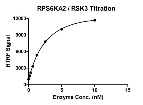
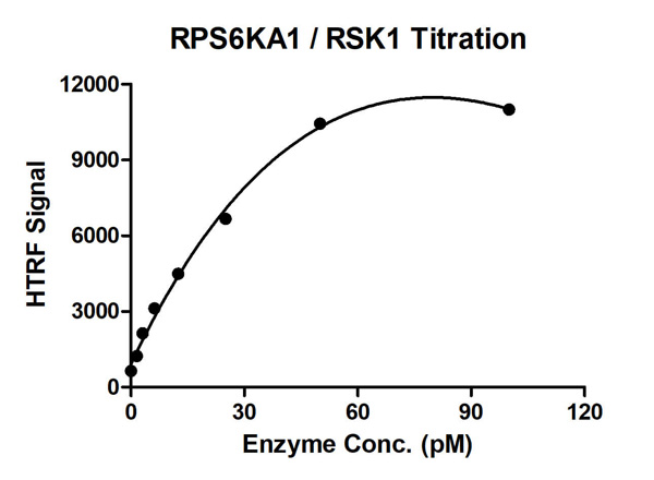
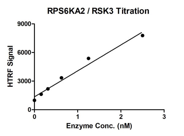 Bioactivity
(HTRF assay for RPS6KA2/RSK3 protein activity 1 uM STK S1 substrate was incubated with different concentrations of RPS6KA2/RSK3 protein in a 10 ul reaction system containing 1×Enzymatic Buffer, 5 mM MgCl2, 1 mM DTT, and 100 uM ATP for 1 hour. The 10 ul detection reagents containing anti-STK antibody (1:2) and SA-XL665 (1:100) diluted with 1× Detection Buffer were added and incubated with the reactions for 30 min. All the operations and reactions were performed at room temperature. HTRF assay was used for detection.)
Bioactivity
(HTRF assay for RPS6KA2/RSK3 protein activity 1 uM STK S1 substrate was incubated with different concentrations of RPS6KA2/RSK3 protein in a 10 ul reaction system containing 1×Enzymatic Buffer, 5 mM MgCl2, 1 mM DTT, and 100 uM ATP for 1 hour. The 10 ul detection reagents containing anti-STK antibody (1:2) and SA-XL665 (1:100) diluted with 1× Detection Buffer were added and incubated with the reactions for 30 min. All the operations and reactions were performed at room temperature. HTRF assay was used for detection.)
RPS6KA2/RSK3, Active Protein (Cat# AAA60321)
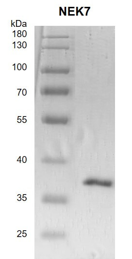
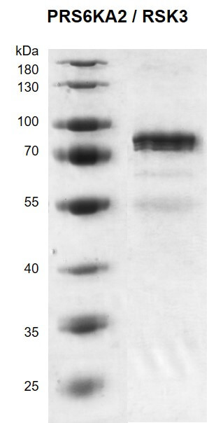
 SDS-PAGE
Glutathione S-Transferase (GST), Active Protein (Cat# AAA48483)


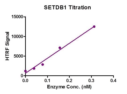 Bioactivity
(HTRF assay for Recombinant SETDB1 protein activity. 1 uM histone peptide H3 (1-21) was incubated with SETDB1 in reaction buffer including 50 mM Tris-HCl pH 8.6, 0.02% Triton X-100, 2 mM MgCl2, 1 mM TCEP and 50 uM SAM for 3 hours at room temperature. H3K9me2 antibody was used to detect reaction product.)
Bioactivity
(HTRF assay for Recombinant SETDB1 protein activity. 1 uM histone peptide H3 (1-21) was incubated with SETDB1 in reaction buffer including 50 mM Tris-HCl pH 8.6, 0.02% Triton X-100, 2 mM MgCl2, 1 mM TCEP and 50 uM SAM for 3 hours at room temperature. H3K9me2 antibody was used to detect reaction product.)
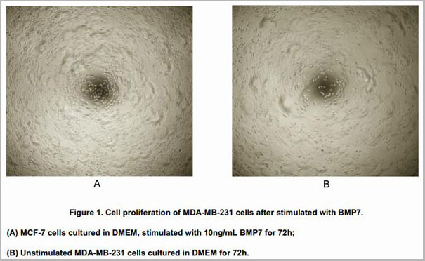
 WB (Western Blot)
(Sample: Recombinant BMP7, Mouse;Antibody: Rabbit Anti-Mouse BMP7 Ab)
WB (Western Blot)
(Sample: Recombinant BMP7, Mouse;Antibody: Rabbit Anti-Mouse BMP7 Ab)
Bone Morphogenetic Protein 7 (BMP7), Active Protein (Cat# AAA21057)
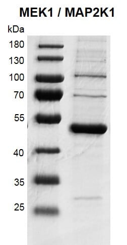
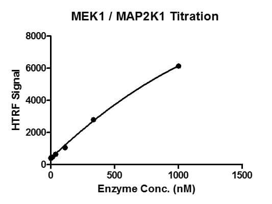 Bioactivity
(HTRF assay for MAP2K1 (MEK1) activity 1 uM TK substrate was incubated with different concentrations of MAP2K1 (MEK1) protein in 10 ul reaction system containing 1×Enzymatic Buffer, 5 mM MgCl2, 1 mM DTT, 5 nM SEB and 100 uM ATP for 1 hour. Then 10 ul detection reagents containing anti-TK antibody and SAXL665 (each of which was 1:100 diluted with 1× Detection Buffer) were added and incubated with the reactions for 30 min. All the operations and reactions were performed at room temperature. HTRF KinEASE TK assay was used to detect the enzymatic activity.)
Bioactivity
(HTRF assay for MAP2K1 (MEK1) activity 1 uM TK substrate was incubated with different concentrations of MAP2K1 (MEK1) protein in 10 ul reaction system containing 1×Enzymatic Buffer, 5 mM MgCl2, 1 mM DTT, 5 nM SEB and 100 uM ATP for 1 hour. Then 10 ul detection reagents containing anti-TK antibody and SAXL665 (each of which was 1:100 diluted with 1× Detection Buffer) were added and incubated with the reactions for 30 min. All the operations and reactions were performed at room temperature. HTRF KinEASE TK assay was used to detect the enzymatic activity.)
MAP2K1 (MEK1), Active Protein (Cat# AAA60313)
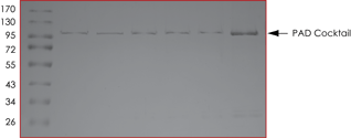 SDS-PAGE
(Purity: SDS-PAGE image of PAD Cocktail Proteins. The five PAD isoform proteins from left to right are: PAD1, PAD4, PAD3, PAD6 and PAD2. The far-right lane contains PAD Cocktail.)
SDS-PAGE
(Purity: SDS-PAGE image of PAD Cocktail Proteins. The five PAD isoform proteins from left to right are: PAD1, PAD4, PAD3, PAD6 and PAD2. The far-right lane contains PAD Cocktail.)
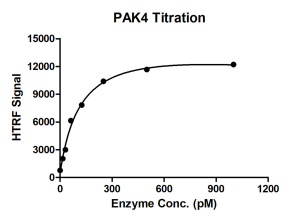
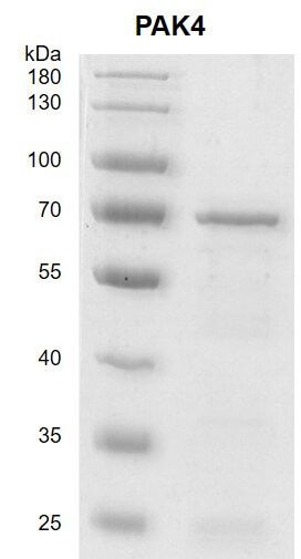
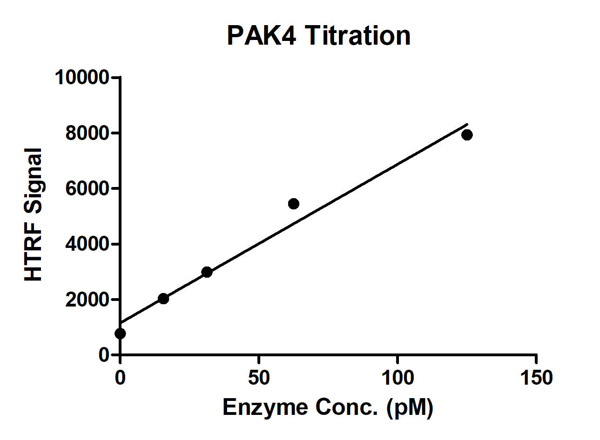 Bioactivity
(HTRF Assay for Recombinant PAK4 activity. 1 uM STK S2 substrate was incubated with different concentrations of PAK4 protein in 10 ul reaction system containing 1×Enzymatic Buffer, 5 mM MgCl2, 1 mM DTT and 100 uM ATP for 1 hour. The 10 ul detection reagents containing anti-STK antibody (1:2) and SA-XL665 (1:100) diluted with 1× Detection Buffer were added and incubated with the reactions for 30 min. All the operations and reactions were performed at room temperature. HTRF assay was used for detection.)
Bioactivity
(HTRF Assay for Recombinant PAK4 activity. 1 uM STK S2 substrate was incubated with different concentrations of PAK4 protein in 10 ul reaction system containing 1×Enzymatic Buffer, 5 mM MgCl2, 1 mM DTT and 100 uM ATP for 1 hour. The 10 ul detection reagents containing anti-STK antibody (1:2) and SA-XL665 (1:100) diluted with 1× Detection Buffer were added and incubated with the reactions for 30 min. All the operations and reactions were performed at room temperature. HTRF assay was used for detection.)

 Bioactivity
 WB (Western Blot)
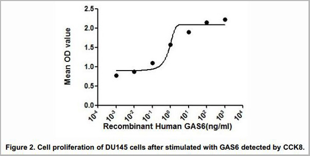
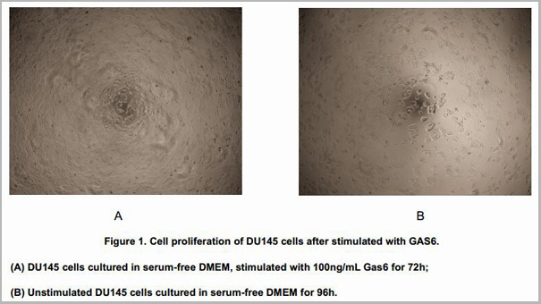
(Sample: Recombinant GAS6, Human;Antibody: Rabbit Anti-Human GAS6 Ab)
WB (Western Blot)
(Sample: Recombinant GAS6, Human;Antibody: Rabbit Anti-Human GAS6 Ab)
Growth Arrest Specific Protein 6 (GAS6), Active Protein (Cat# AAA21055)
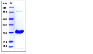 SDS-PAGE
UCHL1, Active Protein (Cat# AAA257988)
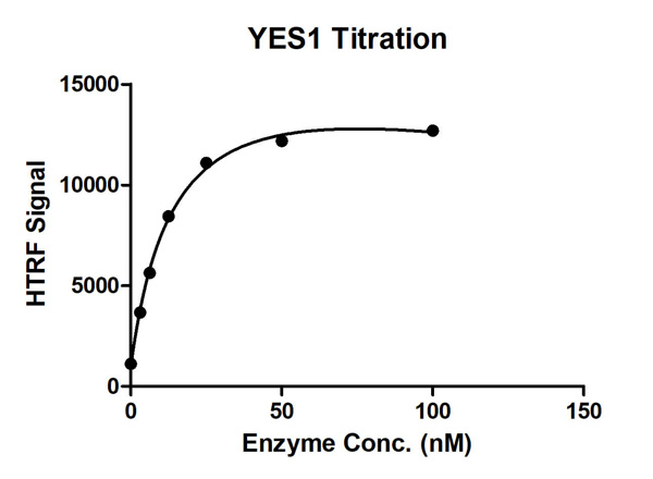
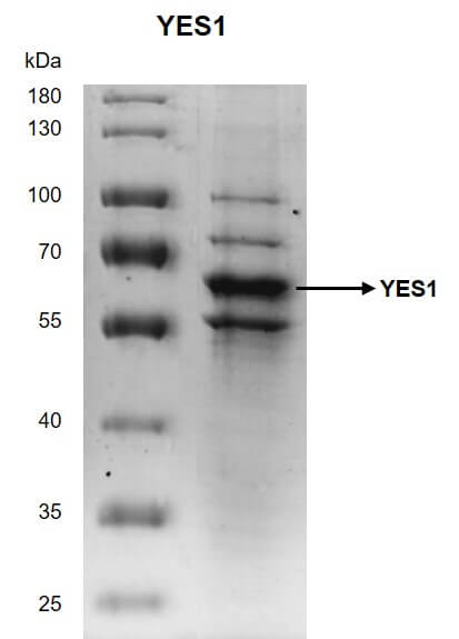
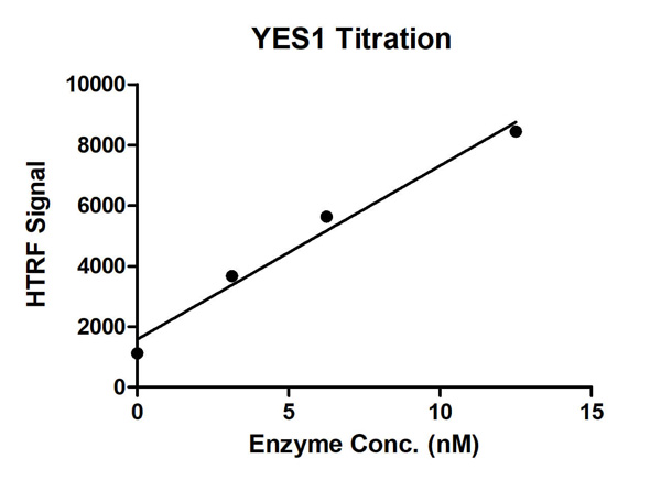 Bioactivity
(HTRF Assay for Recombinant YES1 activity. 1 uM TK substrate was incubated with different concentrations of YES1 protein in 10 ul reaction system containing 1×Enzymatic Buffer, 5 mM MgCl2, 1 mM DTT and 100 uM ATP for 1 hour. The 10 ul detection reagents containing anti-TK antibody (1:2) and SA-XL665 (1:100) diluted with 1× Detection Buffer were added and incubated with the reactions for 30 min. All the operations and reactions were performed at room temperature. HTRF assay was used for detection.)
Bioactivity
(HTRF Assay for Recombinant YES1 activity. 1 uM TK substrate was incubated with different concentrations of YES1 protein in 10 ul reaction system containing 1×Enzymatic Buffer, 5 mM MgCl2, 1 mM DTT and 100 uM ATP for 1 hour. The 10 ul detection reagents containing anti-TK antibody (1:2) and SA-XL665 (1:100) diluted with 1× Detection Buffer were added and incubated with the reactions for 30 min. All the operations and reactions were performed at room temperature. HTRF assay was used for detection.)
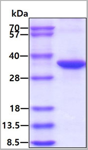
 Purity
(The purity of PDE1B was determined to be >90% by densitometry. Approx. MW 86kDa)
Purity
(The purity of PDE1B was determined to be >90% by densitometry. Approx. MW 86kDa)
PDE1B, Active Protein (Cat# AAA73094)
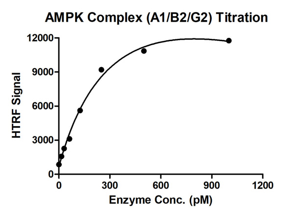
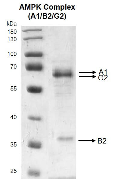
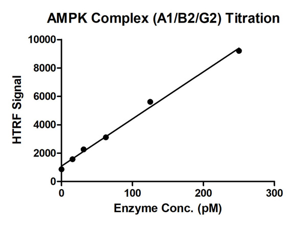 Bioactivity
(HTRF assay for AMPK Complex (A1+B2+G2) activity 1 uM STK S1 substrate was incubated with different concentrations of AMPK Complex (A1+B2+G2) protein in a 10 ul reaction system containing 1×Enzymatic Buffer, 5 mM MgCl2, 1 mM DTT, 50 uM AMP and 100 uM ATP for 1 hr. The 10 ul detection reagents containing anti-STK antibody (1:100) and SA-XL665 (1:100) diluted with 1× Detection Buffer were added and incubated with the reactions for 30 min. All the operations and reactions were performed at room temperature. HTRF assay was used for detection.)
Bioactivity
(HTRF assay for AMPK Complex (A1+B2+G2) activity 1 uM STK S1 substrate was incubated with different concentrations of AMPK Complex (A1+B2+G2) protein in a 10 ul reaction system containing 1×Enzymatic Buffer, 5 mM MgCl2, 1 mM DTT, 50 uM AMP and 100 uM ATP for 1 hr. The 10 ul detection reagents containing anti-STK antibody (1:100) and SA-XL665 (1:100) diluted with 1× Detection Buffer were added and incubated with the reactions for 30 min. All the operations and reactions were performed at room temperature. HTRF assay was used for detection.)
AMPK Complex (A1+B2+G2), Active Protein (Cat# AAA60318)
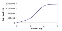
 Bioactivity
(Sample Activity Plot.)
Bioactivity
(Sample Activity Plot.)
COVID 19 Spike S1 Coronavirus, Active Protein (Cat# AAA73161)
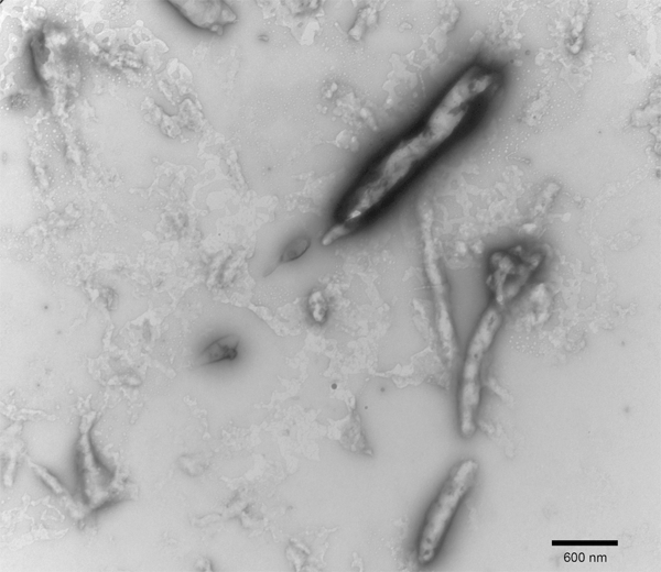
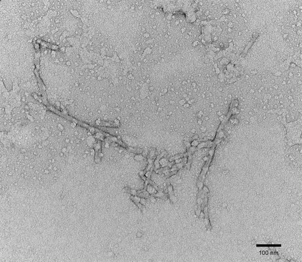
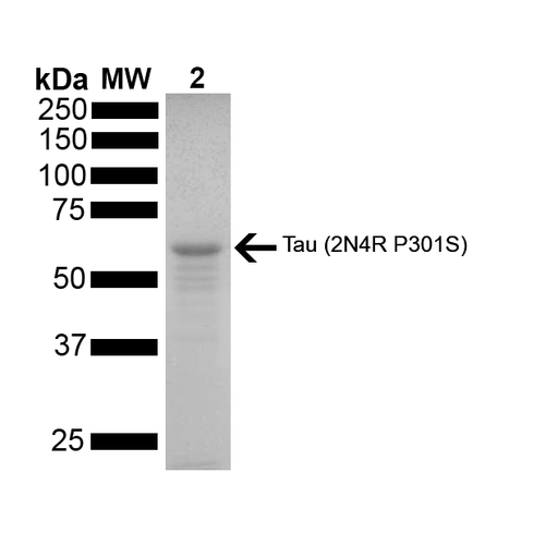
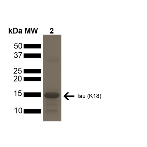 SDS_PAGE
(SDS-PAGE of ~15 kDa Active Human Tau Protein K18 P301L Preformed Fibrils. Lane 1: MW Ladder. Lane 2: Tau Protein Preformed Fibrils.)
SDS_PAGE
(SDS-PAGE of ~15 kDa Active Human Tau Protein K18 P301L Preformed Fibrils. Lane 1: MW Ladder. Lane 2: Tau Protein Preformed Fibrils.)
Tau, Active Protein (Cat# AAA253968)
Ion-Exchange Purified
HVEM-Fc, Active Protein (Cat# AAA14394)
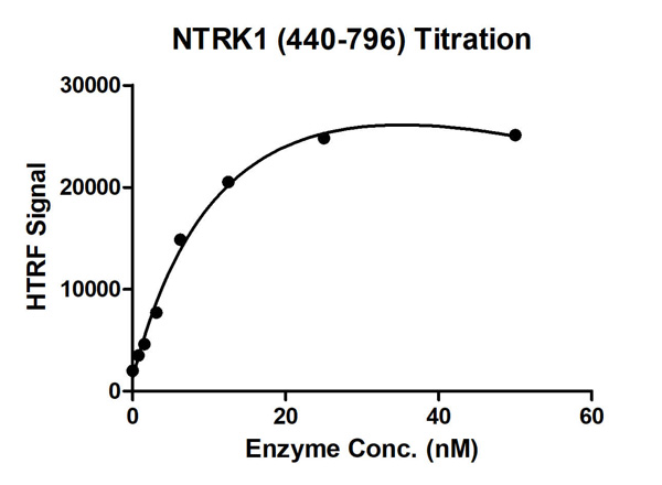
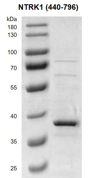
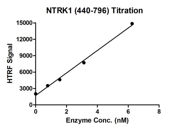 Bioactivity
(HTRF assay for NTRK1 (440-796) activity 1 uM TK substrate was incubated with different concentrations of NTRK1 (440-796) protein in a 10 ul reaction system containing 1×Enzymatic Buffer, 5 mM MgCl2, 1 mM DTT, and 100 uM ATP for 1 hour. Then 10 ul detection reagents containing anti-TK antibody (1:2) and SA-XL665 (1:100) diluted with 1× Detection Buffer were added and incubated with the reactions for 30 min. All the operations and reactions were performed at room temperature. HTRF assay was used for detection.)
Bioactivity
(HTRF assay for NTRK1 (440-796) activity 1 uM TK substrate was incubated with different concentrations of NTRK1 (440-796) protein in a 10 ul reaction system containing 1×Enzymatic Buffer, 5 mM MgCl2, 1 mM DTT, and 100 uM ATP for 1 hour. Then 10 ul detection reagents containing anti-TK antibody (1:2) and SA-XL665 (1:100) diluted with 1× Detection Buffer were added and incubated with the reactions for 30 min. All the operations and reactions were performed at room temperature. HTRF assay was used for detection.)
NTRK1, Active Protein (Cat# AAA60324)
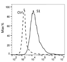
 Application Data
COVID 19 Spike S1 Coronavirus, Active Protein (Cat# AAA60537)
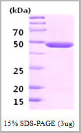
 SDS-PAGE
(3ug by SDS-PAGE under reducing condition and visualized by coomassie blue stain.)
SDS-PAGE
(3ug by SDS-PAGE under reducing condition and visualized by coomassie blue stain.)
NQO1, Active Protein (Cat# AAA11735)
GDF11, Active Protein (Cat# AAA14381)
IFN ALPHA, Active Protein (Cat# AAA12301)
 SDS-PAGE
(3ug by SDS-PAGE under reducing condition and visualized by coomasie blue stain)
SDS-PAGE
(3ug by SDS-PAGE under reducing condition and visualized by coomasie blue stain)
PTP-1B (Protein Tyrosine Phosphatase1B), Active Protein (Cat# AAA11740)
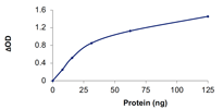
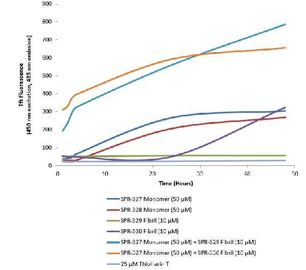
 Application Data
(Thioflavin T is a fluorescent dye that binds to beta sheet-rich structures, such as those in tau fibrils. Upon binding, the emission spectrum of the dye experiences a red-shift and increased fluorescence intensity. Thioflavin T emission curves show increased fluorescence (correlated to tau aggregation) over time in tau monomers. A greater increase in fluorescence is seen when 50 uM monomer is combined with 10 uM PFFs, as the fibrils seed the formation of new fibrils from the pool of monomers. Thioflavin T ex = 450 nm, em = 485 nm.)
Application Data
(Thioflavin T is a fluorescent dye that binds to beta sheet-rich structures, such as those in tau fibrils. Upon binding, the emission spectrum of the dye experiences a red-shift and increased fluorescence intensity. Thioflavin T emission curves show increased fluorescence (correlated to tau aggregation) over time in tau monomers. A greater increase in fluorescence is seen when 50 uM monomer is combined with 10 uM PFFs, as the fibrils seed the formation of new fibrils from the pool of monomers. Thioflavin T ex = 450 nm, em = 485 nm.)
Tau441, Active Protein (Cat# AAA253967)
Ion-Exchange Purified
CD22, Active Protein (Cat# AAA14393)

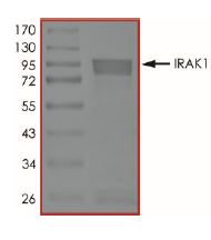 SDS-PAGE
(The purity was determined to be >85% by densitometry. Approx. MW 85-100 kDa.)
SDS-PAGE
(The purity was determined to be >85% by densitometry. Approx. MW 85-100 kDa.)
IRAK1, Active Protein (Cat# AAA73158)

 Bioactivity
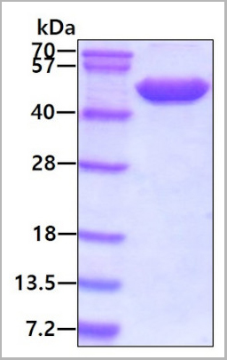 SDS-PAGE
(3ug by SDS-PAGE under reducing condition and visualized by coomassie blue stain)
SDS-PAGE
(3ug by SDS-PAGE under reducing condition and visualized by coomassie blue stain)
Neuron-Specific Enolase (NSE), Active Protein (Cat# AAA11754)
Neuregulin-4, Active Protein (Cat# AAA14428)
IL-11 receptor alpha, Active Protein (Cat# AAA14431)
Peroxiredoxin-1, Active Protein (Cat# AAA10855)
super leptin antagonist, Active Protein (Cat# AAA13623)
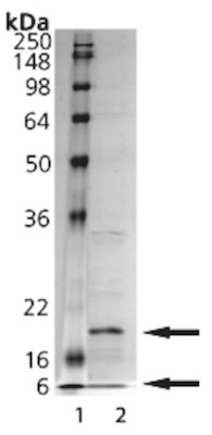
(a) Gel filtration analysis.
(b) Analysis by reducing and non-reducing SDS-PAGE gel.
West Nile Virus E, Active Protein (Cat# AAA24010)
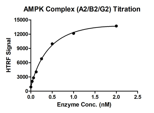

 Bioactivity
(HTRF assay for AMPK Complex (A2+B2+G2) activity 1 uM STK S1 substrate was incubated with different concentrations of AMPK Complex (A2+B2+G2) protein in a 10 ul reaction system containing 1×Enzymatic Buffer, 5 mM MgCl2, 1 mM DTT, 50 uM AMP and 100 uM ATP for 1 hr. The 10 ul detection reagents containing anti-STK antibody (1:100) and SA-XL665 (1:100) diluted with 1× Detection Buffer were added and incubated with the reactions for 30 min. All the operations and reactions were performed at room temperature. HTRF assay was used for detection.)
Bioactivity
(HTRF assay for AMPK Complex (A2+B2+G2) activity 1 uM STK S1 substrate was incubated with different concentrations of AMPK Complex (A2+B2+G2) protein in a 10 ul reaction system containing 1×Enzymatic Buffer, 5 mM MgCl2, 1 mM DTT, 50 uM AMP and 100 uM ATP for 1 hr. The 10 ul detection reagents containing anti-STK antibody (1:100) and SA-XL665 (1:100) diluted with 1× Detection Buffer were added and incubated with the reactions for 30 min. All the operations and reactions were performed at room temperature. HTRF assay was used for detection.)
AMPK Complex (A2+B2+G2), Active Protein (Cat# AAA60320)
CD32a (FcgRIIa), Active Protein (Cat# AAA14417)
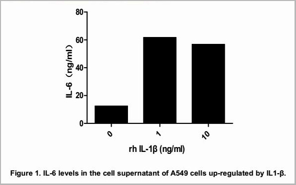
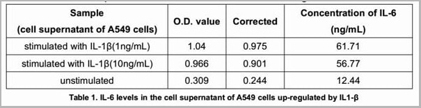
Interleukin 6, Active Protein (Cat# AAA14805)
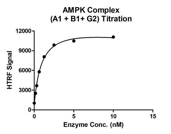

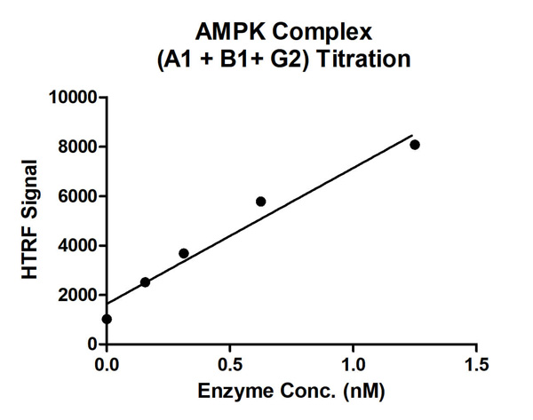 Bioactivity
(HTRF assay for AMPK Complex (A1+B1+G2) activity 1 uM STK S1 substrate was incubated with different concentrations of AMPK Complex (A1+B1+G2) protein in a 10 ul reaction system containing 1×Enzymatic Buffer, 5 mM MgCl2, 1 mM DTT, 50 uM AMP and 100 uM ATP for 1 hr. The 10 ul detection reagents containing anti-STK antibody (1:100) and SA-XL665 (1:100) diluted with 1× Detection Buffer were added and incubated with the reactions for 30 min. All the operations and reactions were performed at room temperature. HTRF assay was used for detection.)
Bioactivity
(HTRF assay for AMPK Complex (A1+B1+G2) activity 1 uM STK S1 substrate was incubated with different concentrations of AMPK Complex (A1+B1+G2) protein in a 10 ul reaction system containing 1×Enzymatic Buffer, 5 mM MgCl2, 1 mM DTT, 50 uM AMP and 100 uM ATP for 1 hr. The 10 ul detection reagents containing anti-STK antibody (1:100) and SA-XL665 (1:100) diluted with 1× Detection Buffer were added and incubated with the reactions for 30 min. All the operations and reactions were performed at room temperature. HTRF assay was used for detection.)
AMPK Complex (A1+B1+G2), Active Protein (Cat# AAA60344)
Transferrin, Active Protein (Cat# AAA10866)
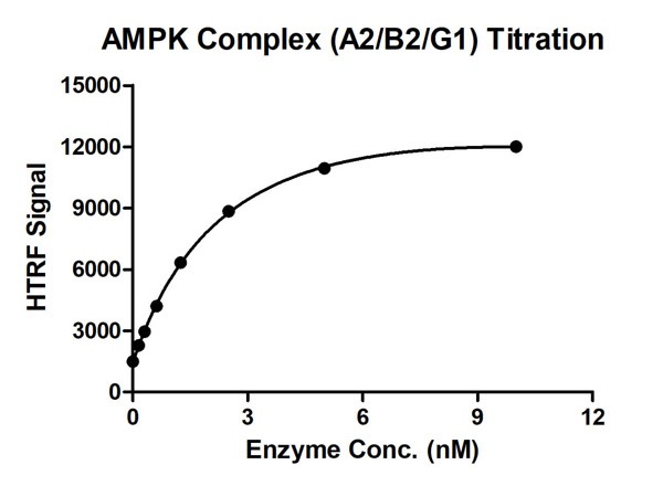
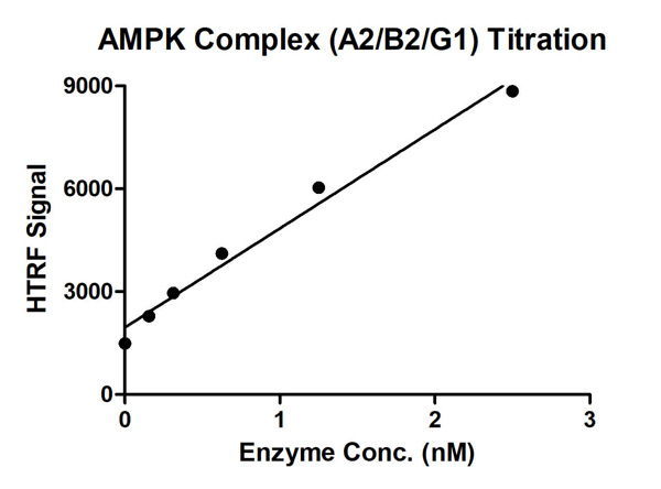 Bioactivity
(HTRF assay for AMPK Complex (A2+B2+G1) activity 1 uM STK S1 substrate was incubated with different concentrations of AMPK Complex (A2+B2+G1) protein in a 10 ul reaction system containing 1×Enzymatic Buffer, 5 mM MgCl2, 1 mM DTT, 50 uM AMP and 100 ul ATP for 1 hr. The 10 ul detection reagents containing anti-STK antibody (1:100) and SA-XL665 (1:100) diluted with 1× Detection Buffer were added and incubated with the reactions for 30 min. All the operations and reactions were performed at room temperature. HTRF assay was used for detection.)
Bioactivity
(HTRF assay for AMPK Complex (A2+B2+G1) activity 1 uM STK S1 substrate was incubated with different concentrations of AMPK Complex (A2+B2+G1) protein in a 10 ul reaction system containing 1×Enzymatic Buffer, 5 mM MgCl2, 1 mM DTT, 50 uM AMP and 100 ul ATP for 1 hr. The 10 ul detection reagents containing anti-STK antibody (1:100) and SA-XL665 (1:100) diluted with 1× Detection Buffer were added and incubated with the reactions for 30 min. All the operations and reactions were performed at room temperature. HTRF assay was used for detection.)
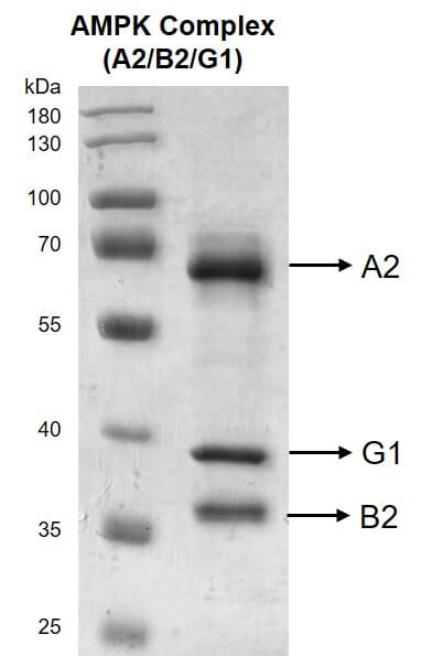
AMPK Complex (A2+B2+G1), Active Protein (Cat# AAA60319)
What Are Active Proteins?
Proteins are large molecules made up of long chains of amino acids.
They will typically fold into a very particular 3-dimensional shape/conformation, that is sometimes referred to as their “native” form, which allows them to work properly in the body. For the purposes of product categorization, AAA Biotech will typically refer to proteins purified from their original animal host as being “native” proteins (this is to signify their difference compared to their “recombinant” or “synthetic” protein counterparts).
If a protein successfully folds into the correct shape, it is will typically display high fidelity characteristics to its original protein in its original animal host, and be classified as an active protein, as it will be able to function “normally” in most enzymatic or binding capacities. If it loses this shape, due to factors such as heat or strong chemicals (such as detergents), it becomes inactive and is no longer able to perform its basic functions. All of the proteins in this category are made under strict quality control, and they are active, pure, low in contaminants, and stable.
Most are stored as freeze-dried powders and come without extra tags, so they’re very close to the actual natural/native form.
Key Applications of Active Proteins
1. Scientific Research
- Aid in the study of how proteins function in the body
- Aid in understanding various disease processes
2. Drug Development
- Powerful tools to investigate how potential drugs interact with specific proteins
- Ideal for identifying drug targets
3. Cell Culture
- Are routinely utilized to support cell growth and function (e.g., using exogenous growth factors)
- Can be used to promote cellular development into specific types (differentiation)
4. Diagnostics
- Regularly utilized in tests to detect diseases or infections (e.g., COVID-19, cancer)
- Note: All products are strictly for research-use only (RUO).
5. Therapeutics
- Some active proteins are used directly as treatments (e.g., insulin, enzymes)
- Note: All products are strictly for research-use only (RUO).
6. Vaccine Development
- Used to create or test vaccines by mimicking parts of viruses or bacteria
7. Biochemical Assays
- They can facilitate the characterization of enzyme activity, binding strength, or protein interactions in lab tests
Why Buy Active Proteins from AAA Biotech?
- High biological activity – Verified to perform as expected or indicated on datasheet
- Strict quality control – We are confident in our active proteins’ reliability and consistency
- High purity & low endotoxin – Ideal for applications involving sensitive or precious samples/components
- Freeze-dried for stability – Long shelf life and straightforward storage
- Mostly tag-free – Closer to natural/native protein form
FAQ
1. What are active proteins used for in research?
Active proteins are used primarily in the study of how proteins function, in characterizing/discovering drug interactions, supporting cell growth, running biochemical assays, and in development of diagnostics or therapeutics.
2. How are AAA Biotech's active proteins validated?
AAA Biotech’s active proteins are validated through strict quality control and functional assays to ensure they are properly folded and active. “Active”, though, can be an ambiguous term, so if a specific “activity” or “binding” capability of a protein is of crucial interest to you, please inquire with us prior to purchase, and we will provide further details on how the “Active” modifier was determined to be applicable.
3. Are these proteins tested for biological activity?
Yes, all active proteins from AAA Biotech are tested to confirm they have the expected biological activity before being offered for use. Though, said “biological activity” can be either “enzymatic”, “binding”, or both.