Filters
▼Clonality
▼Type
▼Reactivity
▼Gene Name
▼Isotype
▼Host
▼Application
▼Clone
▼Active Proteins
AAA Biotech also known as AAA Bio or AAABio provides a variety of high-quality recombinant and natural/native proteins that are proven to work in a wide range of experiments. Explore our products to find the active protein that best fits your needs or experimental model.
Viewing 2250-2300 of 2567 product results
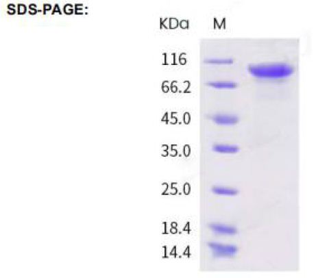
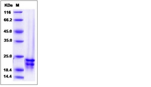
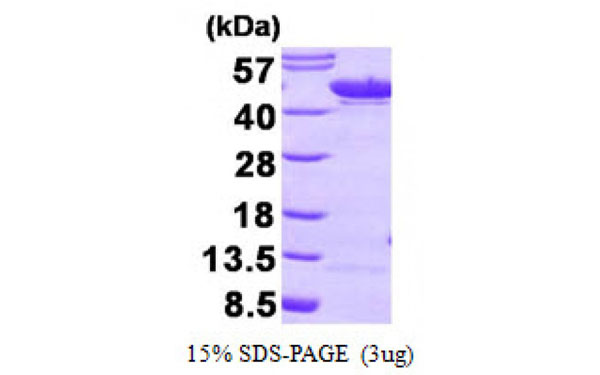 SDS-PAGE
(Figure annotation denotes ug of protein loaded and % gel used.)
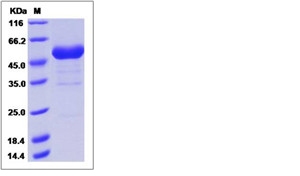
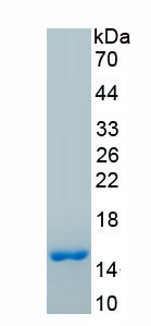
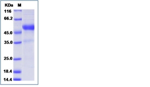
SDS-PAGE
(Figure annotation denotes ug of protein loaded and % gel used.)
Alphaenolase, Active Protein (Cat# AAA75217)
Beta Defensin -3, Active Protein (Cat# AAA38981)
(a) Analysis by RP-HPLC.
(b) Analysis by SDS-PAGE.
Indian Hedgehog, Active Protein (Cat# AAA39005)
Eotaxin, Active Protein (Cat# AAA39096)
(a) Analysis by RP-HPLC
(b) Analysis by SDS-PAGE
Glutamic-Oxaloacetic Transaminase 1, Active Protein (Cat# AAA39141)
Carboxylesterase 1D, Active Protein (Cat# AAA39142)
Fibrinogen, Active Protein (Cat# AAA37797)
Superoxide Dismutase-2, Active Protein (Cat# AAA38823)
Transforming Growth Factor-Beta 1, Active Protein (Cat# AAA38647)
Artemin, Active Protein (Cat# AAA75568)
Stromal Cell-Derived Factor-1 alpha, Active Protein (Cat# AAA38080)
GRO/KC, Active Protein (Cat# AAA38092)
Interleukin-1 beta, Active Protein (Cat# AAA38108)
Persephin, Active Protein (Cat# AAA39062)
(a) Analysis by RP-HPLC.
(b) Analysis by SDS-PAGE.
Glutathione S-Transferase Alpha 4, Active Protein (Cat# AAA39148)
Cathepsin-B, Active Protein (Cat# AAA39158)
Interleukin-15, Active Protein (Cat# AAA39137)
Peroxiredoxin-4, Active Protein (Cat# AAA38812)
Cathepsin G, Active Protein (Cat# AAA44787)
Lactate Dehydrogenase 3, Active Protein (Cat# AAA44811)
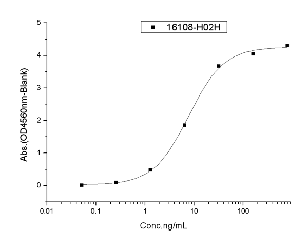 Application Data
(Measured by its binding ability in a functional ELISA. Immobilized Human RSPO3 His at 2 ug/ml (100 ul/well) can bind Human RNF43 hFc, the EC50 of Human RNF43 hFc is 3.0-12.0 ng/mL)
Application Data
(Measured by its binding ability in a functional ELISA. Immobilized Human RSPO3 His at 2 ug/ml (100 ul/well) can bind Human RNF43 hFc, the EC50 of Human RNF43 hFc is 3.0-12.0 ng/mL)
RNF43, Active Protein (Cat# AAA258047)
 Bioactivity
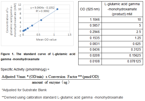
(Transglutaminase 2 (TGM2), encoded by the TGM2 gene, is belongs to the family of transglutaminases that catalyze the posttranslational modification of proteins via calcium dependent cross-linking reactions. In addition to its function in protein cross-linking, TGM2 is also capable of hydrolyzing both GTP and ATP and has intrinsic kinase activity. TGM2 has been implicated in a variety of human diseases including celiac disease, inclusion body myositis, atherosclerosis, and neurodegenerative diseases. The activity of recombinant human TGM2 is measured by its ability to cleave a synthetic peptide Benzyloxycarbonyl-Gln-Gly and NH2OH in the assay buffer 200 mM MES, 10 mM DTT, 10 mM CaCl2, 100 mM Hydroxylamine Hydrochloride, pH 6.0. The rhTGM2 is diluted to 12.5 ug/ml in assay buffer. Loading into a clear well plate 50 uL of 12.5 ug/mL rhTGM2 and start the reaction by adding 50 uL of 100 mM substrate, with a substrate blank containing 50 uL assay buffer, 50 uL substrate, and no rhTGM2. Incubated at 37 degree C for 2 hours and stop the reaction with 400 ul stop solution of 0.37 M FeCl3, 0.67 M HCl, 0.2 M Trichloroacetic Acid. Centrifuge at 2000 rpm for 2 minutes and then load 200 ul of the supernatant into a plate and read at 525 nm (absorbance) in endpoint mode. The specific activity of recombinant human TGM2 is > 800 pmol/min/ug.)
Bioactivity
(Transglutaminase 2 (TGM2), encoded by the TGM2 gene, is belongs to the family of transglutaminases that catalyze the posttranslational modification of proteins via calcium dependent cross-linking reactions. In addition to its function in protein cross-linking, TGM2 is also capable of hydrolyzing both GTP and ATP and has intrinsic kinase activity. TGM2 has been implicated in a variety of human diseases including celiac disease, inclusion body myositis, atherosclerosis, and neurodegenerative diseases. The activity of recombinant human TGM2 is measured by its ability to cleave a synthetic peptide Benzyloxycarbonyl-Gln-Gly and NH2OH in the assay buffer 200 mM MES, 10 mM DTT, 10 mM CaCl2, 100 mM Hydroxylamine Hydrochloride, pH 6.0. The rhTGM2 is diluted to 12.5 ug/ml in assay buffer. Loading into a clear well plate 50 uL of 12.5 ug/mL rhTGM2 and start the reaction by adding 50 uL of 100 mM substrate, with a substrate blank containing 50 uL assay buffer, 50 uL substrate, and no rhTGM2. Incubated at 37 degree C for 2 hours and stop the reaction with 400 ul stop solution of 0.37 M FeCl3, 0.67 M HCl, 0.2 M Trichloroacetic Acid. Centrifuge at 2000 rpm for 2 minutes and then load 200 ul of the supernatant into a plate and read at 525 nm (absorbance) in endpoint mode. The specific activity of recombinant human TGM2 is > 800 pmol/min/ug.)
Transglutaminase 2 (TGM2), Active Protein (Cat# AAA161864)
Creatine Kinase MM (CK-MM) >95% Pure, Active Protein (Cat# AAA58215)
 Application Data
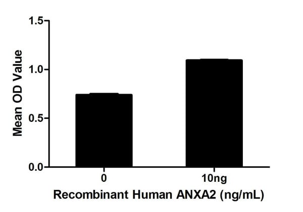
(Figure. Cell proliferation of Hela cells after stimulated with ANXA2.)
Application Data
(Figure. Cell proliferation of Hela cells after stimulated with ANXA2.)
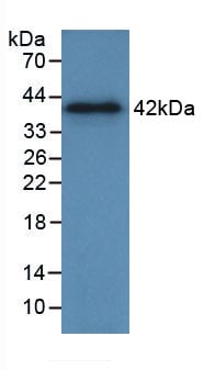
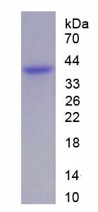
Annexin A2, Active Protein (Cat# AAA150121)
 Bioactivity
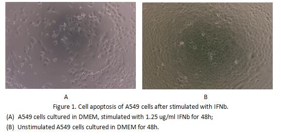
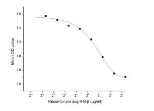
(Figure 2. Cell apoptosis of A549 cells after stimulated with IFNb.)
Bioactivity
(Figure 2. Cell apoptosis of A549 cells after stimulated with IFNb.)
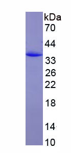
Interferon Beta (IFNb), Active Protein (Cat# AAA153003)
 Application Data
COVID 19 Spike S1 Coronavirus, Active Protein (Cat# AAA268895)
Adiponectin glycosilated, Active Protein (Cat# AAA38203)
Aeromonas Aminopeptidase, Active Protein (Cat# AAA38247)
Matrix Metalloproteinase-2, Active Protein (Cat# AAA39014)
Growth and Differentiation factor 7, Active Protein (Cat# AAA37990)
Phosphoglucomutase, Active Protein (Cat# AAA44730)
 Bioactivity
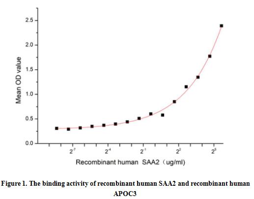
(Serum Amyloid A2 (SAA2) is a multifunctional apolipoprotein that belongs to SAA family. SAA2 is produced by hepatocytes in response to pro-inflammatory cytokines and is the most prominent members of the acute phase response (APR) during which their serum levels rise dramatically after trauma, infection and other stimuli. Besides, Apolipoprotein C3 (APOC3) has been identified as an interactor of SAA2, thus a functional binding ELISA assay was conducted to detect the interaction of recombinant human SAA2 and recombinant human APOC3. Briefly, SAA2 were diluted serially in PBS, with 0.01% BSA (pH 7.4). Duplicate samples of 100 ul were then transferred to APOC3-coated microtiter wells and incubated for 2h at 37 degree C. Wells were washed with PBST and incubated for 1h with anti-SAA2 pAb, then aspirated and washed 3 times. After incubation with HRP labelled secondary antibody, wells were aspirated and washed 5 times. With the addition of substrate solution, wells were incubated 15-25 minutes at 37 degree C. Finally, add 50 uL stop solution to the wells and read at 450 nm immediately. The binding activity of recombinant human SAA2 and recombinant human APOC3 was shown in Figure 1, and this effect was in a dose dependent manner.)
Bioactivity
(Serum Amyloid A2 (SAA2) is a multifunctional apolipoprotein that belongs to SAA family. SAA2 is produced by hepatocytes in response to pro-inflammatory cytokines and is the most prominent members of the acute phase response (APR) during which their serum levels rise dramatically after trauma, infection and other stimuli. Besides, Apolipoprotein C3 (APOC3) has been identified as an interactor of SAA2, thus a functional binding ELISA assay was conducted to detect the interaction of recombinant human SAA2 and recombinant human APOC3. Briefly, SAA2 were diluted serially in PBS, with 0.01% BSA (pH 7.4). Duplicate samples of 100 ul were then transferred to APOC3-coated microtiter wells and incubated for 2h at 37 degree C. Wells were washed with PBST and incubated for 1h with anti-SAA2 pAb, then aspirated and washed 3 times. After incubation with HRP labelled secondary antibody, wells were aspirated and washed 5 times. With the addition of substrate solution, wells were incubated 15-25 minutes at 37 degree C. Finally, add 50 uL stop solution to the wells and read at 450 nm immediately. The binding activity of recombinant human SAA2 and recombinant human APOC3 was shown in Figure 1, and this effect was in a dose dependent manner.)
Serum Amyloid A2 (SAA2), Active Protein (Cat# AAA161860)
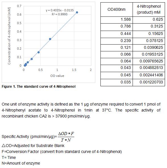 Bioactivity
(Carbonic Anhydrase (CA) catalyzes the reversible reaction of CO2 H2O = HCO3- H, which is fundamental to many processes such as respiration, renal tubular acidification and bone resorption. CA2 is a cytosolic enzyme with the highest activity among all known CAs. Mutations in the CA2 gene result in the CA II deficiency syndrome, an autosomal recessive disorder that produces osteopetrosis, renal tubular acidosis and cerebral calcification. The activity of recombinant chicken CA2 was measured by its ability to hydrolyze 4-Nitrophenyl acetate (4-NPA) to 4-Nitrophenol. The reaction was performed in 12.5 mM Tris, 75 mM NaCl, pH 7.5 (assay buffer), initiated by addition 50 uL of various concentrations of CA2 (diluted by assay buffer) to 50 uL of 2 mM substrate 4-NPA (100 mM stock in Acetone, diluted by assay buffer). Incubated at 37 degree C for 5min, then read at a wavelength of 400 nm.)
Bioactivity
(Carbonic Anhydrase (CA) catalyzes the reversible reaction of CO2 H2O = HCO3- H, which is fundamental to many processes such as respiration, renal tubular acidification and bone resorption. CA2 is a cytosolic enzyme with the highest activity among all known CAs. Mutations in the CA2 gene result in the CA II deficiency syndrome, an autosomal recessive disorder that produces osteopetrosis, renal tubular acidosis and cerebral calcification. The activity of recombinant chicken CA2 was measured by its ability to hydrolyze 4-Nitrophenyl acetate (4-NPA) to 4-Nitrophenol. The reaction was performed in 12.5 mM Tris, 75 mM NaCl, pH 7.5 (assay buffer), initiated by addition 50 uL of various concentrations of CA2 (diluted by assay buffer) to 50 uL of 2 mM substrate 4-NPA (100 mM stock in Acetone, diluted by assay buffer). Incubated at 37 degree C for 5min, then read at a wavelength of 400 nm.)
Carbonic Anhydrase II (CA2), Active Protein (Cat# AAA161775)
 Application Data
(Immobilized human ACE2 protein (mFc tag) at 2 ug/mL (100 uL/well) can bind SARS-CoV-2 (2019-nCoV) Spike S1+S2 (L18F, T20N, P26S, D138Y, R190S, K417T, E484K, N501Y, D614G, H655Y, T1027I) Protein (ECD, His Tag),the EC50 of SARS-CoV-2 (2019-nCoV) Spike S1+S2 (L18F, T20N, P26S, D138Y, R190S, K417T, E484K, N501Y, D614G, H655Y, T1027I) Protein (ECD, His Tag) is 100-600 ng/mL.)
Application Data
(Immobilized human ACE2 protein (mFc tag) at 2 ug/mL (100 uL/well) can bind SARS-CoV-2 (2019-nCoV) Spike S1+S2 (L18F, T20N, P26S, D138Y, R190S, K417T, E484K, N501Y, D614G, H655Y, T1027I) Protein (ECD, His Tag),the EC50 of SARS-CoV-2 (2019-nCoV) Spike S1+S2 (L18F, T20N, P26S, D138Y, R190S, K417T, E484K, N501Y, D614G, H655Y, T1027I) Protein (ECD, His Tag) is 100-600 ng/mL.)
COVID 19 Spike S1+S2 (L18F, T20N, P26S, D138Y, R190S, K417T, E484K, N501Y, D614G, H655Y, T1027I) Protein (ECD, His Tag) Coronavirus, Active Protein (Cat# AAA258082)
 Bioactivity
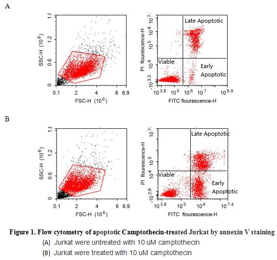
(Annexin V (ANXA5) is a multifunctional protein that is highly expressed on the apical surfaces of syncytiotrophoblasts, and plays an important role in haemostatic regulations, maintaining blood fluidity of the placenta. Lower ANXA5 levels have been observed in M2/ANXA5 haplotype carrying chorion. The association found between the maternal carriage of the M2/ANXA5 haplotype and an elevated risk of IUGR and/or PE supports the hypothesis that carrier status of this haplotype and the consequently reduced placental ANXA5 expression might be responsible, at least partially, for the onset of these gestational vascular complications. Annexin V is a calcium-dependent phospholipid binding protein that can be used to bind Phosphatidylserine (PS) during an early apoptosis event where the PS becomes exposed at the cell surface. Jurkat cells were treated with 10 uM camptothecin for 4h, 2*105 cells which were resuspended in binding buffer were stained with 5 ug recombinant human Annexin V-FITC and 10 ul Propidium iodide (PI) for 20min in dark room temperature. The flow cytometry was used to detect the early apoptotic and late apoptotic of camptothecin-treated Jurkat cells (Figure 1), the combination of Annexin V-FITC and propidium iodide allows for the distinction between early apoptotic cells (Annexin V-FITC positive and propidium iodide negative), late apoptotic and/or necrotic cells (Annexin V-FITC and propidium iodide positive), and viable cells (unstained). Thus, the recombinant human Annexin V-FITC can bind Phosphatidylserine (PS) at early apoptosis of Jurkat.)
Bioactivity
(Annexin V (ANXA5) is a multifunctional protein that is highly expressed on the apical surfaces of syncytiotrophoblasts, and plays an important role in haemostatic regulations, maintaining blood fluidity of the placenta. Lower ANXA5 levels have been observed in M2/ANXA5 haplotype carrying chorion. The association found between the maternal carriage of the M2/ANXA5 haplotype and an elevated risk of IUGR and/or PE supports the hypothesis that carrier status of this haplotype and the consequently reduced placental ANXA5 expression might be responsible, at least partially, for the onset of these gestational vascular complications. Annexin V is a calcium-dependent phospholipid binding protein that can be used to bind Phosphatidylserine (PS) during an early apoptosis event where the PS becomes exposed at the cell surface. Jurkat cells were treated with 10 uM camptothecin for 4h, 2*105 cells which were resuspended in binding buffer were stained with 5 ug recombinant human Annexin V-FITC and 10 ul Propidium iodide (PI) for 20min in dark room temperature. The flow cytometry was used to detect the early apoptotic and late apoptotic of camptothecin-treated Jurkat cells (Figure 1), the combination of Annexin V-FITC and propidium iodide allows for the distinction between early apoptotic cells (Annexin V-FITC positive and propidium iodide negative), late apoptotic and/or necrotic cells (Annexin V-FITC and propidium iodide positive), and viable cells (unstained). Thus, the recombinant human Annexin V-FITC can bind Phosphatidylserine (PS) at early apoptosis of Jurkat.)
Annexin V (ANXA5), Active Protein (Cat# AAA161722)
 Bioactivity
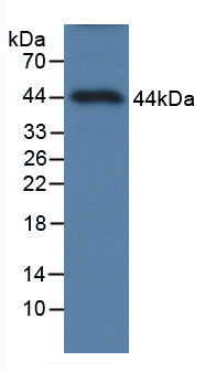
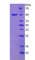
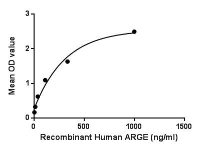
(Amphiregulin, also known as AREG, is an autocrine growth factor as well as a mitogen for astrocytes, Schwann cells, fibroblasts. It is related to epidermal growth factor (EGF) and transforming growth factor alpha (TGF-alpha). This protein interacts with the Epidermal growth factor receptor (EGFR) to promote the growth of normal epithelial cells. Besides, Epidermal Growth Factor Receptor (EGFR) has been identified as an interactor of ARGE, thus a binding ELISA assay was conducted to detect the interaction of recombinant human ARGE and recombinant human EGFR. Briefly, ARGE were diluted serially in PBS, with 0.01% BSA (pH 7.4). Duplicate samples of 100uL were then transferred to EGFR-coated microtiter wells and incubated for 2h at 37. Wells were washed with PBST and incubated for 1h with anti-ARGE pAb, then aspirated and washed 3 times. After incubation with HRP labelled secondary antibody, wells were aspirated and washed 3 times. With the addition of substrate solution, wells were incubated 15-25 minutes at 37. Finally, add 50uL stop solution to the wells and read at 450nm immediately. The binding activity of ARGE and EGFR was shown in Figure 1, and this effect was in a dose dependent manner.Figure. The binding activity of ARGE with EGFR.)
Bioactivity
(Amphiregulin, also known as AREG, is an autocrine growth factor as well as a mitogen for astrocytes, Schwann cells, fibroblasts. It is related to epidermal growth factor (EGF) and transforming growth factor alpha (TGF-alpha). This protein interacts with the Epidermal growth factor receptor (EGFR) to promote the growth of normal epithelial cells. Besides, Epidermal Growth Factor Receptor (EGFR) has been identified as an interactor of ARGE, thus a binding ELISA assay was conducted to detect the interaction of recombinant human ARGE and recombinant human EGFR. Briefly, ARGE were diluted serially in PBS, with 0.01% BSA (pH 7.4). Duplicate samples of 100uL were then transferred to EGFR-coated microtiter wells and incubated for 2h at 37. Wells were washed with PBST and incubated for 1h with anti-ARGE pAb, then aspirated and washed 3 times. After incubation with HRP labelled secondary antibody, wells were aspirated and washed 3 times. With the addition of substrate solution, wells were incubated 15-25 minutes at 37. Finally, add 50uL stop solution to the wells and read at 450nm immediately. The binding activity of ARGE and EGFR was shown in Figure 1, and this effect was in a dose dependent manner.Figure. The binding activity of ARGE with EGFR.)
Amphiregulin, Active Protein (Cat# AAA150047)
Aprotinin, Active Protein (Cat# AAA44781)
tissue plasminogen activator, >85% single chain, Active Protein (Cat# AAA37793)
Glutamic-Pyruvate Transaminase, Active Protein (Cat# AAA38251)
ACE2, Active Protein (Cat# AAA177001)
Activin A, Active Protein (Cat# AAA75456)
Platelet Derived Growth Factor-BB, Active Protein (Cat# AAA38232)
Thrombin, Active Protein (Cat# AAA37801)
 Application Data
(Measured in a cell proliferation assay using BALB/c 3T3 mouse fibroblasts. The ED50 for this effect is typically 0.6-3 ug/mL.)
Application Data
(Measured in a cell proliferation assay using BALB/c 3T3 mouse fibroblasts. The ED50 for this effect is typically 0.6-3 ug/mL.)
FGF17, Active Protein (Cat# AAA258010)
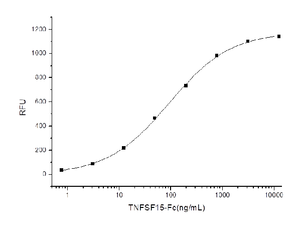 Application Data
(Measured by its ability to induce apoptosis of TF1 human erythroleukemic cells.The ED50 for this effect is typically 20-120 ng/mL.)
Application Data
(Measured by its ability to induce apoptosis of TF1 human erythroleukemic cells.The ED50 for this effect is typically 20-120 ng/mL.)
TL1A, Active Protein (Cat# AAA258057)
 Bioactivity
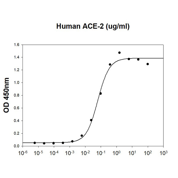
(SARS-CoV-2 Spike RBD is coated at 5 ug/ml (100 ul/well) can bind Human ACE-2. The ED50 range )
Bioactivity
(SARS-CoV-2 Spike RBD is coated at 5 ug/ml (100 ul/well) can bind Human ACE-2. The ED50 range )
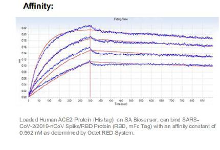
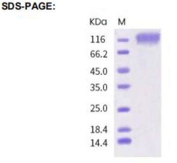
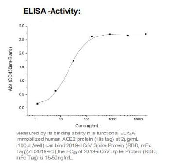
ACE-2, Active Protein (Cat# AAA48372)
 Application Data
COVID 19 Spike S1 Coronavirus, Active Protein (Cat# AAA268893)
Aldose Reductase, Active Protein (Cat# AAA38715)
Interleukin-1 alpha, Active Protein (Cat# AAA38549)
What Are Active Proteins?
Proteins are large molecules made up of long chains of amino acids.
They will typically fold into a very particular 3-dimensional shape/conformation, that is sometimes referred to as their “native” form, which allows them to work properly in the body. For the purposes of product categorization, AAA Biotech will typically refer to proteins purified from their original animal host as being “native” proteins (this is to signify their difference compared to their “recombinant” or “synthetic” protein counterparts).
If a protein successfully folds into the correct shape, it is will typically display high fidelity characteristics to its original protein in its original animal host, and be classified as an active protein, as it will be able to function “normally” in most enzymatic or binding capacities. If it loses this shape, due to factors such as heat or strong chemicals (such as detergents), it becomes inactive and is no longer able to perform its basic functions. All of the proteins in this category are made under strict quality control, and they are active, pure, low in contaminants, and stable.
Most are stored as freeze-dried powders and come without extra tags, so they’re very close to the actual natural/native form.
Key Applications of Active Proteins
1. Scientific Research
- Aid in the study of how proteins function in the body
- Aid in understanding various disease processes
2. Drug Development
- Powerful tools to investigate how potential drugs interact with specific proteins
- Ideal for identifying drug targets
3. Cell Culture
- Are routinely utilized to support cell growth and function (e.g., using exogenous growth factors)
- Can be used to promote cellular development into specific types (differentiation)
4. Diagnostics
- Regularly utilized in tests to detect diseases or infections (e.g., COVID-19, cancer)
- Note: All products are strictly for research-use only (RUO).
5. Therapeutics
- Some active proteins are used directly as treatments (e.g., insulin, enzymes)
- Note: All products are strictly for research-use only (RUO).
6. Vaccine Development
- Used to create or test vaccines by mimicking parts of viruses or bacteria
7. Biochemical Assays
- They can facilitate the characterization of enzyme activity, binding strength, or protein interactions in lab tests
Why Buy Active Proteins from AAA Biotech?
- High biological activity – Verified to perform as expected or indicated on datasheet
- Strict quality control – We are confident in our active proteins’ reliability and consistency
- High purity & low endotoxin – Ideal for applications involving sensitive or precious samples/components
- Freeze-dried for stability – Long shelf life and straightforward storage
- Mostly tag-free – Closer to natural/native protein form
FAQ
1. What are active proteins used for in research?
Active proteins are used primarily in the study of how proteins function, in characterizing/discovering drug interactions, supporting cell growth, running biochemical assays, and in development of diagnostics or therapeutics.
2. How are AAA Biotech's active proteins validated?
AAA Biotech’s active proteins are validated through strict quality control and functional assays to ensure they are properly folded and active. “Active”, though, can be an ambiguous term, so if a specific “activity” or “binding” capability of a protein is of crucial interest to you, please inquire with us prior to purchase, and we will provide further details on how the “Active” modifier was determined to be applicable.
3. Are these proteins tested for biological activity?
Yes, all active proteins from AAA Biotech are tested to confirm they have the expected biological activity before being offered for use. Though, said “biological activity” can be either “enzymatic”, “binding”, or both.