Filters
▼Clonality
▼Type
▼Reactivity
▼Gene Name
▼Isotype
▼Host
▼Application
▼Clone
▼Active Proteins
AAA Biotech also known as AAA Bio or AAABio provides a variety of high-quality recombinant and natural/native proteins that are proven to work in a wide range of experiments. Explore our products to find the active protein that best fits your needs or experimental model.
Viewing 2100-2150 of 2567 product results
soluble CD40 Ligand/TRAP, Active Protein (Cat# AAA38130)
Interleukin-17A, Active Protein (Cat# AAA38132)
Neurotrophin-3, Active Protein (Cat# AAA38136)
Interleukin-4, Active Protein (Cat# AAA38143)
Flt3-Ligand, Active Protein (Cat# AAA38161)
Interleukin-9, Active Protein (Cat# AAA38171)
Vascular Endothelial Growth Factor, Active Protein (Cat# AAA38186)
Complement Component C5a, Active Protein (Cat# AAA39018)
Peroxiredoxin-2, Active Protein (Cat# AAA38698)
Interleukin-8, Active Protein (Cat# AAA38923)
Follistatin, Active Protein (Cat# AAA38937)
Thioredoxin Reductase, Active Protein (Cat# AAA38784)
Malate Dehydrogenase 1, Active Protein (Cat# AAA38835)
Interleukin-27 p28, Active Protein (Cat# AAA39117)
Prolyl 4-Hydroxylase Beta, Active Protein (Cat# AAA39146)
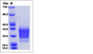
prolactin receptor antagonist, Active Protein (Cat# AAA39185)
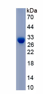
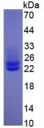
 SDS-PAGE
DNase I, Active Protein (Cat# AAA258026)
 Application Data
(Measured by its binding ability in a functional ELISA. Immobilized human TMIGD2 at 10 ug/mL (100 uL/well) can bind human HHLA2-Fc, the EC50 of human HHLA2-Fc is 0.3-3ug/mL.)
Application Data
(Measured by its binding ability in a functional ELISA. Immobilized human TMIGD2 at 10 ug/mL (100 uL/well) can bind human HHLA2-Fc, the EC50 of human HHLA2-Fc is 0.3-3ug/mL.)
TMIGD2, Active Protein (Cat# AAA258028)

 Bioactivity
Spike glycoprotein (S), Active Protein (Cat# AAA244032)

 Bioactivity
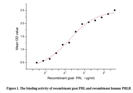
(PRL (prolactin), also known as luteotropin, is a hormone secreted from the pituitary gland and is best known for its role in enabling mammals to produce milk. PRL plays an essential role in metabolism, regulation of the immune system through activating its specific membrane-anchored receptor (PRLR). A functional binding ELISA assay was conducted to detect the interaction of recombinant goat PRL and recombinant human PRLR. Briefly, PRL was diluted serially in PBS with 0.01% BSA (pH 7.4). Duplicate samples of 100 ul were then transferred to PRLR-coated microtiter wells and incubated for 1h at 37 degree C. Wells were washed with PBST and incubated for 1h with anti-PRL pAb, then aspirated and washed 3 times. After incubation with HRP labelled secondary antibody for 1h at 37 degree C, wells were aspirated and washed 5 times. With the addition of substrate solution, wells were incubated 15-25 minutes at 37 degree C. Finally, add 50 uL stop solution to the wells and read at 450/630 nm immediately. The binding activity of recombinant goat PRL and recombinant human PRLR was shown in Figure 1, the EC50 for this effect is 0.1 ug/mL.)
Bioactivity
(PRL (prolactin), also known as luteotropin, is a hormone secreted from the pituitary gland and is best known for its role in enabling mammals to produce milk. PRL plays an essential role in metabolism, regulation of the immune system through activating its specific membrane-anchored receptor (PRLR). A functional binding ELISA assay was conducted to detect the interaction of recombinant goat PRL and recombinant human PRLR. Briefly, PRL was diluted serially in PBS with 0.01% BSA (pH 7.4). Duplicate samples of 100 ul were then transferred to PRLR-coated microtiter wells and incubated for 1h at 37 degree C. Wells were washed with PBST and incubated for 1h with anti-PRL pAb, then aspirated and washed 3 times. After incubation with HRP labelled secondary antibody for 1h at 37 degree C, wells were aspirated and washed 5 times. With the addition of substrate solution, wells were incubated 15-25 minutes at 37 degree C. Finally, add 50 uL stop solution to the wells and read at 450/630 nm immediately. The binding activity of recombinant goat PRL and recombinant human PRLR was shown in Figure 1, the EC50 for this effect is 0.1 ug/mL.)
Prolactin (PRL), Active Protein (Cat# AAA161788)

 Bioactivity
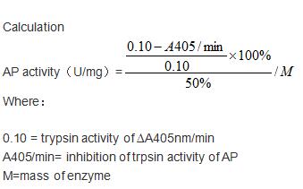
(Aprotinin (AP) is a competitive serine protease inhibitor. Reversibly binds to and blocks the enzymatic active site. Inhibits a range of serine proteases including trypsin, chymotrypsin, kallikrein and plasmin. Inhibits cytopathogenic effect of SARS-CoV-2 and double-stranded RNA formation in SARS-CoV-2-infected cells. The activity of recombinant bovine AP was measured by its ability to inhibit trypsin cleavage of a peptide substrate BAPNA in the assay buffer 200 mM Triethanolamine hydrochloride, 20 mM CaCl2, pH 7.8. The reaction was performed in adding 20 ul 4 mg/mL trypsin diluted by 1mM HCl to 160 ul assay buffer and 20 ul 0.85% (w/v) NaCl and start the reaction by adding 100 ul of 1mg/ml BAPNA. Include a substrate blank containing 160 ul assay buffer, 20 ul 1mM HCl, 20 ul 0.85% (w/v) NaCl and 100 uL of 1mg/ml substrate. Rapidly mixing at 25 degree C, then read at 405 nm in kinetic mode for 5 minutes using a microplate reader controlling the ?A405nm/min=0.08-0.12. The 20 ul different concentrations of recombinant bovine AP was incubated with 20 ul 4 mg/mL trypsin in 160 ul assay buffer at 25 degree C for 10 minutes followed by adding 100 ul substrate, then read at 405 nm in kinetic mode for 5 minutes using a microplate reader. Under these conditions, the enzyme amount of 50% inhibition of trypsin activity per minute is defined as a unit. The specific activity of recombinant bovine AP is >3000 U/mg.)
Bioactivity
(Aprotinin (AP) is a competitive serine protease inhibitor. Reversibly binds to and blocks the enzymatic active site. Inhibits a range of serine proteases including trypsin, chymotrypsin, kallikrein and plasmin. Inhibits cytopathogenic effect of SARS-CoV-2 and double-stranded RNA formation in SARS-CoV-2-infected cells. The activity of recombinant bovine AP was measured by its ability to inhibit trypsin cleavage of a peptide substrate BAPNA in the assay buffer 200 mM Triethanolamine hydrochloride, 20 mM CaCl2, pH 7.8. The reaction was performed in adding 20 ul 4 mg/mL trypsin diluted by 1mM HCl to 160 ul assay buffer and 20 ul 0.85% (w/v) NaCl and start the reaction by adding 100 ul of 1mg/ml BAPNA. Include a substrate blank containing 160 ul assay buffer, 20 ul 1mM HCl, 20 ul 0.85% (w/v) NaCl and 100 uL of 1mg/ml substrate. Rapidly mixing at 25 degree C, then read at 405 nm in kinetic mode for 5 minutes using a microplate reader controlling the ?A405nm/min=0.08-0.12. The 20 ul different concentrations of recombinant bovine AP was incubated with 20 ul 4 mg/mL trypsin in 160 ul assay buffer at 25 degree C for 10 minutes followed by adding 100 ul substrate, then read at 405 nm in kinetic mode for 5 minutes using a microplate reader. Under these conditions, the enzyme amount of 50% inhibition of trypsin activity per minute is defined as a unit. The specific activity of recombinant bovine AP is >3000 U/mg.)
Aprotinin (AP), Active Protein (Cat# AAA161811)
 Bioactivity
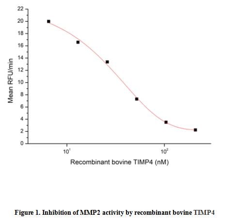
(Tissue Inhibitors of Metalloproteinase 4 (TIMP4) is an enzyme that in humans is encoded by the TIMP4 gene. This gene belongs to the tissue inhibitor of metalloproteinases gene family. The proteins encoded by this gene family are inhibitors of the matrix metalloproteinases, a group of peptidases involved in degradation of the extracellular matrix. The secreted, netrin domain-containing protein encoded by this gene is involved in regulation of platelet aggregation and recruitment and may play role in hormonal regulation and endometrial tissue remodeling. The activity of recombinant bovine TIMP4 was measured by its ability to inhibit rhMMP2 cleavage of a fluorogenic peptide substrate MCA-Pro-Leu-Gly-Leu-DPA-Ala-Arg-NH2 in the assay buffer 50 mM Tris, 10 mM CaCl2, 150 mM NaCl, 0.05% (w/v) Brij-35, pH 7.5. rhMMP2 was diluted to 100 ug/ml and activated with 1 mM APMA at 37 degree C for 1 hour and rbTIMP4 (MW: 24.05 KD) was diluted to different concentrations with the assay buffer. Mix 8 ul of rbTIMP4 curve dilutions, 12.8 ul of activated rhMMP-2, and 59.2 ul of assay buffer, including a control containing assay buffer and the diluted rhMMP-2 and incubate the reactions for 2 hours at 37 degree C. Loading 50 ul of the incubated mixtures which were diluted five-fold in assay buffer into empty wells of a plate, and start the reaction by adding 50 ul of 20 uM substrate. Include a substrate blank containing 50 ul of assay buffer and 50 ul of 20 uM substrate. Then read at excitiation and emission wavelengths of 320 nm and 405 nm, respectively, in kinetic mode for 5 minutes. The result was shown in Figure 1 and it was obvious that recombinant bovine TIMP4 significantly decreased rhMMP2 activity. The inhibition IC50 was )
Bioactivity
(Tissue Inhibitors of Metalloproteinase 4 (TIMP4) is an enzyme that in humans is encoded by the TIMP4 gene. This gene belongs to the tissue inhibitor of metalloproteinases gene family. The proteins encoded by this gene family are inhibitors of the matrix metalloproteinases, a group of peptidases involved in degradation of the extracellular matrix. The secreted, netrin domain-containing protein encoded by this gene is involved in regulation of platelet aggregation and recruitment and may play role in hormonal regulation and endometrial tissue remodeling. The activity of recombinant bovine TIMP4 was measured by its ability to inhibit rhMMP2 cleavage of a fluorogenic peptide substrate MCA-Pro-Leu-Gly-Leu-DPA-Ala-Arg-NH2 in the assay buffer 50 mM Tris, 10 mM CaCl2, 150 mM NaCl, 0.05% (w/v) Brij-35, pH 7.5. rhMMP2 was diluted to 100 ug/ml and activated with 1 mM APMA at 37 degree C for 1 hour and rbTIMP4 (MW: 24.05 KD) was diluted to different concentrations with the assay buffer. Mix 8 ul of rbTIMP4 curve dilutions, 12.8 ul of activated rhMMP-2, and 59.2 ul of assay buffer, including a control containing assay buffer and the diluted rhMMP-2 and incubate the reactions for 2 hours at 37 degree C. Loading 50 ul of the incubated mixtures which were diluted five-fold in assay buffer into empty wells of a plate, and start the reaction by adding 50 ul of 20 uM substrate. Include a substrate blank containing 50 ul of assay buffer and 50 ul of 20 uM substrate. Then read at excitiation and emission wavelengths of 320 nm and 405 nm, respectively, in kinetic mode for 5 minutes. The result was shown in Figure 1 and it was obvious that recombinant bovine TIMP4 significantly decreased rhMMP2 activity. The inhibition IC50 was )
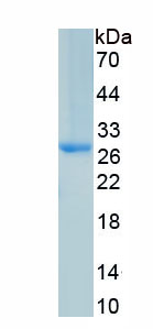
Tissue Inhibitors Of Metalloproteinase 4 (TIMP4), Active Protein (Cat# AAA161701)
 Bioactivity
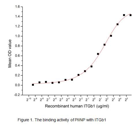
(Procollagen II N-Terminal Propeptide is specific for cartilaginous tissues. It is essential for the normal embryonic development of the skeleton, for linear growth and for the ability of cartilage to resist compressive forces, which was are associated wit)
Bioactivity
(Procollagen II N-Terminal Propeptide is specific for cartilaginous tissues. It is essential for the normal embryonic development of the skeleton, for linear growth and for the ability of cartilage to resist compressive forces, which was are associated wit)
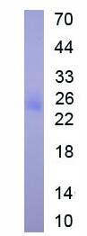
Procollagen II N-Terminal Propeptide (PIINP), Active Protein (Cat# AAA153010)
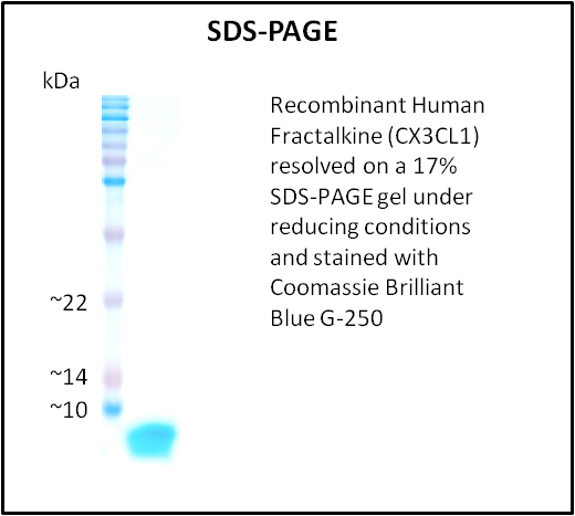
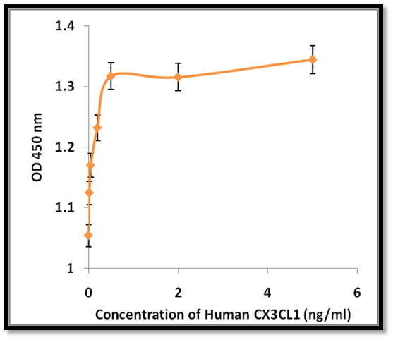 Application Data
Fractalkine (CX3CL1), Active Protein (Cat# AAA214279)
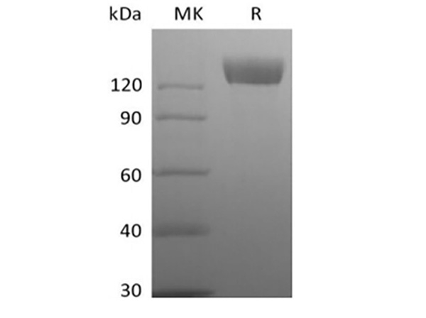
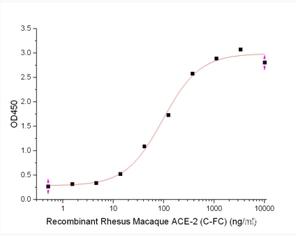 Bioactivity
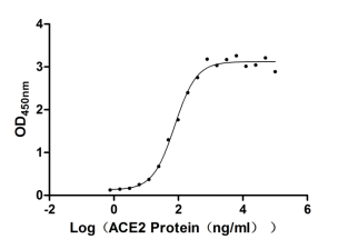
Angiotensin-Converting Enzyme 2/ACE-2, Active Protein (Cat# AAA177979)
Prostate Specific Antigen, Active Protein (Cat# AAA59627)
TGF-beta RI/ALK-5, Active Protein (Cat# AAA48990)
APRIL, Active Protein (Cat# AAA75589)
 Activity
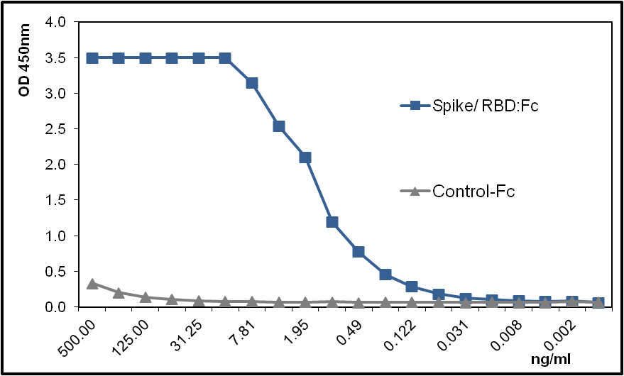
(Figure 1 : ACE2 (human)(rec.) (AAA78521) binds with high affinity to the Spike (RBD) protein of the virus SARS-CoV-2. Method: ACE2 (human)(rec.) (AAA78521 ) is coated on an ELISA plate at 1 ug/ml overnight at 4°C. Spike (SARS-CoV-2):Fc (human)(RBD)(rec.) is added (starting at a concentration of 500 ng/ml with a twofold serial dilution) during one hour at RT and the interaction is then detected using an anti-human IgG (HRP).)
Activity
(Figure 1 : ACE2 (human)(rec.) (AAA78521) binds with high affinity to the Spike (RBD) protein of the virus SARS-CoV-2. Method: ACE2 (human)(rec.) (AAA78521 ) is coated on an ELISA plate at 1 ug/ml overnight at 4°C. Spike (SARS-CoV-2):Fc (human)(RBD)(rec.) is added (starting at a concentration of 500 ng/ml with a twofold serial dilution) during one hour at RT and the interaction is then detected using an anti-human IgG (HRP).)
ACE2, Active Protein (Cat# AAA78521)
Apo Transferrin (ATF), Active Protein (Cat# AAA76530)
 Bioactivity
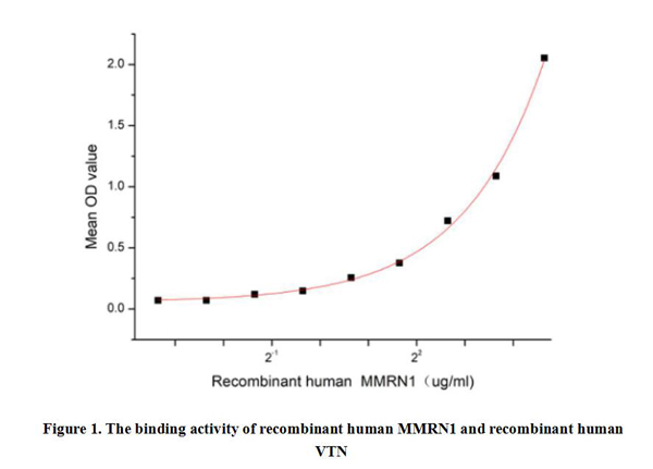
(Multimerin 1 (MMRN1) is a secreted glycoprotein that consists of multiple subunits, forming a complex with a molecular weight of approximately 150 kDa. MMRN1 is a member of the multimerin family and it is mainly expressed in platelets and endothelial cells. MMRN1 plays an important role in maintaining vascular integrity, regulating clotting and inflammatory responses, among others. In addition, the binding of MMRN1 to Vitronectin (VTN) plays an important role in a variety of physiological and pathological processes, including cell adhesion, migration, blood coagulation, and cell signaling. Thus a functional binding ELISA assay was conducted to detect the interaction of recombinant human MMRN1 and recombinant human VTN. Briefly, MMRN1 was diluted serially in PBS with 0.01% BSA (pH 7.4). Duplicate samples of 100 ul were then transferred to VTN-coated microtiter wells and incubated for 1h at 37 degree C. Wells were washed with PBST and incubated for 1h with anti-MMRN1 pAb, then aspirated and washed 3 times. After incubation with HRP labelled secondary antibody for 1h at 37 degree C, wells were aspirated and washed 5 times. With the addition of substrate solution, wells were incubated 15-25 minutes at 37 degree C. Finally, add 50 uL stop solution to the wells and read at 450/630 nm immediately. The binding activity of recombinant human MMRN1 and recombinant human VTN was shown in Figure 1, and this effect was in a dose dependent manner.)
Bioactivity
(Multimerin 1 (MMRN1) is a secreted glycoprotein that consists of multiple subunits, forming a complex with a molecular weight of approximately 150 kDa. MMRN1 is a member of the multimerin family and it is mainly expressed in platelets and endothelial cells. MMRN1 plays an important role in maintaining vascular integrity, regulating clotting and inflammatory responses, among others. In addition, the binding of MMRN1 to Vitronectin (VTN) plays an important role in a variety of physiological and pathological processes, including cell adhesion, migration, blood coagulation, and cell signaling. Thus a functional binding ELISA assay was conducted to detect the interaction of recombinant human MMRN1 and recombinant human VTN. Briefly, MMRN1 was diluted serially in PBS with 0.01% BSA (pH 7.4). Duplicate samples of 100 ul were then transferred to VTN-coated microtiter wells and incubated for 1h at 37 degree C. Wells were washed with PBST and incubated for 1h with anti-MMRN1 pAb, then aspirated and washed 3 times. After incubation with HRP labelled secondary antibody for 1h at 37 degree C, wells were aspirated and washed 5 times. With the addition of substrate solution, wells were incubated 15-25 minutes at 37 degree C. Finally, add 50 uL stop solution to the wells and read at 450/630 nm immediately. The binding activity of recombinant human MMRN1 and recombinant human VTN was shown in Figure 1, and this effect was in a dose dependent manner.)
Multimerin 1 (MMRN1), Active Protein (Cat# AAA161892)
 Bioactivity
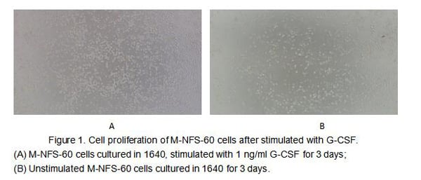
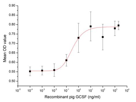
(Figure 2. Cell proliferation of M-NFS-60 cells after stimulated with G-CSF.)
Bioactivity
(Figure 2. Cell proliferation of M-NFS-60 cells after stimulated with G-CSF.)
Colony Stimulating Factor 3, Granulocyte (GCSF), Active Protein (Cat# AAA161665)
Keratinocyte Growth Factor, Active Protein (Cat# AAA38113)
Granulocyte Macrophage-Colony Stimulating Factor, Active Protein (Cat# AAA38115)
Platelet Derived Growth Factor-BB, Active Protein (Cat# AAA38129)
Interleukin-1 beta, Active Protein (Cat# AAA38142)
Carp Growth Hormone, Active Protein (Cat# AAA38148)
Interferon-gamma, Active Protein (Cat# AAA38173)
Transforming Growth Factor-Beta 3, Active Protein (Cat# AAA38178)
Interleukin-3, Active Protein (Cat# AAA38180)
Malate Dehydrogenase 2, Active Protein (Cat# AAA38044)
Granzyme-B, Active Protein (Cat# AAA38045)
I-309, Active Protein (Cat# AAA38084)
Growth Hormone, Active Protein (Cat# AAA38106)
Brain-Derived Neurotrophic Factor, Active Protein (Cat# AAA38107)
FITC Labeled Bovine Fibrinogen, Active Protein (Cat# AAA37784)
Glutathione S-Transferase Alpha 1, Active Protein (Cat# AAA38702)
Trypsin, Active Protein (Cat# AAA38727)
Acid Phosphatase-1, Active Protein (Cat# AAA38771)
Keratinocyte Growth Factor-2, Active Protein (Cat# AAA38945)
What Are Active Proteins?
Proteins are large molecules made up of long chains of amino acids.
They will typically fold into a very particular 3-dimensional shape/conformation, that is sometimes referred to as their “native” form, which allows them to work properly in the body. For the purposes of product categorization, AAA Biotech will typically refer to proteins purified from their original animal host as being “native” proteins (this is to signify their difference compared to their “recombinant” or “synthetic” protein counterparts).
If a protein successfully folds into the correct shape, it is will typically display high fidelity characteristics to its original protein in its original animal host, and be classified as an active protein, as it will be able to function “normally” in most enzymatic or binding capacities. If it loses this shape, due to factors such as heat or strong chemicals (such as detergents), it becomes inactive and is no longer able to perform its basic functions. All of the proteins in this category are made under strict quality control, and they are active, pure, low in contaminants, and stable.
Most are stored as freeze-dried powders and come without extra tags, so they’re very close to the actual natural/native form.
Key Applications of Active Proteins
1. Scientific Research
- Aid in the study of how proteins function in the body
- Aid in understanding various disease processes
2. Drug Development
- Powerful tools to investigate how potential drugs interact with specific proteins
- Ideal for identifying drug targets
3. Cell Culture
- Are routinely utilized to support cell growth and function (e.g., using exogenous growth factors)
- Can be used to promote cellular development into specific types (differentiation)
4. Diagnostics
- Regularly utilized in tests to detect diseases or infections (e.g., COVID-19, cancer)
- Note: All products are strictly for research-use only (RUO).
5. Therapeutics
- Some active proteins are used directly as treatments (e.g., insulin, enzymes)
- Note: All products are strictly for research-use only (RUO).
6. Vaccine Development
- Used to create or test vaccines by mimicking parts of viruses or bacteria
7. Biochemical Assays
- They can facilitate the characterization of enzyme activity, binding strength, or protein interactions in lab tests
Why Buy Active Proteins from AAA Biotech?
- High biological activity – Verified to perform as expected or indicated on datasheet
- Strict quality control – We are confident in our active proteins’ reliability and consistency
- High purity & low endotoxin – Ideal for applications involving sensitive or precious samples/components
- Freeze-dried for stability – Long shelf life and straightforward storage
- Mostly tag-free – Closer to natural/native protein form
FAQ
1. What are active proteins used for in research?
Active proteins are used primarily in the study of how proteins function, in characterizing/discovering drug interactions, supporting cell growth, running biochemical assays, and in development of diagnostics or therapeutics.
2. How are AAA Biotech's active proteins validated?
AAA Biotech’s active proteins are validated through strict quality control and functional assays to ensure they are properly folded and active. “Active”, though, can be an ambiguous term, so if a specific “activity” or “binding” capability of a protein is of crucial interest to you, please inquire with us prior to purchase, and we will provide further details on how the “Active” modifier was determined to be applicable.
3. Are these proteins tested for biological activity?
Yes, all active proteins from AAA Biotech are tested to confirm they have the expected biological activity before being offered for use. Though, said “biological activity” can be either “enzymatic”, “binding”, or both.