Filters
▼Clonality
▼Type
▼Reactivity
▼Gene Name
▼Isotype
▼Host
▼Application
▼Clone
▼Active Proteins
AAA Biotech also known as AAA Bio or AAABio provides a variety of high-quality recombinant and natural/native proteins that are proven to work in a wide range of experiments. Explore our products to find the active protein that best fits your needs or experimental model.
Viewing 1950-2000 of 2567 product results
 Bioactivity
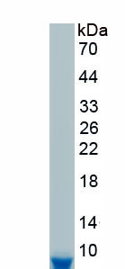
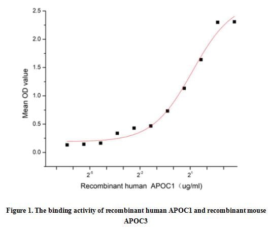
(Apolipoprotein C1 (APOC1), also known as Apo-CI; ApoC-I; apo-CIB; apoC-IB, is the smallest size apolipoprotein of all apolipoprotein C family (Mr = 6.6 kDa) and located at position 19q13.32. APOC1 is primarily expressed in the liver and activated when monocytes differentiate into macrophages. It plays important roles in the innate immune response as effector of glucocorticoid-mediated responses and regulator of the inflammatory process. It has anti-inflammatory activity and also can promote the differentiation of T-cells into Th1 cells and negatively regulates differentiation into Th2 cells. Besides, Apolipoprotein C3 (APOC3) has been identified as an interactor of APOC1, thus a functional binding ELISA assay was conducted to detect the interaction of recombinant human APOC1 and recombinant mouse APOC3. Briefly, APOC1 was diluted serially in PBS with 0.01% BSA (pH 7.4). Duplicate samples of 100 ul were then transferred to APOC3-coated microtiter wells and incubated for 1h at 37 degree C. Wells were washed with PBST and incubated for 1h with anti-APOC1 pAb, then aspirated and washed 3 times. After incubation with HRP labelled secondary antibody for 1h at 37 degree C, wells were aspirated and washed 5 times. With the addition of substrate solution, wells were incubated 15-25 minutes at 37 degree C. Finally, add 50 uL stop solution to the wells and read at 450/630 nm immediately. The binding activity of recombinant human APOC1 and recombinant mouse APOC3 was shown in Figure 1, the EC50 for this effect is 2.19 ug/mL.)
Bioactivity
(Apolipoprotein C1 (APOC1), also known as Apo-CI; ApoC-I; apo-CIB; apoC-IB, is the smallest size apolipoprotein of all apolipoprotein C family (Mr = 6.6 kDa) and located at position 19q13.32. APOC1 is primarily expressed in the liver and activated when monocytes differentiate into macrophages. It plays important roles in the innate immune response as effector of glucocorticoid-mediated responses and regulator of the inflammatory process. It has anti-inflammatory activity and also can promote the differentiation of T-cells into Th1 cells and negatively regulates differentiation into Th2 cells. Besides, Apolipoprotein C3 (APOC3) has been identified as an interactor of APOC1, thus a functional binding ELISA assay was conducted to detect the interaction of recombinant human APOC1 and recombinant mouse APOC3. Briefly, APOC1 was diluted serially in PBS with 0.01% BSA (pH 7.4). Duplicate samples of 100 ul were then transferred to APOC3-coated microtiter wells and incubated for 1h at 37 degree C. Wells were washed with PBST and incubated for 1h with anti-APOC1 pAb, then aspirated and washed 3 times. After incubation with HRP labelled secondary antibody for 1h at 37 degree C, wells were aspirated and washed 5 times. With the addition of substrate solution, wells were incubated 15-25 minutes at 37 degree C. Finally, add 50 uL stop solution to the wells and read at 450/630 nm immediately. The binding activity of recombinant human APOC1 and recombinant mouse APOC3 was shown in Figure 1, the EC50 for this effect is 2.19 ug/mL.)
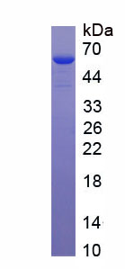
Apolipoprotein C1 (APOC1), Active Protein (Cat# AAA161719)

 Bioactivity
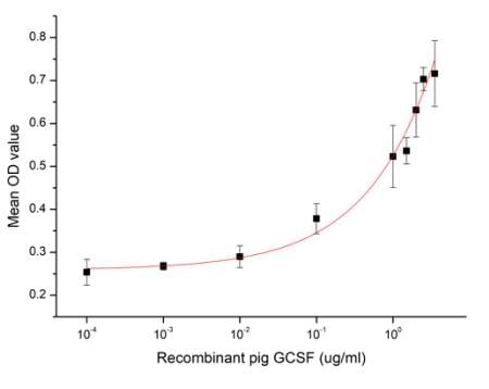
(Figure 2. Cell proliferation of M-NFS-60 cells after stimulated with G-CSF.)
Bioactivity
(Figure 2. Cell proliferation of M-NFS-60 cells after stimulated with G-CSF.)
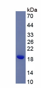
Colony Stimulating Factor 3, Granulocyte (GCSF), Active Protein (Cat# AAA161664)
 Bioactivity
(Catalase (CAT) is an antioxidant enzyme present in all aerobic organisms. It is known to catalyze H2O2 into water and oxygen in an energy-efficient manner in the cells exposed to environmental stress. As we know, H2O2 and (NH4)2MoO4 can produce a stable y)
Bioactivity
(Catalase (CAT) is an antioxidant enzyme present in all aerobic organisms. It is known to catalyze H2O2 into water and oxygen in an energy-efficient manner in the cells exposed to environmental stress. As we know, H2O2 and (NH4)2MoO4 can produce a stable y)
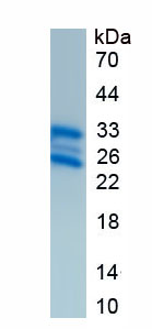
Catalase (CAT), Active Protein (Cat# AAA153115)
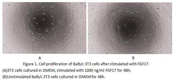 Bioactivity
(Fibroblast growth factor 17(FGF17) is a member of the fibroblast growth factorfamily. FGF family members possess broad mitogenicand cell survival activities and are involved in a varietyof biological processes, including embryonicdevelopment cell growth,)
Bioactivity
(Fibroblast growth factor 17(FGF17) is a member of the fibroblast growth factorfamily. FGF family members possess broad mitogenicand cell survival activities and are involved in a varietyof biological processes, including embryonicdevelopment cell growth,)
Fibroblast Growth Factor 17 (FGF17), Active Protein (Cat# AAA153119)
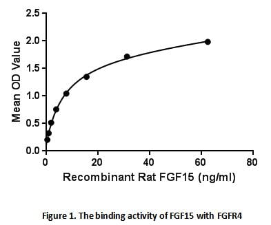 Bioactivity
(Fibroblast growth factor 15 is a protein in mouse encoded by the Fgf15 gene. It is a member of the fibroblast growth factor (FGF) family but, like FGF19, FGF21 and FGF23, has endocrine functions. FGF15 subsequently acts on a cell surface receptor complex)
Bioactivity
(Fibroblast growth factor 15 is a protein in mouse encoded by the Fgf15 gene. It is a member of the fibroblast growth factor (FGF) family but, like FGF19, FGF21 and FGF23, has endocrine functions. FGF15 subsequently acts on a cell surface receptor complex)
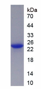
Fibroblast Growth Factor 15 (FGF15), Active Protein (Cat# AAA153137)

 Application Data
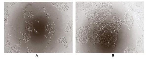
(Transforming growth factor beta (TGF-) is a multifunctional cytokine belonging to the transforming growth factor superfamily. The TGF- superfamily includes endogenous growth inhibiting proteins; an increase in expression of TGF- often correlates with the malignancy of many cancers and a defect in the cellular growth inhibition response to TGF-. Its immunosuppressive functions then come to dominate, contributing to oncogenesis. To test the effect of TGF- on inhibit HGF-dependent proliferation, HepG2 cells were seeded into triplicate wells of 96-well plates at a density of 2,000 cells/well and allowed to attach, replaced with serum-free overnight, then the medium was replaced with 2% serum standard DMEM including 1ng/mL HGF prior to the addition of various concentrations of recombinant human TGF-. After incubated for 96h, cells were observed by inverted microscope and cell proliferation was measured by Cell Counting Kit-8 (CCK-8). Briefly, 10uL of CCK-8 solution was added to each well of the plate, then the absorbance at 450nm was measured using a microplate reader after incubating the plate for 1-4 hours at 37. The inhibitory effect of TGF- on HGF-dependent proliferation of HepG2 cells observed by inverted microscope was shown in Figure 2. Cell viability was assessed by CCK-8 assay after incubation with recombinant TGF- for 96h. The result was shown in Figure 3. It was obvious that TGF- significantly decreased cell viability of HepG2 cells. (A) HepG2 cells cultured in DMEM, stimulated with 1ug/mL TGF- for 96h; (B) Unstimulated HepG2 cells cultured in DMEM for 96h.Figure.The inhibitory effect of TGF- on cell proliferation of HepG2 cells.)
Application Data
(Transforming growth factor beta (TGF-) is a multifunctional cytokine belonging to the transforming growth factor superfamily. The TGF- superfamily includes endogenous growth inhibiting proteins; an increase in expression of TGF- often correlates with the malignancy of many cancers and a defect in the cellular growth inhibition response to TGF-. Its immunosuppressive functions then come to dominate, contributing to oncogenesis. To test the effect of TGF- on inhibit HGF-dependent proliferation, HepG2 cells were seeded into triplicate wells of 96-well plates at a density of 2,000 cells/well and allowed to attach, replaced with serum-free overnight, then the medium was replaced with 2% serum standard DMEM including 1ng/mL HGF prior to the addition of various concentrations of recombinant human TGF-. After incubated for 96h, cells were observed by inverted microscope and cell proliferation was measured by Cell Counting Kit-8 (CCK-8). Briefly, 10uL of CCK-8 solution was added to each well of the plate, then the absorbance at 450nm was measured using a microplate reader after incubating the plate for 1-4 hours at 37. The inhibitory effect of TGF- on HGF-dependent proliferation of HepG2 cells observed by inverted microscope was shown in Figure 2. Cell viability was assessed by CCK-8 assay after incubation with recombinant TGF- for 96h. The result was shown in Figure 3. It was obvious that TGF- significantly decreased cell viability of HepG2 cells. (A) HepG2 cells cultured in DMEM, stimulated with 1ug/mL TGF- for 96h; (B) Unstimulated HepG2 cells cultured in DMEM for 96h.Figure.The inhibitory effect of TGF- on cell proliferation of HepG2 cells.)
Transforming Growth Factor Beta 2, Active Protein (Cat# AAA150084)

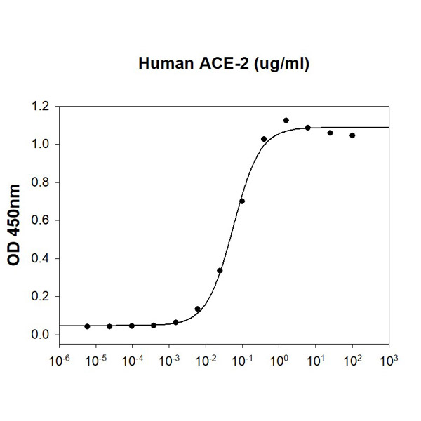 Bioactivity
(Human SARS-CoV-2 Spike RBD is coated at 5ug/ml (100 ul/well) can bind ACE-2 (CAT# ATGP3963) in a Functional ELISA assay)
Bioactivity
(Human SARS-CoV-2 Spike RBD is coated at 5ug/ml (100 ul/well) can bind ACE-2 (CAT# ATGP3963) in a Functional ELISA assay)
COVID 19 Spike RBD Coronavirus, Active Protein (Cat# AAA48384)
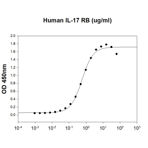 Bioactivity
(Human IL-17E/IL-25 is coated at 2 ug/ml (100 ul/well) can bind Human IL-17 RB (CAT# ATGP4039) in a Functional ELISA assay.)
Bioactivity
(Human IL-17E/IL-25 is coated at 2 ug/ml (100 ul/well) can bind Human IL-17 RB (CAT# ATGP4039) in a Functional ELISA assay.)
IL-17E/IL-25, Active Protein (Cat# AAA48395)
 Bioactivity
(Mouse OX40 Ligand/TNFSF4 (CAT# ATGP4022) is coated at 1 ug/ml (100 ul/well) can bind Human TNFRSF4. The ED50 range )
Bioactivity
(Mouse OX40 Ligand/TNFSF4 (CAT# ATGP4022) is coated at 1 ug/ml (100 ul/well) can bind Human TNFRSF4. The ED50 range )
OX40/TNFRSF4, Active Protein (Cat# AAA48407)
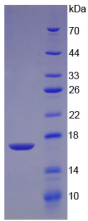
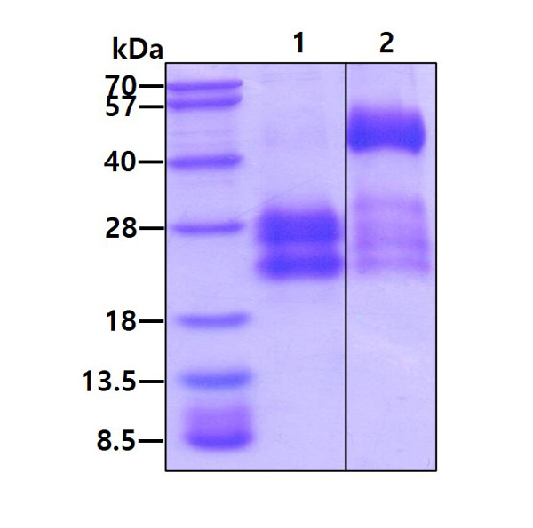
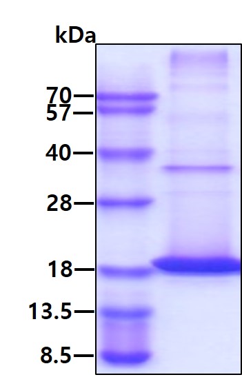
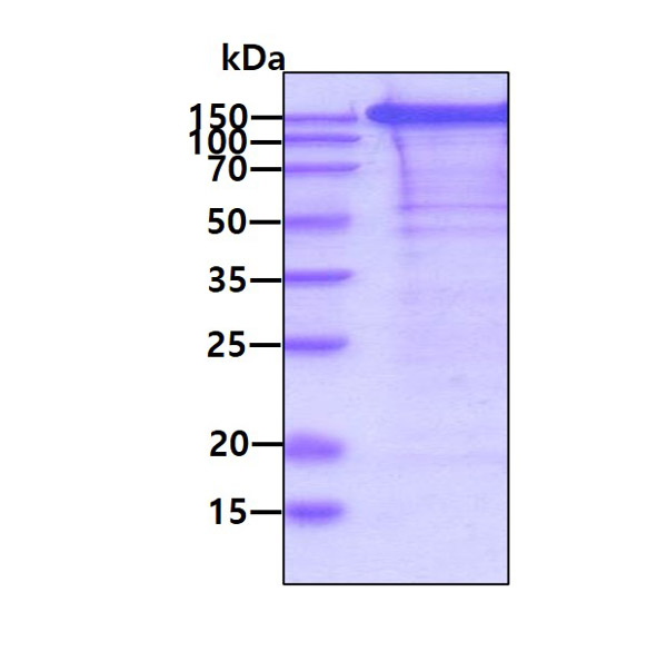
 SDS-PAGE
(3ug by SDS-PAGE under reducing condition and visualized by coomassie blue stain.)
SDS-PAGE
(3ug by SDS-PAGE under reducing condition and visualized by coomassie blue stain.)
Osteopontin/OPN protein, Active Protein (Cat# AAA48974)
OX40/TNFRSF4, Active Protein (Cat# AAA48994)
Flt3 ligand/FLT3LG, Active Protein (Cat# AAA49008)

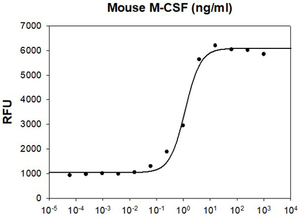 Bioactivity
(Mouse M-CSF stimulates cell proliferation of the M-NFS-60 mouse myelogenous leukemia lymphoblast cells. The ED50 range is )
Bioactivity
(Mouse M-CSF stimulates cell proliferation of the M-NFS-60 mouse myelogenous leukemia lymphoblast cells. The ED50 range is )
M-CSF, Active Protein (Cat# AAA48318)
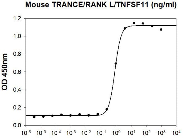 Bioactivity
(Mouse TRANCE/RANK L/TNFSF11 stimulates osteoclast differentiation of the RAW 264.7 mouse monocyte/macrophage cells. The ED50 range )
Bioactivity
(Mouse TRANCE/RANK L/TNFSF11 stimulates osteoclast differentiation of the RAW 264.7 mouse monocyte/macrophage cells. The ED50 range )
TRANCE/RANK L/TNFSF11, Active Protein (Cat# AAA48324)
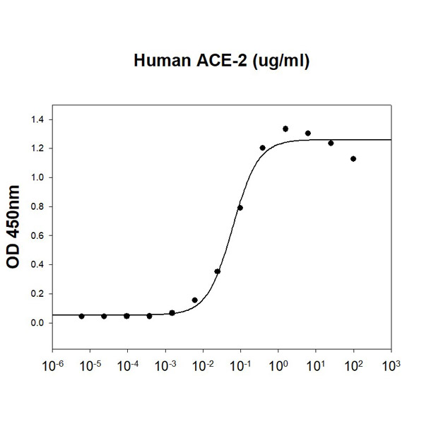
 Bioactivity
(SARS-CoV-2 Spike S1 Subunit is coated at 5ug/ml (100 ul/well) can bind ACE-2 (CAT# ATGP3963) in a Functional ELISA assay.)
Bioactivity
(SARS-CoV-2 Spike S1 Subunit is coated at 5ug/ml (100 ul/well) can bind ACE-2 (CAT# ATGP3963) in a Functional ELISA assay.)
COVID 19 Spike S1 Subunit Coronavirus, Active Protein (Cat# AAA48370)
 Bioactivity
(SARS-CoV Spike is coated at 5ug/ml (100 ul/well) can bind ACE-2 (CAT# ATGP3963) in a Functional ELISA assay.)
Bioactivity
(SARS-CoV Spike is coated at 5ug/ml (100 ul/well) can bind ACE-2 (CAT# ATGP3963) in a Functional ELISA assay.)
COVID 19 Spike Coronavirus, Active Protein (Cat# AAA48375)
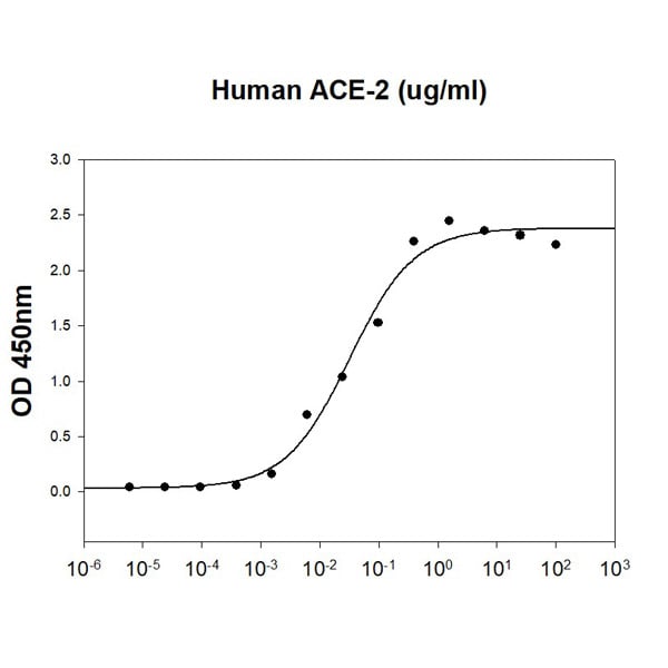
 Bioactivity
(SARS-CoV-2 Spike RBD is coated at 5ug/ml (100 ul/well) can bind ACE-2 (CAT# ATGP3963) in a Functional ELISA assay.)
Bioactivity
(SARS-CoV-2 Spike RBD is coated at 5ug/ml (100 ul/well) can bind ACE-2 (CAT# ATGP3963) in a Functional ELISA assay.)
COVID 19 Spike RBD Coronavirus, Active Protein (Cat# AAA48377)
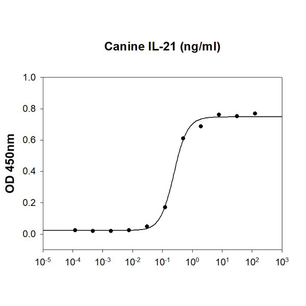 Bioactivity
(Canine IL-21 stimulates IFN-g secretion of the NK-92 human natural killer cells. The ED50 range )
Bioactivity
(Canine IL-21 stimulates IFN-g secretion of the NK-92 human natural killer cells. The ED50 range )
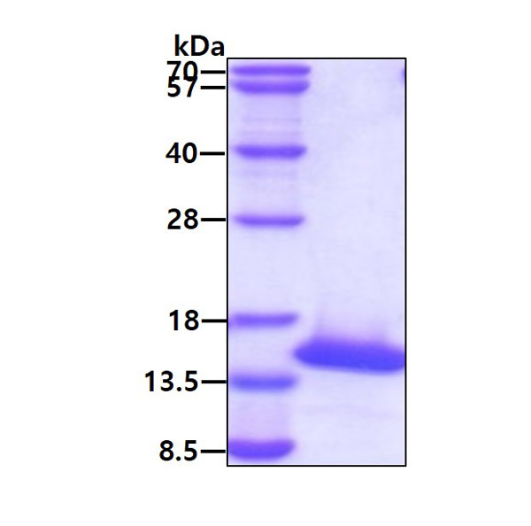
IL-21, Active Protein (Cat# AAA48379)
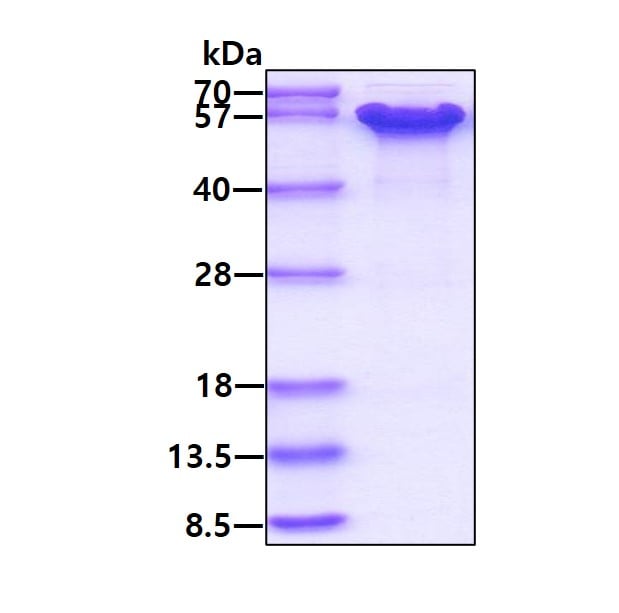
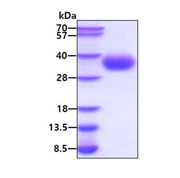
 SDS-PAGE
(3ug by SDS-PAGE under reducing condition and visualized by coomassie blue stain.)
SDS-PAGE
(3ug by SDS-PAGE under reducing condition and visualized by coomassie blue stain.)
PAI-1, Active Protein (Cat# AAA48474)
 SDS-PAGE
Interleukin-4, Active Protein (Cat# AAA48531)
HYALURONIDASE, Active Protein (Cat# AAA50501)
Cancer Antigen 72-4, Active Protein (Cat# AAA44847)
Elastase (ELA-2), Active Protein (Cat# AAA44742)
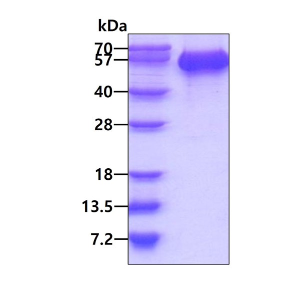
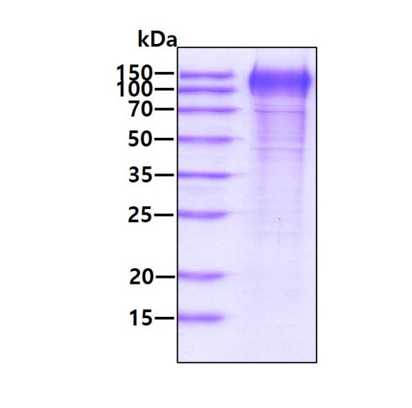
 SDS-PAGE
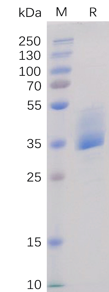
(Figure 1. Human BAFF-R Protein, mFc Tag on SDS-PAGE under reducing condition.)
SDS-PAGE
(Figure 1. Human BAFF-R Protein, mFc Tag on SDS-PAGE under reducing condition.)
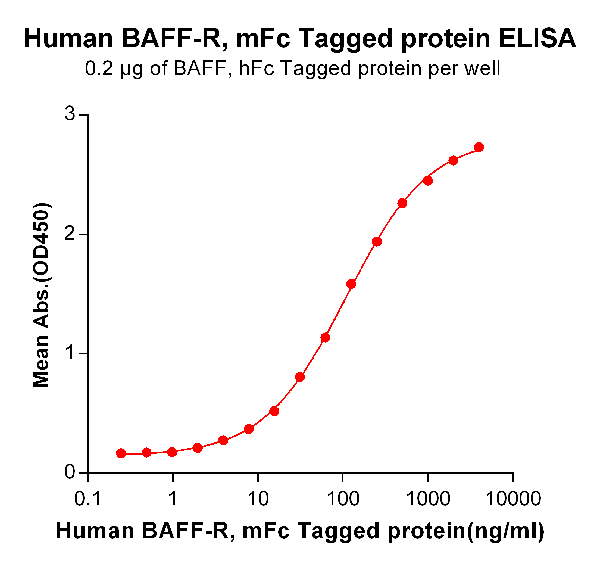
BAFF-R, Active Protein (Cat# AAA47223)
Leptin (Anti-Obesity Protein) (rHuLeptin), Active Protein (Cat# AAA47954)
Purification: Chromatographic
Vascular Endothelial Growth Factor (rHuVEGF), Active Protein (Cat# AAA47955)
Purification: Chromatographic
beta Nerve Growth Factor, Active Protein (Cat# AAA38883)
Interleukin-6, Active Protein (Cat# AAA38887)
Ciliary Neurotrophic Factor, Active Protein (Cat# AAA38894)
Interleukin-3, Active Protein (Cat# AAA38970)
Interleukin-8, Active Protein (Cat# AAA38079)
Monocyte Chemotactic Protein-1, Active Protein (Cat# AAA38086)
MIG, Active Protein (Cat# AAA38091)
Neurotrophin-1, Active Protein (Cat# AAA38018)
Granulocyte Macrophage, Active Protein (Cat# AAA38831)
Flt3-Ligand, Active Protein (Cat# AAA38706)
c-AMP dependant Protein Kinase A catalytic subunit alpha, Active Protein (Cat# AAA38382)
Superoxide Dismutase, Active Protein (Cat# AAA38419)
(a) Analysis by RP-HPLC.
(b) Analysis by SDS-PAGE.
Coagulation Factor IX, Active Protein (Cat# AAA38450)
Insulin Like Growth Factor-1, Active Protein (Cat# AAA38111)
Follistatin, Active Protein (Cat# AAA38122)
Morphogenetic protein-2, Active Protein (Cat# AAA38138)
Insulin Like Growth Factor-2, Active Protein (Cat# AAA38141)
Keratinocyte Growth Factor-2, Active Protein (Cat# AAA38152)
Stem Cell Factor, Active Protein (Cat# AAA38157)
Vascular Endothelial Growth Factor, Active Protein (Cat# AAA38168)
Interleukin-13, Active Protein (Cat# AAA38182)
BBetacellulin, Active Protein (Cat# AAA38191)
Staphylococcal Protein-A, Active Protein (Cat# AAA38452)
MHC class I chain-related gene A, Active Protein (Cat# AAA38458)
What Are Active Proteins?
Proteins are large molecules made up of long chains of amino acids.
They will typically fold into a very particular 3-dimensional shape/conformation, that is sometimes referred to as their “native” form, which allows them to work properly in the body. For the purposes of product categorization, AAA Biotech will typically refer to proteins purified from their original animal host as being “native” proteins (this is to signify their difference compared to their “recombinant” or “synthetic” protein counterparts).
If a protein successfully folds into the correct shape, it is will typically display high fidelity characteristics to its original protein in its original animal host, and be classified as an active protein, as it will be able to function “normally” in most enzymatic or binding capacities. If it loses this shape, due to factors such as heat or strong chemicals (such as detergents), it becomes inactive and is no longer able to perform its basic functions. All of the proteins in this category are made under strict quality control, and they are active, pure, low in contaminants, and stable.
Most are stored as freeze-dried powders and come without extra tags, so they’re very close to the actual natural/native form.
Key Applications of Active Proteins
1. Scientific Research
- Aid in the study of how proteins function in the body
- Aid in understanding various disease processes
2. Drug Development
- Powerful tools to investigate how potential drugs interact with specific proteins
- Ideal for identifying drug targets
3. Cell Culture
- Are routinely utilized to support cell growth and function (e.g., using exogenous growth factors)
- Can be used to promote cellular development into specific types (differentiation)
4. Diagnostics
- Regularly utilized in tests to detect diseases or infections (e.g., COVID-19, cancer)
- Note: All products are strictly for research-use only (RUO).
5. Therapeutics
- Some active proteins are used directly as treatments (e.g., insulin, enzymes)
- Note: All products are strictly for research-use only (RUO).
6. Vaccine Development
- Used to create or test vaccines by mimicking parts of viruses or bacteria
7. Biochemical Assays
- They can facilitate the characterization of enzyme activity, binding strength, or protein interactions in lab tests
Why Buy Active Proteins from AAA Biotech?
- High biological activity – Verified to perform as expected or indicated on datasheet
- Strict quality control – We are confident in our active proteins’ reliability and consistency
- High purity & low endotoxin – Ideal for applications involving sensitive or precious samples/components
- Freeze-dried for stability – Long shelf life and straightforward storage
- Mostly tag-free – Closer to natural/native protein form
FAQ
1. What are active proteins used for in research?
Active proteins are used primarily in the study of how proteins function, in characterizing/discovering drug interactions, supporting cell growth, running biochemical assays, and in development of diagnostics or therapeutics.
2. How are AAA Biotech's active proteins validated?
AAA Biotech’s active proteins are validated through strict quality control and functional assays to ensure they are properly folded and active. “Active”, though, can be an ambiguous term, so if a specific “activity” or “binding” capability of a protein is of crucial interest to you, please inquire with us prior to purchase, and we will provide further details on how the “Active” modifier was determined to be applicable.
3. Are these proteins tested for biological activity?
Yes, all active proteins from AAA Biotech are tested to confirm they have the expected biological activity before being offered for use. Though, said “biological activity” can be either “enzymatic”, “binding”, or both.