Filters
▼Clonality
▼Type
▼Reactivity
▼Gene Name
▼Isotype
▼Host
▼Application
▼Clone
▼Active Proteins
AAA Biotech also known as AAA Bio or AAABio provides a variety of high-quality recombinant and natural/native proteins that are proven to work in a wide range of experiments. Explore our products to find the active protein that best fits your needs or experimental model.
Viewing 2400-2450 of 2567 product results
Fibrinogen, Active Protein (Cat# AAA37779)
 Purity Data
(Sample Purity Data.)
Purity Data
(Sample Purity Data.)
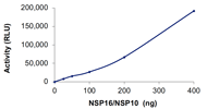
COVID 19 NSP10/NSP16 Methyltransferase Coronavirus, Active Protein (Cat# AAA73168)
 Bioactivity
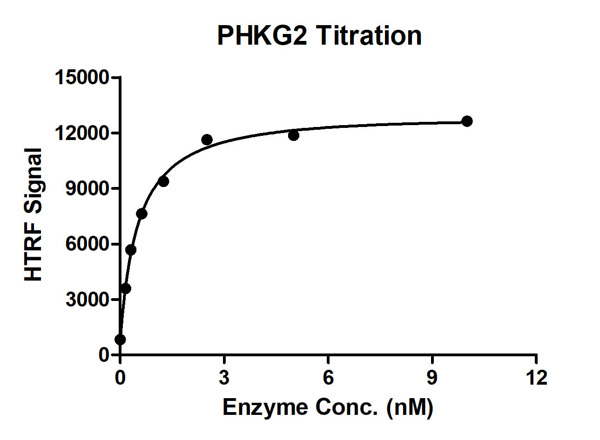
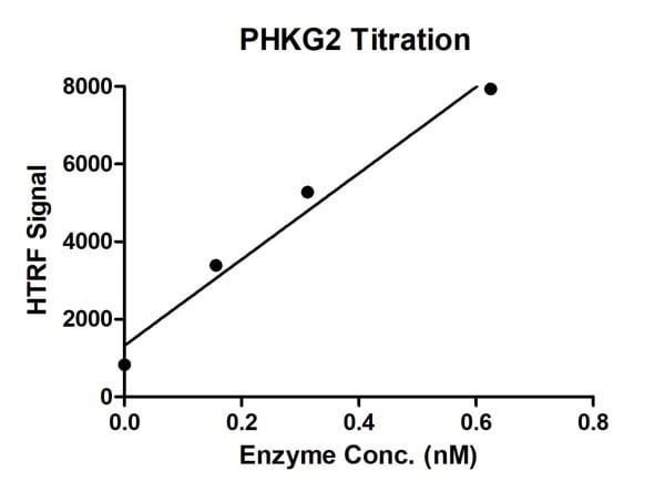
(HTRF Assay for Recombinant PHKG2 activity. 1 uM STK S1 substrate was incubated with different concentrations of PHKG2 protein in 10 ul reaction system containing 1×Enzymatic Buffer, 10 mM MgCl2, 1 mM DTT and 100 uM ATP for 1 hour. The 10 ul detection reagents containing anti-STK antibody (1:2) and SA-XL665 (1:100) diluted with 1× Detection Buffer were added and incubated with the reactions for 30 min. All the operations and reactions were performed at room temperature. HTRF assay was used for detection.)
Bioactivity
(HTRF Assay for Recombinant PHKG2 activity. 1 uM STK S1 substrate was incubated with different concentrations of PHKG2 protein in 10 ul reaction system containing 1×Enzymatic Buffer, 10 mM MgCl2, 1 mM DTT and 100 uM ATP for 1 hour. The 10 ul detection reagents containing anti-STK antibody (1:2) and SA-XL665 (1:100) diluted with 1× Detection Buffer were added and incubated with the reactions for 30 min. All the operations and reactions were performed at room temperature. HTRF assay was used for detection.)
Neuron-specific Enolase, Active Protein (Cat# AAA78132)
 Purity Data
(The purity of CD147 (22-205) was determined to be >90% by densitometry. Approx. MW 30-36 kDa.)
Purity Data
(The purity of CD147 (22-205) was determined to be >90% by densitometry. Approx. MW 30-36 kDa.)
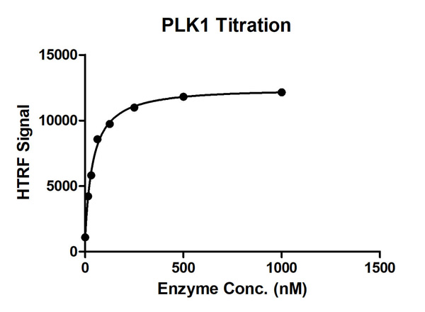
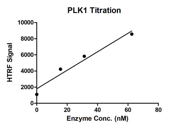 Bioactivity
(HTRF Assay for Recombinant PLK1 activity. 1 uM STK S1 substrate was incubated with different concentrations of PLK1 protein in 10 ul reaction system containing 1×Enzymatic Buffer, 5 mM MgCl2, 1 mM DTT and 100 uM ATP for 1 hour. The 10 ul detection reagents containing anti-STK antibody (1:2) and SA-XL665 (1:100) diluted with 1× Detection Buffer were added and incubated with the reactions for 30 min. All the operations and reactions were performed at room temperature. HTRF assay was used for detection.)
Bioactivity
(HTRF Assay for Recombinant PLK1 activity. 1 uM STK S1 substrate was incubated with different concentrations of PLK1 protein in 10 ul reaction system containing 1×Enzymatic Buffer, 5 mM MgCl2, 1 mM DTT and 100 uM ATP for 1 hour. The 10 ul detection reagents containing anti-STK antibody (1:2) and SA-XL665 (1:100) diluted with 1× Detection Buffer were added and incubated with the reactions for 30 min. All the operations and reactions were performed at room temperature. HTRF assay was used for detection.)
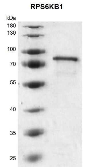
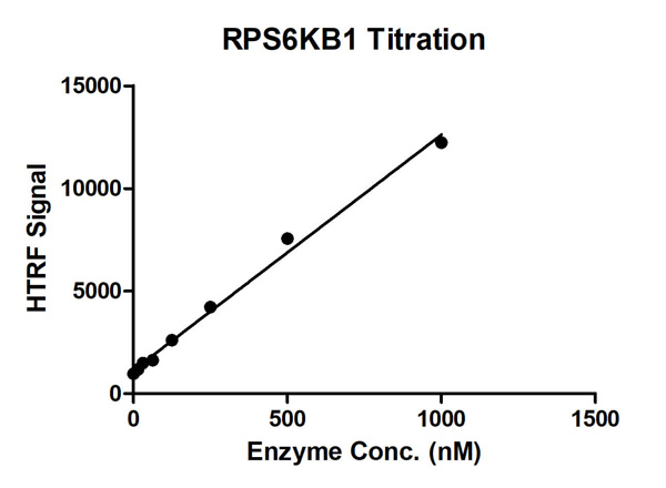 Bioactivity
(HTRF assay for RPS6KB1 activity 1 uM STK S3 substrate was incubated with different concentrations of RPS6KB1 protein in a 10 ul reaction system containing 1×Enzymatic Buffer, 2 mM MgCl2, 1 mM DTT, and 100 uM ATP for 1 hour. The 10 ul detection reagents containing anti-STK antibody (1:2) and SA-XL665 (1:100) diluted with 1× Detection Buffer were added and incubated with the reactions for 30 min. All the operations and reactions were performed at room temperature. HTRF assay was used for detection.)
Bioactivity
(HTRF assay for RPS6KB1 activity 1 uM STK S3 substrate was incubated with different concentrations of RPS6KB1 protein in a 10 ul reaction system containing 1×Enzymatic Buffer, 2 mM MgCl2, 1 mM DTT, and 100 uM ATP for 1 hour. The 10 ul detection reagents containing anti-STK antibody (1:2) and SA-XL665 (1:100) diluted with 1× Detection Buffer were added and incubated with the reactions for 30 min. All the operations and reactions were performed at room temperature. HTRF assay was used for detection.)
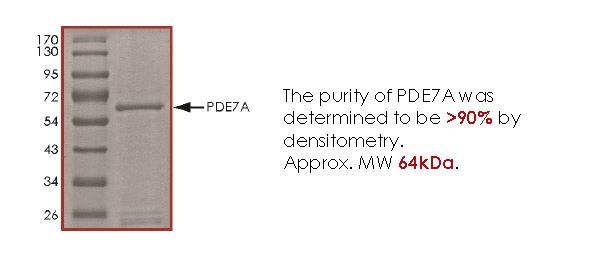 Purity Info
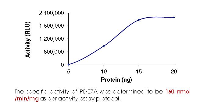
PDE7A, Active Protein (Cat# AAA72929)
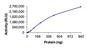
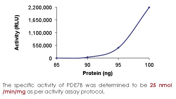
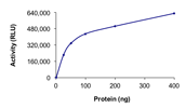
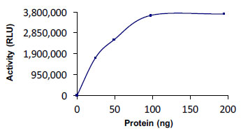
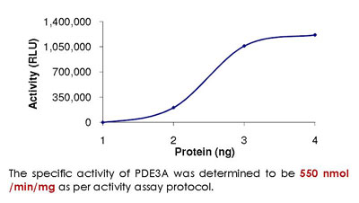
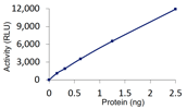 Specific Activity
(Sample Activity Plot.)
Specific Activity
(Sample Activity Plot.)
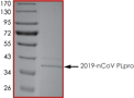
COVID 19 PLpro Coronavirus, Active Protein (Cat# AAA73170)
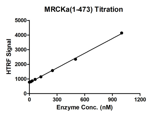 Bioactivity
(HTRF for MRCKa/CDC42BPA (1-473) activity 1 uM STK S3 substrate was incubated with different concentrations of MRCKa/CDC42BPA (1-473) protein in 10 ul reaction system containing 1×Enzymatic Buffer, 5 mM MgCl2, 1 mM DTT and 100 uM ATP for 1 hour. Then 10 ul detection reagents containing anti-STK antibody (1:2) and SA-XL665 (1:100) diluted with 1× Detection Buffer were added and incubated with the reactions for 30 min. All the operations and reactions were performed at room temperature. HTRF assay was used for detection.)
Bioactivity
(HTRF for MRCKa/CDC42BPA (1-473) activity 1 uM STK S3 substrate was incubated with different concentrations of MRCKa/CDC42BPA (1-473) protein in 10 ul reaction system containing 1×Enzymatic Buffer, 5 mM MgCl2, 1 mM DTT and 100 uM ATP for 1 hour. Then 10 ul detection reagents containing anti-STK antibody (1:2) and SA-XL665 (1:100) diluted with 1× Detection Buffer were added and incubated with the reactions for 30 min. All the operations and reactions were performed at room temperature. HTRF assay was used for detection.)
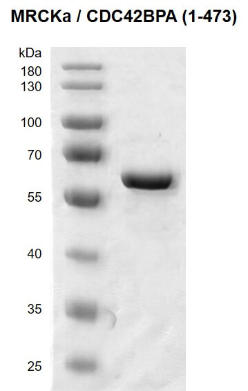
MRCKa/CDC42BPA, Active Protein (Cat# AAA60314)
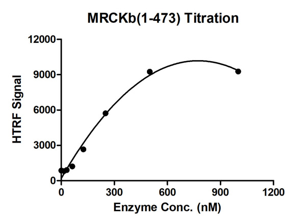
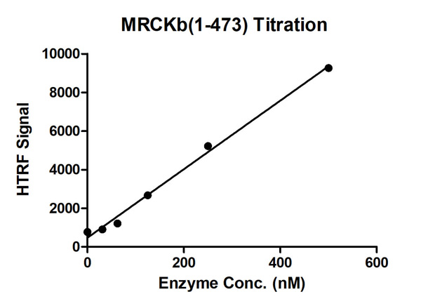 Bioactivity
(HTRF assay for Recombinant MRCKb/CDC42BPB (1-473) protein activity 1 uM STK S3 substrate was incubated with different concentrations of MRCKb/CDC42BPB (1-473) protein in a 10 ul reaction system containing 5 mM MgCl2, 1 mM DTT and 100 uM ATP for 1 hour. Then 10 ul detection reagents containing anti-STK antibody (1:2) GST and SA-XL665 mixture (each 1:100 diluted with 1X Detection Buffer were added and incubated with the reactions for 30 min. All the operations and reactions were performed at room temperature. HTRF assay was used for detection.)
Bioactivity
(HTRF assay for Recombinant MRCKb/CDC42BPB (1-473) protein activity 1 uM STK S3 substrate was incubated with different concentrations of MRCKb/CDC42BPB (1-473) protein in a 10 ul reaction system containing 5 mM MgCl2, 1 mM DTT and 100 uM ATP for 1 hour. Then 10 ul detection reagents containing anti-STK antibody (1:2) GST and SA-XL665 mixture (each 1:100 diluted with 1X Detection Buffer were added and incubated with the reactions for 30 min. All the operations and reactions were performed at room temperature. HTRF assay was used for detection.)
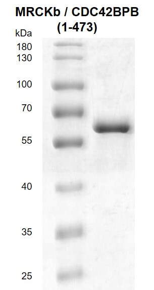
MRCKb/CDC42BPB, Active Protein (Cat# AAA60315)
Soluble Fibrin, Active Protein (Cat# AAA37871)
 Purity Data
(Sample Purity Data.)
Purity Data
(Sample Purity Data.)
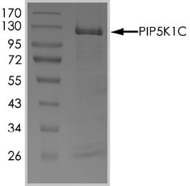 Purity Info
(The purity of PIP5K1C was detrermined to be >70% by densitometry approx. MW 120 kDa.)
Purity Info
(The purity of PIP5K1C was detrermined to be >70% by densitometry approx. MW 120 kDa.)
PIP5K1C, Active Protein (Cat# AAA73045)
 SDS-PAGE
(SDS-PAGE analysis: Lane 1: MW marker, Lane 2: 1ug TRAIL (soluble) (human), (recombinant) (His-tag) (Prod. No. BML-SE721).)
SDS-PAGE
(SDS-PAGE analysis: Lane 1: MW marker, Lane 2: 1ug TRAIL (soluble) (human), (recombinant) (His-tag) (Prod. No. BML-SE721).)
TRAIL, Active Protein (Cat# AAA77052)
Purified by multi-step chromatography
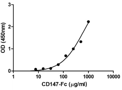
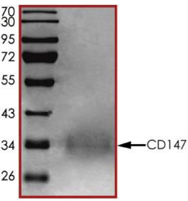
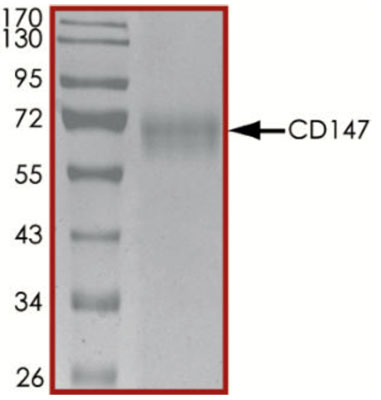 Purity Data
(The purity of CD147 was determined to be >90%by densitometry, approx. MW 70 kDa.)
Purity Data
(The purity of CD147 was determined to be >90%by densitometry, approx. MW 70 kDa.)
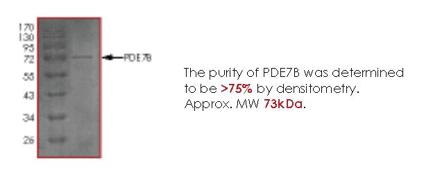 Purity Info
PDE7B, Active Protein (Cat# AAA73106)
 Application Data
(The purity of MLL2 (KMT2D) was determined to be >80% by densitometry, approx. MW 51kDa)
Application Data
(The purity of MLL2 (KMT2D) was determined to be >80% by densitometry, approx. MW 51kDa)
MLL2, Active Protein (Cat# AAA73022)
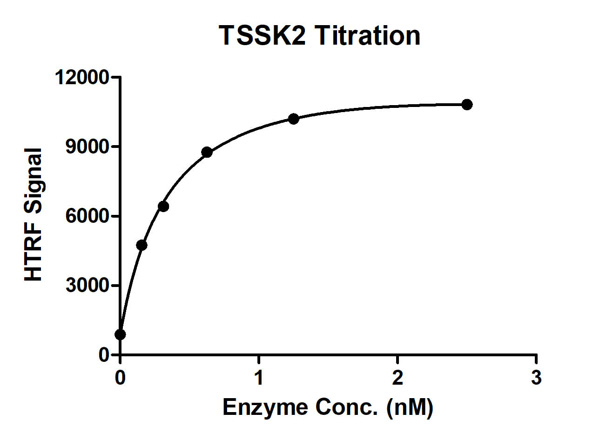
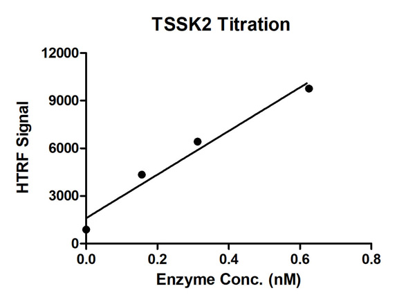 Bioactivity
(HTRF assay for TSSK2 activity 1 uM STK S1 substrate was incubated with different concentrations of TSSK2 protein in a 10 ul reaction system containing 1×Enzymatic Buffer, 10 mM MgCl2, 1 mM DTT, and 100 uM ATP for 1 hour. The 10 ul detection reagents containing anti-STK antibody (1:2) and SA-XL665 (1:100) diluted with 1× Detection Buffer were added and incubated with the reactions for 30 min. All the operations and reactions were performed at room temperature. HTRF assay was used for detection.)
Bioactivity
(HTRF assay for TSSK2 activity 1 uM STK S1 substrate was incubated with different concentrations of TSSK2 protein in a 10 ul reaction system containing 1×Enzymatic Buffer, 10 mM MgCl2, 1 mM DTT, and 100 uM ATP for 1 hour. The 10 ul detection reagents containing anti-STK antibody (1:2) and SA-XL665 (1:100) diluted with 1× Detection Buffer were added and incubated with the reactions for 30 min. All the operations and reactions were performed at room temperature. HTRF assay was used for detection.)
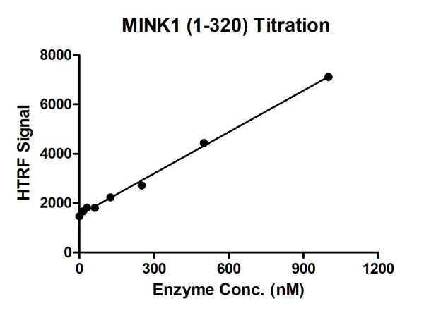 Bioactivity
(HTRF Assay for Recombinant MINK1 (1-320) activity. 1 uM STK S3 substrate was incubated with different concentrations of MINK1 (1-320) protein in 10 ul reaction system containing 1×Enzymatic Buffer, 5 mM MgCl2, 1 mM DTT and 100 uM ATP for 1 hour. The 10 ul detection reagents containing anti-STK antibody (1:2) and SA-XL665 (1:100) diluted with 1× Detection Buffer were added and incubated with the reactions for 30 min. All the operations and reactions were performed at room temperature. HTRF assay was used for detection.)
Bioactivity
(HTRF Assay for Recombinant MINK1 (1-320) activity. 1 uM STK S3 substrate was incubated with different concentrations of MINK1 (1-320) protein in 10 ul reaction system containing 1×Enzymatic Buffer, 5 mM MgCl2, 1 mM DTT and 100 uM ATP for 1 hour. The 10 ul detection reagents containing anti-STK antibody (1:2) and SA-XL665 (1:100) diluted with 1× Detection Buffer were added and incubated with the reactions for 30 min. All the operations and reactions were performed at room temperature. HTRF assay was used for detection.)
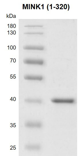
MINK1, Active Protein (Cat# AAA60330)
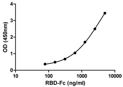
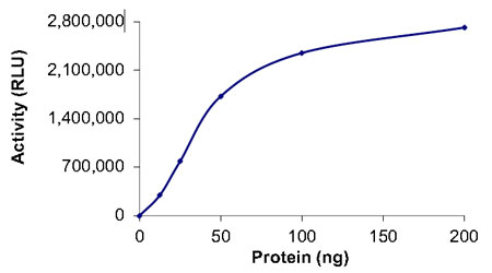
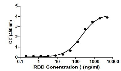 Bioactivity
(Figure 2. Binding ability measured in a functional ELISA. 2019-nCoV spike protein RBD (K417N, E484K, N501Y) binds to immobilized human ACE2 (19-740) protein)
Bioactivity
(Figure 2. Binding ability measured in a functional ELISA. 2019-nCoV spike protein RBD (K417N, E484K, N501Y) binds to immobilized human ACE2 (19-740) protein)
COVID 19 Spike protein RBD (K417N, E484K, N501Y) Coronavirus, Active Protein (Cat# AAA73185)
 Bioactivity
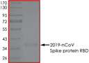
COVID 19 Spike RBD Coronavirus, Active Protein (Cat# AAA73162)

 Purity Data
(Sample purity Data.)
Purity Data
(Sample purity Data.)
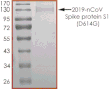
COVID 19 Spike Protein S1 (D614G) Coronavirus, Active Protein (Cat# AAA73174)
SARS Associated Coronavirus Spike, Active Protein (Cat# AAA38073)
 Bioactivity
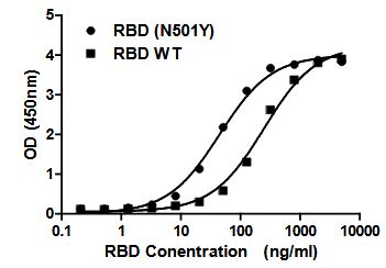
(Figure 2. Binding ability measured in a functional ELISA. Compared to 2019-nCoV spike protein RBD (C19SD-G241H), 2019-nCoV spike protein RBD (N501Y) exhibits increased binding potency forimmobilized human ACE2 (19-740) protein.)
Bioactivity
(Figure 2. Binding ability measured in a functional ELISA. Compared to 2019-nCoV spike protein RBD (C19SD-G241H), 2019-nCoV spike protein RBD (N501Y) exhibits increased binding potency forimmobilized human ACE2 (19-740) protein.)
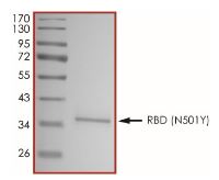
COVID 19 Spike protein RBD (N501Y) Coronavirus, Active Protein (Cat# AAA73184)
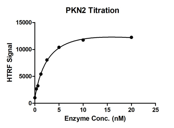
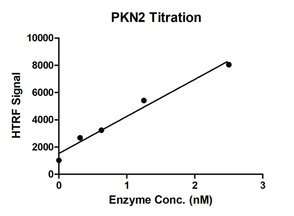 Bioactivity
(HTRF Assay for Recombinant PKN2 activity. 1 uM STK S1 substrate was incubated with different concentrations of PKN2 protein in 10 ul reaction system containing 1×Enzymatic Buffer, 5 mM MgCl2, 1 mM DTT and 100 uM ATP for 1 hour. The 10 ul detection reagents containing anti-STK antibody (1:2) and SA-XL665 (1:100) diluted with 1× Detection Buffer were added and incubated with the reactions for 30 min. All the operations and reactions were performed at room temperature. HTRF assay was used for detection.)
Bioactivity
(HTRF Assay for Recombinant PKN2 activity. 1 uM STK S1 substrate was incubated with different concentrations of PKN2 protein in 10 ul reaction system containing 1×Enzymatic Buffer, 5 mM MgCl2, 1 mM DTT and 100 uM ATP for 1 hour. The 10 ul detection reagents containing anti-STK antibody (1:2) and SA-XL665 (1:100) diluted with 1× Detection Buffer were added and incubated with the reactions for 30 min. All the operations and reactions were performed at room temperature. HTRF assay was used for detection.)
 SDS-PAGE
COVID 19 Spike Protein (RBD) Coronavirus, Active Protein (Cat# AAA176995)
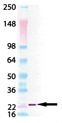
 SDS-PAGE
(3ug by SDS-PAGE under reducing condition and visualized by coomassie blue stain.)
SDS-PAGE
(3ug by SDS-PAGE under reducing condition and visualized by coomassie blue stain.)
IL15RA, Active Protein (Cat# AAA48968)
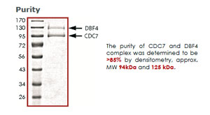
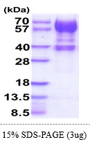
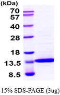
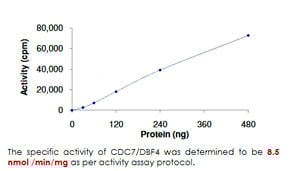 Application Data
CDC7/DBF4, Active Protein (Cat# AAA72963)
 Bioactivity
(Human IL-2 (C145S) stimulates cell proliferation of the CTLL2 mouse cytotoxic T cells. The ED50 range ≥0.65 ng/ml)
Bioactivity
(Human IL-2 (C145S) stimulates cell proliferation of the CTLL2 mouse cytotoxic T cells. The ED50 range ≥0.65 ng/ml)
Interleukin-2, Active Protein (Cat# AAA48459)

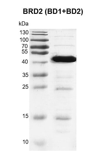
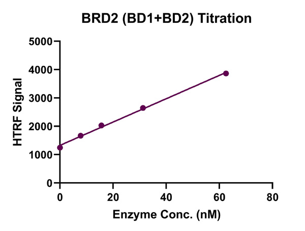 Bioactivity
(HTRF Assay for Recombinant BRD2 (BD1+BD2) activity. 3 uM histone peptide H4K5/8/12/16(ac4) was incubated with BRD2 (BD1+BD2) in reaction buffer including 50 mM HEPES-NaOH pH 7.0, 0.1% BSA for 1 hour at room temperature. Anti-His antibody was used to detect reaction products.)
Bioactivity
(HTRF Assay for Recombinant BRD2 (BD1+BD2) activity. 3 uM histone peptide H4K5/8/12/16(ac4) was incubated with BRD2 (BD1+BD2) in reaction buffer including 50 mM HEPES-NaOH pH 7.0, 0.1% BSA for 1 hour at room temperature. Anti-His antibody was used to detect reaction products.)
BRD2 (BD1+BD2), Active Protein (Cat# AAA60342)
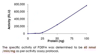
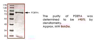 Purity Info
PDE9A, Active Protein (Cat# AAA73107)
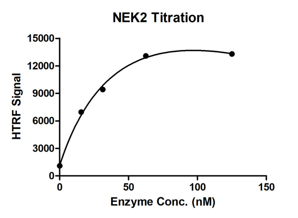
 Bioactivity
(HTRF assay for NEK2 activity 1 uM STK S3 substrate was incubated with different concentrations of NEK2 protein in a 10 ul reaction system containing 1×Enzymatic Buffer, 5 mM MgCl2, 1 mM DTT, and 100 uM ATP for 1 hour. Then 10 ul detection reagents containing anti-STK antibody (1:2) and SA-XL665 (1:100) diluted with 1× Detection Buffer were added and incubated with the reactions for 30 min. All the operations and reactions were performed at room temperature. HTRF assay was used for detection.)
Bioactivity
(HTRF assay for NEK2 activity 1 uM STK S3 substrate was incubated with different concentrations of NEK2 protein in a 10 ul reaction system containing 1×Enzymatic Buffer, 5 mM MgCl2, 1 mM DTT, and 100 uM ATP for 1 hour. Then 10 ul detection reagents containing anti-STK antibody (1:2) and SA-XL665 (1:100) diluted with 1× Detection Buffer were added and incubated with the reactions for 30 min. All the operations and reactions were performed at room temperature. HTRF assay was used for detection.)
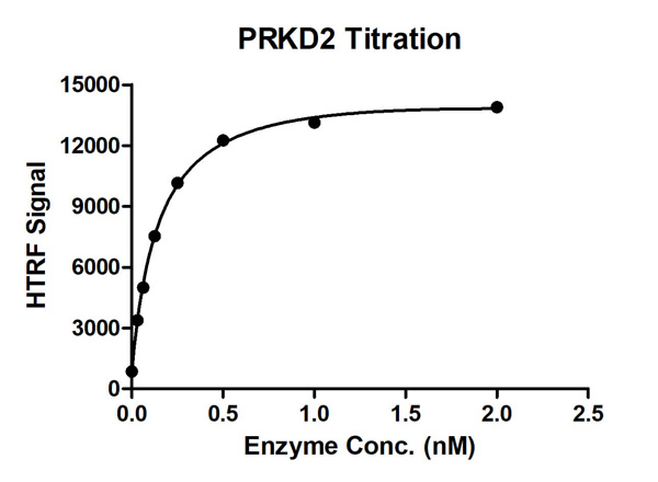

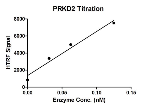 Bioactivity
(HTRF Assay for Recombinant PRKD2 activity. 1 uM STK S1 substrate was incubated with different concentrations of PRKD2 protein in 10 ul reaction system containing 1×Enzymatic Buffer, 5 mM MgCl2, 1 mM DTT and 100 uM ATP for 1 hour. The 10 ul detection reagents containing anti-STK antibody (1:2) and SA-XL665 (1:100) diluted with 1× Detection Buffer were added and incubated with the reactions for 30 min. All the operations and reactions were performed at room temperature. HTRF assay was used for detection.)
Bioactivity
(HTRF Assay for Recombinant PRKD2 activity. 1 uM STK S1 substrate was incubated with different concentrations of PRKD2 protein in 10 ul reaction system containing 1×Enzymatic Buffer, 5 mM MgCl2, 1 mM DTT and 100 uM ATP for 1 hour. The 10 ul detection reagents containing anti-STK antibody (1:2) and SA-XL665 (1:100) diluted with 1× Detection Buffer were added and incubated with the reactions for 30 min. All the operations and reactions were performed at room temperature. HTRF assay was used for detection.)
alpha-thrombin, Active Protein (Cat# AAA37807)
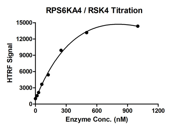
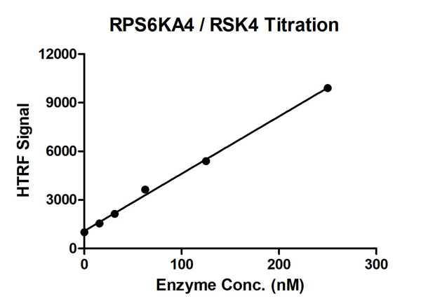 Bioactivity
(HTRF assay for RPS6KA4/RSK4 activity 1 uM STK S3 substrate was incubated with different concentrations of RPS6KA4/RSK4 protein in 10 ul reaction system containing 1×Enzymatic Buffer, 10 mM MgCl2, 1 mM DTT, and 100 uM ATP for 1 hour. The detection reagents were added and incubated with the reactions for 30 min. All operations and reactions were performed at room temperature, and HTRF assay was used to detect the enzymatic activity.)
Bioactivity
(HTRF assay for RPS6KA4/RSK4 activity 1 uM STK S3 substrate was incubated with different concentrations of RPS6KA4/RSK4 protein in 10 ul reaction system containing 1×Enzymatic Buffer, 10 mM MgCl2, 1 mM DTT, and 100 uM ATP for 1 hour. The detection reagents were added and incubated with the reactions for 30 min. All operations and reactions were performed at room temperature, and HTRF assay was used to detect the enzymatic activity.)
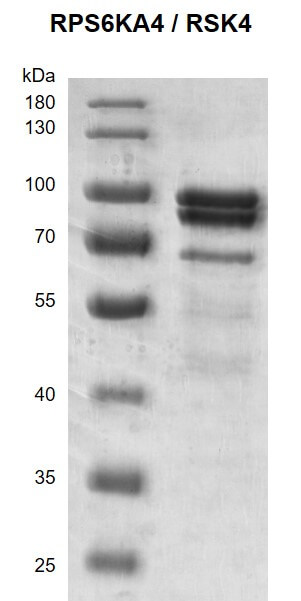
RPS6KA4/RSK4, Active Protein (Cat# AAA60317)
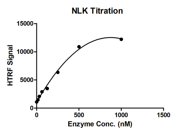
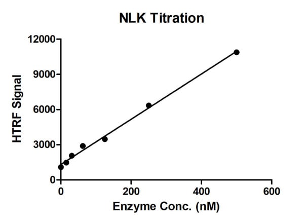 Bioactivity
(HTRF Assay for Recombinant NLK activity. 1 uM STK S3 substrate was incubated with different concentrations of NLK protein in 10 ul reaction system containing 1×Enzymatic Buffer, 5 mM MgCl2, 1 mM DTT and 100 uM ATP for 1 hour. The 10 ul detection reagents containing anti-STK antibody (1:2) and SA-XL665 (1:100) diluted with 1× Detection Buffer were added and incubated with the reactions for 30 min. All the operations and reactions were performed at room temperature. HTRF assay was used for detection.)
Bioactivity
(HTRF Assay for Recombinant NLK activity. 1 uM STK S3 substrate was incubated with different concentrations of NLK protein in 10 ul reaction system containing 1×Enzymatic Buffer, 5 mM MgCl2, 1 mM DTT and 100 uM ATP for 1 hour. The 10 ul detection reagents containing anti-STK antibody (1:2) and SA-XL665 (1:100) diluted with 1× Detection Buffer were added and incubated with the reactions for 30 min. All the operations and reactions were performed at room temperature. HTRF assay was used for detection.)
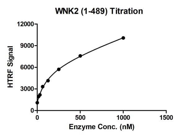
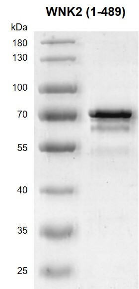
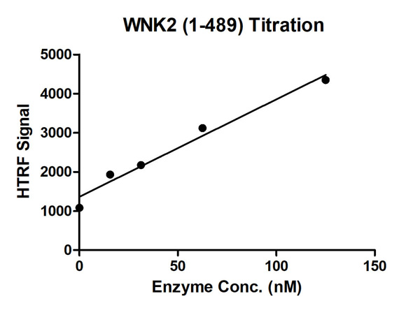 Bioactivity
(HTRF assay for WNK2 (1-489); activity 1 uM STK S3 substrate was incubated with different concentrations of WNK2 (1-489); protein in a 10 ul reaction system containing 1×Enzymatic Buffer, 5 mM MgCl2, 1 mM DTT and 100 uM ATP for 1 hour. The detection reagents were added and incubated with the reactions for 30 min. All the operations and reactions were performed at room temperature.)
Bioactivity
(HTRF assay for WNK2 (1-489); activity 1 uM STK S3 substrate was incubated with different concentrations of WNK2 (1-489); protein in a 10 ul reaction system containing 1×Enzymatic Buffer, 5 mM MgCl2, 1 mM DTT and 100 uM ATP for 1 hour. The detection reagents were added and incubated with the reactions for 30 min. All the operations and reactions were performed at room temperature.)
WNK2, Active Protein (Cat# AAA60337)
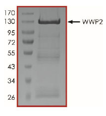 Purity Data
(The purity of WWP2 was determined to be >90% by densitometry, approx. MW 130 kDa.)
Purity Data
(The purity of WWP2 was determined to be >90% by densitometry, approx. MW 130 kDa.)
WWP2, Active Protein (Cat# AAA73157)
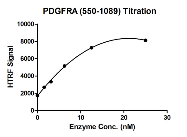

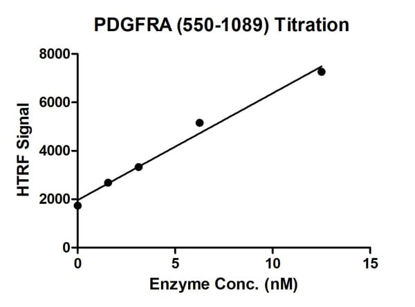 Bioactivity
(HTRF Assay for Recombinant PDGFRA (550-1089) activity. 1 uM TK substrate was incubated with different concentrations of PDGFRA (550-1089) protein in 10 ul reaction system containing 1×Enzymatic Buffer, 5 mM MgCl2, 1 mM DTT, 5 nM SEB and 100 uM ATP for 1 hour. The 10 ul detection reagents containing anti-STK antibody (1:2) and SA-XL665 (1:100) diluted with 1× Detection Buffer were added and incubated with the reactions for 30 min. All the operations and reactions were performed at room temperature. HTRF assay was used for detection.)
Bioactivity
(HTRF Assay for Recombinant PDGFRA (550-1089) activity. 1 uM TK substrate was incubated with different concentrations of PDGFRA (550-1089) protein in 10 ul reaction system containing 1×Enzymatic Buffer, 5 mM MgCl2, 1 mM DTT, 5 nM SEB and 100 uM ATP for 1 hour. The 10 ul detection reagents containing anti-STK antibody (1:2) and SA-XL665 (1:100) diluted with 1× Detection Buffer were added and incubated with the reactions for 30 min. All the operations and reactions were performed at room temperature. HTRF assay was used for detection.)
PDGFRA, Active Protein (Cat# AAA60339)
 SDS-PAGE
COVID 19 Spike S1 Protein Coronavirus, Active Protein (Cat# AAA176997)
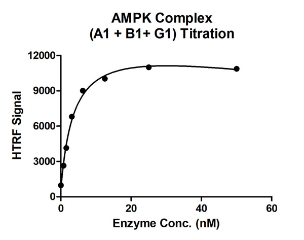
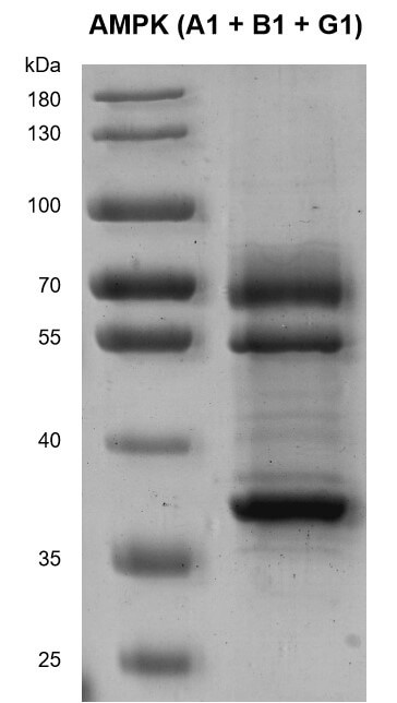
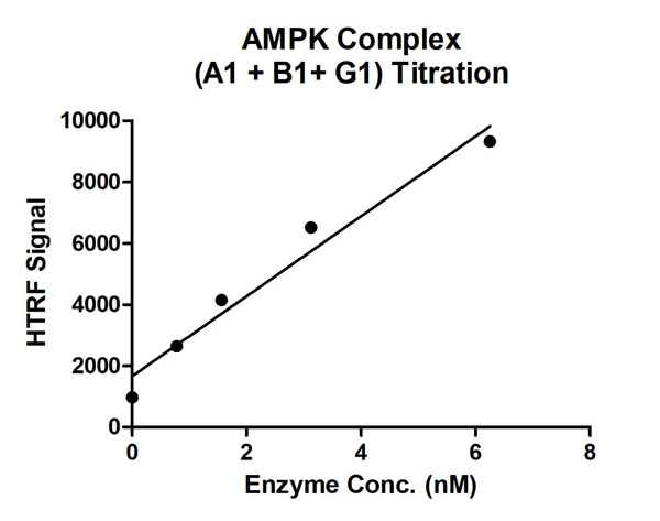 Bioactivity
(HTRF assay for AMPK Complex (A1+B1+G1) activity 1 uM STK S1 substrate was incubated with different concentrations of AMPK Complex (A1+B1+G1) protein in a 10 ul reaction system containing 1×Enzymatic Buffer, 5 mM MgCl2, 1 mM DTT, 50 uM AMP and 100 uM ATP for 1 hr. The 10 ul detection reagents containing anti-STK antibody (1:100) and SA-XL665 (1:100) diluted with 1× Detection Buffer were added and incubated with the reactions for 30 min. All the operations and reactions were performed at room temperature. HTRF assay was used for detection.)
Bioactivity
(HTRF assay for AMPK Complex (A1+B1+G1) activity 1 uM STK S1 substrate was incubated with different concentrations of AMPK Complex (A1+B1+G1) protein in a 10 ul reaction system containing 1×Enzymatic Buffer, 5 mM MgCl2, 1 mM DTT, 50 uM AMP and 100 uM ATP for 1 hr. The 10 ul detection reagents containing anti-STK antibody (1:100) and SA-XL665 (1:100) diluted with 1× Detection Buffer were added and incubated with the reactions for 30 min. All the operations and reactions were performed at room temperature. HTRF assay was used for detection.)
AMPK Complex (A1+B1+G1), Active Protein (Cat# AAA60345)
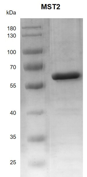
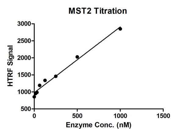 Bioactivity
(HTRF assay for MST2 activity 1 uM STK S3 substrate was incubated with different concentrations of MST2 protein in a 10 ul reaction system containing 1×Enzymatic Buffer, 10 mM MgCl2, 1 mM DTT, and 100 uM ATP for 1 hour. The 10 ul detection reagents containing anti-STK antibody (1:2) and SA-XL665 (1:100) diluted with 1× Detection Buffer were added and incubated with the reactions for 30 min. All the operations and reactions were performed at room temperature. HTRF assay was used for detection.)
Bioactivity
(HTRF assay for MST2 activity 1 uM STK S3 substrate was incubated with different concentrations of MST2 protein in a 10 ul reaction system containing 1×Enzymatic Buffer, 10 mM MgCl2, 1 mM DTT, and 100 uM ATP for 1 hour. The 10 ul detection reagents containing anti-STK antibody (1:2) and SA-XL665 (1:100) diluted with 1× Detection Buffer were added and incubated with the reactions for 30 min. All the operations and reactions were performed at room temperature. HTRF assay was used for detection.)
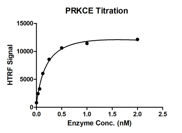
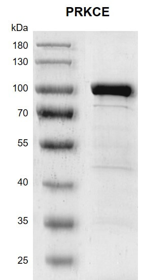
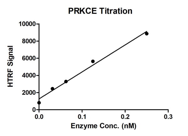 Bioactivity
(HTRF assay for PRKCE; activity 11 uM STK S1 substrate was incubated with different concentrations of PRKCE; protein in a 10 ul reaction system containing 1×Enzymatic Buffer, 10 mM MgCl2, 1 mM DTT and 100 uM ATP for 1 hour. The detection reagents were added and incubated with the reactions for 30 min. All the operations and reactions were performed at room temperature.)
Bioactivity
(HTRF assay for PRKCE; activity 11 uM STK S1 substrate was incubated with different concentrations of PRKCE; protein in a 10 ul reaction system containing 1×Enzymatic Buffer, 10 mM MgCl2, 1 mM DTT and 100 uM ATP for 1 hour. The detection reagents were added and incubated with the reactions for 30 min. All the operations and reactions were performed at room temperature.)
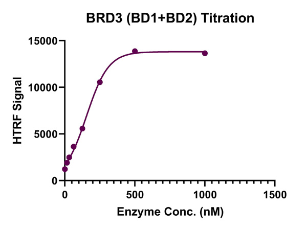
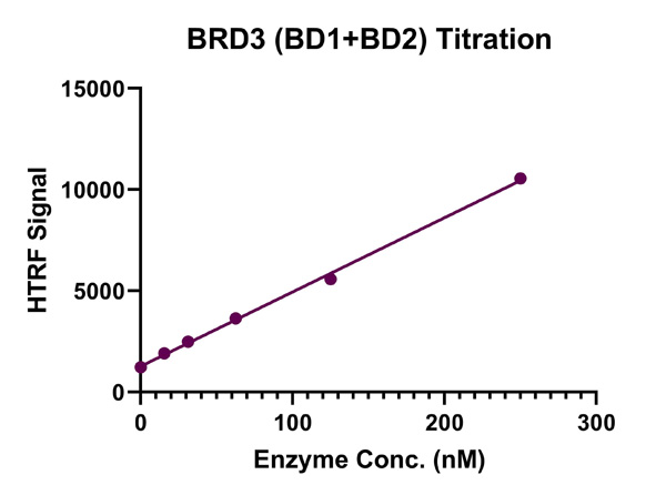 Bioactivity
(HTRF assay for BRD3 (BD1+BD2), Anti-His activity 3 uM histone peptide H4K5/8/12/16(ac4) was incubated with BRD3 (BD1+BD2) in reaction buffer including 50 mM HEPES-NaOH pH 7.4, 0.1% BSA for 1 hour at room temperature. Anti-His antibody was used to detect reaction products. All the operations and reactions were performed at room temperature. HTRF assay was used for detection.)
Bioactivity
(HTRF assay for BRD3 (BD1+BD2), Anti-His activity 3 uM histone peptide H4K5/8/12/16(ac4) was incubated with BRD3 (BD1+BD2) in reaction buffer including 50 mM HEPES-NaOH pH 7.4, 0.1% BSA for 1 hour at room temperature. Anti-His antibody was used to detect reaction products. All the operations and reactions were performed at room temperature. HTRF assay was used for detection.)
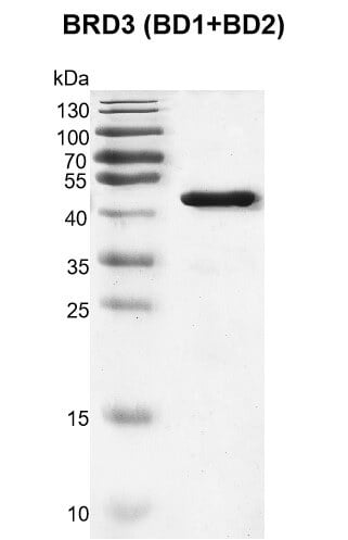
BRD3 (BD1+BD2), Active Protein (Cat# AAA60343)
 SDS-PAGE
GZMB, Active Protein (Cat# AAA48973)
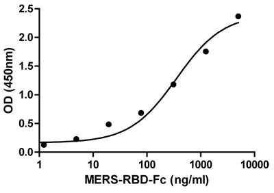
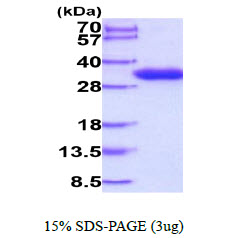
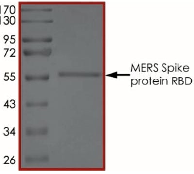 Purity Data
(The purity of MERS Spike Protein RBD was determined to be >90% by densitometry, approx. MW 58 kDa.)
Purity Data
(The purity of MERS Spike Protein RBD was determined to be >90% by densitometry, approx. MW 58 kDa.)
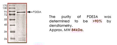 Purity Info
PDE3A, Active Protein (Cat# AAA73099)

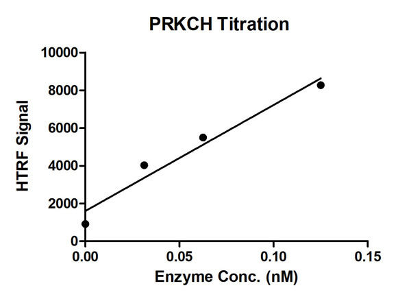 Bioactivity
(HTRF assay for PRKCH; activity 1 uM STK S1 substrate was incubated with different concentrations of PRKCH; protein in a 10 ul reaction system containing 1×Enzymatic Buffer, 10 mM MgCl2, 1 mM DTT and 100 uM ATP for 1 hour. The detection reagents were added and incubated with the reactions for 30 min. All the operations and reactions were performed at room temperature.)
Bioactivity
(HTRF assay for PRKCH; activity 1 uM STK S1 substrate was incubated with different concentrations of PRKCH; protein in a 10 ul reaction system containing 1×Enzymatic Buffer, 10 mM MgCl2, 1 mM DTT and 100 uM ATP for 1 hour. The detection reagents were added and incubated with the reactions for 30 min. All the operations and reactions were performed at room temperature.)
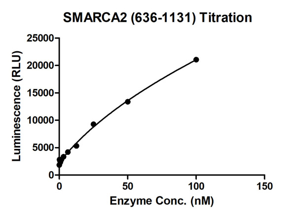 Bioactivity
(ADP-Glo assay for SMARCA (636-1131) activity (Data from ICEB Bioscience Inc.) 100 uM ATP and 10 nM DNA was incubated with different concentrations of SMARCA2 (636-1131) protein in a 10 ul reaction system containing 20 mM HEPES pH 7.5,10 mM MgCl2, 50 mM NaCl, 0.1%Tween-20, 1 mM DTT for 1 hour, 10 ul ADP-Glo Reagent was added to the products and incubated for 1 hour.Then 20 ul Kinase Detection Reagent incubated for 1 hour. All the operations and reactions were performed at RT. Luminescence measurement is collected by BMG.)
Bioactivity
(ADP-Glo assay for SMARCA (636-1131) activity (Data from ICEB Bioscience Inc.) 100 uM ATP and 10 nM DNA was incubated with different concentrations of SMARCA2 (636-1131) protein in a 10 ul reaction system containing 20 mM HEPES pH 7.5,10 mM MgCl2, 50 mM NaCl, 0.1%Tween-20, 1 mM DTT for 1 hour, 10 ul ADP-Glo Reagent was added to the products and incubated for 1 hour.Then 20 ul Kinase Detection Reagent incubated for 1 hour. All the operations and reactions were performed at RT. Luminescence measurement is collected by BMG.)
What Are Active Proteins?
Proteins are large molecules made up of long chains of amino acids.
They will typically fold into a very particular 3-dimensional shape/conformation, that is sometimes referred to as their “native” form, which allows them to work properly in the body. For the purposes of product categorization, AAA Biotech will typically refer to proteins purified from their original animal host as being “native” proteins (this is to signify their difference compared to their “recombinant” or “synthetic” protein counterparts).
If a protein successfully folds into the correct shape, it is will typically display high fidelity characteristics to its original protein in its original animal host, and be classified as an active protein, as it will be able to function “normally” in most enzymatic or binding capacities. If it loses this shape, due to factors such as heat or strong chemicals (such as detergents), it becomes inactive and is no longer able to perform its basic functions. All of the proteins in this category are made under strict quality control, and they are active, pure, low in contaminants, and stable.
Most are stored as freeze-dried powders and come without extra tags, so they’re very close to the actual natural/native form.
Key Applications of Active Proteins
1. Scientific Research
- Aid in the study of how proteins function in the body
- Aid in understanding various disease processes
2. Drug Development
- Powerful tools to investigate how potential drugs interact with specific proteins
- Ideal for identifying drug targets
3. Cell Culture
- Are routinely utilized to support cell growth and function (e.g., using exogenous growth factors)
- Can be used to promote cellular development into specific types (differentiation)
4. Diagnostics
- Regularly utilized in tests to detect diseases or infections (e.g., COVID-19, cancer)
- Note: All products are strictly for research-use only (RUO).
5. Therapeutics
- Some active proteins are used directly as treatments (e.g., insulin, enzymes)
- Note: All products are strictly for research-use only (RUO).
6. Vaccine Development
- Used to create or test vaccines by mimicking parts of viruses or bacteria
7. Biochemical Assays
- They can facilitate the characterization of enzyme activity, binding strength, or protein interactions in lab tests
Why Buy Active Proteins from AAA Biotech?
- High biological activity – Verified to perform as expected or indicated on datasheet
- Strict quality control – We are confident in our active proteins’ reliability and consistency
- High purity & low endotoxin – Ideal for applications involving sensitive or precious samples/components
- Freeze-dried for stability – Long shelf life and straightforward storage
- Mostly tag-free – Closer to natural/native protein form
FAQ
1. What are active proteins used for in research?
Active proteins are used primarily in the study of how proteins function, in characterizing/discovering drug interactions, supporting cell growth, running biochemical assays, and in development of diagnostics or therapeutics.
2. How are AAA Biotech's active proteins validated?
AAA Biotech’s active proteins are validated through strict quality control and functional assays to ensure they are properly folded and active. “Active”, though, can be an ambiguous term, so if a specific “activity” or “binding” capability of a protein is of crucial interest to you, please inquire with us prior to purchase, and we will provide further details on how the “Active” modifier was determined to be applicable.
3. Are these proteins tested for biological activity?
Yes, all active proteins from AAA Biotech are tested to confirm they have the expected biological activity before being offered for use. Though, said “biological activity” can be either “enzymatic”, “binding”, or both.