Filters
▼Clonality
▼Type
▼Reactivity
▼Gene Name
▼Isotype
▼Host
▼Application
▼Clone
▼Active Proteins
AAA Biotech also known as AAA Bio or AAABio provides a variety of high-quality recombinant and natural/native proteins that are proven to work in a wide range of experiments. Explore our products to find the active protein that best fits your needs or experimental model.
Viewing 2550-2567 of 2567 product results


 WB (Western Blot)
(Western BlotSample: Recombinant GAL9, Human;Antibody: Rabbit Anti-Human GAL9 Ab)
WB (Western Blot)
(Western BlotSample: Recombinant GAL9, Human;Antibody: Rabbit Anti-Human GAL9 Ab)
Galectin 9 (GAL9), Active Protein (Cat# AAA21103)


 WB (Western Blot)
(Western Blot Sample: Recombinant LCAT, Mouse;Antibody: Rabbit Anti-Mouse LCAT Ab)
WB (Western Blot)
(Western Blot Sample: Recombinant LCAT, Mouse;Antibody: Rabbit Anti-Mouse LCAT Ab)
Lecithin Cholesterol Acyltransferase (LCAT), Active Protein (Cat# AAA21104)
 SDS-PAGE
BCHE / Butyrylcholinesterase, Active Protein (Cat# AAA22092)


 WB (Western Blot)
(Sample: Recombinant TRAIL, Human;Antibody: Rabbit Anti-Human TRAIL Ab)
WB (Western Blot)
(Sample: Recombinant TRAIL, Human;Antibody: Rabbit Anti-Human TRAIL Ab)
Tumor Necrosis Factor Related Apoptosis Inducing Ligand (TRAIL), Active Protein (Cat# AAA21106)


 WB (Western Blot)
(Western Blot (WB) Sample: Recombinant ADIPOR1, Human; Antibody: Rabbit Anti-Human ADIPOR1 Ab)
WB (Western Blot)
(Western Blot (WB) Sample: Recombinant ADIPOR1, Human; Antibody: Rabbit Anti-Human ADIPOR1 Ab)
Adiponectin Receptor 1 (ADIPOR1), Active Protein (Cat# AAA21107)
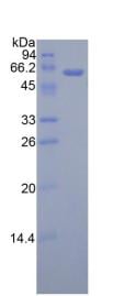
 SDS-PAGE
(3 ug by SDS-PAGE under reducing condition and visualized by coomassie blue stain)
SDS-PAGE
(3 ug by SDS-PAGE under reducing condition and visualized by coomassie blue stain)
NMNAT1, Active Protein (Cat# AAA11834)
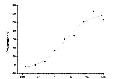 Activity
(The ED(50) was determined by the dose-dependent proliferation of human MCF-7 cells was found to be )
Activity
(The ED(50) was determined by the dose-dependent proliferation of human MCF-7 cells was found to be )
Neuregulin 1 beta, Active Protein (Cat# AAA14425)


 WB (Western Blot)
(Sample: Recombinant PRF1, Human;Antibody: Rabbit Anti-Human PRF1 Ab)
WB (Western Blot)
(Sample: Recombinant PRF1, Human;Antibody: Rabbit Anti-Human PRF1 Ab)
Perforin 1 (PRF1), Active Protein (Cat# AAA21142)
 SDS-PAGE
DPP4, Active Protein (Cat# AAA11760)


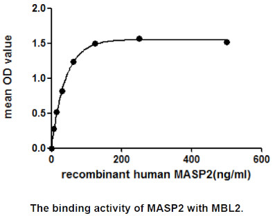

 WB (Western Blot)
(Sample: Recombinant MASP2, Human;Antibody: Rabbit Anti-Human MASP2 Ab)
WB (Western Blot)
(Sample: Recombinant MASP2, Human;Antibody: Rabbit Anti-Human MASP2 Ab)
Mannose Associated Serine Protease 2 (MASP2), Active Protein (Cat# AAA21058)
 SDS-PAGE
Galectin9, Active Protein (Cat# AAA11784)
 SDS-PAGE
Aldolase A, Active Protein (Cat# AAA11774)
 SDS-PAGE
Aldose reductase, Active Protein (Cat# AAA11762)
 SDS-PAGE
PKM2, Active Protein (Cat# AAA11792)


 WB (Western Blot)
(Sample: Recombinant GAL3, Human; Antibody: Rabbit Anti-Human GAL3 Ab)
WB (Western Blot)
(Sample: Recombinant GAL3, Human; Antibody: Rabbit Anti-Human GAL3 Ab)
Galectin 3 (GAL3), Active Protein (Cat# AAA21140)

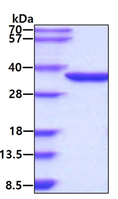
 SDS-PAGE
(SDS-PAGE of ~14 kDa Active Human Recombinant Alpha Synuclein Protein Monomer (SPR-321). Lane 1: Molecular Weight Ladder (MW). Lane 2: BSA (5 ug). Lane 3: BSA (2.5 ug). Lane 4: Active Alpha Synuclein Protein Monomer (5 ug) (SPR-321). Lane 5: Active Alpha Synuclein Protein Monomer (2.5 ug) (SPR-321).)
SDS-PAGE
(SDS-PAGE of ~14 kDa Active Human Recombinant Alpha Synuclein Protein Monomer (SPR-321). Lane 1: Molecular Weight Ladder (MW). Lane 2: BSA (5 ug). Lane 3: BSA (2.5 ug). Lane 4: Active Alpha Synuclein Protein Monomer (5 ug) (SPR-321). Lane 5: Active Alpha Synuclein Protein Monomer (2.5 ug) (SPR-321).)
Alpha Synuclein, Active Protein (Cat# AAA27658)
Purification: Ion-exchange Purified


 WB (Western Blot)
(Western Blot Sample: Recombinant SCF, Porcine; Antibody: Rabbit Anti-Porcine SCF Ab)
WB (Western Blot)
(Western Blot Sample: Recombinant SCF, Porcine; Antibody: Rabbit Anti-Porcine SCF Ab)
Stem Cell Factor (SCF), Active Protein (Cat# AAA21108)
What Are Active Proteins?
Proteins are large molecules made up of long chains of amino acids.
They will typically fold into a very particular 3-dimensional shape/conformation, that is sometimes referred to as their “native” form, which allows them to work properly in the body. For the purposes of product categorization, AAA Biotech will typically refer to proteins purified from their original animal host as being “native” proteins (this is to signify their difference compared to their “recombinant” or “synthetic” protein counterparts).
If a protein successfully folds into the correct shape, it is will typically display high fidelity characteristics to its original protein in its original animal host, and be classified as an active protein, as it will be able to function “normally” in most enzymatic or binding capacities. If it loses this shape, due to factors such as heat or strong chemicals (such as detergents), it becomes inactive and is no longer able to perform its basic functions. All of the proteins in this category are made under strict quality control, and they are active, pure, low in contaminants, and stable.
Most are stored as freeze-dried powders and come without extra tags, so they’re very close to the actual natural/native form.
Key Applications of Active Proteins
1. Scientific Research
- Aid in the study of how proteins function in the body
- Aid in understanding various disease processes
2. Drug Development
- Powerful tools to investigate how potential drugs interact with specific proteins
- Ideal for identifying drug targets
3. Cell Culture
- Are routinely utilized to support cell growth and function (e.g., using exogenous growth factors)
- Can be used to promote cellular development into specific types (differentiation)
4. Diagnostics
- Regularly utilized in tests to detect diseases or infections (e.g., COVID-19, cancer)
- Note: All products are strictly for research-use only (RUO).
5. Therapeutics
- Some active proteins are used directly as treatments (e.g., insulin, enzymes)
- Note: All products are strictly for research-use only (RUO).
6. Vaccine Development
- Used to create or test vaccines by mimicking parts of viruses or bacteria
7. Biochemical Assays
- They can facilitate the characterization of enzyme activity, binding strength, or protein interactions in lab tests
Why Buy Active Proteins from AAA Biotech?
- High biological activity – Verified to perform as expected or indicated on datasheet
- Strict quality control – We are confident in our active proteins’ reliability and consistency
- High purity & low endotoxin – Ideal for applications involving sensitive or precious samples/components
- Freeze-dried for stability – Long shelf life and straightforward storage
- Mostly tag-free – Closer to natural/native protein form
FAQ
1. What are active proteins used for in research?
Active proteins are used primarily in the study of how proteins function, in characterizing/discovering drug interactions, supporting cell growth, running biochemical assays, and in development of diagnostics or therapeutics.
2. How are AAA Biotech's active proteins validated?
AAA Biotech’s active proteins are validated through strict quality control and functional assays to ensure they are properly folded and active. “Active”, though, can be an ambiguous term, so if a specific “activity” or “binding” capability of a protein is of crucial interest to you, please inquire with us prior to purchase, and we will provide further details on how the “Active” modifier was determined to be applicable.
3. Are these proteins tested for biological activity?
Yes, all active proteins from AAA Biotech are tested to confirm they have the expected biological activity before being offered for use. Though, said “biological activity” can be either “enzymatic”, “binding”, or both.