Filters
▼Clonality
▼Type
▼Reactivity
▼Gene Name
▼Isotype
▼Host
▼Application
▼Clone
▼Active Proteins
AAA Biotech also known as AAA Bio or AAABio provides a variety of high-quality recombinant and natural/native proteins that are proven to work in a wide range of experiments. Explore our products to find the active protein that best fits your needs or experimental model.
Viewing 1700-1750 of 2567 product results
 Sequence
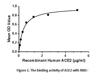
Angiotensin I Converting Enzyme 2 (ACE2), Active Protein (Cat# AAA150976)
 Bioactivity
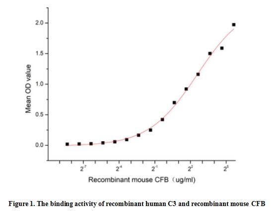
(Complement Component 3 (C3) is a vital component of the complement system that plays a central role in immune response and inflammation. C3 is a beta? globulin synthesized by the liver, whose structure consists of two polypeptide chains, alpha and beta, and is the most abundant complement component in serum. It's been identified that C3 can interact with Complement Factor B (CFB), thus a functional binding ELISA assay was conducted to detect the interaction of recombinant human C3 and recombinant mouse CFB. Briefly, CFB was diluted serially in PBS with 0.01% BSA (pH 7.4). Duplicate samples of 100 ul were then transferred to C3-coated microtiter wells and incubated for 1h at 37 degree C. Wells were washed with PBST and incubated for 1h with anti-CFB pAb, then aspirated and washed 3 times. After incubation with HRP labelled secondary antibody for 1h at 37 degree C, wells were aspirated and washed 5 times. With the addition of substrate solution, wells were incubated 15-25 minutes at 37 degree C. Finally, add 50 uL stop solution to the wells and read at 450/630 nm immediately. When Recombinant C3 is immobilized at 2 ug/mL (100 uL/well), the concentration of CFB that produces 50% optimal binding response is found to be approximately 5.96 ug/mL.)
Bioactivity
(Complement Component 3 (C3) is a vital component of the complement system that plays a central role in immune response and inflammation. C3 is a beta? globulin synthesized by the liver, whose structure consists of two polypeptide chains, alpha and beta, and is the most abundant complement component in serum. It's been identified that C3 can interact with Complement Factor B (CFB), thus a functional binding ELISA assay was conducted to detect the interaction of recombinant human C3 and recombinant mouse CFB. Briefly, CFB was diluted serially in PBS with 0.01% BSA (pH 7.4). Duplicate samples of 100 ul were then transferred to C3-coated microtiter wells and incubated for 1h at 37 degree C. Wells were washed with PBST and incubated for 1h with anti-CFB pAb, then aspirated and washed 3 times. After incubation with HRP labelled secondary antibody for 1h at 37 degree C, wells were aspirated and washed 5 times. With the addition of substrate solution, wells were incubated 15-25 minutes at 37 degree C. Finally, add 50 uL stop solution to the wells and read at 450/630 nm immediately. When Recombinant C3 is immobilized at 2 ug/mL (100 uL/well), the concentration of CFB that produces 50% optimal binding response is found to be approximately 5.96 ug/mL.)
Complement Component 3 (C3), Active Protein (Cat# AAA161793)
 Bioactivity
(Retinol-binding protein 4 (RBP4) is the specific carrier for retinol (also known as vitamin A), and is responsible for the conversion of unstable and insoluble retinol in aqueous solution into stable and soluble complex in plasma through their tight interaction. As a member of the lipocalin superfamily, RBP4 containing a beta-barrel structure with a well-defined cavity is secreted from the liver, and in turn delivers retinol from the liver stores to the peripheral tissues. In plasma, the RBP4-retinol complex interacts with transthyretin (TTR), and this binding is crucial for preventing RBP4 excretion through the kidney glomeruli. RBP4 expressed from an ectopic source efficiently delivers retinol to the eyes, and its deficiency affects night vision largely. Recently, RBP4 as an adipokine, is found to be expressed in adipose tissue and correlated with obesity, insulin resistance (IR) and type 2 diabetes (T2DM). The activity of recombinant mouse RBP4 was measured by its ability to bind all-trans retinoic acid. The binding of retinoic acid results in the quenching of Trp fluorescence in RBP4. RBP4 was diluted to 50 ug/ml in 50 mM Tris, 10 mM CaCl2, 150 mM NaCl, pH 7.5 (assay buffer) and the retinoic acid was diluted to 800, 400, 200, 100, 30, 10, 3, 1, 0.5 and 0.1 uM in 95% ethanol. Mixing 112.5 uL of 50 ug/mL rmRBP4 and 12.5 uL of retinoic acid serial dilutions in microtubes and a blank containing 112.5 uL of 50 ug/mL rmRBP4 and 12.5 uL of 95% ethanol, then incubate at room temperature for 30 minutes. Loading 100 ul of the reaction mixtures and blank and read at excitation and emission wavelengths of 280 nm and 340 nm (top read), respectively, in endpoint mode. The result was shown in figure 1, the 50% binding concentration (BC50) is > 4.6 uM.)
Bioactivity
(Retinol-binding protein 4 (RBP4) is the specific carrier for retinol (also known as vitamin A), and is responsible for the conversion of unstable and insoluble retinol in aqueous solution into stable and soluble complex in plasma through their tight interaction. As a member of the lipocalin superfamily, RBP4 containing a beta-barrel structure with a well-defined cavity is secreted from the liver, and in turn delivers retinol from the liver stores to the peripheral tissues. In plasma, the RBP4-retinol complex interacts with transthyretin (TTR), and this binding is crucial for preventing RBP4 excretion through the kidney glomeruli. RBP4 expressed from an ectopic source efficiently delivers retinol to the eyes, and its deficiency affects night vision largely. Recently, RBP4 as an adipokine, is found to be expressed in adipose tissue and correlated with obesity, insulin resistance (IR) and type 2 diabetes (T2DM). The activity of recombinant mouse RBP4 was measured by its ability to bind all-trans retinoic acid. The binding of retinoic acid results in the quenching of Trp fluorescence in RBP4. RBP4 was diluted to 50 ug/ml in 50 mM Tris, 10 mM CaCl2, 150 mM NaCl, pH 7.5 (assay buffer) and the retinoic acid was diluted to 800, 400, 200, 100, 30, 10, 3, 1, 0.5 and 0.1 uM in 95% ethanol. Mixing 112.5 uL of 50 ug/mL rmRBP4 and 12.5 uL of retinoic acid serial dilutions in microtubes and a blank containing 112.5 uL of 50 ug/mL rmRBP4 and 12.5 uL of 95% ethanol, then incubate at room temperature for 30 minutes. Loading 100 ul of the reaction mixtures and blank and read at excitation and emission wavelengths of 280 nm and 340 nm (top read), respectively, in endpoint mode. The result was shown in figure 1, the 50% binding concentration (BC50) is > 4.6 uM.)
Retinol Binding Protein 4 (RBP4), Active Protein (Cat# AAA161806)
 Bioactivity
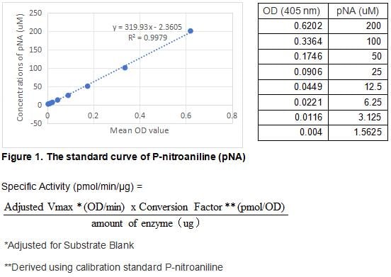
(Kallikrein 3, commonly known as prostate specific antigen (PSA), is a serine protease of the human tissue Kallikrein gene family. PSA is synthesized in the ductal and acinar epithelium of the prostate gland and secreted into the seminal plasma in high concentrations (0.5 - 2 g/L). A small portion of PSA "leaks" into the systemic circulation, the levels of which increase significantly (30-fold) from prostate cancer tissue than normal prostate tissue. PSA has become a well established tumor marker that aids the identification, staging, and follow up of prostate cancer. The activity of recombinant human KLK3 is measured by its ability to cleave a colorimetric peptide substrate Suc-Arg-Pro-Tyr-pNa in the assay buffer 50 mM Tris, 1 M NaCl, pH 8.0. The rhKLK3 is diluted to 200 ug/ml in activation buffer 50 mM Tris, 10 mM CaCl2, 150 mM NaCl, 0.05% Brij-35, pH 7.5, then activated with a final concentration of 1 ug/ml Thermolysin at 37 degree C for 5min. Adding a final concentration of 10 mM 1,10 Phenanthroline to stop the activation. The activated rhKLK3 is diluted to 50 ug/mL in assay buffer. Loading into a clear well plate 50 uL of 50 ug/mL rhKLK3 and start the reaction by adding 50 uL of 2 mM substrate, with a substrate blank containing 50 uL assay buffer, 50 uL substrate, and no rhKLK3. Then read at 405 nm in kinetic mode for 5 minutes. The specific activity of recombinant human KLK3 is > 400 pmol/min/ug.)
Bioactivity
(Kallikrein 3, commonly known as prostate specific antigen (PSA), is a serine protease of the human tissue Kallikrein gene family. PSA is synthesized in the ductal and acinar epithelium of the prostate gland and secreted into the seminal plasma in high concentrations (0.5 - 2 g/L). A small portion of PSA "leaks" into the systemic circulation, the levels of which increase significantly (30-fold) from prostate cancer tissue than normal prostate tissue. PSA has become a well established tumor marker that aids the identification, staging, and follow up of prostate cancer. The activity of recombinant human KLK3 is measured by its ability to cleave a colorimetric peptide substrate Suc-Arg-Pro-Tyr-pNa in the assay buffer 50 mM Tris, 1 M NaCl, pH 8.0. The rhKLK3 is diluted to 200 ug/ml in activation buffer 50 mM Tris, 10 mM CaCl2, 150 mM NaCl, 0.05% Brij-35, pH 7.5, then activated with a final concentration of 1 ug/ml Thermolysin at 37 degree C for 5min. Adding a final concentration of 10 mM 1,10 Phenanthroline to stop the activation. The activated rhKLK3 is diluted to 50 ug/mL in assay buffer. Loading into a clear well plate 50 uL of 50 ug/mL rhKLK3 and start the reaction by adding 50 uL of 2 mM substrate, with a substrate blank containing 50 uL assay buffer, 50 uL substrate, and no rhKLK3. Then read at 405 nm in kinetic mode for 5 minutes. The specific activity of recombinant human KLK3 is > 400 pmol/min/ug.)
Prostate Specific Antigen (PSA), Active Protein (Cat# AAA161711)
 Bioactivity
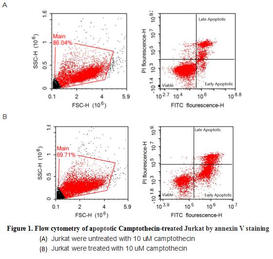
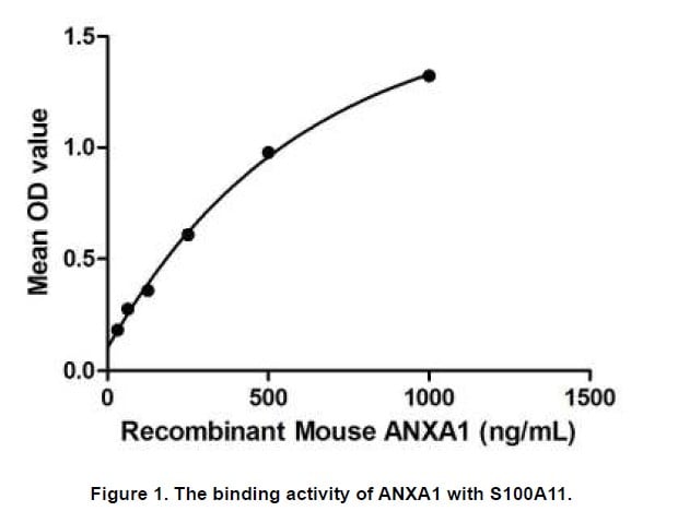
(Annexin V (ANXA5) is a multifunctional protein that is highly expressed on the apical surfaces of syncytiotrophoblasts, and plays an important role in haemostatic regulations, maintaining blood fluidity of the placenta. Lower ANXA5 levels have been observed in M2/ANXA5 haplotype carrying chorion. The association found between the maternal carriage of the M2/ANXA5 haplotype and an elevated risk of IUGR and/or PE supports the hypothesis that carrier status of this haplotype and the consequently reduced placental ANXA5 expression might be responsible, at least partially, for the onset of these gestational vascular complications. Annexin V is a calcium-dependent phospholipid binding protein that can be used to bind Phosphatidylserine (PS) during an early apoptosis event where the PS becomes exposed at the cell surface. Jurkat cells were treated with 10 uM camptothecin for 4h, 2*105 cells which were resuspended in binding buffer were stained with 5 ug recombinant human Annexin V-GFP and 10 ul Propidium iodide (PI) for 20min in dark room temperature. The flow cytometry was used to detect the early apoptotic and late apoptotic of camptothecin-treated Jurkat cells (Figure 1), the combination of Annexin V-GFP and propidium iodide allows for the distinction between early apoptotic cells (Annexin V-FITC positive and propidium iodide negative), late apoptotic and/or necrotic cells (Annexin V-FITC and propidium iodide positive), and viable cells (unstained). Thus, the recombinant human Annexin V-GFP can bind Phosphatidylserine (PS) at early apoptosis of Jurkat.)
Bioactivity
(Annexin V (ANXA5) is a multifunctional protein that is highly expressed on the apical surfaces of syncytiotrophoblasts, and plays an important role in haemostatic regulations, maintaining blood fluidity of the placenta. Lower ANXA5 levels have been observed in M2/ANXA5 haplotype carrying chorion. The association found between the maternal carriage of the M2/ANXA5 haplotype and an elevated risk of IUGR and/or PE supports the hypothesis that carrier status of this haplotype and the consequently reduced placental ANXA5 expression might be responsible, at least partially, for the onset of these gestational vascular complications. Annexin V is a calcium-dependent phospholipid binding protein that can be used to bind Phosphatidylserine (PS) during an early apoptosis event where the PS becomes exposed at the cell surface. Jurkat cells were treated with 10 uM camptothecin for 4h, 2*105 cells which were resuspended in binding buffer were stained with 5 ug recombinant human Annexin V-GFP and 10 ul Propidium iodide (PI) for 20min in dark room temperature. The flow cytometry was used to detect the early apoptotic and late apoptotic of camptothecin-treated Jurkat cells (Figure 1), the combination of Annexin V-GFP and propidium iodide allows for the distinction between early apoptotic cells (Annexin V-FITC positive and propidium iodide negative), late apoptotic and/or necrotic cells (Annexin V-FITC and propidium iodide positive), and viable cells (unstained). Thus, the recombinant human Annexin V-GFP can bind Phosphatidylserine (PS) at early apoptosis of Jurkat.)
Annexin V (ANXA5), Active Protein (Cat# AAA161724)

 Bioactivity
(Fas ligand (FasL) is a 40 kDa type II membrane protein belonging to the TNF family. In the new TNF super family nomenclature, FasL is referred to as TNFSF6. The specific receptor for FasL is Fas (CD95, Apo-1), a 45 kDa type I transmembrane protein that is a member of the TNF receptor family. FasL is predominantly expressed on activated T cells and NK cells, while Fas is expressed on various types of cells. The Fas/FasL system plays a crucial role in modulating immune response by inducing cell apoptosis to maintain homeostasis, self-tolerance of lymphocytes, and immune privilege. FasL was reported to be a potent chemoattractant for neutrophils, suggesting a novel proinflammatory function of this molecule. A functional ELISA assay was conducted to detect the interaction of recombinant human FASL and recombinant human FAS. Briefly, FASL was diluted serially in PBS with 0.01% BSA (pH 7.4). Duplicate samples of 100 ul were then transferred to FAS-coated microtiter wells and incubated for 1h at 37 degree C. Wells were washed with PBST and incubated for 1h with anti-FASL pAb, then aspirated and washed 3 times. After incubation with HRP labelled secondary antibody for 1h at 37 degree C, wells were aspirated and washed 5 times. With the addition of substrate solution, wells were incubated 15-25 minutes at 37 degree C. Finally, add 50 uL stop solution to the wells and read at 450/630nm immediately. The binding activity of recombinant human FASL and recombinant human FAS was shown in Figure 1, the EC50 for this effect is 0.044 ug/mL.)
Bioactivity
(Fas ligand (FasL) is a 40 kDa type II membrane protein belonging to the TNF family. In the new TNF super family nomenclature, FasL is referred to as TNFSF6. The specific receptor for FasL is Fas (CD95, Apo-1), a 45 kDa type I transmembrane protein that is a member of the TNF receptor family. FasL is predominantly expressed on activated T cells and NK cells, while Fas is expressed on various types of cells. The Fas/FasL system plays a crucial role in modulating immune response by inducing cell apoptosis to maintain homeostasis, self-tolerance of lymphocytes, and immune privilege. FasL was reported to be a potent chemoattractant for neutrophils, suggesting a novel proinflammatory function of this molecule. A functional ELISA assay was conducted to detect the interaction of recombinant human FASL and recombinant human FAS. Briefly, FASL was diluted serially in PBS with 0.01% BSA (pH 7.4). Duplicate samples of 100 ul were then transferred to FAS-coated microtiter wells and incubated for 1h at 37 degree C. Wells were washed with PBST and incubated for 1h with anti-FASL pAb, then aspirated and washed 3 times. After incubation with HRP labelled secondary antibody for 1h at 37 degree C, wells were aspirated and washed 5 times. With the addition of substrate solution, wells were incubated 15-25 minutes at 37 degree C. Finally, add 50 uL stop solution to the wells and read at 450/630nm immediately. The binding activity of recombinant human FASL and recombinant human FAS was shown in Figure 1, the EC50 for this effect is 0.044 ug/mL.)
Factor Related Apoptosis Ligand (FASL), Active Protein (Cat# AAA161660)
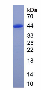
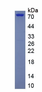
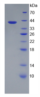
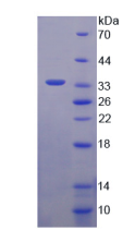
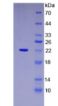
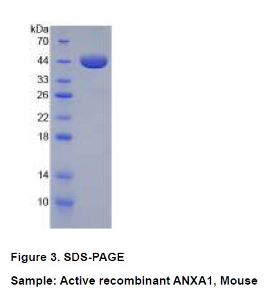
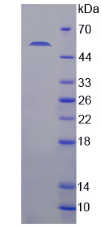
 SDS-PAGE
(Figure. SDS-PAGE)
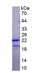
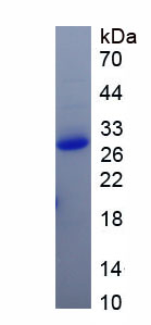
SDS-PAGE
(Figure. SDS-PAGE)
C ReProtein (CRP), Active Protein (Cat# AAA153060)
 Bioactivity
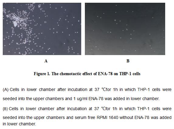
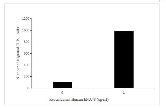
(Figure 2. The chemotactic effect of ENA-78 on THP-1 cells)
Bioactivity
(Figure 2. The chemotactic effect of ENA-78 on THP-1 cells)
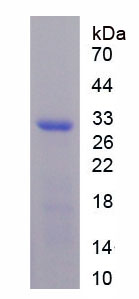
Epithelial Neutrophil Activating Peptide 78 (ENA78), Active Protein (Cat# AAA153063)
 Bioactivity
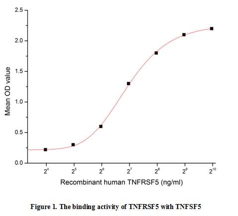
(CD40, also known as Tumor Necrosis Factor Receptor Superfamily, Member 5 (TNFRSF5), is a 45-50 kDa type I transmembrane glycoprotein member of the TNF receptor superfamily. Mature human CD40 consists of a 173 amino acid (aa) extracellular domain, a transm)
Bioactivity
(CD40, also known as Tumor Necrosis Factor Receptor Superfamily, Member 5 (TNFRSF5), is a 45-50 kDa type I transmembrane glycoprotein member of the TNF receptor superfamily. Mature human CD40 consists of a 173 amino acid (aa) extracellular domain, a transm)
Tumor Necrosis Factor Receptor Superfamily, Member 5 (CD40), Active Protein (Cat# AAA153079)
 Bioactivity
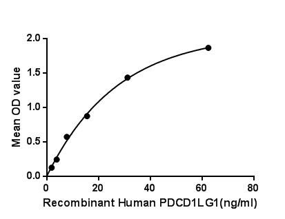
(Programmed Cell Death Protein 1 Ligand 1 (PDCD1LG1) also known as cluster of differentiation 274 (CD274) or B7 homolog 1 (B7-H1) is a 40kDa type 1 transmembrane protein that has been speculated to play a major role in suppressing the immune system during particular events such as pregnancy, tissue allografts, autoimmune disease and other disease states such as hepatitis. Besides, Programmed Cell Death Protein 1 (PDCD1) has been identified as an interactor of PDCD1LG1, thus a binding ELISA assay was conducted to detect the interaction of recombinant human PDCD1LG1 and recombinant human PDCD1. Briefly, PDCD1LG1 were diluted serially in PBS, with 0.01% BSA (pH 7.4). Duplicate samples of 100uL were then transferred to PDCD1-coated microtiter wells and incubated for 2h at 37. Wells were washed with PBST and incubated for 1h with anti-PDCD1LG1 pAb, then aspirated and washed 3 times. After incubation with HRP labelled secondary antibody, wells were aspirated and washed 3 times. With the addition of substrate solution, wells were incubated 15-25 minutes at 37. Finally, add 50uL stop solution to the wells and read at 450nm immediately. The binding activity of PDCD1LG1 and PDCD1 was shown in Figure 1, and this effect was in a dose dependent manner.Figure. The binding activity of PDCD1LG1 with PDCD1.)
Bioactivity
(Programmed Cell Death Protein 1 Ligand 1 (PDCD1LG1) also known as cluster of differentiation 274 (CD274) or B7 homolog 1 (B7-H1) is a 40kDa type 1 transmembrane protein that has been speculated to play a major role in suppressing the immune system during particular events such as pregnancy, tissue allografts, autoimmune disease and other disease states such as hepatitis. Besides, Programmed Cell Death Protein 1 (PDCD1) has been identified as an interactor of PDCD1LG1, thus a binding ELISA assay was conducted to detect the interaction of recombinant human PDCD1LG1 and recombinant human PDCD1. Briefly, PDCD1LG1 were diluted serially in PBS, with 0.01% BSA (pH 7.4). Duplicate samples of 100uL were then transferred to PDCD1-coated microtiter wells and incubated for 2h at 37. Wells were washed with PBST and incubated for 1h with anti-PDCD1LG1 pAb, then aspirated and washed 3 times. After incubation with HRP labelled secondary antibody, wells were aspirated and washed 3 times. With the addition of substrate solution, wells were incubated 15-25 minutes at 37. Finally, add 50uL stop solution to the wells and read at 450nm immediately. The binding activity of PDCD1LG1 and PDCD1 was shown in Figure 1, and this effect was in a dose dependent manner.Figure. The binding activity of PDCD1LG1 with PDCD1.)
Programmed Cell Death Protein 1 Ligand 1, Active Protein (Cat# AAA150101)

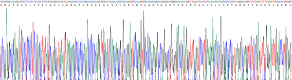
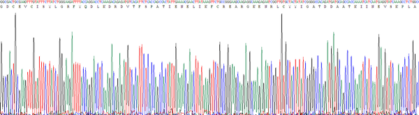
 Application Data
(Figure. Gene Sequencing (Extract))
Application Data
(Figure. Gene Sequencing (Extract))
Glutathione S Transferase Pi, Active Protein (Cat# AAA150106)
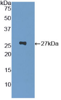
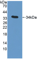 WB (Western Blot)
(Figure. Western Blot)
WB (Western Blot)
(Figure. Western Blot)
Apolipoprotein C3, Active Protein (Cat# AAA150118)
 Application Data
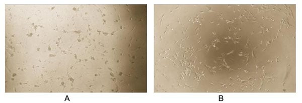
(MANF (Mesencephalic astrocyte-derived neurotrophic factor) is a prosurvival protein that protects the cells when applied intracellularly in vitro or extracellularly in vivo. Recently, MANF has also been proven to inhibit cell proliferation and ER stress-induced cell death and also affects cell size and morphology under certain conditions. Thus the bioactivity of human recombinant MANF were detected using U87-MG. Briefly, U87-MG cells were seeded into triplicate wells of 96-well plates at a density of 2,000 cells/well and allowed to attach overnight, then the medium was replaced with serum-free standard DMEM prior to the addition of various concentrations of MANF. After incubated for 48h, cells were observed by inverted microscope and were shown in Figure 1. It was obvious that MANF inhibited cell proliferation of U87-MG and affected cell size and morphology. (A)U7-MG cells cultured in DMEM, stimulated with 0.01ng/mL MANF for 48h; (B)Unstimulated U87-MG cells cultured in DMEM for 48h.Figure 1. Inhibition of U87-MG cell proliferation after stimulated with MANF.)
Application Data
(MANF (Mesencephalic astrocyte-derived neurotrophic factor) is a prosurvival protein that protects the cells when applied intracellularly in vitro or extracellularly in vivo. Recently, MANF has also been proven to inhibit cell proliferation and ER stress-induced cell death and also affects cell size and morphology under certain conditions. Thus the bioactivity of human recombinant MANF were detected using U87-MG. Briefly, U87-MG cells were seeded into triplicate wells of 96-well plates at a density of 2,000 cells/well and allowed to attach overnight, then the medium was replaced with serum-free standard DMEM prior to the addition of various concentrations of MANF. After incubated for 48h, cells were observed by inverted microscope and were shown in Figure 1. It was obvious that MANF inhibited cell proliferation of U87-MG and affected cell size and morphology. (A)U7-MG cells cultured in DMEM, stimulated with 0.01ng/mL MANF for 48h; (B)Unstimulated U87-MG cells cultured in DMEM for 48h.Figure 1. Inhibition of U87-MG cell proliferation after stimulated with MANF.)
Mesencephalic Astrocyte Derived Neurotrophic Factor, Active Protein (Cat# AAA150127)
 Bioactivity
(Figure. The binding activity of PSA with PDIA3.Puromycin Sensitive Aminopeptidase (PSA) also known as cytosol alanyl aminopeptidase or alanine aminopeptidase (AAP) is used as a biomarker to detect damage to the kidneys, and that may be used to help idenfity certain kidney disorders. It is found at high levels in the urine when there are kidney problems. PSA has been proposed to function in a variety of processes, including metabolism of neuropeptidase, regulation of the cell cycle, and hydrolysis of proteasomal products to amino acids. Besides, Protein Disulfide Isomerase A3 (PDIA3) has been identified as an interactor of PSA, thus a binding ELISA assay was conducted to detect the interaction of recombinant human PSA and recombinant human PDIA3. Briefly, PSA were diluted serially in PBS, with 0.01% BSA (pH 7.4). Duplicate samples of 100uL were then transferred to PDIA3-coated microtiter wells and incubated for 2h at 37. Wells were washed with PBST and incubated for 1h with anti-PSA pAb, then aspirated and washed 3 times. After incubation with HRP labelled secondary antibody, wells were aspirated and washed 3 times. With the addition of substrate solution, wells were incubated 15-25 minutes at 37. Finally, add 50uL stop solution to the wells and read at 450nm immediately. The binding activity of PSA and PDIA3 was shown in Figure 1, and this effect was in a dose dependent manner.)
Bioactivity
(Figure. The binding activity of PSA with PDIA3.Puromycin Sensitive Aminopeptidase (PSA) also known as cytosol alanyl aminopeptidase or alanine aminopeptidase (AAP) is used as a biomarker to detect damage to the kidneys, and that may be used to help idenfity certain kidney disorders. It is found at high levels in the urine when there are kidney problems. PSA has been proposed to function in a variety of processes, including metabolism of neuropeptidase, regulation of the cell cycle, and hydrolysis of proteasomal products to amino acids. Besides, Protein Disulfide Isomerase A3 (PDIA3) has been identified as an interactor of PSA, thus a binding ELISA assay was conducted to detect the interaction of recombinant human PSA and recombinant human PDIA3. Briefly, PSA were diluted serially in PBS, with 0.01% BSA (pH 7.4). Duplicate samples of 100uL were then transferred to PDIA3-coated microtiter wells and incubated for 2h at 37. Wells were washed with PBST and incubated for 1h with anti-PSA pAb, then aspirated and washed 3 times. After incubation with HRP labelled secondary antibody, wells were aspirated and washed 3 times. With the addition of substrate solution, wells were incubated 15-25 minutes at 37. Finally, add 50uL stop solution to the wells and read at 450nm immediately. The binding activity of PSA and PDIA3 was shown in Figure 1, and this effect was in a dose dependent manner.)
Puromycin Sensitive Aminopeptidase, Active Protein (Cat# AAA150133)

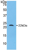
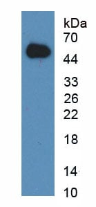
 WB (Western Blot)
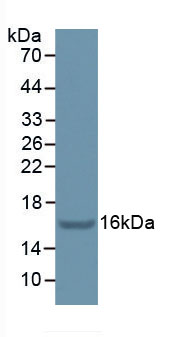
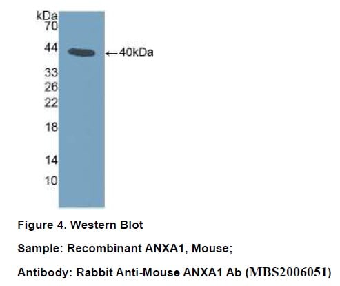
(Figure 4. Western BlotSample: Recombinant ANXA1, Mouse;Antibody: Rabbit Anti-Mouse ANXA1 Ab)
WB (Western Blot)
(Figure 4. Western BlotSample: Recombinant ANXA1, Mouse;Antibody: Rabbit Anti-Mouse ANXA1 Ab)
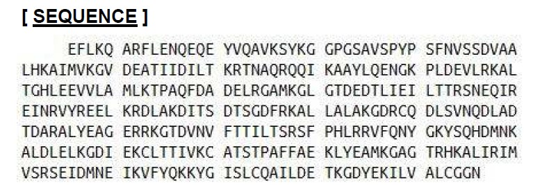
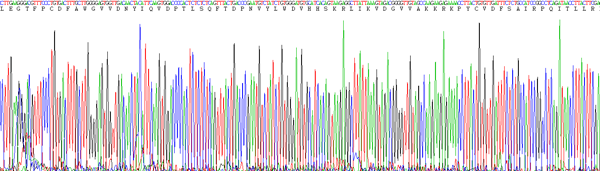
Annexin A1, Active Protein (Cat# AAA150137)
 Bioactivity
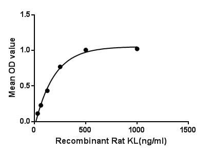
(Klotho (KL) is a transmembrane protein that, in addition to other effects, provides some control over the sensitivity of the organism to insulin and appears to be involved in aging. The Klotho protein is a novel -glucuronidase capable of hydrolyzing steroid -glucuronides. Genetic variants in KLOTHO have been associated with human aging, and Klotho protein has been shown to be a circulating factor detectable in serum that declines with age. The binding of certain fibroblast growth factors (FGFs) viz., FGF19, FGF20, and FGF23, to their fibroblast growth factor receptors, is promoted via their interactions as co-receptors with -Klotho. Besides, Fibroblast Growth Factor 23 (FGF23) has been identified as an interactor of KL, thus a binding ELISA assay was conducted to detect the interaction of recombinant rat KL and recombinant rat FGF23. Briefly, KL were diluted serially in PBS, with 0.01% BSA (pH 7.4). Duplicate samples of 100L were then transferred to FGF23-coated microtiter wells and incubated for 2h at 37. Wells were washed with PBST and incubated for 1h with anti-KL pAb, then aspirated and washed 3 times. After incubation with HRP labelled secondary antibody, wells were aspirated and washed 3 times. With the addition of substrate solution, wells were incubated 15-25 minutes at 37. Finally, add 50uL stop solution to the wells and read at 450nm immediately. The binding activity of KL and FGF23 was shown in Figure 1, and this effect was in a dose dependent manner.Figure. The binding activity of KL with FGF23.)
Bioactivity
(Klotho (KL) is a transmembrane protein that, in addition to other effects, provides some control over the sensitivity of the organism to insulin and appears to be involved in aging. The Klotho protein is a novel -glucuronidase capable of hydrolyzing steroid -glucuronides. Genetic variants in KLOTHO have been associated with human aging, and Klotho protein has been shown to be a circulating factor detectable in serum that declines with age. The binding of certain fibroblast growth factors (FGFs) viz., FGF19, FGF20, and FGF23, to their fibroblast growth factor receptors, is promoted via their interactions as co-receptors with -Klotho. Besides, Fibroblast Growth Factor 23 (FGF23) has been identified as an interactor of KL, thus a binding ELISA assay was conducted to detect the interaction of recombinant rat KL and recombinant rat FGF23. Briefly, KL were diluted serially in PBS, with 0.01% BSA (pH 7.4). Duplicate samples of 100L were then transferred to FGF23-coated microtiter wells and incubated for 2h at 37. Wells were washed with PBST and incubated for 1h with anti-KL pAb, then aspirated and washed 3 times. After incubation with HRP labelled secondary antibody, wells were aspirated and washed 3 times. With the addition of substrate solution, wells were incubated 15-25 minutes at 37. Finally, add 50uL stop solution to the wells and read at 450nm immediately. The binding activity of KL and FGF23 was shown in Figure 1, and this effect was in a dose dependent manner.Figure. The binding activity of KL with FGF23.)
Klotho, Active Protein (Cat# AAA150143)
Bovine Type I Collagen Substrate, Active Protein (Cat# AAA60516)
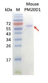
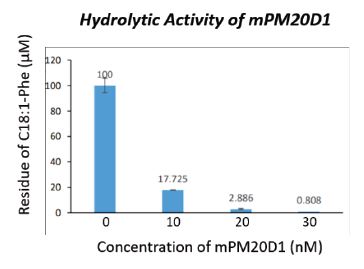 Bioactivity
Peptidase M20 Domain-containing Protein 1 (mPM20D1), Active Protein (Cat# AAA60568)
interleukin 22 receptor antagonist, Active Protein (Cat# AAA60572)
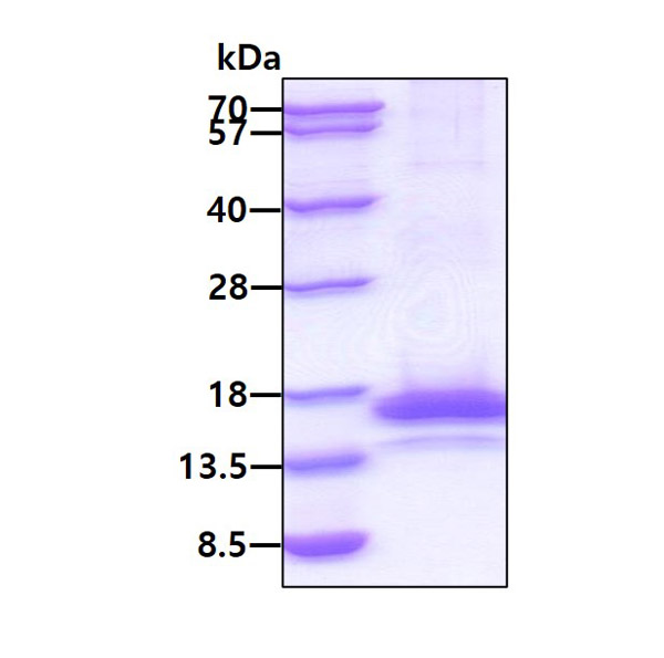
(a) Analysis by reducing and non-reducing SDS-PAGE Silver Stained gel.
(b) Gel filtration chromatography under non-denaturing conditions.
 Bioactivity
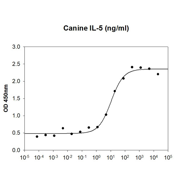
(Canine IL-5 in a cell proliferation assay using TF-1 human erythroleukemic cell. The ED50 range )
Bioactivity
(Canine IL-5 in a cell proliferation assay using TF-1 human erythroleukemic cell. The ED50 range )
IL-5, Active Protein (Cat# AAA48383)
 Bioactivity
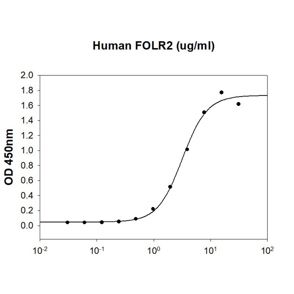
(Folic Acid-BSA is coated at 10ug/ml (100 ul/well) can bind Human FOLR2. The ED50 range )
Bioactivity
(Folic Acid-BSA is coated at 10ug/ml (100 ul/well) can bind Human FOLR2. The ED50 range )
FOLR2, Active Protein (Cat# AAA48385)
 Bioactivity
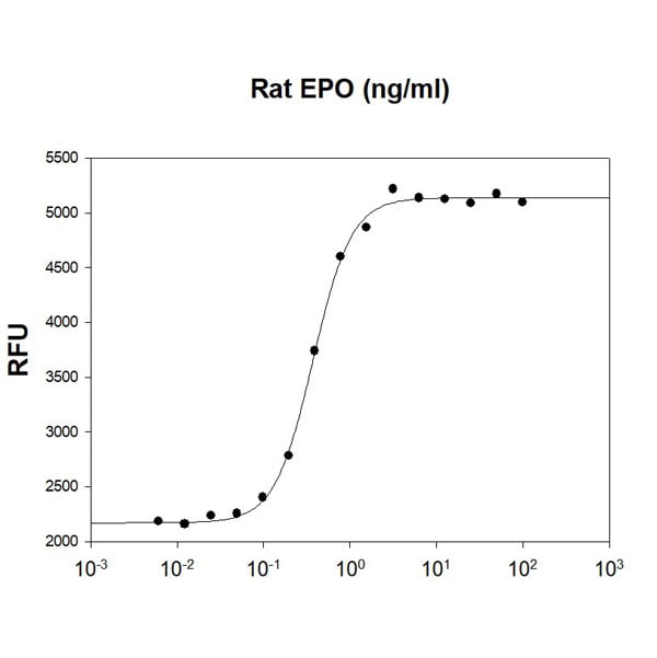
(Rat EPO stimulates cell proliferation of the TF-1 human erythroleukemic cells. The ED50 range )
Bioactivity
(Rat EPO stimulates cell proliferation of the TF-1 human erythroleukemic cells. The ED50 range )
Erythropoietin/EPO, Active Protein (Cat# AAA48388)
 Bioactivity
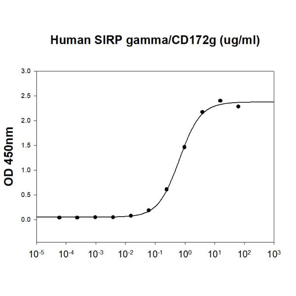
(Human CD47 (CAT# ATGP4015) is coated at 1 ug/ml (100 ul/well) can bind Human SIRP gamma/CD172g. The ED range )
Bioactivity
(Human CD47 (CAT# ATGP4015) is coated at 1 ug/ml (100 ul/well) can bind Human SIRP gamma/CD172g. The ED range )
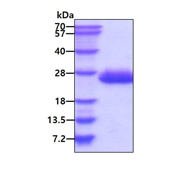
SIRP gamma/CD172g, Active Protein (Cat# AAA48402)
 Bioactivity
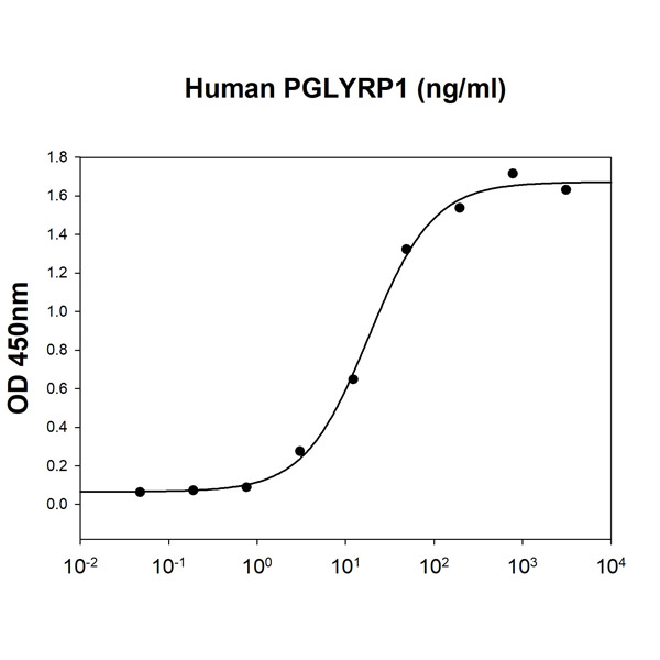
(Peptidoglycan is coated at 10 ug/ml (100 ul/well) can bind Human PGLYRP1. The ED50 range )
Bioactivity
(Peptidoglycan is coated at 10 ug/ml (100 ul/well) can bind Human PGLYRP1. The ED50 range )
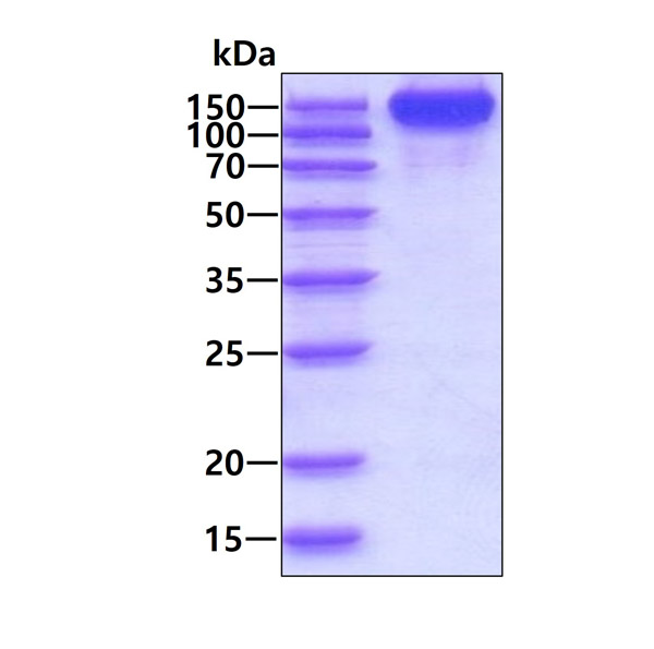
PGLYRP1/PGRP-S, Active Protein (Cat# AAA48403)
 Bioactivity
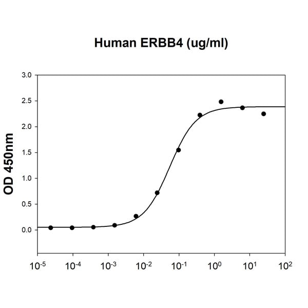
(Human NRG1/HRG1 (CAT# ATGP3990) is coated at 10 ug/ml (100 ul/well) can bind Human ERBB4. The ED50 range )
Bioactivity
(Human NRG1/HRG1 (CAT# ATGP3990) is coated at 10 ug/ml (100 ul/well) can bind Human ERBB4. The ED50 range )
ErbB4/Her4, Active Protein (Cat# AAA48406)
 Bioactivity
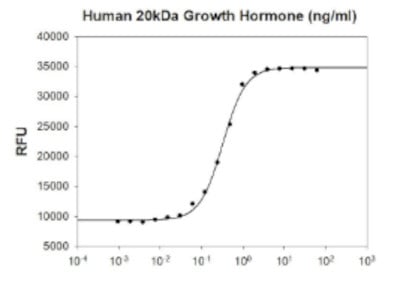
(Human 20kDa Growth Hormone stimulates cell proliferation of the Nb2-11 Rat lymphoma cells. The ED50 range ?1 ng/ml.)
Bioactivity
(Human 20kDa Growth Hormone stimulates cell proliferation of the Nb2-11 Rat lymphoma cells. The ED50 range ?1 ng/ml.)
20kDa Growth Hormone, Active Protein (Cat# AAA48455)
 SDS-PAGE
IGF-1, Active Protein (Cat# AAA48458)
IL-17A, Active Protein (Cat# AAA48986)
PIGF-2, Active Protein (Cat# AAA48996)
 SDS-PAGE
SOD2, Active Protein (Cat# AAA48636)
 SDS-PAGE
Carbonic anhydrase 1, Active Protein (Cat# AAA48917)
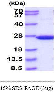
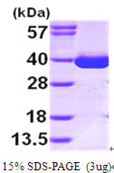
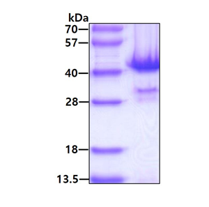
 SDS-PAGE
(3ug by SDS-PAGE under reducing condition and visualized by coomassie blue stain)
SDS-PAGE
(3ug by SDS-PAGE under reducing condition and visualized by coomassie blue stain)
GOT1, Active Protein (Cat# AAA48924)
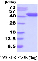
 SDS-PAGE
(3ug by SDS-PAGE under reducing condition and visualized by coomassie blue stain)
SDS-PAGE
(3ug by SDS-PAGE under reducing condition and visualized by coomassie blue stain)
Thioredoxin-1, Active Protein (Cat# AAA48313)
 Bioactivity
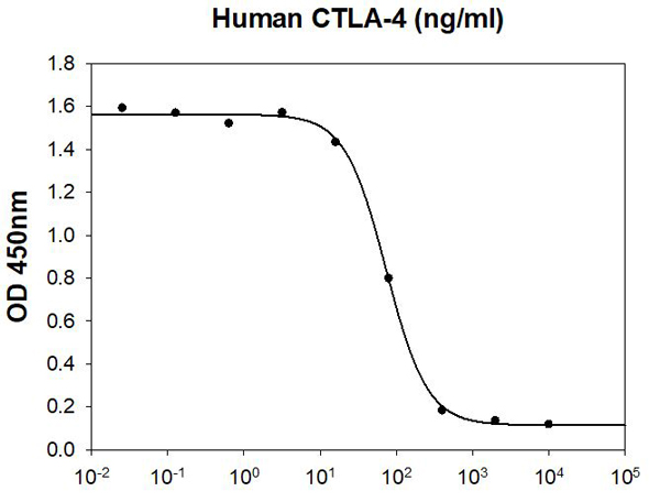
(Human CTLA-4 inhibits Human B7-1/CD80 induced IL-2 secretion of the Jurkat human acute T cell leukemia cells. The ED50 range 150 ng/ml.)
Bioactivity
(Human CTLA-4 inhibits Human B7-1/CD80 induced IL-2 secretion of the Jurkat human acute T cell leukemia cells. The ED50 range 150 ng/ml.)
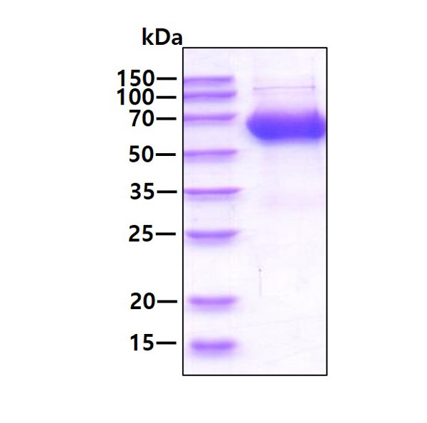
CTLA-4, Active Protein (Cat# AAA48326)
 Bioactivity
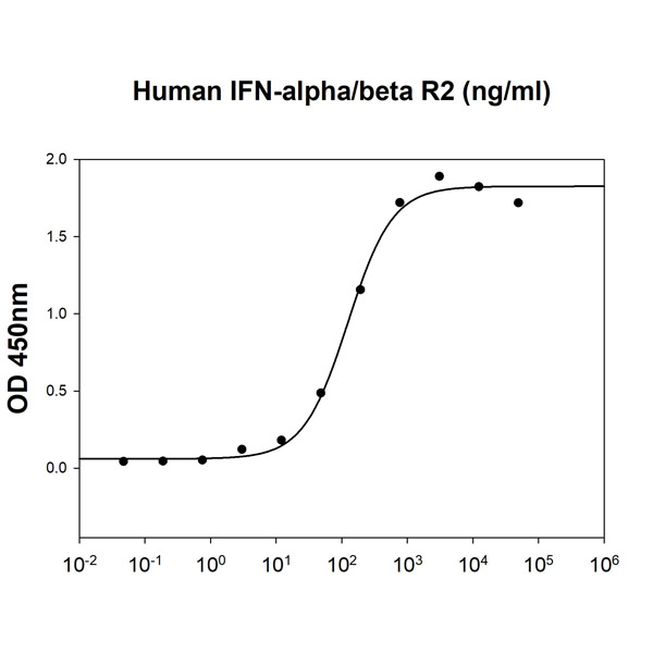
(Human IFN-alpha 2 (CAT# IFN0502) is coated at 10 ug/ml (100 ul/well) can bind Human IFN-alpha/beta R2 in the presence of Human IFN-alpha/beta R1 (CAT# ATGP3894). The ED50 range )
Bioactivity
(Human IFN-alpha 2 (CAT# IFN0502) is coated at 10 ug/ml (100 ul/well) can bind Human IFN-alpha/beta R2 in the presence of Human IFN-alpha/beta R1 (CAT# ATGP3894). The ED50 range )
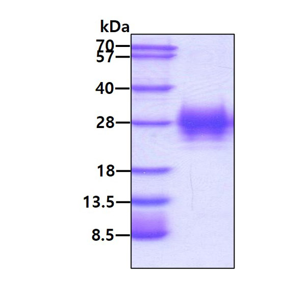
IFN-alpha/beta R2, Active Protein (Cat# AAA48365)
 Bioactivity
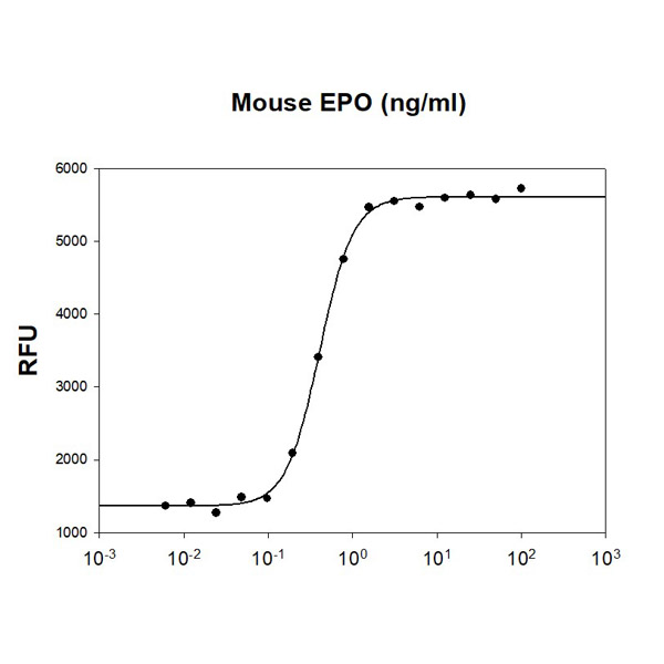
(Mouse EPO stimulates cell proliferation of the TF-1 human erythroleukemic cells. The ED50 range )
Bioactivity
(Mouse EPO stimulates cell proliferation of the TF-1 human erythroleukemic cells. The ED50 range )
Erythropoietin/EPO, Active Protein (Cat# AAA48368)
 Bioactivity
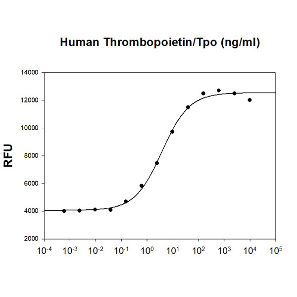
(Human Thrombopoietin stimulates cell proliferation of the MO7e human megakaryocytic leukemic cells. The ED50 range )
Bioactivity
(Human Thrombopoietin stimulates cell proliferation of the MO7e human megakaryocytic leukemic cells. The ED50 range )
Thrombopoietin, Active Protein (Cat# AAA48378)
 SDS-PAGE
IL-1beta, Active Protein (Cat# AAA48475)
 SDS-PAGE
MDH1, Active Protein (Cat# AAA48570)
 SDS-PAGE
MDH2, Active Protein (Cat# AAA48596)
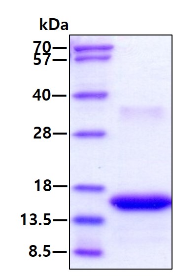
 SDS-PAGE
(3ug by SDS-PAGE under reducing condition and visualized by coomassie blue stain.)
SDS-PAGE
(3ug by SDS-PAGE under reducing condition and visualized by coomassie blue stain.)
PPP1CA, Active Protein (Cat# AAA48757)
GALECTIN-1, Active Protein (Cat# AAA50540)
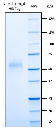
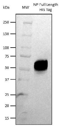 WB (Western Blot)
(Western Blot: NP HIS Tag detected with Covid-19 positive patient serum)
WB (Western Blot)
(Western Blot: NP HIS Tag detected with Covid-19 positive patient serum)
COVID 19 Nucleocapsid (NP) Coronavirus, Active Protein (Cat# AAA58642)
Creatine Kinase MB - Type I, Active Protein (Cat# AAA44773)
Recombinant rat KGF (FGF-7), Active Protein (Cat# AAA47956)
Purification: Purified by heparin affinity chromatography.
BD2, Active Protein (Cat# AAA75442)
Cathepsin G, Active Protein (Cat# AAA75222)
Interleukin-3, Active Protein (Cat# AAA38782)
Zebrafish Growth Hormone, Active Protein (Cat# AAA38832)
What Are Active Proteins?
Proteins are large molecules made up of long chains of amino acids.
They will typically fold into a very particular 3-dimensional shape/conformation, that is sometimes referred to as their “native” form, which allows them to work properly in the body. For the purposes of product categorization, AAA Biotech will typically refer to proteins purified from their original animal host as being “native” proteins (this is to signify their difference compared to their “recombinant” or “synthetic” protein counterparts).
If a protein successfully folds into the correct shape, it is will typically display high fidelity characteristics to its original protein in its original animal host, and be classified as an active protein, as it will be able to function “normally” in most enzymatic or binding capacities. If it loses this shape, due to factors such as heat or strong chemicals (such as detergents), it becomes inactive and is no longer able to perform its basic functions. All of the proteins in this category are made under strict quality control, and they are active, pure, low in contaminants, and stable.
Most are stored as freeze-dried powders and come without extra tags, so they’re very close to the actual natural/native form.
Key Applications of Active Proteins
1. Scientific Research
- Aid in the study of how proteins function in the body
- Aid in understanding various disease processes
2. Drug Development
- Powerful tools to investigate how potential drugs interact with specific proteins
- Ideal for identifying drug targets
3. Cell Culture
- Are routinely utilized to support cell growth and function (e.g., using exogenous growth factors)
- Can be used to promote cellular development into specific types (differentiation)
4. Diagnostics
- Regularly utilized in tests to detect diseases or infections (e.g., COVID-19, cancer)
- Note: All products are strictly for research-use only (RUO).
5. Therapeutics
- Some active proteins are used directly as treatments (e.g., insulin, enzymes)
- Note: All products are strictly for research-use only (RUO).
6. Vaccine Development
- Used to create or test vaccines by mimicking parts of viruses or bacteria
7. Biochemical Assays
- They can facilitate the characterization of enzyme activity, binding strength, or protein interactions in lab tests
Why Buy Active Proteins from AAA Biotech?
- High biological activity – Verified to perform as expected or indicated on datasheet
- Strict quality control – We are confident in our active proteins’ reliability and consistency
- High purity & low endotoxin – Ideal for applications involving sensitive or precious samples/components
- Freeze-dried for stability – Long shelf life and straightforward storage
- Mostly tag-free – Closer to natural/native protein form
FAQ
1. What are active proteins used for in research?
Active proteins are used primarily in the study of how proteins function, in characterizing/discovering drug interactions, supporting cell growth, running biochemical assays, and in development of diagnostics or therapeutics.
2. How are AAA Biotech's active proteins validated?
AAA Biotech’s active proteins are validated through strict quality control and functional assays to ensure they are properly folded and active. “Active”, though, can be an ambiguous term, so if a specific “activity” or “binding” capability of a protein is of crucial interest to you, please inquire with us prior to purchase, and we will provide further details on how the “Active” modifier was determined to be applicable.
3. Are these proteins tested for biological activity?
Yes, all active proteins from AAA Biotech are tested to confirm they have the expected biological activity before being offered for use. Though, said “biological activity” can be either “enzymatic”, “binding”, or both.