Filters
▼Clonality
▼Type
▼Reactivity
▼Gene Name
▼Isotype
▼Host
▼Application
▼Clone
▼Polyclonal Antibodies
At AAA Biotech also known as AAA Bio or AAABio, we provide a broad range of purified polyclonal antibodies (pAbs) that are able to all be browsed online through our website. Due to their high specificity and strong binding affinity, these antibodies are ideal for wide swathes of research and experimental applications.
Our polyclonal antibodies can easily support your work, whether you use them for Western Blotting, Immunocytochemistry (with or without Immunofluorescence used in conjunction), Immunohistochemistry, Immunoprecipitation, and ELISA tests. We highly encourage you to browse our range of pAbs and choose the one that best suits your experimental model.
Viewing 9400-9450 of 96805 product results
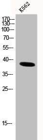
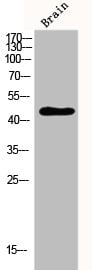
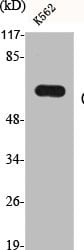
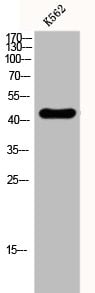
 WB (Western Blot)
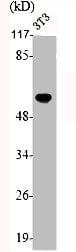
(Western Blot analysis of K562 cells using DNA pol beta Polyclonal Antibody)
WB (Western Blot)
(Western Blot analysis of K562 cells using DNA pol beta Polyclonal Antibody)
POLB, Polyclonal Antibody (Cat# AAA236099)
 WB (Western Blot)
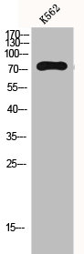
(Western Blot analysis of K562 LOVO cells using Dok-2 Polyclonal Antibody)
WB (Western Blot)
(Western Blot analysis of K562 LOVO cells using Dok-2 Polyclonal Antibody)
DOK2, Polyclonal Antibody (Cat# AAA236101)
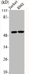
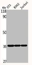
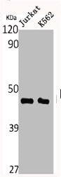 WB (Western Blot)
(Western Blot analysis of Jurkat K562 cells using DRAK2 Polyclonal Antibody)
WB (Western Blot)
(Western Blot analysis of Jurkat K562 cells using DRAK2 Polyclonal Antibody)
STK17B, Polyclonal Antibody (Cat# AAA236103)
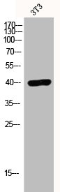
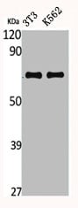
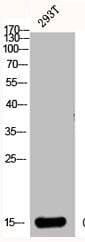
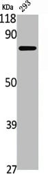
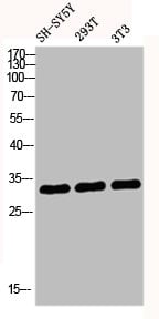 WB (Western Blot)
(Western blot analysis of SH-SY5Y 293T 3T3 lysis using DREAM antibody.)
WB (Western Blot)
(Western blot analysis of SH-SY5Y 293T 3T3 lysis using DREAM antibody.)
KCNIP3, Polyclonal Antibody (Cat# AAA236104)
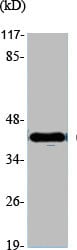
 WB (Western Blot)
(Western Blot analysis of various cells using EDG-7 Polyclonal Antibody)
WB (Western Blot)
(Western Blot analysis of various cells using EDG-7 Polyclonal Antibody)
LPAR3, Polyclonal Antibody (Cat# AAA236115)
 WB (Western Blot)
(Western Blot analysis of various cells using eEF2K Polyclonal Antibody)
WB (Western Blot)
(Western Blot analysis of various cells using eEF2K Polyclonal Antibody)
EEF2K, Polyclonal Antibody (Cat# AAA236116)
 WB (Western Blot)
(Western Blot analysis of Jurkat K562 cells using EMR4 Polyclonal Antibody)
WB (Western Blot)
(Western Blot analysis of Jurkat K562 cells using EMR4 Polyclonal Antibody)
EMR4P, Polyclonal Antibody (Cat# AAA236125)
 WB (Western Blot)
(Western Blot analysis of K562 cells using Endoplasmin Polyclonal Antibody)
WB (Western Blot)
(Western Blot analysis of K562 cells using Endoplasmin Polyclonal Antibody)
HSP90B1, Polyclonal Antibody (Cat# AAA236126)
 WB (Western Blot)
(Western Blot analysis of PC12 HELA cells using EphA5 Polyclonal Antibody)
WB (Western Blot)
(Western Blot analysis of PC12 HELA cells using EphA5 Polyclonal Antibody)
EPHA5, Polyclonal Antibody (Cat# AAA236132)

 WB (Western Blot)
(Western Blot analysis of NIH-3T3 K562 Jurkat cells using ERdj3 Polyclonal Antibody)
WB (Western Blot)
(Western Blot analysis of NIH-3T3 K562 Jurkat cells using ERdj3 Polyclonal Antibody)
DNAJB11, Polyclonal Antibody (Cat# AAA236140)
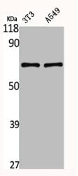
 WB (Western Blot)
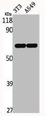
(Western Blot analysis of NIH-3T3 A549 cells using ERF Polyclonal Antibody)
WB (Western Blot)
(Western Blot analysis of NIH-3T3 A549 cells using ERF Polyclonal Antibody)
ERF, Polyclonal Antibody (Cat# AAA236141)
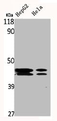 WB (Western Blot)
(Western Blot analysis of HepG2 HELA cells using ERK 1/2 Polyclonal Antibody)
WB (Western Blot)
(Western Blot analysis of HepG2 HELA cells using ERK 1/2 Polyclonal Antibody)
MAPK3/MAPK1, Polyclonal Antibody (Cat# AAA236143)
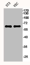 WB (Western Blot)
(Western Blot analysis of NIH-3T3 VEC cells using ERalpha Polyclonal Antibody)
WB (Western Blot)
(Western Blot analysis of NIH-3T3 VEC cells using ERalpha Polyclonal Antibody)
ESR1, Polyclonal Antibody (Cat# AAA236144)
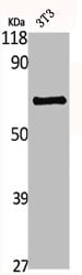
 WB (Western Blot)
(Western Blot analysis of NIH-3T3 cells using EWS Polyclonal Antibody)
WB (Western Blot)
(Western Blot analysis of NIH-3T3 cells using EWS Polyclonal Antibody)
EWSR1, Polyclonal Antibody (Cat# AAA236146)
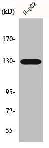
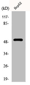
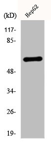
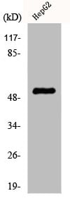
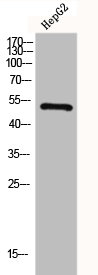
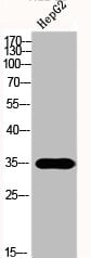 WB (Western Blot)
(Western Blot analysis of HepG2 cells using FAS-L Polyclonal Antibody)
WB (Western Blot)
(Western Blot analysis of HepG2 cells using FAS-L Polyclonal Antibody)
FASLG, Polyclonal Antibody (Cat# AAA236148)
 WB (Western Blot)
(Western Blot analysis of NIH-3T3 cells using Fli-1 Polyclonal Antibody)
WB (Western Blot)
(Western Blot analysis of NIH-3T3 cells using Fli-1 Polyclonal Antibody)
FLI1, Polyclonal Antibody (Cat# AAA236153)
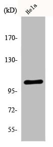
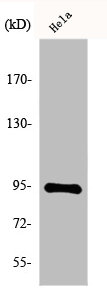
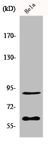
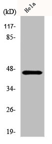
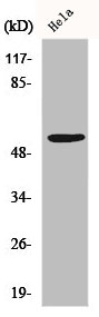
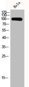
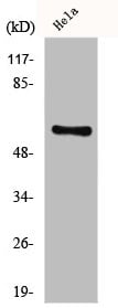
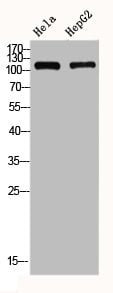
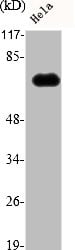 WB (Western Blot)
(Western Blot analysis of Hela cells using FSHR Polyclonal Antibody)
WB (Western Blot)
(Western Blot analysis of Hela cells using FSHR Polyclonal Antibody)
FSHR, Polyclonal Antibody (Cat# AAA236160)
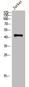
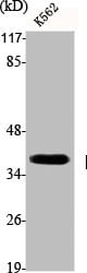
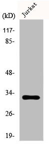
 WB (Western Blot)
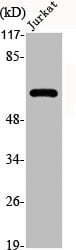
(Western Blot analysis of Jurkat cells using Creatine Kinase M Polyclonal Antibody)
WB (Western Blot)
(Western Blot analysis of Jurkat cells using Creatine Kinase M Polyclonal Antibody)
CKM, Polyclonal Antibody (Cat# AAA236042)
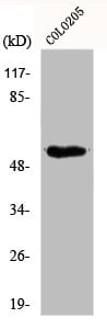
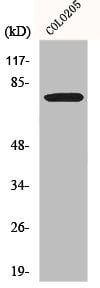
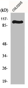
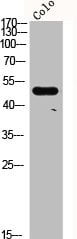 WB (Western Blot)
(Western Blot analysis of COLO cells using CRF-RII Polyclonal Antibody)
WB (Western Blot)
(Western Blot analysis of COLO cells using CRF-RII Polyclonal Antibody)
CRHR2, Polyclonal Antibody (Cat# AAA236045)

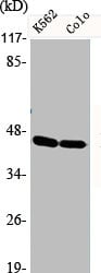
 WB (Western Blot)
(Western Blot analysis of NIH-3T3 K562 cells using CTPS Polyclonal Antibody)
WB (Western Blot)
(Western Blot analysis of NIH-3T3 K562 cells using CTPS Polyclonal Antibody)
CTPS1, Polyclonal Antibody (Cat# AAA236053)
 WB (Western Blot)
(Western Blot analysis of various cells using CXCR-3 Polyclonal Antibody)
WB (Western Blot)
(Western Blot analysis of various cells using CXCR-3 Polyclonal Antibody)
CXCR3, Polyclonal Antibody (Cat# AAA236056)
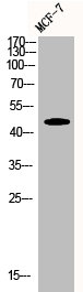
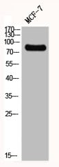
 WB (Western Blot)
(Western Blot analysis of MCF7 cells using Cyclin E1 Polyclonal Antibody)
WB (Western Blot)
(Western Blot analysis of MCF7 cells using Cyclin E1 Polyclonal Antibody)
CCNE1, Polyclonal Antibody (Cat# AAA236061)
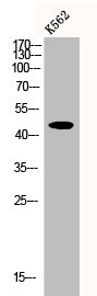
 WB (Western Blot)
(Western Blot analysis of K562 cells using CYP19A1 Polyclonal Antibody)
WB (Western Blot)
(Western Blot analysis of K562 cells using CYP19A1 Polyclonal Antibody)
CYP19A1, Polyclonal Antibody (Cat# AAA236064)
 WB (Western Blot)
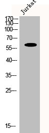
(Western Blot analysis of Jurkat cells using CYP1B1 Polyclonal Antibody)
WB (Western Blot)
(Western Blot analysis of Jurkat cells using CYP1B1 Polyclonal Antibody)
CYP1B1, Polyclonal Antibody (Cat# AAA236065)
 WB (Western Blot)
(Western Blot analysis of HELA 293T cells using CYP2C19 Polyclonal Antibody)
WB (Western Blot)
(Western Blot analysis of HELA 293T cells using CYP2C19 Polyclonal Antibody)
CYP2C19, Polyclonal Antibody (Cat# AAA236068)
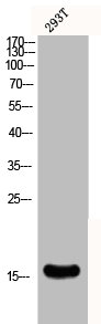
 WB (Western Blot)
(Western Blot analysis of 293T cells using Cystatin C Polyclonal Antibody)
WB (Western Blot)
(Western Blot analysis of 293T cells using Cystatin C Polyclonal Antibody)
CST3, Polyclonal Antibody (Cat# AAA236075)
 WB (Western Blot)
(Western Blot analysis of various cells using Cytokeratin 13 Polyclonal Antibody)
WB (Western Blot)
(Western Blot analysis of various cells using Cytokeratin 13 Polyclonal Antibody)
KRT13, Polyclonal Antibody (Cat# AAA236078)
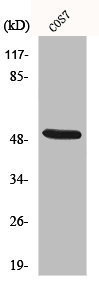
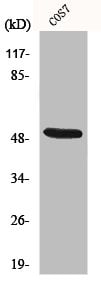 WB (Western Blot)
(Western Blot analysis of COS7 cells using Cytokeratin 15 Polyclonal Antibody)
WB (Western Blot)
(Western Blot analysis of COS7 cells using Cytokeratin 15 Polyclonal Antibody)
KRT15, Polyclonal Antibody (Cat# AAA236079)
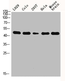 WB (Western Blot)
(Western Blot analysis of L929 COLO 293T HELA MOUSE-brain cells using Cytokeratin 18 Polyclonal Antibody)
WB (Western Blot)
(Western Blot analysis of L929 COLO 293T HELA MOUSE-brain cells using Cytokeratin 18 Polyclonal Antibody)
KRT18, Polyclonal Antibody (Cat# AAA236081)
 WB (Western Blot)
(Western Blot analysis of Jurkat cells using Cytokeratin 5 Polyclonal Antibody)
WB (Western Blot)
(Western Blot analysis of Jurkat cells using Cytokeratin 5 Polyclonal Antibody)
KRT5, Polyclonal Antibody (Cat# AAA236083)
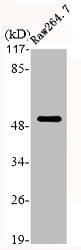 WB (Western Blot)
(Western Blot analysis of RAE264.7 cells using Cytokeratin 7 Polyclonal Antibody)
WB (Western Blot)
(Western Blot analysis of RAE264.7 cells using Cytokeratin 7 Polyclonal Antibody)
KRT7, Polyclonal Antibody (Cat# AAA236084)
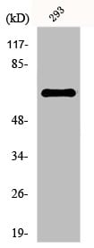
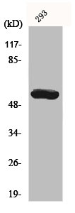
 WB (Western Blot)
(Western Blot analysis of 293 cells using Daxx Polyclonal Antibody)
WB (Western Blot)
(Western Blot analysis of 293 cells using Daxx Polyclonal Antibody)
DAXX, Polyclonal Antibody (Cat# AAA236090)
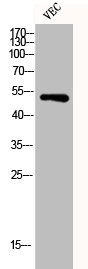
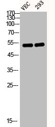 WB (Western Blot)
(Western Blot analysis of VEC 293 cells using Desmin Polyclonal Antibody)
WB (Western Blot)
(Western Blot analysis of VEC 293 cells using Desmin Polyclonal Antibody)
DES, Polyclonal Antibody (Cat# AAA236093)
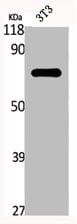
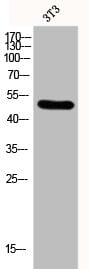
 WB (Western Blot)
(Western Blot analysis of NIH-3T3 cells using Fyn Polyclonal Antibody)
WB (Western Blot)
(Western Blot analysis of NIH-3T3 cells using Fyn Polyclonal Antibody)
FYN, Polyclonal Antibody (Cat# AAA236163)
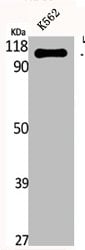
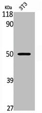
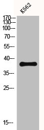
 WB (Western Blot)
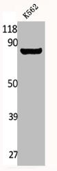
(Western Blot analysis of K562 cells using Gab 2 Polyclonal Antibody)
WB (Western Blot)
(Western Blot analysis of K562 cells using Gab 2 Polyclonal Antibody)
GAB2, Polyclonal Antibody (Cat# AAA236165)

 WB (Western Blot)
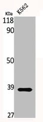
(Western Blot analysis of K562 cells using Gads Polyclonal Antibody)
WB (Western Blot)
(Western Blot analysis of K562 cells using Gads Polyclonal Antibody)
GRAP2, Polyclonal Antibody (Cat# AAA236168)
 WB (Western Blot)
(Western Blot analysis of various cells using GATA-4 Polyclonal Antibody)
WB (Western Blot)
(Western Blot analysis of various cells using GATA-4 Polyclonal Antibody)
GATA4, Polyclonal Antibody (Cat# AAA236171)
 WB (Western Blot)
(Western Blot analysis of NIH-3T3 cells using GBP3 Polyclonal Antibody)
WB (Western Blot)
(Western Blot analysis of NIH-3T3 cells using GBP3 Polyclonal Antibody)
GBP3, Polyclonal Antibody (Cat# AAA236172)
 WB (Western Blot)
(Western Blot analysis of various cells using GCN5 Polyclonal Antibody)
WB (Western Blot)
(Western Blot analysis of various cells using GCN5 Polyclonal Antibody)
KAT2A, Polyclonal Antibody (Cat# AAA236174)
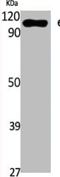
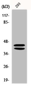
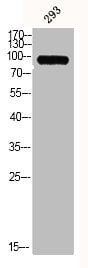 WB (Western Blot)
(Western Blot analysis of 293 cells using MMP-9 Polyclonal Antibody)
WB (Western Blot)
(Western Blot analysis of 293 cells using MMP-9 Polyclonal Antibody)
MMP9, Polyclonal Antibody (Cat# AAA236176)
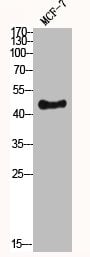
 WB (Western Blot)
(Western Blot analysis of MCF7 cells using GIT2 Polyclonal Antibody)
WB (Western Blot)
(Western Blot analysis of MCF7 cells using GIT2 Polyclonal Antibody)
GIT2, Polyclonal Antibody (Cat# AAA236179)
 WB (Western Blot)
(Western Blot analysis of NIH-3T3 A549 cells using GK2 Polyclonal Antibody)
WB (Western Blot)
(Western Blot analysis of NIH-3T3 A549 cells using GK2 Polyclonal Antibody)
GK2, Polyclonal Antibody (Cat# AAA236180)
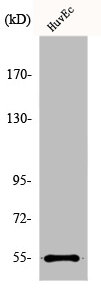
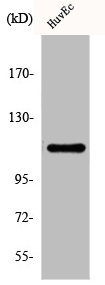
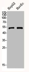 WB (Western Blot)
(Western Blot analysis of HepG2 HuvEc cells using GLP-1R Polyclonal Antibody)
WB (Western Blot)
(Western Blot analysis of HepG2 HuvEc cells using GLP-1R Polyclonal Antibody)
GLP1R, Polyclonal Antibody (Cat# AAA236181)
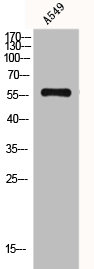
 WB (Western Blot)
(Western Blot analysis of A549 cells using Glucosidase IIbeta Polyclonal Antibody)
WB (Western Blot)
(Western Blot analysis of A549 cells using Glucosidase IIbeta Polyclonal Antibody)
PRKCSH, Polyclonal Antibody (Cat# AAA236183)
 WB (Western Blot)
(Western Blot analysis of HELA HEPG2 using GluR-1 Polyclonal Antibody)
WB (Western Blot)
(Western Blot analysis of HELA HEPG2 using GluR-1 Polyclonal Antibody)
GRIA1, Polyclonal Antibody (Cat# AAA236184)
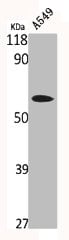
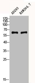 WB (Western Blot)
(Western Blot analysis of AD293 RAW using gp91-phox Polyclonal Antibody)
WB (Western Blot)
(Western Blot analysis of AD293 RAW using gp91-phox Polyclonal Antibody)
CYBB, Polyclonal Antibody (Cat# AAA236189)
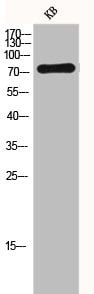
 IHC (Immunohistochemisry)
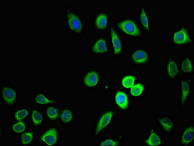
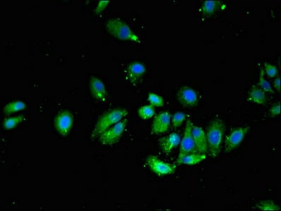
(Immunofluorescent analysis of A549 cells using AAA232974 at a dilution of 1:100 and Alexa Fluor 488-congugated AffiniPure Goat Anti-Rabbit IgG(H+L))
IHC (Immunohistochemisry)
(Immunofluorescent analysis of A549 cells using AAA232974 at a dilution of 1:100 and Alexa Fluor 488-congugated AffiniPure Goat Anti-Rabbit IgG(H+L))
ZDHHC1, Polyclonal Antibody (Cat# AAA232974)
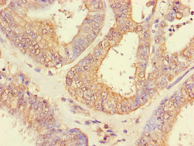
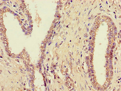
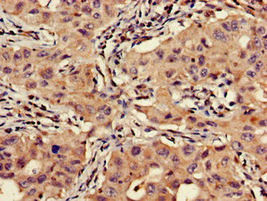
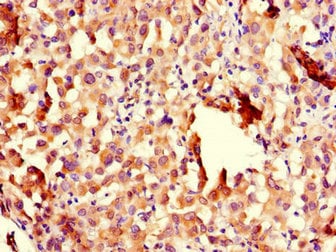 IHC (Immunohistochemisry)
(Immunohistochemistry of paraffin-embedded human lung cancer using AAA232976 at dilution of 1:100)
IHC (Immunohistochemisry)
(Immunohistochemistry of paraffin-embedded human lung cancer using AAA232976 at dilution of 1:100)
CHFR, Polyclonal Antibody (Cat# AAA232976)
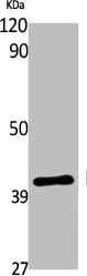
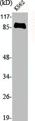
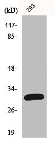
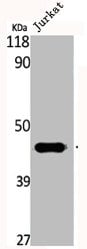
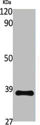
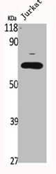
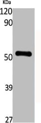
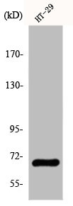
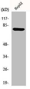
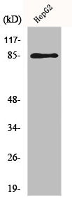
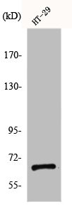
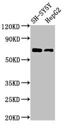 WB (Western Blot)
(Western BlotPositive WB detected in: SH-SY5Y whole cell lysate,HepG2 whole cell lysateAll lanes: C7orf31 antibody at 2.5ug/mlSecondaryGoat polyclonal to rabbit IgG at 1/50000 dilutionPredicted band size: 69 KDaObserved band size: 69 KDa)
WB (Western Blot)
(Western BlotPositive WB detected in: SH-SY5Y whole cell lysate,HepG2 whole cell lysateAll lanes: C7orf31 antibody at 2.5ug/mlSecondaryGoat polyclonal to rabbit IgG at 1/50000 dilutionPredicted band size: 69 KDaObserved band size: 69 KDa)
C7orf31, Polyclonal Antibody (Cat# AAA232979)
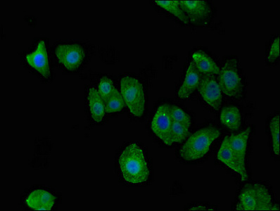
 IHC (Immunohistochemisry)
(Immunofluorescent analysis of HepG2 cells using AAA232980 at a dilution of 1:100 and Alexa Fluor 488-congugated AffiniPure Goat Anti-Rabbit IgG(H+L))
IHC (Immunohistochemisry)
(Immunofluorescent analysis of HepG2 cells using AAA232980 at a dilution of 1:100 and Alexa Fluor 488-congugated AffiniPure Goat Anti-Rabbit IgG(H+L))
VTI1A, Polyclonal Antibody (Cat# AAA232980)
What are Polyclonal Antibodies?
Polyclonal antibodies are antibodies that come from multiple B cell clones of a host animal. The typical hosts used for the majority of polyclonal antibody production are rabbits, goats, sheep, and donkeys. These polyclonal antibodies, once having identified their target, will bind to different epitopes located at different regions or sequences on the same protein/antigen. As a result, they are ideal at locating and binding to the target, even if the target is in very low concentrations (due to many different antibodies being able to bind to the same target molecule, which allows for significant amplification of a downstream signal).
Polyclonal antibodies are typically produced by injecting an antigen into a host animal, which causes the animal’s immune system to attack the foreign antigen by mass generating antibodies against it. After a period of time, serum is collected from the animal and purified using physicochemical fractionation, class-specific affinity purification, and/or antigen-affinity purification.
Key Uses of Polyclonal Antibodies
- Western Blotting: This method is used to find specific proteins in biological samples after separating them by size.
- Immunohistochemistry: IHC helps visualize the location of proteins in tissue sections using various staining techniques.
- ELISA: (Enzyme-Linked Immunosorbent Assay) is typically used to identify specific protein quantities in a sample. ELISAs can be either “Quantitative” or “Qualitative”.
- Flow Cytometry: technique that identifies and measures the specific protein on the surface or inside the cells in a fluid suspension.
- Immunoprecipitation: IP isolates and studies a specific protein from a complex mixture using antibodies.
Why Buy Polyclonal Antibodies from AAA Biotech?
1. Ideal for Various Applications
Our antibodies are generally going to be validated for use in multiple types of assays, including ELISA, Western Blotting, Immunohistochemistry, Immunoprecipitation, amongst others. They are ideal for a wide range of research applications.
2. Rigorous Quality Control
All of the antibodies in our catalog undergo strict quality testing to ensure specificity, sensitivity, and consistent performance. We are confident in the ability of our antibodies to provide you with accurate results.
3. Wide Assortment of Antibodies
Antibodies in are catalog can be found for both common and exotic species, and these antibodies are also available in both conjugated and recombinant forms to suit many diverse experimental needs.
4. Highly Purified
Our antibodies are available in purified forms with over 85% purity, as confirmed by SDS-PAGE. They are also available with tags such as His, Flag, GST, or MBP. We cater to customers worldwide.
FAQ
1. How are polyclonal antibodies produced?
Traditionally, polyclonal antibodies are produced by injecting an antigen into a host animal (such as a rabbit or goat), which then triggers an immune response from the host animal. The animal’s B cells produce antibodies that will recognize different parts of the injected antigen. These antibodies are then collected from the animal’s blood and purified for use.
2. How do polyclonal antibodies differ from monoclonal antibodies?
Polyclonal antibodies are a mix of antibodies that bind to different locations (epitopes) of the same antigen, while monoclonal antibodies are identical and bind to just one specific epitope. This makes polyclonal antibodies more versatile and better at detecting proteins that may be present in low quantities or in altered/modified forms.
3. How should I store polyclonal antibodies?
Polyclonal antibodies should be stored at 4°C for short-term use (up to a few weeks) and at -20°C or -80°C for long-term storage. Avoid repeated freeze-thaw cycles by dividing them into small aliquots. Always check the datasheet for specific storage instructions.